Article

The Relationship between Gait Velocity and Walking Pattern in Hemiplegic Patients

1
ASL 11—San Miniato Hospital, 50053 Empoli, Italy
2
Physical Therapy and Rehabilitation Unit, University of Bologna, IRCCS Istituto Ortopedico Rizzoli, Via Pupilli 1, 40136 Bologna, Italy
3
Physical Medicine and Rehabilitation Unit, AUSL Romagna, 48121 Ravenna, Italy
*
Author to whom correspondence should be addressed.
Appl. Sci. 2023, 13(2), 934; https://doi.org/10.3390/app13020934
Submission received: 25 November 2022 / Revised: 23 December 2022 / Accepted: 7 January 2023 / Published: 10 January 2023
(This article belongs to the Special Issue Musculoskeletal Rehabilitation and Clinical Biomechanics)

Abstract

:
Background Gait speed represents a functional predictor and an impairment severity index in stroke survivors; gait analysis parameters are descriptors of walking strategies used to compensate for the muscle impairment such as vaulting, circumduction and hip hiking. The aim of this study was to assess if there is a relationship between the gait compensatory strategy and gait speed of progression. Methods A sample of 30 patients with post-stroke hemiparesis was assessed for gait compensatory patterns through gait analysis and videorecording. BMI, pain-VAS, Barthel Index, Nottingham Extended ADL Scale, Motricity Index, lower limb muscles strength and aROMs were also included in the assessment. Results In 19 patients it was possible to identify one or more compensatory strategies; in 11 patients no specific gait pattern was found. The vaulting and hip hiking combined gait strategy had an effect on gait speed. Gait speed was directly related to Barthel Index, Nottingham Extended ADL Scale, Motricity Index of the paretic side and in particular with quadriceps and iliopsoas strength and hip extension aROM. Gender, age and paretic side did not influence gait speed. Conclusion Compensatory gait strategies influence gait speed but studies with larger sample size are needed to better highlight their impact.

1. Introduction

Stroke is a major public health issue worldwide [1], as it is the second leading cause of death and the third leading cause of disability [2]. The mobility limitations resulting from a stroke, due mostly to hemiparesis, have a significant impact on a patient’s independent ambulation [1].
Several studies have shown the effectiveness of rehabilitation in the acute, post-acute and chronic phases of stroke, as it improves walking speed, physical fitness and balance and reduces the risk of falling, fractures and a further decline in mobility [3,4,5]. Furthermore, in chronic stroke survivors, an adaptive physical activity (APA) program is also an effective approach to maintain and improve activities of daily living and reduce falls and recourse to rehabilitation services. Additionally, it makes it possible to perform the exercises at home, which increases patient compliance and improves quality of life perception [6].
One of the primary goals in stroke rehabilitation is regaining locomotor ability. Change in gait speed is the most commonly used parameter to evaluate patients because it is easy to measure, cost effective and reliable. Moreover, it is a great functional walking status predictor and an impairment severity index [7].
Spatiotemporal, kinematic and kinetic parameters, widely studied in the existing literature, show a decreased cadence, prolonged swing duration on the paretic side, prolonged stance duration on the non-paretic side and step length asymmetry in post-stroke patients [8]. Previous studies also support that gait asymmetry ratios are more sensitive outcome measures [9,10,11]. Furthermore, the inadequate propulsion force is correlated to an alteration of the leg extension angle, knee flexion angle and foot dorsiflexion in the paretic side [12].
Strength deficit in the lower limbs appears to be one of the main factors slowing the recovery of walking ability, strictly dependent on abnormal posture and stretching reflexes and loss of voluntary movement resulting from paresis [13]. Hemiparesis leads to impaired motion patterns, abnormal muscle activities and abnormal joint trajectories. Common features are decreased peak hip flexion, peak knee flexion-extension and foot dorsiflexion [14], which generally determine a problem with foot–floor clearance. Compensatory gait strategies to enhance progression and foot-floor clearance, such as circumduction, hip hiking and vaulting, have been described and recognized to increase energy expenditure [14,15,16] (Figure 1).
All these gait patterns have some common features, but also some significant variations in terms of group muscles weakness and spasticity. In the daily clinical practice, usually hemiplegic or hemiparetic patients adopt one of these gait patterns, but it is not unusual to find their combinations, with a prevalence of some features despite others in the activation sequence of muscles and strategy of progression. Gait modification is part of all three plans for movement of lower limbs. To attain a proper understanding, it is essential to focus on each one.
Circumduction gait is an abnormal walking pattern in which a patient, to avoid hitting the ground with the foot in order to achieve its adequate clearance, lifts and moves away from the body the affected lower limb and takes it forward, performing a semi-circular movement. It occurs during the swing phase on the affected side, the compensation consisting in the activation of the leg abduction, and the principal muscles involved are the glutei minimus and medius, assisted by the tensor fasciae latae and adductor muscles. Circumduction is commonly seen in hemiplegic patients but could occur also in severe knee osteoarthritis/arthrosis, peripheral nerve injuries with foot drop and hetero-metry of lower limbs. In all these cases, the patient cannot produce a sufficient hip or knee flexion or ankle dorsiflexion. In hemiplegic muscle weakness, spasticity and spastic synergistic activation patterns interact and also affect the trunk and pelvis trajectories; we can observe the mechanical consequences during the clinical evaluation as gait impairment [17].
As stated, hip hiking gait is a strategy used to compensate for the insufficient flexion of the hip joint during the swing phase, as well as knee flexion and ankle dorsiflexion, thus shortening the paretic limb. To achieve forward progression of the lower limb, the patient lifts the pelvis on the affected side more than in the normal walking pattern and in the meantime he bends the trunk laterally on the opposite side. That movement is made possible by the activation of paravertebral and lateral abdominal muscles, increasing the trunk inclination and pelvic rotation. It is common to observe an associated posterior trunk inclination at the beginning of the swing phase in this gait pattern, often at lower speed and with the prevalence of weakness of the hamstring muscles. In the pure hip hiking gait, there are no rotation or abduction movements of the hip joint, but the combination of this pattern and the circumduction pattern is unusual [17].
The last is vaulting. In this case the patient rises up on his toe on the unaffected side to achieve adequate foot–floor clearance of the paretic side during its swing phase to promote the leg progression. In a normal gait, the moving forward/swinging leg becomes shorter to allow this by keen flexion, hip flexion and ankle dorsiflexion. In an hemiparetic/hemiplegic patient, due to spasticity and involuntary activation synergy, knee bending could not occur and he could present a foot drop or an equinus foot (overactivation of triceps surae), so the progression of the paretic limb is made possible by lengthening the contralateral. Overall, the vaulting pattern is the most inefficient and expensive in terms of energy cost [17].
Depending on the group of muscles involved and the intensity of spasticity and flaccidity, as already stated, post-stroke patients could perform combined walking strategies but without specific patterns, unclassifiable and indescribable in terms of the known ones.
The aim of this study was to assess, in a sample of hemiparetic patients, whether these compensatory patterns influence walking speed. The relationship between gait speed and impairment and disability scores, selective muscle strength and range of motion of the lower limb joints were also considered, in order to bring possible rehabilitation implications to light.

2. Materials and Methods

2.1. Study Design

This is an observational, retrospective study carried out on a sample of patients with post-stroke hemiplegia, examined by means of clinical-functional assessment and gait analysis, according to the clinical practice.

2.2. Patient Sample

This study took place at AUSL 11 in the Tuscany Region of Italy on a sample of chronic stroke patients with hemiplegia as the primary disability at a minimum follow-up of 24 months.
The only inclusion criteria were that patients were able to walk independently and without cognitive impairment. After stroke, all patients received a rehabilitation course and, at the time of the study, they were included in an APA program.

2.3. Assessment

Each patient underwent a clinical evaluation where demographic parameters and body mass index (BMI) were recorded and several scales were used: Visual Analogic Scale (VAS) for pain, paretic side Motricity Index (MI), Barthel Index (BI) and Nottingham Extended ADL Scale (NEAS).
To record the pain, a Visual Analogue Scale (VAS) was chosen as an instrument that can measure a characteristic or attitude that ranges across a continuum of values [18]. The assessment of pain in rehabilitation is one of the central clinical features, depending on its correlation to the level of patients’ activities and limitations, and it is widely used as an outcome target in both clinical practice and treatment efficacy [19]. The VAS for chronic pain has demonstrated sensitivity to changes in pain assessed at different times and to the variables that increase or decrease its intensity, presents a high test–retest reliability and repeatability and finally shows a strong association with activities influenced by pain.
To test the hemiparetic patients’ motor impairment, the Motricity Index (MI) was selected for its feasible application [20]. MI is a brief measure of motor function and in the literature was effective as predictor of mobility outcome poststroke [20]. It consists of testing the muscle strength in upper and lower limbs. We used only the section for the lower limb, representing the main focus of our study. The three movements checked were hip flexion, knee extension and ankle dorsiflexion [20]. To graduate muscle force, the Medical Research Council scale [21] was used, an ordinal six points scale (0 no movement to 5 normal power), then the scores were converted to the corresponding motricity score and summed and added, resulting in a total value ranging from 0 (complete paresis) to 100 (normal strength) [20]. MI has high reliability as measure of muscle strength, is simple to apply and does not require special training or equipment [22].
The Barthel Index (BI) is a common scale used to quantify the poststroke patient’s disability and impairment in ADL function [23,24,25]. The range scoring is from 0 to 100 (maximum disability to high independence) and each item assesses different features of ADL such as bowel and bladder control, grooming, toilet use, feeding, dressing, chair/bed transfers, walking, climbing stairs and bathing [26]. It represents a good outcome measure for functional evaluation and is one of the most recommended measures, showing good results in test–retest reliability [22,23,26].
The Nottingham Extended ADL scale (NEAS) is composed of four subsections with defined subscales that evaluates poststroke patient’s mobility, household ability and leisure activity [27]. NEAS is a measure of ADL that goes beyond basic self-care as BI [28]. The scale has 22 items, each one having four possible answers representing the help needed during the specific activity (from “not at all”—0 to “on my own”—3) [27]. The final score (range 0–66) represents an instant frame of disability level and values of 44 or higher indicate no need for assistance in more complex self-care abilities [22]. NEAS was developed to best assess poststroke patients’ ADL during the follow up after discharge from hospital [28,29].
The goniometric measurements of lower limb active range of motion (aROM) for hip (flexion, extension, abduction, adduction), knee (flexion, extension) and ankle (dorsiflexion, plantarflexion) joints was also performed. Lower limb muscular strength was measured for the paretic and unaffected limb using a hand-held dynamometer (IMADA Muscle Force Measurement Device). The iliopsoas was tested as hip flexor, the gluteus maximus as hip extensor, the gluteus medius as hip abductor, the quadriceps as knee extensor, the hamstring and semitendinosus as knee flexors and the tibialis anterior as ankle dorsiflexor.
In order to perform the gait assessment (spatio-temporal and kinematic parameters) and identify the main compensatory gait strategies, three gait analysis acquisitions per patient were recorded, applying IOR GAIT experimental protocol [30] using a stereophotogrammetric system for movement acquisition (Oxford Metrics VICON 612). The video record was also checked. Compensatory gait patterns were identified using the kinematic data for the lower limbs and checked via video recording by two expert gait analysts (MGB and BN).
This system tracks several 10-mm-diameter spherical markers applied in specific anatomical landmarks as follows: the anterior and posterior margin of the iliac spines, the prominence of the great trochanter, the lateral femorus epicondyle, the head of the fibula, the anterior border of the tibial tuberosity, the lateral malleolus, the Achilles tendon insertion on the calcaneous, the dorsal margins of the first and fifth metatarsal heads [30]. According to various international recommendations [31,32] and previous work by Cappozzo et al. [33], anatomical reference frames for each body segment are defined. For each joint a standard coordinate system was adopted to define their movements as flexion/extension, intra/extra-rotation and ab/adduction, except for the ankle, where a special terminology is needed to define the movements as dorsi/plantarflexion, adduction/abduction and inversion/eversion [30].

2.4. Data Analysis

Continuous data were expressed as mean and standard deviation from the mean. To test the normality of continuous variables, the Kolmogorov–Smirnov and Shapiro-Wilk tests were performed. The Mann–Whitney test was used to assess whether gender has any effect on gait speed and hemiparetic side (right/left). The correlation of the patients’ remaining data to gait speed values, taking into account their abnormal distribution, was evaluated through Spearman rank correlation. However, due to the small number of patients in each group, it was not possible to obtain a statistical analysis for correlation of patient’s data within single groups of gait pattern.
Furthermore, for the same reason, in order to better evaluate the effects of gait patterns on the speed of progression, a Wald Chi-Square test was carried out. All statistical analyses were considered significant for p < 0.05 and were performed using SPSS v.19.0 (IBM Corp., Armonk, NY, USA).

3. Results

Of the 30 patients enrolled, 17 were men and 13 were women, with an average age of 69.7 ± 7.1 years and an average BMI of 28.8 ± 5.2 (Table 1).
The post-stroke sample presents hemiparesis as the primary disability (20 involving the left side, 10 the right side), with an average Motricity Index for the paretic side of 76.5 ± 25.0. Functional evaluations of the activities of daily living showed an average BI of 71.60 ± 16.4 and a NEAS of 10.3 ± 4.3. Evaluation of kinematic gait analysis data and video recording made it possible to identify the following compensatory gait strategies: vaulting in two patients; leg circumduction in two patients; hip hiking in five patients; hip hiking and vaulting in four patients; hip hiking, vaulting and circumduction in five patients; 11 patients had no specific compensatory gait strategy (non-classifiable); and one patient was unable to perform gait analysis.
The average speed gait values are reported in Table 2.
Considering the small number of patients in each group, the gait strategy that allowed for a faster gait speed was the circumduction pattern alone (58.9 cm/s ± 11.2). Patients with a non-classifiable pattern showed the second fastest speed, although with higher variance (51.5 cm/s ± 23.6).
The slower gait was that of the Hip hiking group (28.3 cm/s ± 14.8), close to the speed of patients who used vaulting (30.4 cm/s ± 8.7).
However, when the Wald Chi-Square test was performed to evaluate the effects of gait patterns on the speed of progression, only the vaulting and hip hiking gait strategy affected gait speed (p = 0.003), while circumduction alone was not included in the model.
Gait speed was directly related to the Motricity Index of the paretic side (p = 0.024, Rho 0.4), as well as to the BI (Rho 0.7, p < 0.000) and the NEAS (Rho 0.6, p < 0.000). Gender, age and paretic side (left or right hemiparesis) did not affect gait speed. The VAS did not relate to speed. Furthermore, the selective muscular strength of the quadriceps of the paretic side significantly and directly related to gait speed (Rho 0.6, p = 0.001) and there was a slight direct correlation, with borderline statistical significance, between selective muscular strength of the iliopsoas of the paretic side and speed while walking (Rho 0.4, p = 0.061). No selective strength of other muscles on the paretic side was found to affect gait speed (Table 3).
Paretic side hip extension aROM was directly correlated with gait speed (p = 0.037, Rho 0.5). No other correlations between paretic side lower limb articular aROMs were found. Gait speed was, as expected, correlated with stance time, stride length, cycle time and cadence (Table 4).

4. Discussion

The assessment of specific compensatory gait strategies as it relates to gait speed in hemiparetic post-stroke patients is a relatively new feature in the literature. Several features of stroke gait were assessed to understand their relationship to gait speed, with particular interest in the presence of global, multi-joint, multi-planar abnormal patterns which would strongly influence the impaired locomotor patterns post-stroke [16].
Gait speed reported in the current study is within the range of mean comfortable gait speed reported for chronic stroke patients in literature, which reports a range between 0.23 m/s and 0.96 m/s [34]. The vaulting and hip hiking combination strategy showed an impact on gait speed in the Wald Chi-Square test. If we look at speed in the single groups, this pattern, however, does not have the highest speed value, but when associated to circumduction the speed value increases. Circumduction alone, present in only two patients, had in fact the highest speed value, but did not feature in the Wald Chi-Square model, probably due to the small number of patients.
The lack of a precise gait pattern means a gait more similar to normal gait, since both from gait analysis data and videorecording it was not possible to individuate major specific compensations in any plane. Actually, this group of patients has a large variance of gait speed ranging from 0.22 m/s to 1.07 m/s. Probably, other factors such as the severity of paralysis in terms of spasticity/flaccidity, sensory problems or others should have been considered.
According to Vive et al., Nascimento et al. and Patterson et al. [34,35,36], gender and age did not have any effect on walking speed in hemiparesis or the paretic side. The correlation we found between walking speed and severity of motor paralysis as measured by the Motricity Index, confirms the literature findings, even if this is not the only factor to affect speed, and biomechanical and neurological factors need to be considered [37]. Among neurological factors that affect walking speed, sensory disorder, muscular co-contractions and spasticity, coordination and muscular weakness [35,37,38,39,40,41,42] are all involved. In particular, in terms of the influence of selective muscle strength on the paretic side on walking speed, we found a correlation between quadriceps strength as a knee extensor and a slight correlation with the iliopsoas strength as a hip flexor. The implication of these muscles for gait speed agrees with previous findings for hip flexors and quadriceps [42]. Among other muscles of the lower limbs affecting gait speed, the literature also reports ankle dorsiflexor, hip extensor, ankle plantar flexor and knee flexor strength [16,38,39,42,43,44], not found in the current study. Unfortunately, the correlation of single muscle strength with each gait pattern was not possible due to the small number of patients in each group. Considering the muscles resulted related to gait speed in the present work (quadriceps and ileopasoas), none are involved in the gait patterns highly related to speed, i.e., hip hiking and vaulting.
Furthermore, measuring selective muscle strength in stroke patients is not an easy task due to the presence of spasticity, co-contraction or synergies. This implies that, notwithstanding the use of a dynamometer, the measure may not have been taken appropriately. In addition to the hand-held dynamometer, other authors reported MMT (Manual Muscle Test) and MST (Modified Sphygmomanometer Test) as measuring muscle strength systems. They demonstrated proper strength assessment with optimal reliability, but reported the same limitations in term of presence of muscle spastic impairment influencing evaluation. In addition, in certain circumstances severe hemiparetic patients were unable to generate sufficient isometric force and, in particular for MST, some post-stoke patients might be stronger than the instrument’s reading capacity [13,35,38,41,42,43].
Only hip extension aROM on the paretic side was found to have a relationship to gait speed. The previous literature neglects to consider hip extension directly, but only abnormalities in pelvic tilt or reduced hip flexion were reported. Cruz et al. [16] found that gait speed was positively associated with paretic hip extension strength in musculoskeletal models and, in addition to the ankle plantar-flexors, contributes to the forward acceleration of the trunk during walking. This finding is somewhat inconsistent with the finding of a correlation between hip flexors and speed. The kinematics of all lower limb joints in the sagittal plane and its relationship with spastic biarticular muscles should be studied further.
Data from the current study confirms the findings of Chang et al. regarding the correlation between BI and gait speed, even if they used the Modified Barthel Index with sub-score (ambulation and stair climbing) [45]. There have been no previous studies analyzing a patient’s functional capacities using the NEAS scale.
As regards spatio-temporal gait parameters, a systematic review conducted by Wonsetler et al. [9] analyzed the mechanisms of gait speed change post-stroke and the recovery measures after physical therapy and showed how, in some studies, changes in spatiotemporal parameter results significantly correlated, as expected, with gait speed both for the hemiparetic and unaffected side. Further studies compared hemiparetic and healthy subjects’ gait speed (normal and slow), confirming significant differences in the step length and duration of the double stance phase, the single-stance phase and the swing phase and cadence at the slow speed [9]. The results of the current study are in good agreement with these findings.
The assessment of gait speed in stroke survivors is of paramount relevance because it is a valuable way to evaluate function and quality of life [46], and rehabilitation should aim to reach the best gait speed performance to these patients. A recent Cochrane review [6] highlighted how cardiorespiratory training alone or associated with resistance training can significantly increase the maximum walking speed in post-stroke hemiparetic patients. All the interventions analyzed, including specific gait-related training, resulted in positive effects, reflecting the task-related nature of training specificity, strictly related to measurements such as the BI. Patients in the current study were all included in an inpatient rehabilitation program after the stroke, followed later by inclusion in an APA program for managing residual chronic impairment. APA for stroke is a low-cost, feasible and well-accepted intervention, showing gain or stabilization in terms of walking speed and ADLs [47].
The current study has limitations. The small sample size of patients in each compensatory gait strategy group did not allow us to perform a statistical comparison directly with muscle strength and ROM, thus justifying patient’s choice of one strategy over another from a biomechanical point of view. Furthermore, the selection of the patterns, although shared between two expert gait analysists, could have been biased by individual interpretation of data, since it is very difficult to spot a single pattern with confidence. An inter-rater repeatability study could be helpful in finding a systematic approach to compensatory strategy in gait-based kinematic patterns. Additionally, measuring selective strength and aROM in stroke patients is not easy due to their muscular impairment, so much so that, for many patients, it was not possible to measure, shrinking the sample even further. Lastly, several biomechanical and clinical features of stroke patients were not considered in the current study, even though their role in influencing gait has been recognized [37]. Since the main objective of this study was to explore the relationship between compensatory gait pattern and gait speed, only kinematic impairment and muscular strength were considered. Further studies should be carried out.

5. Conclusions

This study was aimed at evaluating the relationship between gait pattern and gait speed in chronic, post-stroke, hemiparetic patients. With the small sample of data for each gait pattern (vaulting, circumduction, hip hiking) in the sample examined, it was not possible to strongly determine the best pattern influencing speed. Vaulting and hip hiking strategy for progression affected gait speed compared to other strategies. However, the higher progression speed was observed in patients for whom a specific gait strategy using the kinematic gait analysis data was not determined. A wider sample of hemiparetic stroke patients should be analyzed in order to confirm the findings of this study.
In agreement with previous studies, a correlation between MI, BI and NEAS and walking speed was found, with neither gender nor age making any difference. Our study also confirmed the previously described relationship of the spatio-temporal parameters with walking speed in hemiparetic patients, such as the direct correlation with swing time, stride length, stride length normalized and cadence, and an inverse correlation with stance time and cycle time. An inverse correlation of walking speed with severity of motor paralysis, particularly the paretic side quadriceps and iliopsoas strength and hip extension, was also found. Further studies based on a larger sample size are needed to better explore these findings and, in particular, to understand which and how the presence or absence of a walking compensatory strategy gives an advantage in terms of gait speed, which muscles are involved and how rehabilitation programs could enhance the increase of gait speed.

Author Contributions

F.B., B.N., A.T. and M.G.B. conception and design of the study; B.N., A.T. data collection; L.D., G.Z., D.P. and R.B. literature retrieving, content contribution and draft of the paper; B.N. and M.G.B. finalizing the manuscript. All authors have read and agreed to the published version of the manuscript.

Funding

This research had the support of Ministry of Health, within the project ex art. 56 L 289/2002.

Institutional Review Board Statement

The study was conducted in accordance with the Declaration of Helsinki, and approved by the Ethics Committee Istituto Ortopedico Rizzoli (Code 0021257, data approval 21.08.2008).

Data Availability Statement

Data supporting the findings of this study are available from the corresponding author upon reasonable request.

Acknowledgments

We thank Elettra Pignotti statistical Engineer, for support in statistical analysis.

Conflicts of Interest

The authors declare that the research was conducted in the absence of any commercial or financial relationships that could be construed as a potential conflict of interest.

References

  1. Keenan, M.A.; Perry, J.; Jordan, C. Factors affecting balance and ambulation following stroke. Clin. Orthop. Relat. Res. 1984, 182, 165–171. [Google Scholar] [CrossRef]
  2. GBD 2019 Stroke Collaborators. Global, regional, and national burden of stroke and its risk factors, 1990–2019: A systematic analysis for the Global Burden of Disease Study 2019. Lancet Neurol. 2021, 20, 795–820. [Google Scholar] [CrossRef] [PubMed]
  3. Pollock, A.; Baer, G.; Campbell, P.; Choo, P.L.; Forster, A.; Morris, J.; Pomeroy, V.M.; Langhorne, P. Physical rehabilitation approaches for the recovery of function and mobility following stroke. Cochrane Database Syst. Rev. 2014, 2014, CD001920. [Google Scholar] [CrossRef] [PubMed] [Green Version]
  4. Saunders, D.H.; Sanderson, M.; Hayes, S.; Johnson, L.; Kramer, S.; Carter, D.D.; Jarvis, H.; Brazzelli, M.; E Mead, G. Physical fitness training for stroke patients. Cochrane Database Syst. Rev. 2020, 2020, CD003316. [Google Scholar] [CrossRef] [Green Version]
  5. Saunders, D.H.; Greig, C.A.; Mead, G.E. Physical Activity and Exercise After Stroke: Review of multiple meaningful benefits. Stroke 2014, 45, 3742–3747. [Google Scholar] [CrossRef] [Green Version]
  6. Calugi, S.; Taricco, M.; Rucci, P.; Fugazzaro, S.; Stuart, M.; Dallolio, L.; Pillastrini, P.; Fantini, M.P. Effectiveness of adaptive physical< activity combined with therapeutic patient education in stroke survivors at twelve months: A non-randomized parallel group study. Eur. J. Phys. Rehabil. Med. 2016, 52, 72–80. [Google Scholar]
  7. Wonsetler, E.C.; Bowden, M.G. A systematic review of mechanisms of gait speed change post-stroke. Part 2: Exercise capacity, muscle activation, kinetics, and kinematics. Top. Stroke Rehabil. 2017, 24, 394–403. [Google Scholar] [CrossRef]
  8. Wang, Y.; Mukaino, M.; Ohtsuka, K.; Otaka, Y.; Tanikawa, H.; Matsuda, F.; Tsuchiyama, K.; Yamada, J.; Saitoh, E. Gait characteristics of post-stroke hemiparetic patients with different walking speeds. Int. J. Rehabil. Res. 2020, 43, 69–75. [Google Scholar] [CrossRef]
  9. Wonsetler, E.C.; Bowden, M.G. A systematic review of mechanisms of gait speed change post-stroke. Part 1: Spatiotemporal parameters and asymmetry ratios. Top. Stroke Rehabil. 2017, 24, 435–446. [Google Scholar] [CrossRef]
  10. Iosa, M.; Benedetti, M.G.; Antonucci, G.; Paolucci, S.; Morone, G. Artificial Neural Network Detects Hip Muscle Forces as Determinant for Harmonic Walking in People after Stroke. Sensors 2022, 22, 1374. [Google Scholar] [CrossRef]
  11. Cleland, B.; Madhavan, S. Changes in Walking Speed After High-Intensity Treadmill Training Are Independent of Changes in Spatiotemporal Symmetry After Stroke. Front. Neurol. 2021, 12, 647338. [Google Scholar] [CrossRef]
  12. Matsuzawa, Y.; Miyazaki, T.; Takeshita, Y.; Higashi, N.; Hayashi, H.; Araki, S.; Nakatsuji, S.; Fukunaga, S.; Kawada, M.; Kiyama, R. Effect of Leg Extension Angle on Knee Flexion Angle during Swing Phase in Post-Stroke Gait. Medicina 2021, 57, 1222. [Google Scholar] [CrossRef]
  13. Wist, S.; Clivaz, J.; Sattelmayer, K.M. Muscle strengthening for hemiparesis after stroke: A meta-analysis. Ann. Phys. Rehabil. Med. 2016, 59, 114–124. [Google Scholar] [CrossRef]
  14. Haruyama, K.; Kawakami, M.; Okada, K.; Okuyama, K.; Tsuzuki, K.; Liu, M. Pelvis-Toe Distance: 3-Dimensional Gait Characteristics of Functional Limb Shortening in Hemiparetic Stroke. Sensors 2021, 21, 5417. [Google Scholar] [CrossRef]
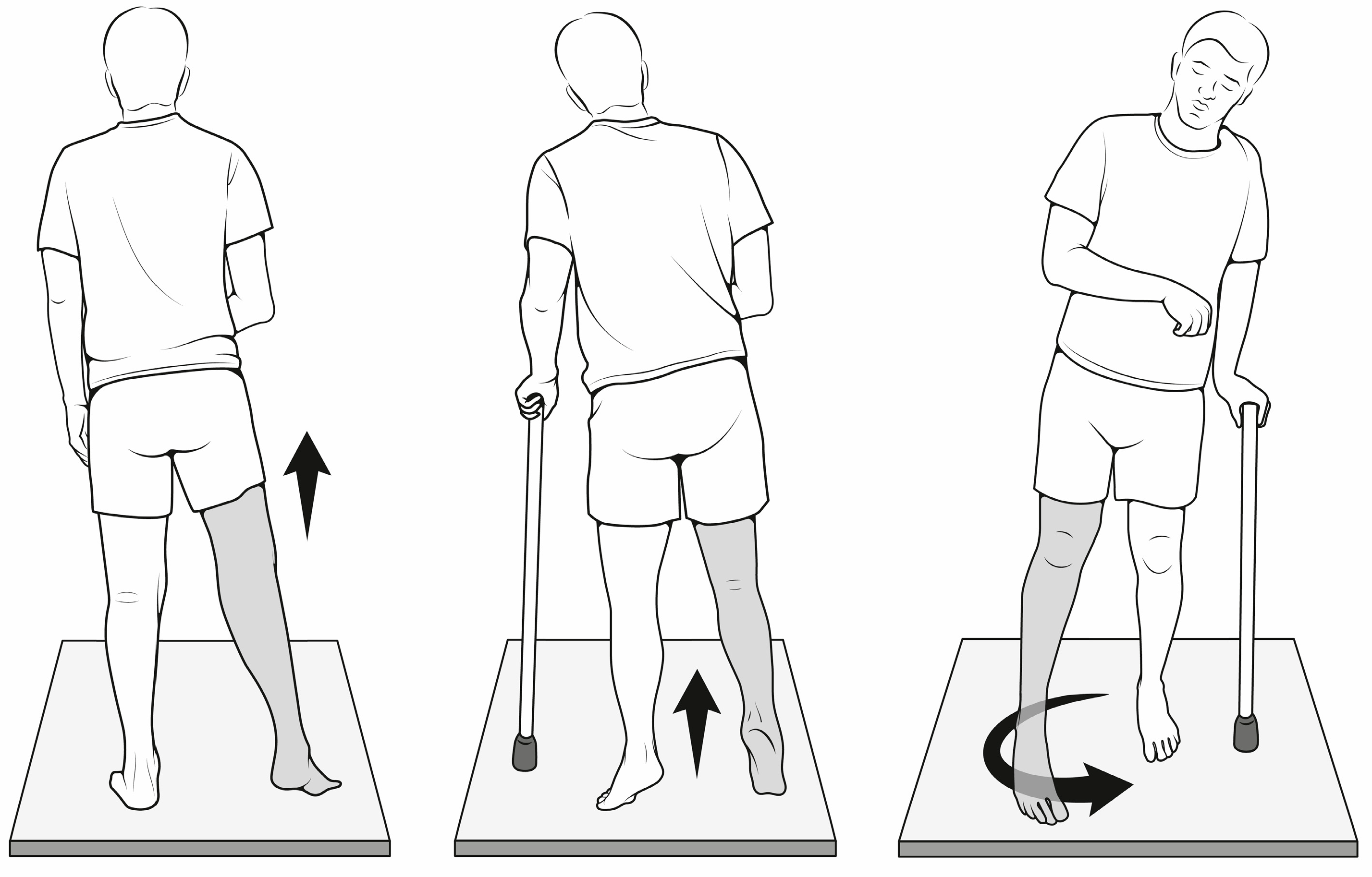
  15. Kerrigan, D.C.; Frates, E.P.; Rogan, S.; Riley, P.O. Hip Hiking and Circumduction. Am. J. Phys. Med. Rehabil. 2000, 79, 247–252. [Google Scholar] [CrossRef]
  16. Cruz, T.H.; Lewek, M.D.; Dhaher, Y.Y. Biomechanical impairments and gait adaptations post-stroke: Multi-factorial associations. J. Biomech. 2009, 42, 1673–1677. [Google Scholar] [CrossRef] [Green Version]
  17. Perry, J.; Burnfield, J. Section Three: Pathological Gait. In Gait Analysis: Normal and Pathological Function, 2nd ed.; SLACK Incorporated: West Deptford, NJ, USA, 2010; pp. 256, 260, 267, 339. [Google Scholar]
  18. Gould, D.; Kelly, D.; Goldstone, L.; Gammon, J. Examining the validity of pressure ulcer risk assessment scales: Developing and using illustrated patient simulations to collect the data INFORMATION POINT: Visual Analogue Scale. J. Clin. Nurs. 2001, 10, 697–706. [Google Scholar] [CrossRef]
  19. Boonstra, A.M.; Preuper, H.R.S.; Reneman, M.F.; Posthumus, J.B.; Stewart, R.E. Reliability and validity of the visual analogue scale for disability in patients with chronic musculoskeletal pain. Int. J. Rehabil. Res. 2008, 31, 165–169. [Google Scholar] [CrossRef] [Green Version]
  20. Demeurisse, G.; Demol, O.; Robaye, E. Motor evaluation in vascular hemiplegia. Eur. Neurol. 1980, 19, 382–389. [Google Scholar] [CrossRef]
  21. Medical Research Council. Aids to the Investigation of Peripheral Nerve Injuries; HMSO: London, UK, 1942. [Google Scholar]
  22. Fayazi, M.; Dehkordi, S.N.; Dadgoo, M.; Salehi, M. Test-retest reliability of Motricity Index strength assessments for lower extremity in post stroke hemiparesis. Med. J. Islam. Repub. Iran 2012, 26, 27–30. [Google Scholar]
  23. Shah, S.; Vanclay, F.; Cooper, B. Improving the sensitivity of the Barthel Index for stroke rehabilitation. J. Clin. Epidemiol. 1989, 42, 703–709. [Google Scholar] [CrossRef] [PubMed]
  24. E Kasner, S. Clinical interpretation and use of stroke scales. Lancet Neurol. 2006, 5, 603–612. [Google Scholar] [CrossRef]
  25. Quinn, T.J.; Dawson, J.; Walters, M.R.; Lees, K.R. Functional Outcome Measures in Contemporary Stroke Trials. Int. J. Stroke 2009, 4, 200–205. [Google Scholar] [CrossRef] [PubMed]
  26. Hsueh, I.-P.; Lee, M.M.; Hsieh, C.-L. Psychometric characteristics of the Barthel activities of daily living index in stroke patients. J. Formos. Med. Assoc. 2001, 100, 526–532. [Google Scholar] [PubMed]
  27. Lincoln, N.B.; Gladman, J. The Extended Activities of Daily Living scale: A further validation. Disabil. Rehabil. 1992, 14, 41–43. [Google Scholar] [CrossRef]
  28. Sarker, S.-J.; Rudd, A.G.; Douiri, A.; Wolfe, C.D. Comparison of 2 Extended Activities of Daily Living Scales with the Barthel Index and Predictors of Their Outcomes. Stroke 2012, 43, 1362–1369. [Google Scholar] [CrossRef] [Green Version]
  29. Green, J.; Young, J. A test-retest reliability study of the Barthel Index, the Rivermead Mobility Index, the Nottingham extended Activities of Daily Living Scale and the Frenchay Activities Index in stroke patients. Disabil. Rehabil. 2001, 23, 670–676. [Google Scholar] [CrossRef]
  30. Leardini, A.; Sawacha, Z.; Paolini, G.; Ingrosso, S.; Nativo, R.; Benedetti, M.G. A new anatomically based protocol for gait analysis in children. Gait Posture 2007, 26, 560–571. [Google Scholar] [CrossRef]
  31. Wu, G.; Cavanagh, P.R. ISB recommendations for standardization in the reporting of kinematic data. J. Biomech. 1995, 28, 1257–1261. [Google Scholar] [CrossRef]
  32. Wu, G.; Siegler, S.; Allard, P.; Kirtley, C.; Leardini, A.; Rosenbaum, D.; Whittle, M.; D’Lima, D.D.; Cristofolini, L.; Witte, H.; et al. ISB recommendation on definitions of joint coordinate system of various joints for the reporting of human joint motion—Part I: Ankle, hip, and spine. J. Biomech. 2002, 35, 543–548. [Google Scholar] [CrossRef]
  33. Cappozzo, A.; Catani, F.; Della Croce, U.; Leardini, A. Position and orientation in space of bones during movement: Anatomical frame definition and determination. Clin. Biomech. 1995, 10, 171–178. [Google Scholar] [CrossRef]
  34. Vive, S.; Elam, C.; Bunketorp-Käll, L. Comfortable and Maximum Gait Speed in Individuals with Chronic Stroke and Community-Dwelling Controls. J. Stroke Cerebrovasc. Dis. 2021, 30, 106023. [Google Scholar] [CrossRef] [PubMed]
  35. Nascimento, L.R.; de Menezes, K.K.P.; Scianni, A.A.; Faria-Fortini, I.; Teixeira-Salmela, L.F. Deficits in motor coordination of the paretic lower limb limit the ability to immediately increase walking speed in individuals with chronic stroke. Braz. J. Phys. Ther. 2019, 24, 496–502. [Google Scholar] [CrossRef]
  36. Patterson, K.K.; Nadkarni, N.K.; Black, S.E.; McIlroy, W.E. Gait symmetry and velocity differ in their relationship to age. Gait Posture 2012, 35, 590–594. [Google Scholar] [CrossRef] [Green Version]
  37. Mizuta, N.; Hasui, N.; Nakatani, T.; Takamura, Y.; Fujii, S.; Tsutsumi, M.; Taguchi, J.; Morioka, S. Walking characteristics including mild motor paralysis and slow walking speed in post-stroke patients. Sci. Rep. 2020, 10, 1–10. [Google Scholar] [CrossRef]
  38. Menezes, K.K.; Nascimento, L.R.; Faria, C.; Avelino, P.R.; A Scianni, A.; Polese, J.C.; Faria-Fortini, I.; Teixeira-Salmela, L.F. Deficits in motor coordination of the paretic lower limb best explained activity limitations after stroke. Physiother. Theory Pr. 2018, 36, 417–423. [Google Scholar] [CrossRef]
  39. Sánchez, N.; Acosta, A.M.; Lopez-Rosado, R.; Stienen, A.H.A.; Dewald, J.P.A. Lower Extremity Motor Impairments in Ambulatory Chronic Hemiparetic Stroke: Evidence for Lower Extremity Weakness and Abnormal Muscle and Joint Torque Coupling Patterns. Neurorehabilit. Neural Repair 2017, 31, 814–826. [Google Scholar] [CrossRef]
  40. Kwan, M.S.-M.; Hassett, L.M.; Ada, L.; Canning, C.G. Relationship between lower limb coordination and walking speed after stroke: An observational study. Braz. J. Phys. Ther. 2018, 23, 527–531. [Google Scholar] [CrossRef]
  41. Hiba, S.; Raphael, Z.; Jonathan, B.; Nicolas, R.; Pauline, G. Co-contraction around the knee and the ankle joints during post-stroke gait. Eur. J. Phys. Rehabil. Med. 2018, 54, 380–387. [Google Scholar] [CrossRef]
  42. Mentiplay, B.F.; Williams, G.; Tan, D.; Adair, B.; Pua, Y.-H.; Bok, C.W.; Bower, K.J.; Cole, M.H.; Ng, Y.S.; Lim, L.S.; et al. Gait Velocity and Joint Power Generation After Stroke. Am. J. Phys. Med. Rehabil. 2018, 98, 841–849. [Google Scholar] [CrossRef]
  43. Aguiar, L.T.; Camargo, L.B.A.; Estarlino, L.D.; Teixeira-Salmela, L.F.; Faria, C.D.C.D.M. Strength of the lower limb and trunk muscles is associated with gait speed in individuals with sub-acute stroke: A cross-sectional study. Braz. J. Phys. Ther. 2018, 22, 459–466. [Google Scholar] [CrossRef] [PubMed]
  44. Dorsch, S.; Ada, L.; Canning, C.G.; Al-Zharani, M.; Dean, C. The Strength of the Ankle Dorsiflexors Has a Significant Contribution to Walking Speed in People Who Can Walk Independently After Stroke: An Observational Study. Arch. Phys. Med. Rehabil. 2012, 93, 1072–1076. [Google Scholar] [CrossRef] [PubMed]
  45. Chang, M.C.; Lee, B.J.; Joo, N.-Y.; Park, D. The parameters of gait analysis related to ambulatory and balance functions in hemiplegic stroke patients: A gait analysis study. BMC Neurol. 2021, 21, 1–8. [Google Scholar] [CrossRef]
  46. Wing, K.; Lynskey, J.; Bosch, P.R. Walking Speed in Stroke Survivors: Considerations for Clinical Practice. Top. Geriatr. Rehabil. 2012, 28, 113–121. [Google Scholar] [CrossRef]
  47. Stuart, M.; Dromerick, A.W.; Macko, R.; Benvenuti, F.; Beamer, B.; Sorkin, J.; Chard, S.; Weinrich, M. Adaptive Physical Activity for Stroke: An Early-Stage Randomized Controlled Trial in the United States. Neurorehabilit. Neural Repair 2019, 33, 668–680. [Google Scholar] [CrossRef]
Figure 1. Compensatory gait patterns allowing paretic limb clearance during swing. From left: hip hiking, vaulting and circumduction.
Applsci 13 00934 g001
Table 1. Sample characteristics.
Age 69.7 (SD 7.1)
Sex (M:F) 17:13
BMI 28.8 (SD 5.2)
Paretic side (R:L) 10:20
Compensatory gait strategy 2 Vaulting
5 Hip hiking
2 Circumduction
4 Vaulting + hip hiking
5 Vaulting + hip hiking + circumduction
11 Non classifiable
VAS 19.7 (SD 24.9)
BI 71.6 (SD 16.4)
MI paretic side 76.5 (SD 25.0)
NEAS 10.3 (SD 4.3)
Table 2. Gait pattern.
PatternN PatientsMean Speed cm/sSD
Vaulting 230.38.7
Hip hiking 528.314.8
Circumduction 258.911.2
Hip hiking + vaulting 443.29.9
Hip hiking + vaulting + circumduction 546.05.1
Non classifiable 1151.523.6
Table 3. Correlation with gait speed.
Joint aROM (n pts)
Hemiplegic Side
MeanSDSpearman Rank Correlation (p)
Hip flexion (30)106.310.1Ns
Hip extension (19)15.05.27Rho 0.480, p = 0.03
Hip Abduction (30)27.9319.9Ns
Hip Adduction (30)15.15.5Ns
Knee flexion (30)121.518.7Ns
Knee extension (20)−9.98.8Ns
Dorsiflexion TT (28)9.32.9Ns
Plantarflexion TT (30)29.310.9Ns
Muscular Strength (n pts)
Hemiplegic side
meanSDSpearman rank correlation (p)
Quadriceps (30)72.431.3Rho 0.604, p = 0.001
Ilio-psoas (29)57.326.6Rho 0.358, p = 0.061
Tibialis anterior (10)25.513.3Ns
Gluteus Maximus (15)34.115.4Ns
Gluteus Medius (22)55.423.7Ns
Biceps femoris (17)32.417.2Ns
Semitendinosus (20)34.718.2Ns
Table 4. Correlation between gait speed (cm/s) and time–distance parameters.
MeanSDSpearman Rank Correlation
Stance time (% stride) Plegic
Unaffected
65.7
72.5
6.6
5.4
Rho −0.631, p < 0.000
Rho −0.865, p < 0.000
Stride lenght normalized (%h) 38.110.8Rho 0.879, p < 0.000
Cycle time 1.50.4Rho 0.568, p = 0.001
Cadence (stride/min) 42.610.7Rho 0.561, p = 0.002
Disclaimer/Publisher’s Note: The statements, opinions and data contained in all publications are solely those of the individual author(s) and contributor(s) and not of MDPI and/or the editor(s). MDPI and/or the editor(s) disclaim responsibility for any injury to people or property resulting from any ideas, methods, instructions or products referred to in the content.

