Article

Growth of Escherichia coli in Human Milk and Powdered Infant Formula under Various Treatments and Feeding Conditions in Neonatal Units

by
María Carmen López-Mendoza
1,*,
Marta Lozano
1,
Edgar García-Romero
1,
Pilar Ruiz-García
1,
María Gormaz
2,
Rafael Jordano
3,
Luis Manuel Medina
3,
Amparo Ramón-Beltrán
2 and
Dolores Silvestre
4
1
Department of Animal Production and Food Science and Technology, University CEU-Cardenal Herrera, CEU Universities, C/Tirant lo Blanc 7, 46115 Valencia, Spain
2
Division of Neonatology, University and Polytechnic Hospital La Fe, Avenida Fernando Abril Martorell, 106, 46026 Valencia, Spain
3
Department of Food Science and Technology, University of Córdoba, Campus of Rabanales, Ctra. Madrid-Cádiz km. 396, 14071 Córdoba, Spain
4
Department of Pharmacy, University CEU-Cardenal Herrera, CEU Universities, C/Lluis Vives 1, 46115 Valencia, Spain
*
Author to whom correspondence should be addressed.
Appl. Sci. 2023, 13(15), 8978; https://doi.org/10.3390/app13158978
Submission received: 12 May 2023 / Revised: 28 July 2023 / Accepted: 3 August 2023 / Published: 4 August 2023
(This article belongs to the Special Issue Microbiological Contamination: Latest Advances and Prospects)

Abstract

:
Milk supplied to neonates in neonatal units is kept at room temperature for some time, which could influence microbial growth. This study aims to evaluate the growth of Escherichia coli in HM and PIF under various treatments and conditions, as well as to determine the influence of different thawing methods on microbial growth in HM. The number of E. coli generations appearing over a 4 h period at 22 °C in HM (frozen; frozen and pasteurized; and frozen, pasteurized, and fortified) and in PIF (four brands) was determined. E. coli counts in HM inoculated and thawed using different methods were also compared. In frozen HM and in pasteurized and frozen HM, significant differences were found after 2.5 h and 1.5 h, respectively. In PIF, differences were found between 1.5 and 3 h. With regard to the thawing process, the lowest microorganism counts were obtained at 4 °C overnight; thus, it seems advisable to store milk at room temperature for a maximum of 1 h during administration in neonatal units. Thawing HM at 4 °C overnight should be the method of choice.

1. Introduction

Human milk (HM) is the preferred food for infants, at least for the first six months of life, since it is one of the most effective ways to ensure children’s health and survival [1]. In addition to its optimal nutrient supply, HM transfers immune factors to protect newborns during their first years of life. For this reason, breastfeeding is the preferred method for feeding infants, including those who are preterm and hospitalized.
In the case of preterm infants admitted to neonatal intensive care units (NICUs), it is often not possible to directly breastfeed the infant; therefore, bottle feeding or feeding through an enteral feeding tube are used instead [2]. If the mother’s milk is insufficient or unavailable, donated HM can be used; thus, many hospitals have incorporated human milk banks (HMBs) in their facilities. In these facilities, milk usually undergoes different processes that can vary between the different HMBs. In general, some steps are common to all HMBs: freezing, thawing, and Holder pasteurization [3,4,5]. Lastly, before administration, milk is fortified with proteins, fats, minerals, and vitamins that are necessary in the case of preterm infants [6]. With regard to thawing, the recommended methods from different organizations include carrying thawing out overnight under refrigeration, at room temperature, or in a thermal bath [3,4]. In some of these methods, the milk remains at a temperature that could allow microbial growth for a considerable period of time; therefore, it is necessary to evaluate the different methods in relation to this issue.
In addition, it must be considered that treatments such as freezing and pasteurization can affect the antimicrobial capacity of milk [7,8,9]. Preterm infants are an extremely vulnerable population; therefore, it is essential to control hygiene during all treatments and handling of HM, as well as powdered infant formula (PIF), which is used as an alternative in some cases [8].
Finally, during the milk administration process (both HM and PIF) through an enteral feeding tube in the NICU, milk is kept at room temperature for some time. Guidelines from various countries [10,11] and several authors [5,12,13] recommend that milk delivered through enteral feeding tubes should be kept for a maximum of 4 h and containers must also be changed every 4 h. However, the United States Centers for Diseases Control and Prevention [14] recommend keeping thawed HM at room temperature for a maximum of 1–2 h at 25 °C. These differences in recommendations justify the need to obtain experimental information about the microbial growth in HM and PIF over time, under the conditions of administration in NICUs, to rationalize these recommendations. The holding time of milk at room temperatures is key, because if it exceeds the duration of the lag phase of a microorganism, it can lead to undesirable microbial growth. Among the different microorganisms that can contaminate milk, Escherichia coli is one of the most common pathogens that causes neonatal infections [15,16], and is a cause of outbreaks due to the consumption of unpasteurized human milk in NICUs [17,18]. The growth of E. coli in HM and PIF and its handling practices have implications for neonatal health and the management of neonatal units. Therefore, it is of interest to study the maximum time that milk can be kept at room temperature during its administration in NICUs without posing a risk of possible microbial growth.
Furthermore, evidence-based standards for recommending the optimal HM thawing method and feeding temperature for infants are limited [19].
This study aims to evaluate the growth of E. coli in HM and PIF under various treatments and conditions, as well as to determine the influence of different thawing methods on microbial growth in HM.

2. Materials and Methods

2.1. Sample Collection

For this research, HM samples donated to the HMB of the University and Polytechnic Hospital La Fe, Valencia (Spain) were used. In this HMB, milk is donated by lactating mothers voluntarily and altruistically, without any financial compensation [20]. The samples were collected from January to April 2018. A total of 42 donors participated in this study. All the participants gave their informed consent for their milk to be used in this study. The study was conducted according to the guidelines of the Declaration of Helsinki [21] and was approved by the Research and Ethics Committee of the Instituto de Investigación Sanitaria La Fe.
In order to ensure sample homogeneity, inclusion criteria for the participants were considered: lactating mothers of any age, with a lactation period not exceeding 30 days, with healthy habits (Mediterranean diet, moderate physical activity of at least 30 min per day), and negative results in the microbiological screening tests performed during the donor selection process. Donors were excluded from the study if they met any of the following criteria: currently under medical treatment, having restrictive diets or known addictions (including smokers), having sleep-related disorders, or having a lactation period shorter than 30 days.
Milk was expressed at home by the donors following a standardized protocol of extraction and conservation, which included hand washing with soap and drying before extraction. The milk was expressed with sterile material provided by the HMB, using an electric (Lactina, Medela, Switzerland) or manual (Harmony, Medela, Switzerland) breast pump with sterile parts attached to a sterile polypropylene container with a hermetic closure, thereby eliminating the risk of accidental contamination. After each use, the extraction equipment was washed with water and detergent and disinfected in the microwave, in sterilization bags (Quick Clean, Medela). The complete milk extraction from one breast was always obtained to take advantage of all the properties of the milk, as its composition varies throughout the extraction process. The frequency of donation was weekly. The milk samples were immediately frozen at −20 °C for transport to the HMB. Once there, the milk was stored at the same temperature until it was transported to the laboratories for processing, always maintaining the cold chain and sterile conditions. Once at the laboratories, the milk was kept under frozen conditions until analysis. No more than 24 h elapsed between the sample collection and the start of sample processing.
In the case of PIF, 5 containers of 4 different brands purchased at different pharmacies were utilized. The brands were randomly chosen from among the most popular ones, and all the brands complied with the regulations regarding nutritional composition [22] and included docosahexaenoic acid and arachidonic acid. The bovine fortifier was provided by the HMB.
The sample size was calculated based on a previous study, using mean and standard deviation of the counts (CFU/mL) of an enterobacteria (Cronobacter sakazakii) reported from inoculated HM and PIF (power = 95% and significance level = 0.05) [23].

2.2. Study of Growth of E. coli in HM and PIF Held at 22 °C for 4 h

For this study, a total of 12 mature HM samples of 60 mL from 12 participants were used. A bovine fortifier (PreNAM-FM 85, Nestlé®, Vevey, Switzerland) was also used, with an iron concentration of 26 mg/100 g, which was prepared and added in accordance with the manufacturer’s specifications.
The same procedure was carried out for the PIF, for which 5 samples of 4 different brands (A, B, C, and D) were used. All the brands studied contained docosahexaenoic acid (DHA) and arachidonic acid (ARA). These samples were prepared with sterile water following the manufacturer’s specifications, in a volume of 60 mL for each sample of each brand.
Once in the laboratory, the HM samples were thawed in a refrigerator (12 h at 4 °C) and all of them were divided into 3 aliquots of 20 mL to be used for each treatment: (A) milk frozen for 3 months; (B) milk with Holder pasteurization (62 °C for 30 min) and frozen for 3 months; (C) milk with Holder pasteurization, frozen for 3 months, and fortified by adding bovine fortifier after thawing.
Each aliquot of PIF and HM, once subjected to the indicated treatments, was inoculated with a concentration of 104 CFU/mL of E. coli NCTC 9111 serovar 0111:K58 (B4):H. The bacteria were cultured overnight on plate count agar (PCA) (Scharlab®, Barcelona, Spain). After incubation, one or two colonies were selected and suspended in 0.1% peptone water (Scharlab®, Barcelona, Spain), adjusting the concentration to an absorbance of 0.145 at 546 nm (approximately 3 × 10⁸ CFU/mL) using a Jenway 7200 spectrophotometer (reliability: ±0.005 A/h at 0.04 A; validity: ±0.01 A/h at 1.0 A). Three decimal dilutions were made to reach a concentration of 3 × 10⁵ CFU/mL. Finally, 0.7 mL and 2 mL of this dilution were added to each aliquot of HM and PIF, respectively, to reach a final concentration of 104 CFU/mL.
After inoculation, the samples were kept at 22 °C for 4 h, simulating one of the possible conditions for the milk supply in neonatal units. During this period, E. coli counts were carried out at 0 h, 0.5 h, 1 h, 1.5 h, 2 h, 2.5 h, 3 h, 3.5 h, and 4 h.
To obtain the E. coli counts in HM and PIF at different times, 1 mL was taken from each milk sample. The corresponding decimal dilutions were made in 0.1% peptone broth (Scharlab®, Barcelona, Spain) and the plates were seeded via duplication in violet red bile agar (VRBA, Scharlab®, Barcelona, Spain), which was incubated at 37 °C for 24 h [24].

2.3. Study of the Influence of Different Thawing Methods on the Growth of E. coli in HM

In this study, a total volume of 5000 mL of HM from 30 donors was used. Each donor contributed about 150–200 mL of milk. The HM samples were thawed at 4 °C for 12 h. The samples from three participants were mixed and 10 pools of 500 mL each were prepared. Each pool was distributed in 4 aliquots of 125 mL. This is the common storing volume for frozen milk in the HMB, which must be thawed later. Each aliquot was inoculated with a concentration of 104 CFU/mL of E. coli NCTC 9111 serovar 0111:K58 (B4):H, obtained as described above. Subsequently, all the aliquots were frozen at −20 °C for 7 days. Once the 7 days had elapsed, the aliquots were thawed using four different methods: (A) room temperature (22 °C) for 3 h; (B) thermostatic bath at 35 °C for 30 min; (C) thermostatic bath at 25 °C for 40 min; and (D) refrigeration at 4 °C for 12 h. All four thawing methods were applied at the same time on different aliquots of the same sample. After these treatments, an E. coli count was performed.
The count of E. coli was carried out by taking 1 mL of each milk sample, performing the corresponding decimal dilutions in 0.1% peptone broth (Scharlab®, Barcelona, Spain) and plate seeding via duplication in violet red bile agar (VRBA, Scharlab®, Barcelona, Spain), which was incubated at 37 °C for 24 h [24].

2.4. Data Analysis

In the study of the growth of E. coli in HM and PIF, after obtaining E. coli counts at each time point, the number of E. coli generations (n) appearing at a given time was calculated in the different sample types. One generation is considered a microorganism count doubling; thus, the calculation was as follows: n = (log N − logN0)/0.301, where N is the count (CFU/mL) at that given time and N0 is the baseline microorganism count. The generation time (g), or the time it takes for a microbial population to double, was also calculated for each type of milk: g = t/n, where t is the time elapsed (240 min in our study) and n is the generation number that appeared during that time [25].
In the study of different thawing methods, E. coli counts were obtained for each of the thawed samples according to the four methods used. The results were obtained in CFU/mL and were transformed to log10 CFU/mL.
The statistical analysis of the results was performed using IBM SPSS software (Version 27). The differences were considered statistically significant at p < 0.05. A mixed analysis of variance (ANOVA) was used to study the effect of maintaining milk in the neonatal room conditions on the generations that appeared at the different times, and to determine whether any change in the number of E. coli generations was the result of the interaction between the type of milk (different treatments or different brands) and time. For HM, the two factors considered were the milk treatment (between-subjects factor), with three levels for HM (frozen milk; pasteurized and frozen milk; pasteurized, frozen and fortified milk), and the time (within-subjects factor), with eight levels (0 h, 0.5 h, 1 h, 1.5 h, 2 h, 2.5 h, 3 h, 3.5 h, and 4 h). For PIF, the two factors considered were the milk brand (between-subjects factor), with four levels, and the time (within-subjects factor), with eight levels (0 h, 0.5 h, 1 h, 1.5 h, 2 h, 2.5 h, 3 h, 3.5 h, and 4 h).
To compare the results of the number of generations obtained in HM to PIF, a mixed ANOVA was also used. One factor was the milk type (between-subjects factor), with two levels (powdered infant formula and human milk), and the other factor was time (within-subjects factor), with eight levels (0 h, 0.5 h, 1 h, 1.5 h, 2 h, 2.5 h, 3 h, 3.5 h, and 4 h).
To determine the differences between the groups, a post hoc analysis was used with the Bonferroni adjustment test. To verify the data normality, the Kolmogorov–Smirnov test was used (p > 0.05). The homogeneity of the variance assumption within the subjects and between the subjects was examined using Mauchly’s test of sphericity (p > 0.05) and Levene’s test (p > 0.05), respectively. In the cases where the sphericity hypothesis was not met, the multivariate adjustment was applied.
For the comparison of E. coli generation times in the HM subjected to different treatments, a one-way ANOVA was used. Similarly, a one-way ANOVA was performed to compare the generation times obtained in the different brands of PIF.
To evaluate the thawing methods, a one-way repeated-measures ANOVA with four levels (thawing at room temperature (20 °C) for 2 h; in a thermostatic bath at 35 °C for 30 min; in a thermostatic bath at 25 °C for 40 min; and refrigerated at 4 °C for 12 h) was performed.

3. Results

3.1. Study of Growth of E. coli in HM and PIF Held at 22 °C for 4 h

The results of the growth of E. coli obtained after maintaining HM and PIF under neonatal nursery conditions, at 22 °C for 4 h, are presented below.
Table 1 shows E. coli’s growth evolution in HM subjected to different treatments and maintained at 22 °C for 4 h, expressed as the number of generations that appeared at each time, and the generation time (time it takes for the E. coli population to double) for each type of milk treated.
The mixed ANOVA showed that there were significant differences between the number of generations obtained at different times and that these differences were not the same for each type of HM treated. Regarding the generation number obtained at different times, for the frozen milk (treatment A), significant differences were found after 2.5 h. In the case of frozen and pasteurized milk (treatment B), and frozen, pasteurized, and fortified milk (treatment C), differences in the generations were detected at 1.5 h and 2 h, respectively.
For the generation time, the one-way ANOVA showed that there were significant differences between the generation times obtained in the HM subjected to the different treatments (Table 1). Specifically, there were differences between frozen milk and frozen and pasteurized milk, with the generation time being shorter in the case of frozen and pasteurized milk (126.65 min).
Table 2 shows E. coli’s growth evolution in PIF maintained at 22 °C for 4 h, expressed as the number of generations that appeared at each time and the generation time for each brand.
The mixed ANOVA showed that there were significant differences between the number of generations obtained at different times and that these differences were not the same for each brand of PIF. The times from which significant differences in the number of generations were obtained were 3 h for brands A, C, and D, and 1.5 h for brand B.
Regarding the generation times, the one-way ANOVA showed that there were significant differences between the different milk brands, obtaining the greatest generation time for brand A (313.61 min) and the shortest for brand D (179.24 min).
The comparative study between both milk types (HM and PIF) did not show significant differences (p = 0.053) in the generations obtained at different times between the two milk types studied (Table 3).

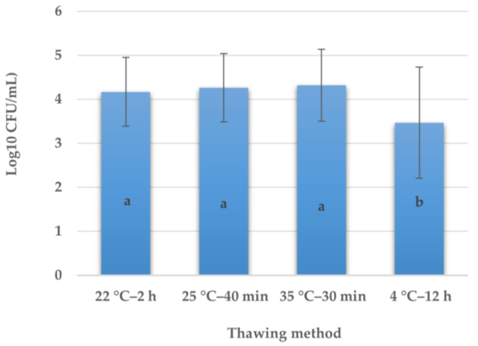
3.2. Influence of Different Thawing Methods on the Development of E. coli in HM

The analysis of variance for E. coli counts obtained after the different thawing methods of inoculated HM indicates that there were significant differences between the different methods evaluated (Figure 1). The lowest counts were obtained when thawing at 4 °C for 12 h, with significant differences (p = 0.03) being obtained compared to all the other studied thawing methods.

4. Discussion

Various guidelines [10,11] and authors [5,12,13,26] recommend that the maximum residence time of milk administered to infants under nursery conditions (20–22 °C) should be 4 h. However, the results obtained in the present study showed that under conditions of E. coli contamination of 104 CFU/mL, there were differences in the counts at earlier time points. In the case of frozen HM, count differences were obtained after 2.5 h, and for frozen and pasteurized milk, the times were reduced to 1.5 h. It must be considered that authors such as Lemons [26], who is used as a bibliographic reference in some guides, such as the British Dietetic Association [10], carried out a study with fresh HM and frozen HM for six weeks and their counts were only performed from natural contamination. However, in the present study, for frozen HM, the count differences were obtained after 2.5 h, but the freezing time was longer (3 months), and E. coli inoculation was also performed to obtain homogeneity in the contamination of the samples. In addition, it was of interest to study the microbial behavior in milk under conditions of potential contamination.
The obtained results are consistent with the United States CDC recommendation [14], which states that HM should not be kept thawed for more than 1–2 h at a temperature of 25 °C. Otherwise, Handa [27] found a significant increase in the bacterial count of HM that was frozen for 7 d at −20 °C, thawed, refrigerated for 24 h, heated, and kept at room temperature for 4 h; thus, these authors propose reducing this time to 3 h and recommend re-evaluating the possibility of keeping milk at room temperature. This is consistent with the results obtained in the present study, as we also obtained increases in the milk counts before 4 h.
Moreover, it is important to consider the freezing time, as longer times (3 months) affect the antimicrobial capacity of the HM [8] and could allow for faster microbial growth under NICU conditions.
Gao [6] also obtained a significant increase in E. coli counts in HM that was previously frozen and maintained at 37 °C for 24 h. At 6 h and 24 h, E. coli increased its number 107-fold and 109-fold, respectively. In this case, microbial growth was very high at 6 h, but it must be taken into account that the holding temperature was 37 °C.
In HMBs, milk is frozen for long periods for a maximum of two times (before and after pasteurization), but it is also pasteurized. According to the results of the present work, the E. coli generation times were significantly reduced (p = 0.012) when the milk was frozen and pasteurized; thus, the time at which the differences in the counts were obtained was reduced to 1.5 h. Various authors [7,8] have shown that pasteurization significantly reduces the bactericidal capacity of HM, and according to the studies by Paulaviciene [28] and Arroyo [29], pasteurization and freezing/thawing processes cause a significant loss of lactoferrin, lysozyme, IgG, and IgA, components with important antibacterial activity, which could explain the reduction of this capacity in HM subjected to these treatments. It is evident that the pasteurization process ensures the absence of pathogenic microorganisms in the milk that is supplied to the infant, but if milk contamination occurs during subsequent handling, microorganisms could multiply when the milk supply is kept at room temperature. In addition, feeding tubes can contain high amounts of potentially pathogenic and antibiotic-resistant bacteria, which could develop under NICU feeding conditions [2,30]. It is important, therefore, to maximize hygiene conditions throughout milk processing and administration and to re-evaluate the timing of the milk supply. It seems key, therefore, to reduce the time of milk administration to less than the 4 h recommended in several guidelines.
Regarding fortification, in the present study, no significant differences were found in the E. coli generation time between HM with and without fortifier. These results are consistent with those of Telang [31], who found no differences in microbial growth at 22 °C for 6 h between fortified milk (with iron-rich and iron-poor fortifiers) and unfortified milk. However, these authors did not observe significant microbial growth during those 6 h in any of the milk types studied. This difference, as compared to our results, may be due to the fact that the HM used was fresh, not frozen or pasteurized, and they also inoculated another microorganism (Cronobacter sakazakii). As indicated above, milk treatments seem to be determinants of possible microbial growth.
Different results were obtained by Lenati [23], who investigated the growth of different strains of C. sakazakii in HM previously frozen at −80 °C, fortified (1.44 mg of iron/100 mL), and unfortified, kept at 23 °C for 24 h. In this case, the obtained results differed depending on the investigated strain; therefore, differences in growth were obtained for some strains but not for others. This indicates that the type of microorganism also plays a role, which may justify the differences compared to this study, since in the present study, E. coli was used. In addition, recent studies have shown that lactoferrin’s antibacterial capacity is not totally dependent on its iron saturation. This capacity is rather related to direct interaction between lactoferrin and bacteria [32]. In addition, in HM, there are many other components with antimicrobial capacity, such as oligosaccharides, lipids (e.g., glycerol monolaurate), other proteins (casein201), peptides, and enzymes (e.g., lysozymes, peroxidases, and xanthine oxidase) [33,34,35,36,37].
For PIF and with regard to the time at which differences appear in E. coli generations, variable results were obtained; these times ranged between 1.5 and 3 h, depending on the milk brand. Gao [6] obtained similar results in PIF with 103 CFU/mL of E. coli added, although in this study, the incubation temperature was 37 °C. At this temperature, the authors detected rapid microorganism growth, whose number was 10 times and 104 times greater at 2 h and at 4 h, respectively. In our case, the microbial growth was not as fast, but the holding temperature was also lower (22 °C).
If the generation times obtained in PIF are compared with those of HM, it can be observed that there are no significant differences between them. This is a possible consequence of human milk antimicrobial capacity loss due to the applied treatments. Otherwise, Gao [6] observed significant differences in the E. coli counts obtained in PIF and HM maintained at 37 °C for 24 h at all the evaluated time points. In this case, the HM used was frozen at −20 °C (no freezing time was indicated) but not pasteurized, which, as shown above, seems to affect the HM antimicrobial capacity.
In summary, in both the treated HM and PIF, the results obtained show that differences in E. coli counts are obtained before 4 h. Consequently, new studies are needed to evaluate microbial growth in milk under NICU administration conditions, considering different microbial loads and with other microorganisms of interest in this food, such as C. sakazakii. This bacterium has posed an emerging danger in recent years in the feeding of infants, both in PIF and improperly handled HM [38,39].
The thawing process is also an important phase of HM processing before neonate administration, as it provides another opportunity for contamination or microbial growth. The obtained results showed that there were significant differences between the evaluated thawing methods, obtaining the smallest count increase for thawing at 4 °C for 12 h. Of the studied methods, it was the only one in which the milk remained at refrigeration temperatures that inhibit microbial growth throughout. No differences were found between the other three studied methods (room temperature and warm baths at 25 °C and 37 °C), which is consistent with other studies. For example, Handa [27] found no differences between the total bacterial counts obtained in thawed HM via two different thawing methods (tepid water at 37 °C for 20 min and a waterless warmer at 37 °C for the required time depending on milk volume). It should be considered that treatments involving a rapid rise in temperature in the outer layer of the milk could favor microbial growth in the area where the temperature rises faster and is maintained until the inside thaws. Moreover, Li [19] investigated the effect on the presence of immunoglobulin A and lysozyme of thawing HM in a refrigerator at 4 °C for 12 h and using warm water at 25 °C and 37 °C for 15 min. As a result, they obtained greater IgA and lysozyme preservation when thawing HM at 4 °C than thawing in warm water at 37 °C, while thawing in warm water at 25 °C is the method that produced a greater reduction in both biocomponent levels. In light of this, it seems that the thawing method can affect different milk components that provide its antibacterial capacity. Likewise, Arroyo [29] studied the effect of HM processing on its IgA, IgM, and lactoferrin concentrations. Specifically, they applied the treatments used in the HMB to HM, namely, initial freezing/thawing, pasteurization, and second freezing/thawing, and found significant differences in the amount of IgM after the second HM freezing/thawing. These authors did not indicate the thawing method used, but these results indicate that freezing/thawing processes affect various biocomponents of HM that provide its antimicrobial capacity and, therefore, can affect microorganism growth in the product.
To summarize, it seems necessary to re-evaluate the timings of milk administration to infants through an enteral feeding tube in the NICU, considering the previous treatments that have been performed on the milk. It would also be of interest to test the behavior of other pathogenic microorganisms of interest in HM, such as C. sakazakki, and to use different levels of contamination. A relatively high level of contamination (104 CFU/mL) was chosen in this study, but it would be interesting to study what happens at lower levels (10–100 CFU/mL). Finally, future research on HM and PIF without inoculation and with other types of HM (colostrum and transition) are needed.

4.1. Significance and Practical Application of Results

This study has attempted to simulate the possible contamination of milk with the microorganism E. coli, which is widely used in food as an indicator microorganism and is among the most frequent contaminants of food. The treatments to which the HM was subjected for the present study are those normally used in HMBs, and allow us to evaluate the effect of possible contamination on the type of milk that is administered to the newborn.
This study warns of the risk to neonatal health posed by certain practices carried out during feeding in NICUs. Limited studies have been published that discuss the influence of milk holding times under NICU conditions (temperature and holding time) on microbial growth. The present study provides information on the growth of E. coli under certain conditions (22 °C during 4 h) in HM subjected to various treatments, which may help to re-evaluate milk holding times. It also demonstrates the importance of avoiding contamination of milk during handling, since microorganisms such as E. coli easily develop in milk. These conclusions allow for immediate clinical application.

4.2. Limitations

The results obtained in this study must be interpreted in the context of the limitations they present. These results can only be applied to HM obtained from a certain profile of participants as described above. The conclusions are valid only for one microorganism (E. coli) and under the specified conditions. Milk inoculation with E. coli was carried out in a specified amount (104 CFU/mL), and the growth of this microorganism could be different according to different inoculation doses or without inoculation. Moreover, this study was carried out with mature HM; therefore, we cannot draw conclusions about the evolution of E. coli growth in transition milk or colostrum. Finally, only four PIF brands were evaluated, and differences were found between them. Microorganism growth in other brands could be different.

5. Conclusions

According to the obtained results, it does not seem advisable to keep milk at room temperature for 4 h during its enteral feeding tube administration in the NICU. The maximum holding times for milk in these conditions seem to be conditioned by the previous treatment performed on the HM and the infant formula brand. It would be advisable to avoid keeping milk to be administered to neonates in the NICU for more than 1 h at room temperature, since microbial growth could be present after that time. However, to avoid having to discard a product of such high value, the volume of the milk doses should be adapted to the timing of the milk supply. Hygienic conditions during milk handling are essential because if milk is contaminated, microbial growth under NICU conditions could pose a risk. As for milk thawing, we feel that it might be wise to perform this process in the refrigerator at 4 °C overnight.

Author Contributions

Conceptualization, M.C.L.-M., M.G. and D.S.; methodology, M.C.L.-M. and M.L.; software, M.C.L.-M. and P.R.-G.; validation, D.S. and M.C.L.-M.; formal analysis, M.C.L.-M. and P.R.-G.; investigation, M.L. and E.G.-R.; resources, M.G. and A.R.-B.; data curation, M.L., E.G.-R. and A.R.-B.; writing—original draft preparation, M.C.L.-M. and P.R.-G.; writing—review and editing, D.S. and M.G.; visualization, L.M.M. and R.J.; supervision, L.M.M. and R.J.; project administration, M.C.L.-M.; funding acquisition, M.C.L.-M. All authors have read and agreed to the published version of the manuscript.

Funding

This research was funded by University CEU-Cardenal Herrera, grant number IDOC 1606.

Institutional Review Board Statement

The study was conducted in accordance with the Declaration of Helsinki and was approved by the Research and Ethics Committee of the Instituto de Investigación Sanitaria La Fe, Valencia, Spain (protocol code 0524, 11 July 2017).

Informed Consent Statement

Informed consent was obtained from all the subjects involved in the study.

Data Availability Statement

The data presented in this study are available on request from the corresponding author. The data are not publicity available due to data protection regulations.

Acknowledgments

All the equipment used in this study belongs to CEU-Cardenal Herrera University.

Conflicts of Interest

The authors declare no conflict of interest.

References

  1. World Health Organization. Breastfeeding. Available online: https://www.who.int/health-topics/breastfeeding (accessed on 16 January 2023).
  2. Taft, D.H.; Salinero, L.K.; Vongbhavit, K.; Kalanetra, K.M.; Masarweh, C.; Yu, A.; Underwood, M.A.; Mills, D.A. Bacterial colonization and antimicrobial resistance genes in neonatal enteral feeding tubes. FEMS Microbiol. Ecol. 2019, 95, fiz039. [Google Scholar] [CrossRef]
  3. Calvo, J.; García, N.R.; Gormaz, M.; Peña, M.; Martínez, M.J.; Ortiz, P.; Brull, J.M.; Samaniego, C.M.; Gaya, A. Recomendaciones para la creación y el funcionamiento de los bancos de leche materna en España [Recommendations for the creation and operation of maternal milk banks in Spain]. An. Pediatr. 2018, 89, 65.e1–65.e6. [Google Scholar] [CrossRef] [PubMed]
  4. Scott, H.; Sweet, L.; Strauch, L.; Muller, A. Expressed breastmilk handling and storage guidelines available to mothers in the community: A scoping review. Women Birth 2020, 33, 426–432. [Google Scholar] [CrossRef] [PubMed]
  5. Steele, C.; Collins, E. (Eds.) Infant and Pediatric Feedings: Guidelines for Preparation of Human Milk and Formula in Health Care Facilities, 3rd ed.; Academy of Nutrition and Dietetics: Chicago, IL, USA, 2018; pp. 1–248. [Google Scholar]
  6. Gao, X.; Li, Y.; Olin, A.B.; Nguyen, D.N. Fortification with bovine colostrum enhances antibacterial activity of human milk. J. Parenter. Enter. Nutr. 2021, 45, 1417–1424. [Google Scholar] [CrossRef] [PubMed]
  7. Barbarska, O.; Strom, K.; Oledzka, G.; Calvo, J.; Gayà, A.; López-Mendoza, M.C.; Rutkowska, M.; Rosiak, E.; Wesolowska, A.M. Effect of nonthermal processing on human milk bactericidal activity against Escherichia coli. J. Pediatr. Gastroenterol. Nutr. 2020, 70, 864–867. [Google Scholar] [CrossRef]
  8. Fernández-Pastor, S.; Silvestre, D.; López-Mendoza, M.C. Stability of the antimicrobial capacity of human milk against Cronobacter sakazakii during handling. J. Hum. Lact. 2021, 37, 139–146. [Google Scholar] [CrossRef]
  9. Schlotterer, H.R.; Parvez, B.; Perrin, M.T. The Effects of Fortification and Refrigerated Storage on Bioactive Proteins in Holder-pasteurized Donor Human Milk. J. Pediatr. Gastroenterol. Nutr. 2019, 69, 370–374. [Google Scholar] [CrossRef]
  10. British Dietetic Association. Guidelines for the Preparation and Handling of Expressed and Donor Breast Milk and Specialist Feeds for Infants and Children in Neonatal and Paediatric Health Care Settings. Available online: https://www.bda.uk.com/uploads/assets/913a1f78-c805-42c1-8d85e37ca75e0fc0/2019sfuguidelines.pdf (accessed on 16 January 2023).
  11. PATH. Strengthening Human Milk Banking: A Resource Toolkit for Establishing and Integrating Human Milk Bank Programs—A Global Implementation Framework. Version 2.0; PATH: Seattle, WA, USA, 2019; Available online: https://www.aeblh.org/images/publicaciones/2019/20191009_path_hmb_toolkit_0.global_implementation_framework.pdf (accessed on 16 January 2023).
  12. Eglash, A.; Simon, L. ABM Clinical Protocol #8: Human milk storage information for home use for full-term infants, Revised 2017. Breastfeed. Med. 2017, 12, 390–395, Erratum in Breastfeed. Med. 2018, 13, 459. [Google Scholar]
  13. Boullata, J.I.; Carrera, A.L.; Harvey, L.; Escuro, A.A.; Hudson, L.; Mays, A.; McGinnis, C.; Wessel, J.J.; Bajpai, S.; Beebe, M.L.; et al. ASPEN safe practices for enteral nutrition therapy. J. Parenter. Enter. Nutr. 2017, 41, 15–103. [Google Scholar] [CrossRef] [Green Version]
  14. United States Centers for Diseases Control and Prevention. Proper Storage and Preparation of Breast Milk. Available online: https://www.cdc.gov/breastfeeding/recommendations/handling_breastmilk.htm (accessed on 1 March 2023).
  15. Flannery, D.D.; Akinboyo, I.C.; Mukhopadhyay, S.; Tribble, A.C.; Song, L.; Chen, F.; Li, Y.; Gerber, J.S.; Puopolo, K.M. Antibiotic susceptibility of Escherichia coli among infants admitted to neonatal intensive care units across the US from 2009 to 2017. JAMA Pediatr. 2021, 175, 168–175. [Google Scholar] [CrossRef]
  16. Xiao, R.; Li, Y.; Liu, X.; Ding, Y.; Lai, J.; Li, Y.; Kang, W.; Zou, P.; Wang, J.; Du, Y.; et al. Antibiotic susceptibility of Escherichia coli isolated from neonates admitted to neonatal intensive care units across China from 2015 to 2020. Front. Cell. Infect. Microbiol. 2023, 13, 1183736. [Google Scholar] [CrossRef] [PubMed]
  17. Chishiki, M.; Nishiyama, K.; Suzutani, T.; Hiruta, S.; Ichikawa, H.; Haneda, K.; Maeda, H.; Shimizu, H.; Kanai, Y.; Ogasawara, K.; et al. Sterilization efficacy of a new water-free breast milk pasteurizer. Pediatr. Int. 2023, 65, e15574. [Google Scholar] [CrossRef] [PubMed]
  18. Nakamura, K.; Kaneko, M.; Abe, Y.; Yamamoto, N.; Mori, H.; Yoshida, A.; Ohashi, K.; Miura, S.; Yang, T.T.; Momoi, N.; et al. Outbreak of extended-spectrum β-lactamase-producing Escherichia coli transmitted through breast milk sharing in a neonatal intensive care unit. J. Hosp. Infect. 2016, 92, 42–46. [Google Scholar] [CrossRef] [PubMed]
  19. Li, X.; Siviroj, P.; Ruangsuriya, J.; Yousaibua, N.; Ongprasert, K. Effects of the thawing rate and heating temperature on immunoglobulin A and lysozyme activity in human milk. Int. Breastfeed. J. 2022, 17, 52. [Google Scholar] [CrossRef] [PubMed]
  20. Asociación Española de Bancos de Leche Humana. Donación de Leche. [Milk Donation]. Available online: https://www.aeblh.org/banco-de-leche/como-donar-leche (accessed on 3 March 2023).
  21. World Medical Association. World Medical Association Declaration of Helsinki. Ethical principles for medical research involving human subjects. Bull. World Health Organ. 2001, 79, 373–374. [Google Scholar]
  22. Spanish Ministry of the Presidency. Royal Decree 867/2008 of 23 May 2008 Approving the Specific Technical Health Regulations for Infant Formulae and Follow-on Formulae. Available online: https://www.boe.es/diario_boe/txt.php?id=BOE-A-2008-9289 (accessed on 13 July 2023).
  23. Lenati, R.F.; O’Connor, D.L.; Hébert, K.C.; Farber, J.M.; Pagotto, F.J. Growth and survival of Enterobacter sakazakii in human breast milk with and without fortifiers as compared to powdered infant formula. Int. J. Food Microbiol. 2008, 122, 171–179. [Google Scholar] [CrossRef]
  24. International Organization for Standardization. Microbiology of the Food Chain—Horizontal Method for the Detection and Enumeration of Enterobacteriaceae—ISO Standard No. 21528-2:2017; International Organization for Standardization: Geneva, Switzerland, 2017.
  25. Madigan, M.T.; Bender, K.S.; Bucley, W.; Sattley, M.; Stahl, D.A. Brock Biology of Microorganisms, 15th ed.; Pearson: London, UK, 2018. [Google Scholar]
  26. Lemons, P.M.; Miller, K.; Eitzen, H.; Strodtbeck, F.; Lemons, J.A. Bacterial growth in human milk during continuous feeding. Am. J. Perinatol. 1983, 1, 76–80. [Google Scholar] [CrossRef]
  27. Handa, D.; Ahrabi, A.F.; Codipilly, C.N.; Shah, S.; Ruff, S.; Potak, D.; Williams, J.E.; McGuire, M.A.; Schanler, R.J. Do thawing and warming affect the integrity of human milk? J. Perinatol. 2014, 34, 863–866. [Google Scholar] [CrossRef]
  28. Paulaviciene, I.J.; Liubsys, A.; Eidukaite, A.; Molyte, A.; Tamuliene, L.; Usonis, V. The effect of prolonged freezing and holder pasteurization on the macronutrient and bioactive protein compositions of human milk. Breastfeed. Med. 2020, 15, 583–588. [Google Scholar] [CrossRef]
  29. Arroyo, G.; Ortiz Barrientos, K.A.; Lange, K.; Nave, F.; Miss Mas, G.; Lam Aguilar, P.; Soto Galindo, M.A. Effect of the various steps in the processing of human milk in the concentrations of IgA, IgM, and lactoferrin. Breastfeed. Med. 2017, 12, 443–445. [Google Scholar] [CrossRef]
  30. Vongbhavit, K.; Salinero, L.K.; Kalanetra, K.M.; Masarweh, C.; Yu, A.; Taft, D.H.; Mills, D.A.; Underwood, M.A. A comparison of bacterial colonization between nasogastric and orogastric enteral feeding tubes in infants in the neonatal intensive care unit. J. Perinatol. 2022, 42, 1446–1452. [Google Scholar] [CrossRef] [PubMed]
  31. Telang, S.; Berseth, C.L.; Ferguson, P.W.; Kinder, J.M.; DeRoin, M.; Petschow, B.W. Fortifying fresh human milk with commercial powdered human milk fortifiers does not affect bacterial growth during 6 hours at room temperature. J. Am. Diet. Assoc. 2005, 105, 1567–1572. [Google Scholar] [CrossRef] [PubMed]
  32. Wang, B.; Timilsena, Y.P.; Blanch, E.; Adhikari, B. Lactoferrin: Structure, function, denaturation and digestion. Crit. Rev. Food Sci. Nutr. 2017, 59, 580–596. [Google Scholar] [CrossRef]
  33. Ackerman, D.L.; Craft, K.M.; Doster, R.S.; Weitkamp, J.H.; Aronoff, D.M.; Gaddy, J.A.; Townsend, S.D. Antimicrobial and antibiofilm activity of human milk oligosaccharides against Streptococcus agalactiae, Staphylococcus aureus, and Acinetobacter baumannii. ACS Infect. Dis. 2018, 4, 315–324. [Google Scholar] [CrossRef]
  34. Ochoa, T.J.; Mendoza, K.; Carcamo, C.; Zegarra, J.; Bellomo, S.; Jacobs, J.; Cossey, V. Is mother’s own milk lactoferrin intake associated with reduced neonatal sepsis, necrotizing enterocolitis, and death? Neonatology 2020, 117, 167–174. [Google Scholar] [CrossRef] [PubMed]
  35. Schlievert, P.M.; Kilgore, S.H.; Seo, K.S.; Leung, D.Y.M. Glycerol Monolaurate Contributes to the Antimicrobial and Anti-inflammatory Activity of Human Milk. Sci. Rep. 2019, 9, 14550. [Google Scholar] [CrossRef] [PubMed] [Green Version]
  36. Sun, Y.; Zhou, Y.; Liu, X.; Zhang, F.; Yan, L.; Chen, L.; Wang, X.; Ruan, H.; Ji, C.; Cui, X.; et al. Antimicrobial activity and mechanism of PDC213, an endogenous peptide from human milk. Biochem. Biophys. Res. Commun. 2017, 484, 132–137. [Google Scholar] [CrossRef]
  37. Thai, J.D.; Gregory, K.E. Bioactive factors in human breast milk attenuate intestinal inflammation during early life. Nutrients 2020, 12, 581. [Google Scholar] [CrossRef] [Green Version]
  38. Odeyemi, O.A.; Sani, N.A. The prevention of Cronobacter infections in hospital neonatal intensive care units. J. Infect. Public Health 2016, 9, 110–112. [Google Scholar] [CrossRef] [Green Version]
  39. Sundararajan, M.; Enane, L.A.; Kidwell, L.A.; Gentry, R.; Danao, S.; Bhumbra, S.; Lehmann, C.; Teachout, M.; Yeadon-Fagbohun, J.; Krombach, P.; et al. Notes from the field: Cronobacter sakazakii meningitis in a full-term neonate fed exclusively with breast milk. Indiana, 2018. Morb. Mortal Wkly. Rep. 2018, 67, 1248–1249. [Google Scholar] [CrossRef] [Green Version]
Figure 1. E. coli counts (Log10 CFU/mL) obtained after different thawing methods in inoculated HM (n = 10). Different letters inside the columns indicate significant differences (p = 0.03). Bars indicate SD.
Applsci 13 08978 g001
Table 1. Escherichia coli growth evolution in human milk (HM) maintained at 22 °C for 4 h, expressed as number of generations at different times and generation times (mean ± standard deviation).
Number of Generations Generation Time 2
Treatment 10.5 h1 h1.5 h2 h2.5 h3 h3.5 h4 hp(Minutes)
A (n = 12)−0.09 ± 0.15−0.02 ± 0.160.14 ± 0.110.06 ± 0.130.39 ± 0.28 *0.45 ± 0.220.75 ± 0.301.04 ± 0.190.013229.72 ± 37.84 a
B (n = 12)−0.12 ± 0.490.13 ± 0.550.58 ± 0.46 *0.71 ± 0.590.93 ± 0.621.20 ± 0.731.79 ± 0.791.89 ± 0.710.001126.65 ± 53.32 b
C (n = 12)−0.17 ± 0.240.20 ± 0.440.15 ± 0.600.65 ± 0.69 *0.73 ± 0.540.90 ± 0.631.30 ± 0.461.53 ± 0.580.001157.05 ± 66.17 a,b
1 A = freezing at −20 °C for 3 months; B = pasteurization and freezing at −20 °C for 3 months; C = pasteurization, freezing at −20 °C for 3 months, and fortification. 2 Different superscripts (a, b) in this column represent significant differences between treatments (p = 0.007). * Indicates that from this time, significant differences were obtained in the number of generations that appeared (p < 0.05). The p-values of these differences are shown in the corresponding column.
Table 2. E. coli growth evolution in powdered infant formula (PIF) maintained at 22 °C during 4 h, expressed as number of generations at different times and generation times (mean ± standard deviation).
Number of Generations Generation Time 1
Brand0.5 h1 h1.5 h2 h2.5 h3 h3.5 h4 hp(Minutes)
A (n = 5)−0.11 ± 0.260.19 ± 0.170.20 ± 0.130.07 ± 0.070.20 ± 0.170.47 ± 0.04 *0.74 ± 0.040.77 ± 0.080.029313.61 ± 33.16 a
B (n = 5)−0.17 ± 0.39−0.08 ± 0.090.18 ± 0.19 *0.30 ± 0.160.38 ± 0.160.67 ± 0.150.76 ± 0.130.98 ± 0.220.035256.95 ± 61.34 a.b
C (n = 5)0.16 ± 0.170.10 ± 0.090.31 ± 0.160.42 ± 0.200.58 ± 0.140.75 ± 0.14 *0.78 ± 0.131.04 ± 0.300.002244.94 ± 67.17 a.b
D (n = 5)0.09 ± 0.080.16 ± 0.080.10 ± 0.080.19 ± 0.140.40 ± 0.110.71 ± 0.07 *0.99 ± 0.381.39 ± 0.280.009179.24 ± 40.18 b
1 Different superscripts (a, b) in this column represent significant differences between brands of PIF (p = 0.019). * Indicates that from this time, significant differences were obtained in the number of generations that appeared (p < 0.05). The p-values of these differences are shown in the corresponding column.
Table 3. E. coli growth evolution at 22 °C during 4 h in HM (n = 36), subjected to different treatments, compared with PIF (n = 20), expressed as number of generations (mean ± standard deviation).
Time at 22 °C
0.5 h1 h1.5 h2 h2.5 h3 h3.5 h4 hp
HM 1 −0.12 ± 0.20.10 ± 0.410.29 ± 0.490.47 ± 0.590.68 ± 0.540.85 ± 0.641.28 ± 0.691.48 ± 0.750.054
PIF 2−0.001 ± 0.270.09 ± 0.150.20 ± 0.150.24 ± 0.200.39 ± 0.210.65 ± 0.150.82 ± 0.221.05 ± 0.32
1 Data obtained in HM subjected to different treatments (freezing at −20 °C for 3 months; pasteurization and freezing at −20 °C for 3 months; pasteurization, freezing at −20 °C for 3 months, and fortification; n = 12 for each treatment). 2 Data obtained from four brands of powdered infant milk (n = 5 for each brand).
Disclaimer/Publisher’s Note: The statements, opinions and data contained in all publications are solely those of the individual author(s) and contributor(s) and not of MDPI and/or the editor(s). MDPI and/or the editor(s) disclaim responsibility for any injury to people or property resulting from any ideas, methods, instructions or products referred to in the content.

