Article

Feasibility of Osseous Landmarks for ACL Reconstruction—A Macroscopic Anatomical Study

1
Division of Anatomy, Center for Anatomy and Cell Biology, Medical University of Vienna, 1090 Vienna, Austria
2
Alfried Krupp Hospital Essen, 45131 Essen, Germany
3
Knie & Sport, 1010 Vienna, Austria
*
Author to whom correspondence should be addressed.
Appl. Sci. 2023, 13(12), 7345; https://doi.org/10.3390/app13127345
Submission received: 12 May 2023 / Revised: 14 June 2023 / Accepted: 20 June 2023 / Published: 20 June 2023

Abstract

:

Featured Application

Osseous landmarks are of great importance in the anatomical placement of the graft in ACL reconstruction. Differentiating which of them are more reliable than others thus improves surgical procedures.

Abstract

During knee arthroscopy, easy orientation is important, and possible landmarks include the lateral intercondylar ridge (LIR) and the lateral bifurcate ridge (LBR). The objective was to show the feasibility of the LIR and the LBR as landmarks of the femoral attachment of the anterior cruciate ligament (ACL) among subjects with different levels of training. Thirty-six formalin-phenol-fixed lower extremities were acquired for this prospective macroscopic anatomical study. All soft tissue apart from the ligaments was removed. The two bundles of the ACL and their origins were identified, marked and photographed. Photographs were taken in an arthroscopic setting. An orthopedic surgeon, an anatomist and a medical student identified the ridges. The LIR existed in 80.6% of samples, while the LBR existed in 13.8% of samples. A significant difference existed between the raters in correctly identifying the LIR (p < 0.01). Due to its high frequency, the LIR seems more reliable than the LBR, especially as the LBR has the potential for false positive identification. Nevertheless, as these ridges are not easily discernible, the surgeon has to know the anatomy of the intercondylar notch perfectly to stand even a small chance of correctly placing drill holes in ACL reconstruction. New guidelines for more easily recognizing LIR and LBR arthroscopically are proposed.

1. Introduction

Osseous landmarks are very important for the correct positioning of the drill holes in the reconstruction of the anterior cruciate ligament (ACL). The most frequent mistake in ACL reconstruction is the incorrect placement of the femoral tunnel, which then can lead to dysfunction or instability of the joint [1,2,3,4].
Two osseous landmarks have been reported frequently in the literature: the lateral intercondylar ridge (LIR), also called the “resident’s ridge”, a term introduced by William Clancy Jr., and the lateral bifurcate ridge (LBR) (see Figure 1). The LIR has been described as the anterior border of the ACL [5,6,7,8], whereas Ferretti et al. [5] showed the LBR to be the border between the two bundles of the ACL, the anteromedial (AM) and posterolateral (PL) bundle [9,10,11,12,13,14,15,16].
The LIR, as the anterior border of the ACL footprint in 90° knee flexion, is a very important tool for the correct positioning of the graft in single-bundle reconstruction [8,17]. With this reconstruction, most of the function of the AM bundle is restored [18], but the rotational stability of the knee is reduced [19,20]. The reconstruction method said to be more anatomical is the double-bundle technique, aiming to repair the two bundles of the ACL through individual placement of the graft at the respective osseous insertion. In this, the LBR thus represents an important landmark [21]. This procedure aims for a more stable and anatomically correct reconstruction, but is combined with a rather more complicated surgical procedure [21]. Even more complicated are revisions of double-bundle ACL reconstructions, as an additional set of tunnels have to be added to the already existing two femoral and tibial tunnels [22,23].
As the two bundles of the ACL cannot be discerned macroscopically with certainty without functional tests, we are especially in need of a reliable landmark for the correct positioning of the femoral tunnels. The LIR was overall found in 85 to 100% [5,8,13,16,17,24,25], and therefore was deemed as a reliable osseous landmark for arthroscopic ACL reconstruction. In contrast, reports of the frequency of the LBR vary substantially, ranging from 15 to 90% [5,13,16,24,26]. This would suggest that the LBR is highly variable, probably depending both on the study population and the investigators, and therefore is not a secure point of orientation during surgery.
In reviewing the available existing literature, the abovementioned large range of incidence of the two osseous ridges was noted. Of course, there are several possible explanations for this, such as anatomical variability, study design or the kind of specimens and mode of dissection in anatomical studies. However, the question also arose as to whether the experience of the investigator could influence the correct identification of the LIR and the LBR.
Therefore, the hypothesis for this study was that the correct identification of the two relevant ridges at the femoral insertion of the ACL correlates with the experience of the investigator and is therefore a tool for orientation during knee arthroscopy for experienced knee surgeons, and not for beginners.

2. Materials and Methods

2.1. Specimens

Thirty-six formaldehyde-phenol-fixed non-paired lower extremities (left = 15 (41.7%), right = 21 (58.3%)) were obtained from voluntary body donations to the Division of Anatomy, Center for Anatomy and Cell Biology of the Medical University of Vienna. Sample size depended on the availability of the specimens and the inclusion criteria. Specimens showing osteophytes extending into the notch, a damaged or nonexistent ACL, or signs of prior surgical procedures at the knee joint were not included.
The age of the specimens was on average 69.3 ± 9.5 years—16 lower extremities originated from male specimens (7 left and 9 right) and 20 lower extremities (8 left, 12 right) originated from female specimens.
An ethical approval for this study was obtained from the ethics committee of the Medical University of Vienna according to the declaration of Helsinki (EK Nr 1300/2013).

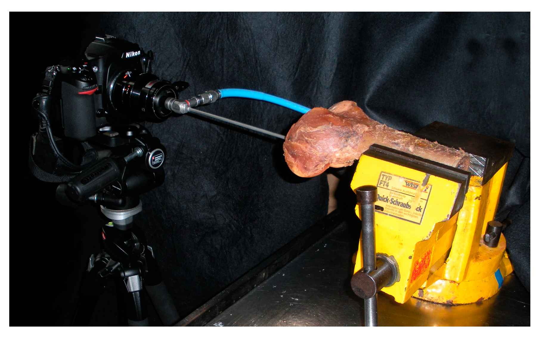
2.2. Photographic Setting and Specimen Preparation

First, all extraarticular soft tissue of the 36 specimens was removed and the knee joint was exposed. The ACL was located and divided in its two functional bundles (AM- and PL-bundle) by evaluating their differing tautness during knee movement [27,28]. Afterwards, the ACL was cautiously removed to expose its bony insertion site. The origin of the two bundles was documented photographically using a standard reflex camera (Nikon Corporation, Tokio, Japan; Type D300s, lens: AF-S DX NIKKOR 18–200 mm 1:3.5–5. G IF-ED VR) (see Figure 2).
Then, photographs from an arthroscopic view of the bare intercondylar region were taken in a standardized setting (see Figure 3). Photographs were taken using a commonly available reflex camera (Nikon Corporation, Tokio, Japan; Type D300s) and a custom-made adaptor for arthroscopes. The camera was mounted on a Manfrotto (Cassola, Italy) 190XPROB-tripod with an 804RC2 three-way head. The camera was aligned horizontally with the help of a bubble level attached to the tripod. To simulate the conditions during knee joint arthroscopy, a standard arthroscope (Arthrex, Wiener Neustadt, Austria; 4 mm, 30°) was connected via the custom-made adaptor to the camera lens. The specimens were secured horizontally using a bench vice on a table. The arthroscope was exactly positioned in the same level as the intercondylar notch, imitating a 90° flexion of the knee, as is standard during arthroscopy, and simulating the view into the knee joint via the anterolateral portal [29].
The position of the bench vice as well as the position of the camera and its tripod were marked on the table and on the ground, respectively. Thus, the same recording conditions were guaranteed for each picture. The picture of each knee was pasted on a DIN A4 page (36 photo series) and then printed on high-quality photo paper (see Figure 4).

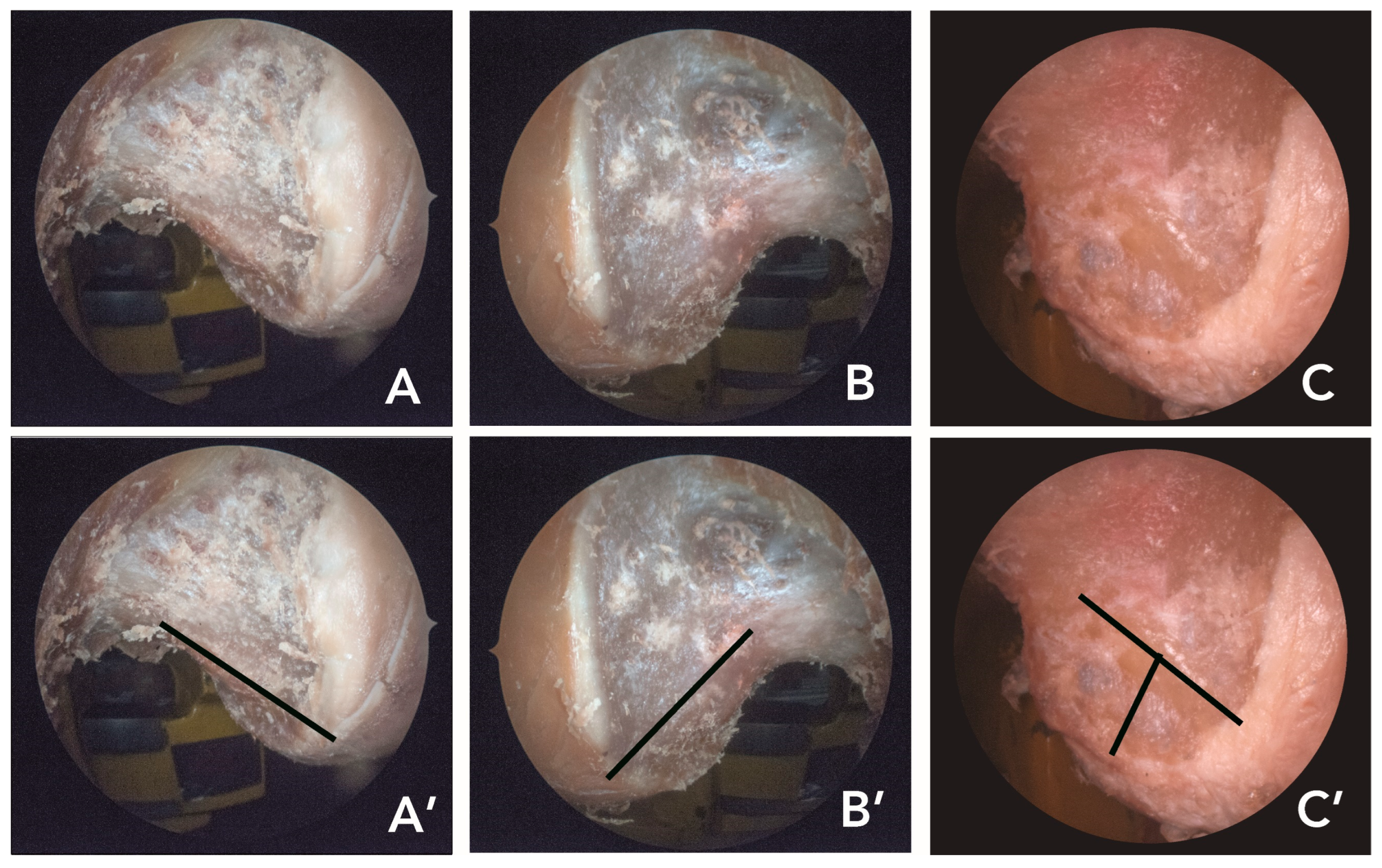
2.3. Identification of the Ridges as Control

First, the LIR and LBR, if identifiable, were marked on each of the 36 knee photo series in dual control. These markings were then verified with the help of the actual anatomical specimen.
The following variables were evaluated to create the standard for the evaluation: the absolute and the relative frequency of the LIR and LBR, the angle between the LIR and the image horizontal, and the angle between the LBR and the image horizontal.
The two angles were used as a decision guide to decide whether the ridges were correctly marked in the photographs or not.

2.4. Evaluation

A medical student, an anatomist and an orthopedic surgeon were then asked to mark both ridges, if identifiable, on the photographs. Those three subjects had the following experience level:
  • Medical student (6th year): Anatomical tutor experienced in general macroscopic anatomy, who was given a short introduction regarding the existence and location of the two ridges.
  • Anatomist: Research focus is on bone and joint anatomy, has some in-depth knowledge of the ridges but minor experience in arthroscopy.
  • Orthopedic surgeon: Specialized in knee arthroscopy with a focus on ACL reconstruction, high level of surgical experience.
To compare the three different experience levels, each subject was given 54 knee photo series. This included one set of all 36 specimens and one set of 18 randomly chosen specimens which were shown twice to test the intra-rater reliability.
The frequency of the correctly marked, incorrectly marked and non-marked photo series among the three subjects was evaluated and then compared with one another.

2.5. Statistical Analysis

Statistical analysis was performed using SPSS Statistics Version 21.0.0.0 (IBM). First, descriptive statistical analysis was conducted. For all metric variables, mean and standard deviation were documented. The McNemar test was used for the comparison of paired nonparametric data. Due to three comparisons for each data record, a Bonferroni correction was applied, setting the level of significance at p < 0.017.

2.6. Intra-Rater Reliability

To verify intra-rater reliability, the individual subjects’ results for the twice shown photo series were compared using Brennan and Prediger’s kappa-coefficient [30]. According to the definition of Landis and Koch [31], kappa values were interpreted as follows: 0.81–1.00 showed very good agreement, 0.61–0.80 represented good agreement, 0.41–0.60 indicated moderate agreement, 0.21–0.40 suggested fair agreement and below 0.20 represented poor agreement.

3. Results

3.1. Control Data

In the 36 specimens, the LIR existed in 29 (80.6%) and the LBR existed in 7 (13.9%) of all specimens.
To differentiate between the correct and incorrect ridge markings of the three subjects, the angles of the ridges were measured in the respective arthroscopic perspectives. The angle between the LIR and the image horizontal was 45.12 ± 11.03°. The angle between the LBR and the image horizontal was 94 ± 8.49° (Figure 5).

3.2. Comparison of the Subjects

With the help of the prior defined angles of the ridges, the subjects’ correct markings were then identified. The angles between the LIR/LBR and the image horizontal in the subjects’ series had to be within the standard deviation of the angles in the dual control group. The results of the evaluation of correct, incorrect and unmarked ridges are represented in Table 1.
The frequency of correct LIR markings and the frequency of incorrect LBR markings were then compared between the medical student, the anatomist and the orthopedic surgeon. The differences between the medical student’s correct LIR markings and the correct LIR marks of the anatomist as well as these of the orthopedic surgeon were highly significant (p < 0.01). Meanwhile, the difference between the orthopedic surgeon’s correct LIR markings and the anatomist’s correct LIR markings was not significant (p = 0.05). Regarding the incorrect LBR markings, there was a significant difference between the orthopedic surgeon and the anatomist, as well as the medical student (p < 0.01). No significant difference could be found between the anatomist’s and the medical student’s incorrect LBR markings.

3.3. Intra-Rater Reliability

To specify the agreement among the individual subjects’ results of the twice shown photo series, Brennan and Prediger’s kappa-coefficient was applied. The kappa value regarding the medical student was 0.583, indicating moderate agreement, the kappa value regarding the anatomist was 0.63, indicating good agreement, and the kappa value concerning the orthopedic surgeon was 0.917, indicating very good agreement.

4. Discussion

This study clearly shows the feasibility of the use of LIR and LBR as osseous landmarks for ACL reconstruction. Additionally, it points out the strong correlation of correctly identifying these landmarks with the level of experience of the investigators, verifying the primary hypothesis of this study.
With a prevalence of 13.9% in the dual control group and low percentages (<10%) of correct LBR markings among the three subjects, the LBR cannot be used as a reliable landmark during knee joint arthroscopy. Meanwhile, the LIR, with a prevalence of 80.6% in the dual control group and high percentages (>70%) for the anatomist and the orthopedic surgeon, can be used as a reliable landmark for the arthroscopic reconstruction of the ACL. Additionally, with the false positive rate of 58.3% for the experienced orthopedic surgeon, this study points out the problem of an expectance of the LBR based on its reported high frequency in the literature (see Table 2).
Furthermore, based on the results of this study, the authors propose a guideline for correctly identifying the LIR and LBR if present: in a 90° flexed knee, with a 30°-arthroscopic view of the medial wall of the lateral femoral condyle, the angulation of the LIR to the horizon of the camera is approximately 45° (45.12 ± 11.03°) and that of the LBR is 90° (94.0 ± 8.49) (Figure 4).
Ferretti et al. [5] described the LBR for the first time. They demonstrated a high prevalence (>80%) of the LBR in their findings, so they suggested that it can be used as a reliable landmark during arthroscopy. The LIR was found in nearly 100% of all specimens. In a macroscopic anatomical study by Ziegler et al. [16], the LIR was detected in all specimens. The LBR could be found in 80% of all specimens. Zauleck et al. [24] investigated a large number of cases in their study. The overall prevalence of the LIR was 94%, whereas the prevalence of the LBR was just 21.4%. Additionally, Norman et al. [34] showed in their study some different values. They were the first to use Micro-CT scanning and surface extraction to objectively evaluate the existence of the LIR and the LBR in the femoral footprint of the ACL. Although they had a very low sample size (n = 7), they showed in the quantitative analysis a frequency of 57% of the LIR and 29% of the LBR. In the qualitative analysis, the respective frequency was 86% and 14%. Additionally, Li et al. [25] reported a frequency of the LIR in their computed tomography study of 87.5%. The results of 80.6% for the LIR and especially 13.9% for the LBR in this study are therefore more in line with the data of Zauleck et al. [24] and Norman et al. [34] than with the other previous studies (see Table 2).
This study is also the first one to put the quantitative data of the ridges’ frequency in the context of different experience levels in applied knee anatomy.
As the LIR is present in over 80% of the cases, this ridge is the perfect tool to point out a possible difference between the three experience levels investigated in this study. The results show that there was a significant difference in correct identifications of the LIR between the medical student and the anatomist and the orthopedic surgeon (p < 0.01). The medical student, despite having sufficient anatomical knowledge, has a lack of experience concerning the detailed anatomy of the intercondylar notch and knee joint arthroscopy. This made it more difficult for them to find the LIR on the pictures. The anatomist and orthopedic surgeon both had an equal number of correct markings, suggesting that the identification of the LIR and therefore its feasibility as a landmark in arthroscopic reconstruction of the ACL requires profound anatomical knowledge of the intercondylar notch, but does not depend on experience in arthroscopic knee surgery.
This is especially important, as the LIR is used to identify the anterior border of the femoral osseous insertion of the ACL. In reconstruction, the femoral tunnel has to be positioned posterior to this landmark to result in adequate postoperative joint biomechanics [35,36,37,38,39,40,41,42,43,44]. If it were positioned anterior of the LIR, and thus in a non-anatomical location, the result would be a biomechanically unstable knee [45]. As this incorrect positioning of the femoral tunnel most frequently happens in inexperienced surgeons, it was also called the “resident’s ridge” by William Clancy Jr. [5,6,7,46]. This terminology also exactly reflects the results presented in this study: profound anatomical knowledge of the intercondylar notch is important for correct tunnel placement.
The results of the markings of the LBR presented another interesting finding. Here, the overall frequency reported in newer studies [24] is lower than the LIR (less than 25%). Additionally, the low number of correct markings of the LBR made a statistical comparison between the three investigators impossible. However, the prevalence of incorrect LBR markings differed significantly between the orthopedic surgeon and the medical student and the anatomist. The orthopedic surgeon incorrectly marked the LBR in 58.3% of all cases. The subject with the greatest expertise had the highest prevalence of incorrect LBR markings. This result shows that a high experience level in combination with the knowledge of the high detectability of the LBR in previous studies could cause a subject to detect the LBR more often. This could perhaps also be the reason for the high number of LBR findings in previous studies [5,13,16,26].
The kappa-coefficients of the intra-rater reliability tests among the three subjects indicate a good repeatability of finding the LIR and LBR. Here, the experience level had an important influence on the intra-rater reliability. The most experienced subject, the orthopedic surgeon, was the one with the highest kappa-coefficient (κ = 0.917). Here, the high number of arthroscopic procedures performed by the orthopedic surgeon clearly had an impact on the high repeatability. Improving the surgical performance of residents and increasing their skills through options like training in a laboratory setting with anatomical specimens have to be considered [47]
This study is one of the very few evaluating the identification of the osseous ridges in different subjects and focuses on different experience levels, which therefore defines the main strength of the study. Norman et al. [34] showed the evaluation of the LIR and LBR by ten clinicians (eight orthopedic surgeons and two musculoskeletal radiologists), resulting in similar findings as presented above. The clinicians were overall more comfortable identifying the LIR than the LBR [34].
The low prevalence and the great difference in the detectability of the LBR in this study and also in previous studies [24,34] show that it should not be used as a reliable landmark during knee joint arthroscopy. If one needs to identify the border between the two bundles of the ACL, one should instead follow the suggestions of Zantop and Petersen [21] and Petersen [48], who recommend the intersection of the intercondylar line and the osteochondral border as a landmark to subdivide the footprint of the ACL into the attachment areas of the two functional bundles.
Finally, the authors were able to propose a new guideline for correctly identifying the LIR and LBR during arthroscopy at approximately 45° (45.12 ± 11.03°) for the LIR and 90° (94.0 ± 8.49) for the LBR in relation to the horizontal plane (Figure 4), which may improve femoral tunnel placement in ACL reconstruction, especially for surgeons in training.
This proposed guideline is the first in recent research based on the angulation of the ridges to the horizontal plane of the camera during arthroscopy, which characterizes these guidelines as more easily practicable intraoperatively. Although several authors describe the angle of the ridges to the anatomical axis of the femur [24,49,50] and to the roof of the intercondylar notch [32], this information seems to only be informative for anatomical description of the landmarks, and not for application during surgery.

Limitations

The hypothesis for this study was that the correct identification correlates with the experience of the investigator. To answer this question, the number of specimens used in this study was deemed sufficient, especially as the prevalence of the two ridges has already been reported adequately in a large number of specimens [24] and the number of specimens was limited by their availability and the inclusion criteria. As the age in anatomical specimens is on average higher, a large number of specimens could not be included due to degenerative changes to the knee joint.
Additionally, the rather high mean age of the specimens could be questioned, particularly when following the argumentation of Ferretti et al. [26], who showed a high number of LBRs in fetal lower extremities and hypothesized the possibility that the LBR is lost during aging. If, however, this argumentation were true, we would expect an equally diminished number of LBRs and LIRs in the specimen. As in this study the LIR was present in 80.6% of sample, we argue that this line of argumentation [9,26] is highly questionable. Based on the reported existence of LIR and LBR, aging seems not to be the most influential factor.
In contrast to the study of Zauleck et al. [24], where the low detectability of the LBR was shown on the basis of macroscopic photos from the intercondylar notch, in this study, the intercondylar notch was photographed from an arthroscopic perspective, with similar results.
The photographic setting in this study could also be questioned. Using a reflex camera with a custom-made adaptor to attach the arthroscope in place of an arthroscopic camera does not reflect circumstances in a clinical setting, and therefore could also challenge the applicability of the results presented in this study. However, to achieve standardized photographs which guaranteed the same position in each specimen, the authors decided in favor of the combination of a reflex camera and tripod to minimize this bias in the photographic setting.
Regarding the comparison of the different experience levels, the low number of investigators could be seen as a limiting factor. In addition, one important group, orthopedic residents, was missing. However, this group would be very difficult to rate, as the experience level varies widely regarding the different levels of qualification and experience in arthroscopy.
Furthermore, the knowledge of the existence of the LIR and LBR, as we were able to show in this study, may bias the results. Therefore, the bony ridges were located in a dual control step with the help of the anatomical specimens, which reduced falsification.

5. Conclusions

In this study, the LIR existed in 80.6% of all cases, and it therefore can be used as a reliable landmark for the placement of the femoral tunnel in arthroscopic ACL reconstruction. This result concurs with recent research. The LBR was only present in 13.9% of all cases, and therefore its secure use as a tool for identifying the border between the two bundles of the ACL during arthroscopy is highly questionable. For correct identification of all the osseous landmarks of the intercondylar region, adequate anatomical knowledge thereof is of great importance, and looking at the large difference between the medical student and the other investigators, this is made especially clear. Another interesting finding was the high number of incorrect markings provided by the orthopedic surgeon in identifying the LBR. Here, the high expectance of its existence as reported in the literature, for example by Ferretti et al. [5] and Ziegler et al. [16], could be a reason for this. The clinical significance of the LBR, even though it sometimes exists, therefore needs discussion. By introducing the guidelines for correctly identifying the LIR (45°) and LBR (90°), the surgeon should be able to recognize these osseous ridges more easily if present.

Author Contributions

Conceptualization, L.H. and P.W.; methodology, L.H., D.R. and P.W.; validation, L.H.; formal analysis, L.H. and D.R.; investigation, L.H. and D.R.; resources, L.H.; data curation, L.H.; writing—original draft preparation, L.H.; writing—review and editing, L.H., D.R., S.A.K. and P.W.; visualization, L.H.; supervision, L.H.; project administration, L.H. All authors have read and agreed to the published version of the manuscript.

Funding

This research received no external funding.

Institutional Review Board Statement

The study was conducted in accordance with the Declaration of Helsinki, and approved by the Institutional Ethics Committee of Medical University of Vienna (1300/2023, 7 May 2013).

Informed Consent Statement

Not applicable.

Data Availability Statement

The data presented in this study are available on request from the corresponding author. The data are not publicly available due to ethical reasons.

Acknowledgments

The authors would like to thank the body donors and their families. Without their selfless donation, this study would not be possible.

Conflicts of Interest

The authors declare no conflict of interest.

Abbreviations

ACL (anterior cruciate ligament), AM-bundle (anteromedial bundle), LBR (lateral bifurcate ridge), LIR (lateral intercondylar ridge), PL-bundle (posterolateral bundle).

References

  1. Cheatham, S.A.; Johnson, D.L. Anatomic Revision ACL Reconstruction. Sports Med. Arthrosc. Rev. 2010, 18, 33–39. [Google Scholar] [CrossRef] [PubMed]
  2. Forkel, P.; Achtnich, A.; Metzlaff, S.; Zantop, T.; Petersen, W. Causes for revision after arthoscopic reconstruction of the anterior cruciate ligament: A retrospective review of preoperative findings of 100 cases of revision. Unfallchirurg 2011, (Suppl. S2), 119. [Google Scholar]
  3. Jorge, P.B.; Escudeiro, D.; Severino, N.R.; Santili, C.; Cury, R.D.P.L.; Junior, A.D.; Guglielmetti, L.G.B. Positioning of the femoral tunnel in anterior cruciate ligament reconstruction: Functional anatomical reconstruction. BMJ Open Sport Exerc. Med. 2018, 4, e000420. [Google Scholar] [CrossRef] [PubMed]
  4. Rowan, F.A.; Marshall, T.; Gombosh, M.R.; Farrow, L.D. Utilization of Osseous Landmarks for Anatomic Anterior Cruciate Ligament Femoral Tunnel Placement. J. Knee Surg. 2017, 30, 359–363. [Google Scholar] [CrossRef] [PubMed]
  5. Ferretti, M.; Ekdahl, M.; Shen, W.; Fu, F.H. Osseous Landmarks of the Femoral Attachment of the Anterior Cruciate Ligament: An Anatomic Study. Arthrosc. J. Arthrosc. Relat. Surg. 2007, 23, 1218–1225. [Google Scholar] [CrossRef]
  6. Petersen, W.; Zantop, T. Anatomy of the Anterior Cruciate Ligament with Regard to Its Two Bundles. Clin. Orthop. Relat. Res. 2007, 454, 35–47. [Google Scholar] [CrossRef] [PubMed]
  7. Bicer, E.K.; Lustig, S.; Servien, E.; Selmi, T.A.S.; Neyret, P. Current knowledge in the anatomy of the human anterior cruciate ligament. Knee Surg. Sports Traumatol. Arthrosc. 2010, 18, 1075–1084. [Google Scholar] [CrossRef] [PubMed]
  8. Hutchinson, M.R.; Ash, S.A. Resident’s ridge: Assessing the cortical thickness of the lateral wall and roof of the intercondylar notch. Arthrosc. J. Arthrosc. Relat. Surg. 2003, 19, 931–935. [Google Scholar] [CrossRef]
  9. Fu, F.H.; Jordan, S.S. The lateral intercondylar ridge—A key to anatomic anterior cruciate ligament reconstruction. J. Bone Jt. Surg. Am. 2007, 89, 2103–2104. [Google Scholar]
  10. Kopf, S.; Musahl, V.; Tashman, S.; Szczodry, M.; Shen, W.; Fu, F.H. A systematic review of the femoral origin and tibial insertion morphology of the ACL. Knee Surg. Sports Traumatol. Arthrosc. 2009, 17, 213–219. [Google Scholar] [CrossRef]
  11. Forsythe, B.; Kopf, S.; Wong, A.K.; Martins, C.A.; Anderst, W.; Tashman, S.; Fu, F.H. The Location of Femoral and Tibial Tunnels in Anatomic Double-Bundle Anterior Cruciate Ligament Reconstruction Analyzed by Three-Dimensional Computed Tomography Models. J. Bone Jt. Surg. 2010, 92, 1418–1426. [Google Scholar] [CrossRef] [Green Version]
  12. Abrams, D.A.; Nicol, T.; Zecker, S.; Kraus, N. Rapid acoustic processing in the auditory brainstem is not related to cortical asymmetry for the syllable rate of speech. Clin. Neurophysiol. 2010, 121, 1343–1350. [Google Scholar] [CrossRef] [PubMed] [Green Version]
  13. van Eck, C.F.; Morse, K.R.; Lesniak, B.P.; Kropf, E.J.; Tranovich, M.J.; van Dijk, C.N.; Fu, F.H. Does the lateral intercondylar ridge disappear in ACL deficient patients? Knee Surg. Sports Traumatol. Arthrosc. 2010, 18, 1184–1188. [Google Scholar] [CrossRef] [PubMed] [Green Version]
  14. Van Eck, C.F.; Schreiber, V.M.; Liu, T.T.; Fu, F.H. The anatomic approach to primary, revision and augmentation anterior cruciate ligament reconstruction. Knee Surg. Sports Traumatol. Arthrosc. 2010, 18, 1154–1163. [Google Scholar] [CrossRef]
  15. Van Eck, C.F.; Schreiber, V.M.; Mejia, H.A.; Samuelsson, K.; van Dijk, C.N.; Karlsson, J.; Fu, F.H. “Anatomic” anterior cruciate ligament reconstruction: A systematic review of surgical techniques and reporting of surgical data. Arthroscopy 2010, 26, S2–S12. [Google Scholar] [CrossRef] [PubMed]
  16. Ziegler, C.G.; Pietrini, S.D.; Westerhaus, B.D.; Anderson, C.J.; Wijdicks, C.A.; Johansen, S.; Engebretsen, L.; LaPrade, R.F. Arthroscopically Pertinent Landmarks for Tunnel Positioning in Single-Bundle and Double-Bundle Anterior Cruciate Ligament Reconstructions. Am. J. Sports Med. 2010, 39, 743–752. [Google Scholar] [CrossRef]
  17. Farrow, L.D.; Gillespie, R.J.; Victoroff, B.N.; Cooperman, D.R. Radiographic location of the lateral intercondylar ridge: Its relationship to Blumensaat’s line. Am. J. Sports Med. 2008, 36, 2002–2006. [Google Scholar] [CrossRef]
  18. Zantop, T.; Wellmann, M.; Fu, F.H.; Petersen, W. Tunnel positioning of anteromedial and posterolateral bundles in anatomic anterior cruciate ligament reconstruction: Anatomic and radiographic findings. Am. J. Sports Med. 2008, 36, 65–72. [Google Scholar] [CrossRef]
  19. Tashman, S.; Kopf, S.; Fu, F.H. The Kinematic Basis of ACL Reconstruction. Oper. Tech. Sports Med. 2008, 16, 116–118. [Google Scholar] [CrossRef] [Green Version]
  20. Tajima, G.; Iriuchishima, T.; Ingham, S.J.; Shen, W.; van Houten, A.H.; Aerts, M.M.; Shimamura, T.; Smolinski, P.; Fu, F.H. Anatomic Double-Bundle Anterior Cruciate Ligament Reconstruction Restores Patellofemoral Contact Areas and Pressures More Closely Than Nonanatomic Single-Bundle Reconstruction. Arthrosc. J. Arthrosc. Relat. Surg. 2010, 26, 1302–1310. [Google Scholar] [CrossRef]
  21. Zantop, T.; Peterson, W. Anatomical reconstruction of the anterior cruciate ligament. Arthroskopie 2007, 20, 94–104. [Google Scholar] [CrossRef]
  22. Hofbauer, M.; Muller, B.; Murawski, C.D.; Baraga, M.; van Eck, C.F.; Fu, F.H. Strategies for revision surgery after primary double-bundle anterior cruciate ligament (ACL) reconstruction. Knee Surg. Sports Traumatol. Arthrosc. 2013, 21, 2072–2080. [Google Scholar] [CrossRef]
  23. Hofbauer, M.; Murawski, C.D.; Muller, B.; van Eck, C.F.; Fu, F.H. Revision surgery after primary double-bundle ACL reconstruction: AAOS exhibit selection. J. Bone Jt. Surg. Am. 2014, 96, e30. [Google Scholar] [CrossRef] [PubMed]
  24. Zauleck, M.K.; Gabriel, S.; Fischmeister, M.F.; Hirtler, L. Origin of the anterior cruciate ligament and the surrounding osseous landmarks of the femur. Clin. Anat. 2014, 27, 1103–1110. [Google Scholar] [CrossRef] [PubMed]
  25. Li, Z.; Zhang, W.; Ren, S.; Zhou, R.; Zhang, X.; You, T.; Bai, L. Relationship Between Number of Lateral Intercondylar Ridges and Area of Denser Bone on the Lateral Intercondylar Wall. Orthop. J. Sports Med. 2022, 10, 23259671221091332. [Google Scholar] [CrossRef] [PubMed]
  26. Ferretti, M.; Levicoff, E.A.; Macpherson, T.A.; Moreland, M.S.; Cohen, M.; Fu, F.H. The Fetal Anterior Cruciate Ligament: An Anatomic and Histologic Study. Arthrosc. J. Arthrosc. Relat. Surg. 2007, 23, 278–283. [Google Scholar] [CrossRef]
  27. Brantigan, O.C.; Voshell, A.F. The Mechanics of the Ligaments and Menisci of the Knee Joint. J. Bone Jt. Surg. Am. 1941, 23, 44–66. [Google Scholar]
  28. Girgis, F.G.; Marshall, J.L.; Monajem, A. The cruciate ligaments of the knee joint. Anatomical, functional and experimental analysis. Clin. Orthop. Relat. Res. 1975, 106, 216–231. [Google Scholar] [CrossRef]
  29. Aviles, S.A.; Allen, C.R. Knee Arthroscopy: The Basics. In Operative Techniques in Orthopaedic Surgery; Wiesel, S.W., Ed.; Lippincott Williams & Wilkins: Philadelphia, PA, USA, 2011; pp. 248–256. [Google Scholar]
  30. Brennan, R.L.; Prediger, D.J. Coefficient Kappa: Some Uses, Misuses, and Alternatives. Educ. Psychol. Meas. 1981, 41, 687–699. [Google Scholar] [CrossRef]
  31. Landis, J.R.; Koch, G.G. The Measurement of Observer Agreement for Categorical Data. Biometrics 1977, 33, 159–174. [Google Scholar] [CrossRef] [Green Version]
  32. Tsukada, S.; Fujishiro, H.; Watanabe, K.; Nimura, A.; Mochizuki, T.; Mahakkanukrauh, P.; Yasuda, K.; Akita, K. Anatomic variations of the lateral intercondylar ridge: Relationship to the anterior margin of the anterior cruciate ligament. Am. J. Sports Med. 2014, 42, 1110–1117. [Google Scholar] [CrossRef]
  33. Norman, D.; Metcalfe, A.J.; Barlow, T.; Hutchinson, C.E.; Thompson, P.J.; Spalding, T.J.; Williams, M.A. Cortical Bony Thickening of the Lateral Intercondylar Wall: The Functional Attachment of the Anterior Cruciate Ligament. Am. J. Sports Med. 2017, 45, 394–402. [Google Scholar] [CrossRef] [Green Version]
  34. Norman, D.G.; Getgood, A.; Thornby, J.; Bird, J.; Turley, G.A.; Spalding, T.; Williams, M.A. Quantitative topographic anatomy of the femoral ACL footprint: A micro-CT analysis. Med. Biol. Eng. Comput. 2014, 52, 985–995. [Google Scholar] [CrossRef] [PubMed]
  35. Fu, F.H.; Van Eck, C.F.; Tashman, S.; Irrgang, J.J.; Moreland, M.S. Anatomic anterior cruciate ligament reconstruction: A changing paradigm. Knee Surg. Sports Traumatol. Arthrosc. 2015, 23, 640–648. [Google Scholar] [CrossRef]
  36. Lim, H.-C.; Yoon, Y.-C.; Wang, J.-H.; Bae, J.-H. Anatomical versus Non-Anatomical Single Bundle Anterior Cruciate Ligament Reconstruction: A Cadaveric Study of Comparison of Knee Stability. Clin. Orthop. Surg. 2012, 4, 249–255. [Google Scholar] [CrossRef] [PubMed] [Green Version]
  37. Amis, A.A.; Dawkins, G.P. Functional anatomy of the anterior cruciate ligament. Fibre bundle actions related to ligament replacements and injuries. J. Bone Jt. Surg. 1991, 73, 260–267. [Google Scholar] [CrossRef] [PubMed] [Green Version]
  38. Loh, J.C.; Fukuda, Y.; Tsuda, E.; Steadman, R.J.; Fu, F.H.; Woo, S.L.-Y. Knee stability and graft function following anterior cruciate ligament reconstruction: Comparison between 11 o’clock and 10 o’clock femoral tunnel placement. Arthrosc. J. Arthrosc. Relat. Surg. 2003, 19, 297–304. [Google Scholar] [CrossRef] [PubMed]
  39. Zavras, T.D.; Race, A.; Amis, A.A. The effect of femoral attachment location on anterior cruciate ligament reconstruction: Graft tension patterns and restoration of normal anterior-posterior laxity patterns. Knee Surg. Sports Traumatol. Arthrosc. 2005, 13, 92–100. [Google Scholar] [CrossRef]
  40. Karlsson, J.; Irrgang, J.J.; Van Eck, C.F.; Samuelsson, K.; Mejia, H.A.; Fu, F.H. Anatomic Single- and Double-Bundle Anterior Cruciate Ligament Reconstruction, Part 2. Am. J. Sports Med. 2011, 39, 2016–2026. [Google Scholar] [CrossRef]
  41. Kondo, E.; Merican, A.M.; Yasuda, K.; Amis, A.A. Biomechanical Comparison of Anatomic Double-Bundle, Anatomic Single-Bundle, and Nonanatomic Single-Bundle Anterior Cruciate Ligament Reconstructions. Am. J. Sports Med. 2011, 39, 279–288. [Google Scholar] [CrossRef]
  42. Kondo, E.; Yasuda, K.; Azuma, H.; Tanabe, Y.; Yagi, T. Prospective Clinical Comparisons of Anatomic Double-Bundle versus Single-Bundle Anterior Cruciate Ligament Reconstruction Procedures in 328 Consecutive Patients. Am. J. Sports Med. 2008, 36, 1675–1687. [Google Scholar] [CrossRef]
  43. Yagi, M.; Wong, E.K.; Kanamori, A.; Debski, R.E.; Fu, F.H.; Woo, S.L.-Y. Biomechanical Analysis of an Anatomic Anterior Cruciate Ligament Reconstruction. Am. J. Sports Med. 2002, 30, 660–666. [Google Scholar] [CrossRef] [PubMed]
  44. Yasuda, K.; Kondo, E.; Ichiyama, H.; Tanabe, Y.; Tohyama, H. Clinical Evaluation of Anatomic Double-Bundle Anterior Cruciate Ligament Reconstruction Procedure Using Hamstring Tendon Grafts: Comparisons Among 3 Different Procedures. Arthrosc. J. Arthrosc. Relat. Surg. 2006, 22, 240–251. [Google Scholar] [CrossRef] [PubMed]
  45. Marchant, B.G.; Noyes, F.R.; Barber-Westin, S.D.; Fleckenstein, C. Prevalence of Nonanatomical Graft Placement in a Series of Failed Anterior Cruciate Ligament Reconstructions. Am. J. Sports Med. 2010, 38, 1987–1996. [Google Scholar] [CrossRef]
  46. Shino, K.; Suzuki, T.; Iwahashi, T.; Mae, T.; Nakamura, N.; Nakata, K.; Nakagawa, S. The resident’s ridge as an arthroscopic landmark for anatomical femoral tunnel drilling in ACL reconstruction. Knee Surg. Sports Traumatol. Arthrosc. 2010, 18, 1164–1168. [Google Scholar] [CrossRef]
  47. Pirri, C.; Stecco, C.; Porzionato, A.; Boscolo-Berto, R.; Fortelny, R.H.; Macchi, V.; Konschake, M.; Merigliano, S.; De Caro, R. Forensic Implications of Anatomical Education and Surgical Training with Cadavers. Front. Surg. 2021, 8, 641581. [Google Scholar] [CrossRef]
  48. Petersen, W. Tunnel Positioning. In The Anteruir Cruciate Ligament; Petersen, W., Zantop, T., Eds.; Deutscher Ärzte-Verlag: Köln, Germany, 2009. [Google Scholar]
  49. Giron, F.; Cuomo, P.; Aglietti, P.; Bull, A.M.J.; Amis, A.A. Femoral attachment of the anterior cruciate ligament. Knee Surg. Sports Traumatol. Arthrosc. 2005, 14, 250–256. [Google Scholar] [CrossRef]
  50. Arnoczky, S.P. Anatomy of the anterior cruciate ligament. Clin. Orthop. Relat. Res. 1983, 172, 19–25. [Google Scholar] [CrossRef]
Figure 1. Schematic drawing of the medial wall of the lateral femoral condyle. The osseous insertion of the anteromedial bundle is depicted in orange, and the osseous insertion of the posterolateral bundle is depicted in green. The lateral intercondylar ridge (LIR) is depicted in blue as anterior border of the ACL, while the lateral bifurcate ridge (LBR) is depicted in red as a border between the two bundles.
Applsci 13 07345 g001
Figure 2. Macroscopic view of the lateral femoral condyle. The osseous attachment of the ACL was mapped in the photograph. (A) Left femur. (B) Right femur. Scale represents 1 cm.
Applsci 13 07345 g002
Figure 3. Photographic setting. The camera was mounted on a tripod, via a custom-made adapter, and a 30° 4 mm arthroscope was connected. An arthroscopic light source provided adequate illumination. The specimen was fixed in a vice.
Applsci 13 07345 g003
Figure 4. Arthroscopic view of the lateral femoral condyle. (A,B) are the same specimen as in Figure 2. (A′,B′) show the same photograph with the LIR marked after evaluation. In these specimens, no LBR was identified. (C) shows an additional specimen with an LBR present. (C′) shows the same photograph with its LIR and LBR marked after evaluation.
Applsci 13 07345 g004
Figure 5. Approximation of the angulation of the ridges towards the horizon of the camera with the knee at 90° and a 30° arthroscope. (A) shows the summary of the results, (B) displays an exemplary specimen with osseous ACL origin and LIR marked.
Applsci 13 07345 g005
Table 1. Interpretation of the three subjects’ results. The 36 photo-series are divided into 3 groups: the frequency of correctly and incorrectly marked pictures, as well as the frequency of unmarked pictures, concerning the LIR and the LBR.
LIR (%)LBR (%)
CorrectUnmarkedIncorrectCorrectUnmarkedIncorrect
Medical student19.4 *47.2 *33.4083.316.7
Anatomist72.213.913.95.67519.4
Orthopedic surgeon77.82.819.48.433.358.3 *
* p < 0.017.
Table 2. Overview of the existence of LIR and LBR reported in the literature.
nLIR (%)LBR (%)Material
Ferretti et al. [5]7-85.7Fetus
60100.081.7Arthroscopic
16100.081.6Anatomical specimens
Van Eck et al. [13]5088.048.0Arthroscopic
Ziegler et al. [16]26100.092.3Anatomical specimens
Zauleck et al. [24]16697.624.7Bones
6985.313.2Anatomical specimens
Tsukada et al. [32]31894.0-Bones
Norman et al. [33]757.029.0Anatomical specimens
Li et al. [25]8987.5%-Patients
This study3680.613.8Anatomical specimens
Disclaimer/Publisher’s Note: The statements, opinions and data contained in all publications are solely those of the individual author(s) and contributor(s) and not of MDPI and/or the editor(s). MDPI and/or the editor(s) disclaim responsibility for any injury to people or property resulting from any ideas, methods, instructions or products referred to in the content.

