Article

Neuroprotective Effect of the Mixture of Gastrodiae elata and Glycyrrhizae uralensis In Vitro

1
Department of Korean Pharmacy, Woosuk University, 443 Samnye-ro, Samnye-eup, Wanju-Gun 55338, Jeollabuk-do, Republic of Korea
2
MJ Health Food Co., Ltd., 5 Sinchon-gil, Anseong-myeon, Muju-gun 55536, Jeollabuk-do, Republic of Korea
3
Department of Technical Research, Agricultural Technology Center, 416 Hanpungru-ro, Muju-eup, Muju-gun 55517, Jeollabuk-do, Republic of Korea
*
Author to whom correspondence should be addressed.
Appl. Sci. 2023, 13(1), 190; https://doi.org/10.3390/app13010190
Submission received: 13 November 2022 / Revised: 17 December 2022 / Accepted: 19 December 2022 / Published: 23 December 2022
(This article belongs to the Special Issue New Trends in Biosciences III)

Abstract

:
Background: This study investigated the effect of a mixture of Gastrodiae elata and Glycyrrhizae uralensis (GGW) on β-amyloid-induced neuronal damage in vitro. Methods: For finding the appropriate GGW ratio, we performed MTT assays using the ratios of 5:5, 6:4, 7:3, 8:2 and 9:1 in SK-N-SH cell and SH-SY-5Y cell. Treatment with β-amyloid (10 μM) caused cell death and overexpression of acetylcholinesterase (ACHE) in SH-SY-5Y cells. β-amyloid treatment increased the expression of mitogen-activated protein kinases (MAPKs). In addition, we detected the antioxidant activity of GGW using DCF-DA staining of SK-N-SH cells. To determine the effect of GGW on oxidative stress, we used a hydrogen peroxidase-induced in vitro model. Results: We selected the ratios of 5:5 and 7:3 mixtures with the least cytotoxicity. The 7:3 ratio of GGW (GGW73) decreased the mRNA expression of ACHE in SH-SY-5Y cell more than the 5:5 ratio of GGW (GGW55). GGW73 suppressed p-ERK protein expression in β-amyloid-treated SH-SY-5Y cells. Additionally, GGW73 regulated reactive oxygen species (ROS) in vitro. GGW73 treatment regulated apoptotic factors in β-amyloid-stimulated neuronal cells. Conclusions: These results suggest that GGW73 exerts neuroprotective and anti-inflammatory effects in vitro. These results also showed that GGW73 had a protective effect against H2O2 induced cell damage in an in vitro model. These results indicate the potential of GGW as a therapeutic agent for cognitive disorders.

1. Introduction

Worldwide population aging is emerging as a major long-term problem [1]. As population aging progresses worldwide, the number of people above the age of 65 are expected to steadily increase to two billion by 2050 [2], and the patients with neurodegenerative disease including dementia and cognitive disorder are also expected to increase to over 130 million [3]. Among the numerous age-related diseases, degenerative brain diseases, such as Alzheimer’s disease (AD), cause many additional diseases and excessive medical expenses [4]. AD is characterized by cognitive impairment and memory loss [5]. These degenerative brain disorders reduce learning and memory, affecting not only patients, but also their families and social behavior [6]. AD with cognitive function disorders is etiologically characterized by neuroinflammation, causing neuronal loss, amyloid deposition, and nerve fiber entanglement [7]. It is well known that β-amyloid (Aβ) and oxidative stress are involved in the pathogenesis of cognitive function disorder [8]. The oligomeric form of Aβ peptide plays a major role in neuronal and glial activation of inflammation. After activation, neuronal cells produce inflammatory agents, including chemokines and cytokines [9]. It induces apoptosis and destabilizes calcium homeostasis by Aβ peptide, ultimately leading to neurotoxicity both in vitro and in vivo [10]. Aβ peptide is released from amyloid precursor protein (APP) by β-and γ-secretases, which form senile plaques [11]. Therapies that modulate the Aβ pathway are promising targets for drug development for cognitive function disorders. In connection with this method, approaches are being developed to regulate the Aβ pathway by inhibition of β- or γ-secretase or anti-Aβ monoclonal antibodies in cognitive function disorders [12].
Acetylcholine (Ach), a neurotransmitter in the nervous system and brain, plays a key role in memory and learning [13]. General medications might be used for degenerative brain disorders, such as AD and cognitive function disorders, by inhibiting acetylcholinesterase (AChE), nicotinic acetylcholine receptors, and muscarinic acetylcholine receptor (mAChR) [14]. The expression of AChE, which breaks down ACh, is activated in patients with cognitive disorders or AD [15]. Generally, in clinical treatment, donepezil, tacrine, and rivastigmine are used to inhibit AChE. However, these medicines have side effects, such as somnipathy and nausea, and short half-lives [16]. In the amyloid pathway, β-secretase (BACE-1) is localized in endosomes and cleaved APP [17]. Therapeutically, inhibition of BACE-1 is a better treatment for AD or cognitive function disorders, aimed at suppressing the production of Aβ [18]. Additionally, Aβ peptide has been known to inhibit mitochondrial activity and alter intercellular calcium levels and induce cell death through oxidative and inflammatory stimulation [19].
Gastrodia elata (GE), a traditional herbal medicine in South Korea, and its constituents have been used to treat various diseases, such as cardiovascular system disorders [20], convulsions [21], neurotransmitter disorders [22], and cognitive function disorders by regulating APP cleavage [23]. Gastrodin, one of the major constituents of Gastrodia elata, has been reported to have beneficial effects on the nervous system, including suppression of microglial activation, anti-inflammation, regulation of oxidative stress, and modulation of neurotransmitters [24]. Furthermore, gastrodin showed a protective effect on the midbrain by increasing the levels of GSH, HO-1, and SOD in MPTP intoxicated mice [25]. These results suggest that GE has the potential to treat cognitive function disorders. Glycytthiza ularesis (GU), belonging to the Leguminosae family [26], has been used as an herbal medicine and a natural sweetener [27]. In addition, GU has traditionally been used to treat skin inflammation, digestive system diseases, and hepatitis [28]. GU has been reported to contain bioactive compounds such as triterpenoids (glycyrrhizic acid, glycyrrhetinic acid) and flavonoids (liquilitin, liquilitigenin, and licochalcone A) [29,30], which possess a variety of biological activities [28]. A previous study showed that GU decreases Aβ-induced neurotoxicity by regulating oxidative stress in cultured cortical neurons [31]. In addition, GU has been reported to ameliorate cognitive dysfunction in an AD mice model [32]. The constituents of GU have been studied for their effects on neuronal diseases by attenuating kainic acid-induced neuronal cell death [33] and anti-apoptotic activity in dopaminergic neurons [34]. Various effects of GE and GU on neurological diseases have been reported; however, the efficacy of a mixture of GE and GU has not yet been evaluated. The mixture of GE and GU as herbal medicines, each with effects on neuronal diseases, is expected to have a better improvement effect on cognitive function disorders.
This study aimed to determine whether a mixture of GE and GU could ameliorate neuronal damage in vitro. In addition, we investigated the mechanism of AChE and apoptosis in Aβ-stimulated cell damage by applying a mixture of GE and GU. We evaluated the protection effect against Aβ-induced cell death and AChe and BDNF expression using SH-SY-5Y cells, and regulatory effect against Aβ-induced apoptotic factors such as BCl-2 and Bax using SK-N-SY neuroblastoma cells.

2. Materials and Methods

2.1. Reagents

Minimum Essential Medium (MEM) used in cell culture was purchased from Welgene (Gyoungsan, Gyoungsangbuk-do, Republic of Korea). Dulbecco’s modified Eagle’s medium (DEME), fetal bovine serum (FBS), and penicillin streptomycin were purchased from Gibco (Grand Island, NY, USA). The Amyloid β-Protein Fragment 25–35 (Aβ25–35) (purity 95%) and lipopolysaccharide (LPS) from Escherichia coli 0111:B4 were purchased from Sigma-Aldrich (St. Louis, MO, USA), and 1–42 (Human) HFIP-treated Aβ 1–42 peptide (purity 95%) was purchased from GenicBio Synthetic Peptide (Shanghai, China). All primary antibodies used for Western blotting were purchased from Cell Signaling Technology (Danvers, MA, USA). Secondary antibodies for Western blot analysis were purchased from Jackson ImmunoResearch (West Grove, PA, USA).

2.2. Preparation of GE and GU Extracts

The rhizome of gastrodia elata (GER) was provided by MJ Health Food Co., Ltd. (Muju-gun, Jeollabuk-do, Republic of Korea). Then, 100 g of GER was decocted into 10 times volume of distilled water (1 L) at 90 °C for 6 h. The extract was filtered through 25 μm polyethersulfone (PES) syringe filter, then vacuum evaporated at 65 °C. Subsequently, the concentrate was frozen at −75 °C for 24 h, and then frozen-dried at −85 °C and 5 mTorr condition for 60 h. The GER yield was 13.8%. G. ularensis radix of glycyrrhiza ularensis (GUR) was provided by Mj Health Food Co., Ltd. (Muju-gun, Jeollabuk-do, Republic of Korea). Next, 100 g of GUR was decocted with 10 times volume of distilled water (1 L) at 100 °C for 3 h. The extract was filtered through 25 μm polyethersulfone (PES) syringe filter, then vacuum evaporated at 65 °C. Subsequently, the concentrate was frozen at −75 °C for 24 h, and then frozen at −85 °C and 5 mTorr condition for 60 h. The GUR yield was 17.4%. Various ratios (GER:GUR = 5:5, 6:4, 7:3, 8:2, 9:1) of a mixture of GER and GUR were prepared by mixing GER and GUR.

2.3. Cell Culture

Human neuroblastoma cells (SH-SY5Y and SK-N-SH) and a macrophage cell line (RAW 264.7) were purchased from the Korea Cell Line Bank (Jongno-gu, Seoul, Republic of Korea). SH-SY5Y and SK-N-SH were cultured in MEM containing 10% FBS and 1% penicillin-streptomycin, and the RAW 264.7 cells were cultured in DMEM containing 10% FBS and 1% penicillin-streptomycin. All cell lines were incubated at 37 °C with 5% CO2.

2.4. Cell Viability

The cytotoxicity of the complex mixture was measured using the MTT assay. SH-SY-5Y cells (3 × 10⁴ cells per well) were added to a 96-well plate and incubated at 37 °C and 5% CO2 for 24 h. After that, the mixtures were added for 4 h, and β-amyloid (25–35) was added at a concentration of 10 μM, incubated for 24 h, and 5 mg/mL of 1-(4,5-Dimethylthiazol-2-yl)-3,5-diphenylformazan (MTT; Sigma-Aldrich Co.) solution was added to each well and incubated for an additional 5 h, after which the supernatant from each well was removed. To completely dissolve the remaining formazan crystals, 200μL of dimethyl sulfoxide (DMSO) was added to the wells, and the absorbance was measured at 540 nm using a microplate reader (Synergy HTX Multi-Mode Reader, BioTek, Santa Clara, CA, USA).

2.5. Western Blot Analysis

Cells were seeded at a density of 3 × 10⁶ in 6 cm dishes. After 24 h, it was replaced with 0% medium, and various concentrations of GGW were added. After 2 h, 2 μM of Aβ was added. After 45 min, the cells were washed with 1 × PBS (ELPIS BIOTECH, Seo-gu, Daejeon, Republic of Korea), collected, and centrifuged for 5 min at 3515× g. The supernatant was removed, and the cells were lysed with PRO-PREP (iNtRON Biotech, Seongnam, Gyeonggi-do, Republic of Korea). Centrifugation was performed at 18,894× g for 10 min at 4 °C.
The supernatant was collected and transferred into a sterile tube. Proteins were quantified using the DC Protein Assay Reagent (BIO-RAD Laboratories, Hercules, Califonia, USA).
Quantified samples (5 μg) were subjected to SDS-polyacrylamide gel electrophoresis (SDS-PAGE). After blocking with 5% BSA for 2 h, the membranes were washed with 0.05% TBST at 15 min intervals for 1 h 30 min. Primary antibodies were incubated overnight with the transferred membrane. The membranes were washed with 0.05% TBST at intervals of 15 min for 1 h 30 min. After stabilization with 2.5% BSA, the secondary antibody diluted 1/10 with sterile distilled water and shaken for 2 h. The membranes were washed with 0.05% TBST at 10-min intervals for 1 h 30 min. After using Western blotting luminol reagent (Santa Cruz, CA, USA), it was visualized using LuminoGraph III Lite (ATTO, Tokyo, Japan).

2.6. Reactive Oxygen Species (ROS) Analysis

ROS activities in RAW 264.7, and SK-N-SH cells were determined using a DCFDA/H2DCFDA-cellular ROS assay kit (Abcam, London, UK). The cells (3 × 10⁵) were seeded in a 24-well plate. After 24 h, various concentrations of GGW were added, and 4 h later, 200 μM hydrogen peroxide (H2O2) or 1 μM Aβ was added. The cells were washed with sterile 1 × PBS and observed under a fluorescence microscope (Carl Zeiss, Berlin, Germany).

2.7. Quantitative Real Time Polymerase Chain Reaction (qPCR)

The cells were seeded into a 6 cm dish at a density of 3 × 10⁶ and incubated at 37 °C, 5% CO2 for 20 h. After that, various concentrations of GGWs were treated for 8 h, and then β-amyloid (25–35) was added at a concentration of 2 μM and incubated for 24 h.
Total RNA was isolated using an easy-BLUE™ Total RNA Extraction Kit (iNtRON Biotechnology, Seongnam, Gyeonggi, Republic of Korea). cDNA was synthesized using the HelixCript™ Easy cDNA Synthesis Kit (NanoHelix, Republic of Korea).
Real-time PCR (QuantGene 9600 real-time system, Bioer Tech, Hangzhou, China) analysis was performed using the RealHelix™ Premier qPCR Kit (NanoHelix, Republic Korea) and gene-specific primers (Table 1). Primers were purchased from Bioneer (Daejeon, Republic of Korea). The samples were denatured at 95 °C for 2 min, followed by 40 cycles of 20 s denaturation at 95 °C and 15 min of annealing/extension at 63 °C. All comparative data were normalized to those for Glyceraldehyde 3-phosphate dehydrogenase (GAPDH).

2.8. Statistical Analysis

GraphPad Prism (version 5.0) was used for all analyses. Results are presented as the mean ± standard error of the mean (SEM) of at least three independent experiments. Statistical evaluation was performed using one-way analysis of variance (ANOVA) and Bonferroni’s post hoc analysis. p-values of < 0.05 was considered to be statistically significant.

3. Results

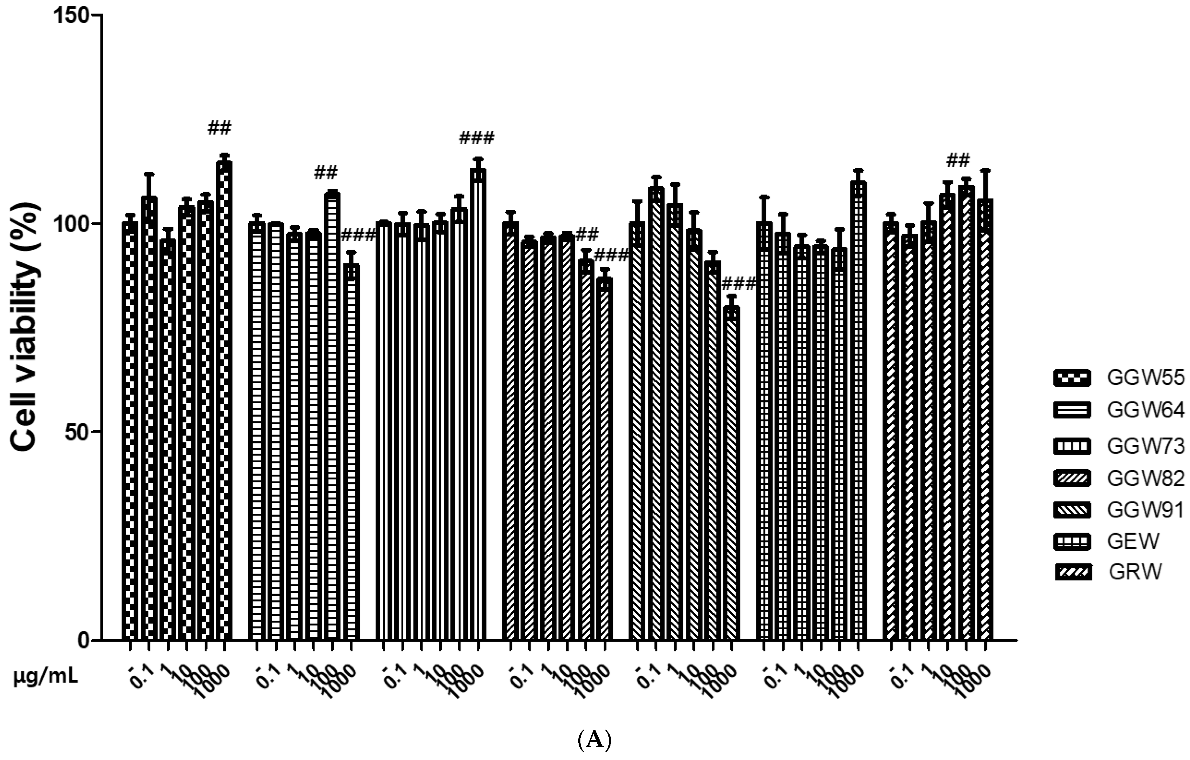
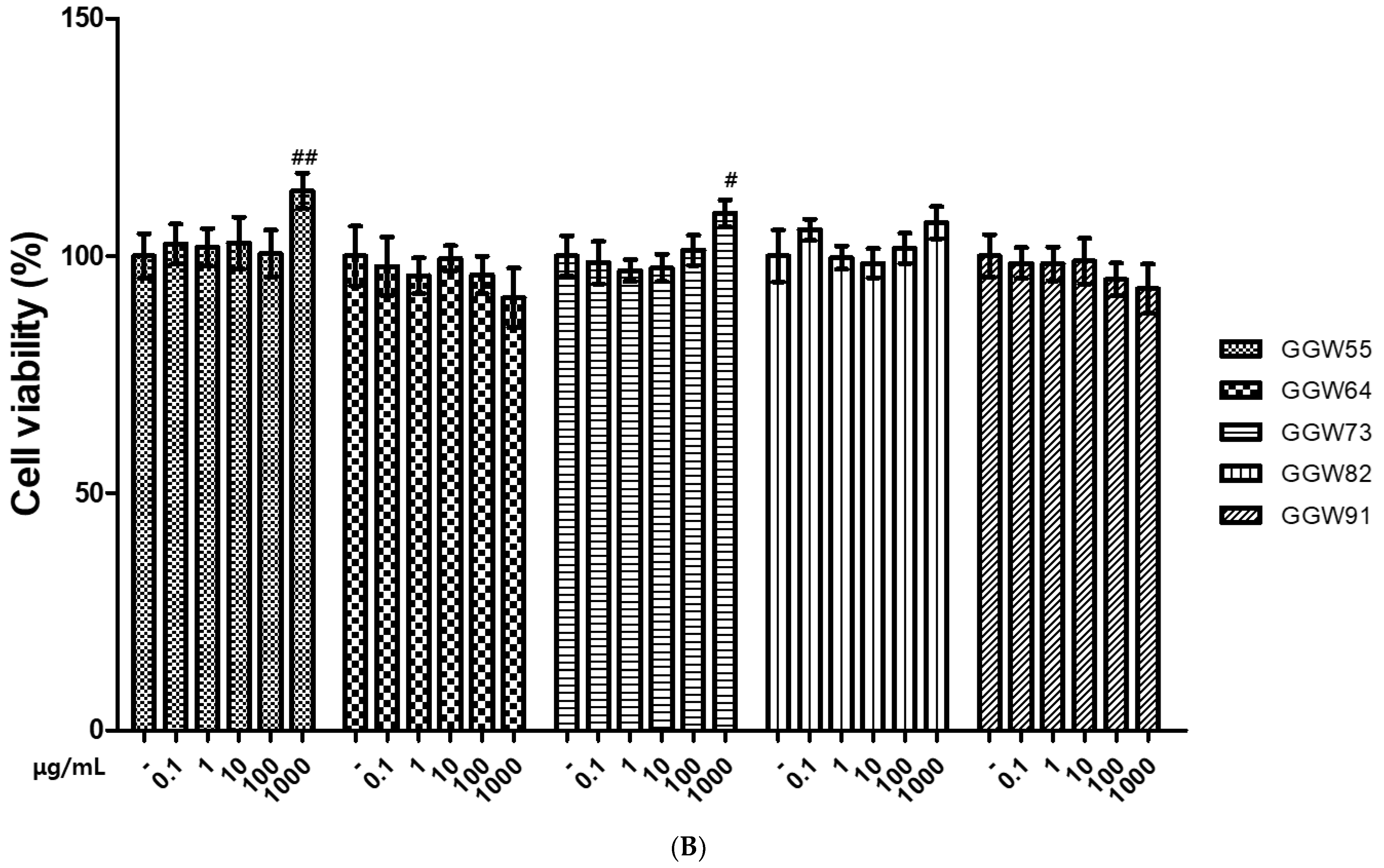
3.1. The Cytotoxicity of GGWs on Neuronal Cells

To choose the mix ratio of GEW and GUW, the authors evaluated cytotoxicity in neuronal cells using an MTT assay. The various ratio of GGWs (5:5, 6:4, 7:3, 8:2, 9:1) were treated on SH-SY-5Y cell. GGW64, GGW82 and GGW91 had cytotoxicity on SH-SY-5Y cell at concentrations of 100 and 1000 μg/mL. However, GGW55 and GGW73 had non-cytotoxicity on SH-SY-5Y cell. Moreover, cell viability increased following GGW55 and GGW73 treatment (Figure 1A). In SK-N-SH cells, GGW64 and GGW91 were cytotoxic. The cell viability was not affected by GGW55 or GGW73 treatment (Figure 1B). Therefore, the ratios of GGW55 and GGW73 were used for the in vitro study.

3.2. The Protective Effect of GGW on Aβ-Treated Neuronal Cells

Aβ 10 μM treatment significantly reduced the cell viability of both SH-SY-5Y cell and SK-N-SH cell. Cytotoxicity caused by Aβ treatment was increased by treatment with GGW55 and GGW73. Especially, GGW55 100 μg/mL and GGW73 100 μg/mL increased the cell viability significantly. In SK-N-SH cells, Aβ treatment reduced the cell viability, and GGW55 and GGW73 treatment increased thr cell viability, respectively (Figure 2). Compared to the protective effects of GGW55 and GGW73, GGW73 had better efficacy.

3.3. The Regulatory Effect of GGW on Aβ-Stimulated ACHE Pathway on SH-SY-5Y Cells

In order to confirm the mechanism of the neuroprotective effects of GGW55 and GGW73, the expression levels of Aβ-stimulated ACHE, BACE-1, and Brain-derived neurotrophic factor (BDNF) in the SH-SY-5Y cell were determined by real-time qPCR and ELISA. The mRNA expression of ACHE and BACE-1 was stimulated by Aβ 2 μM. Both GGW55 and GGW73 treatment downregulated the mRNA expression of ACHE and BACE-1 at a concentration of 100 μg/mL (Figure 3A,B). BDNF expression was decreased by Aβ stimulation. However, decreased expression of BDNF was recovered by dose-dependent GGW55 and GGW73 treatments (Figure 3C). These results suggest that GGW73 had a better neuroprotective effect than GGW55.

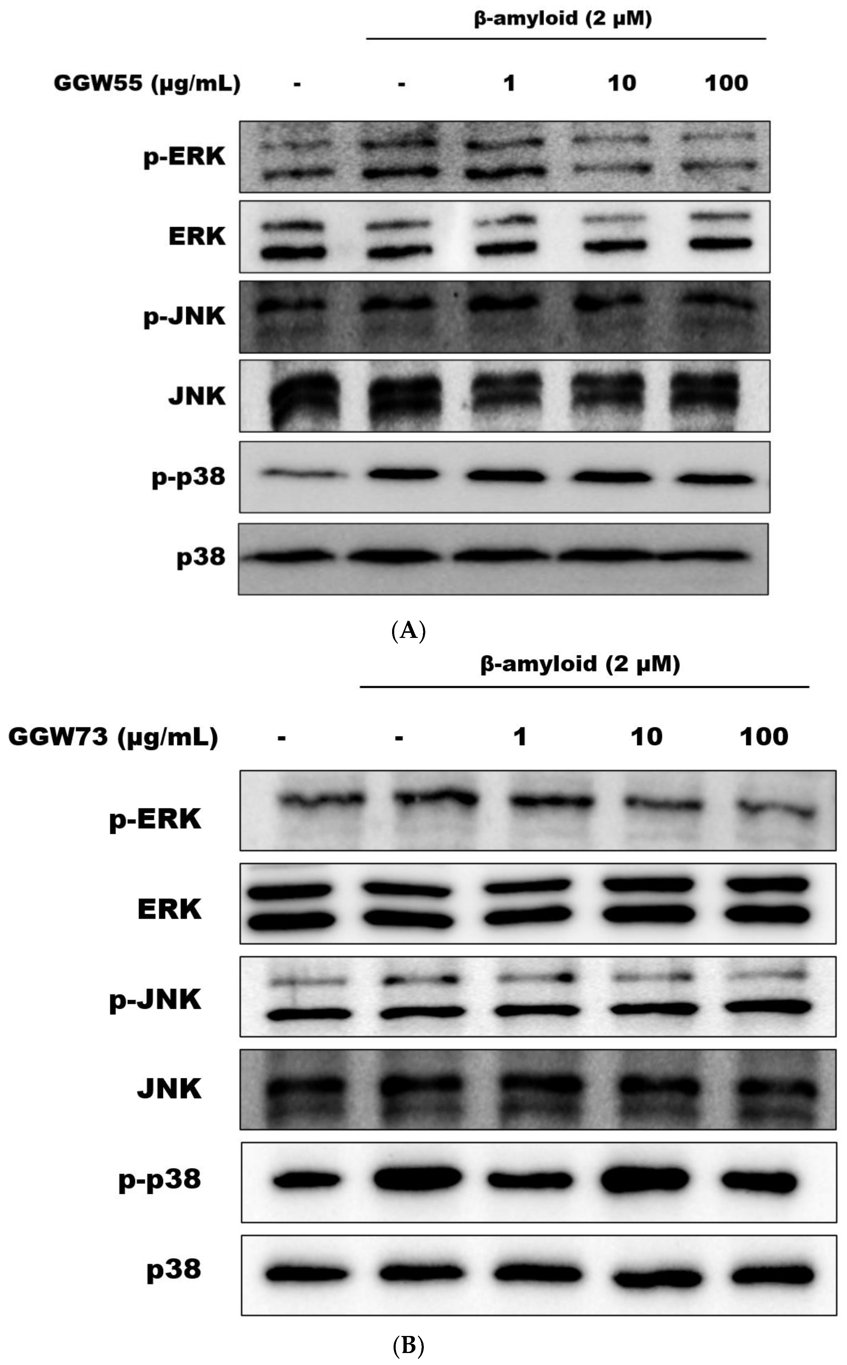
3.4. The Regulatory Effect of GGW on Aβ-Stimulatedmitogen Activated Protein Kinases MAPKs) Expression on Neuronal Cells

To determine the mechanism underlying the neuroprotective effect of GGW, the regulation of Aβ-induced phosphorylation of MAPKs was determined by Western blotting. In SH-Sy-5Y cells, GGW55 treatment regulated only phosphorylation of extracellular signal regulated kinase (ERK). However, GGW73 treatment attenuated the phosphorylation of ERK and p38 (Figure 4A,B). In SK-N-SH cells, GGW55 treatment only regulated phosphorylation of c-Jun N-terminal kinase JNK). However, GGW73 treatment attenuated the phosphorylation of ERK and JNK (Figure 4C,D).

3.5. The Attenuation Effect of GGW on Aβ-Stimulated Apoptotic Factors on SK-N-SH Cells

To clarify the neuroprotective effect of GGW, apoptotic factors were investigated using real-time qPCR and Western blotting. The mRNA expression of Bcl-2 was reduced by Aβ stimulation. GGW55 and GGW73 treatment increased Bcl-2 mRNA expression (Figure 5A). In addition, increased beclin-1 mRNA levels were decreased by GGW55 and GGW73 treatments (Figure 5B). Moreover, the protein expression of Bax and Bcl-2 was regulated by GGW55 and GGW73 treatments (Figure 5C,D). Therefore, from these results, GGW73 was more effective than GGW55.

3.6. The Regulatory Effect of GGW on H2O2-Stimulated ROS on SK-N-SH Cell and RAW264.7 Cell

DCF-DA staining was performed to determine the antioxidant effects of GGW on the neuronal cells. ROS were released by H2O2 or Aβ (data not shown) treatment of SK-N-SH cells. However, ROS production induced by H2O2 was decreased by treatment with GGW55 and GGW73 (Figure 6). RAW264.7 cells were stained for comparison with normal cells. These results indicated that GGW regulates oxidative stress in neuronal cells.

4. Discussion

The present study demonstrated that GGW inhibited cell death by Aβ-treatment in the neuronal cell lines of SH-SY-5Y cell and SK-N-SH cell. Additionally, GGW55 and GGW73 modulated the AChE pathway and MAPKs to prove the neuroprotective effect in SH-SY-5Y cell. Moreover, GGW55 and GGW73 regulated apoptotic factors such as BCl-2 and Bax.
GU has been shown to prevent oxidative damage and cognitive impairment in Aβ-treated mice [35]. In addition, memory deficiencies in mice, induced by diazepam and scopolamine, were improved by the administration of GU water extract [36]. GE has been reported to exert neuroprotective effects by inhibiting Aβ-induced PC12 cell death and ameliorating acute hypomnesia [19]. In addition, GE water extract recovered memory impairment in the Morris water maze test and passive avoidance test in a chronic restraint stress-stimulated mice model [37]. Gastrodin, a major compound of GE, decreased protein kinase/eukaryotic initiation factor-2alpha and BACE-1 expression in an AD mouse model [38]. These studies suggest that GE and GU are effective in improving cognitive function disorders, degenerative neurological disease, and that better effects could be expected using a combination of GE and GU. Among GGWs, GE, and GU treatments, GGW55, GGW73, GEW, and GRW did not show cytotoxicity in the neuronal cell line SH-SY-5Y cell (Figure 1A). Therefore, we chose GGW55 and GGW73 to explore the mechanisms underlying their neuroprotective effects. In addition, GGW55 and GGW73 significantly reduced cell death following Aβ treatment in SH-SY-5Y cell (normal group; 100.00 ± 6.67%, Aβ group; 67.52 ± 9.19%; GGW55 100 group; 79.57 ± 5.96%, GGW73 100 group; 87.04 ± 9.19%) and SK-N-SH cell (normal group: 100 ± 4.06%, GGW55 100 group; 87.72 ± 4.81%, GGW73 100 group; 88.26 ± 5.46%) (Figure 2). In particular, these results suggested that GGW73 treatment had more protective effect on neuronal damage than GGW55.
Impairment of cholinergic function, reduced ACh levels, and a role in the pathogenesis of dementia have been mainly reported in degenerative diseases such as AD and cognitive function disorder [39]. A previous study suggested that AChE, which downregulates ACh, is a biomarker of neurotoxicity [40]. In addition, AChE is associated with neuronal mineralization and differentiation [41]. Attempts to treat cognitive function disorders have been made using AChE inhibitors such as donepezil, tacrine, and rivastigmine [42]. Both GGW55 and GGW73 attenuated AChE mRNA expression, and GGW73 at 100 μg/mL significantly reduced AChE mRNA expression (Figure 3A).
BDNF is primarily responsible for maintaining the central nervous system and is widely expressed in the brain [43]. However, neurological inflammation significantly reduces BDNF expression in the brain, leading to cognitive function disorder [44]. β-secretase (BACE-1), together with γ-secretase, sequentially cleave APP to produce Aβ. Inhibiting the expression of BACE-1 to prevent degenerative disease is considered a strategy to remedy cognitive function disorders by lowering cerebral Aβ levels [45]. GGW55 and GGW73 increased BDNF expression upregulation by Aβ treatment (GGW55: normal group; 1855.60 ± 494.11 pg/mL, Aβ group; 1219.65 ± 119.52 pg/mL, GGW55 100 group; 1422.26 ± 450.03 pg/mL, GGW73: normal group; 2041.90 ± 213.48 pg/mL, Aβ group; 1019.04 ± 152.24 pg/mL, GGW73 100 group; 1608.13 ± 239.95 pg/mL) (Figure 3C) and decreased BACE-1 mRNA expression (Figure 3B). These results indicate that GGWs have a protective effect against the Aβ-induced neuronal damage. In addition, from the view point of central nervous system disorder, GGW is expected to have a positive effect on hydrocephalus, which occurs in the adult population with a ratio of 0.011% [46]. The previous study discovered the course of the hydrocephalus patients after shunting by observing brain volume and pressure change [47]. It requires more research into hydrocephalus using GGW as a natural origin-medicine treatment.
MAPKs signaling, especially ERK1/2, is necessary to activate transcription during long and short-term memory formation [48]. MAPKs, including ERK, (JNK), and p38, play important roles in attenuating neuronal inflammation and plasticity. Moreover, these separate pathways are related to memory and learning functions [49]. In this study, GGW55 and GGW73 did not significantly downregulate the phosphorylation of MAPKs signaling, but slightly decreased phosphorylation (Figure 4). These results suggest that the mixtures of GE and GU (GGW55 and GGW73) regulate Aβ-induced neuronal inflammation.
Increased expression of Bax and decreased expression of Bcl-2 have been observed in a memory and learning impairment mouse model [50]. In addition, dopamine-mediated neuronal cell death is associated with neurodegenerative disorders. In particular, the Bcl-2 signaling pathway affects dopaminergic synaptic apoptosis [51]. A previous study reported that regulation of the Bcl-2 signaling pathway is a way to treat neuronal damage [52]. Both GGW55 and GGW73 increased mRNA expression and protein expression of Bcl-2 that had been decreased by Aβ. It was also confirmed that GGW73 ameliorates the Bcl-2 signaling pathway at the protein level (Figure 5). These results indicate that GGW73 exerts a protective effect against neuronal cell apoptosis.
Oxidative stress is observed in early stages of neurodegenerative diseases [53]. Aβ induces ROS [54] and affects ROS activation, thus inducing NADPH oxidase (NOX) and neuronal cell death [55]. The previous study demonstrated that the antioxidant such as edaravone improved cognitive impairment in hypoxia-stimulated rat model by regulation oxidative stress [56]. In this study, DCFDA staining confirmed ROS expression in RAW264.7 cells and SK-N-SH cells. GGW55 and GGW73 inhibited ROS overexpression induced by Aβ and H2O2 stimulation (Figure 6). These results suggest that GGW55 and GGW73 exert protective effects against oxidative stress in the nervous system.

5. Conclusions

We investigated the protective effects of GGW55 and GGW73 against Aβ-induced neuronal cell damage. Both GGW55 and GGW73 helped repair Aβ-stimulated cell death and the Bcl-2 pathway. GGW55 and GGW73 treatment attenuated mechanisms in cognitive function disorder by regulating AChE and BACE-1 expression. In addition, GGW55 and GGW73 protected against apoptosis through the Bcl-2 signaling pathway and ROS regulation. Considering these results, GGW55 and GGW73 could be used in the medical treatment of cognitive function disorders, such as neurodegenerative diseases.

Author Contributions

Y.-D.J., J.-W.L. and M.-R.P. designed the experiments. S.-H.H., S.-B.P., D.-Y.Y., W.-W.L. and J.-Y.C. conducted the experiments. Y.-D.J. analyzed the experimental data. S.-H.H., S.-B.P. and Y.-D.J. wrote the manuscript. J.-W.L. and M.-R.P. edited the manuscript. All authors have read and agreed to the published version of the manuscript.

Funding

This research was financially supported by the Ministry of Trade, Industry and Energy (MOTIE) and the Korea Institute for Advancement of Technology (KIAT) through the National Innovation Cluster R&D Program (P0016236).

Institutional Review Board Statement

Not applicable.

Informed Consent Statement

Not applicable.

Data Availability Statement

Not applicable.

Conflicts of Interest

The authors declare no conflict of interest.

References

  1. Pen, A.E.; Jensen, U.B. Current status of treating neurodegenerative disease with induced pluripotent stem cells. Acta Neurol. Scand. 2016, 135, 57–72. [Google Scholar] [CrossRef] [PubMed]
  2. Department of Economic and Social Affairs, Population Division. World Population Ageing, United Nations 2019; United Nations: New York, NY, USA, 2019; p. 5. [Google Scholar]
  3. Gavrilova, S.I.; Alvarez, A. Cerebrolysin in the Therapy of Mild Cognitive Impairment and Dementia Due to Alzheimer’s Disease: 30 Years of Clinical Use. Med. Res. Rev. 2021, 41, 2775–2803. [Google Scholar] [CrossRef] [PubMed]
  4. Wimo, A.; Jönsson, L.; Bond, J.; Prince, M.; Winblad, B.; International, A.D. Worldwide economic impact of dementia 2010. Alzheimers Dement. 2013, 9, 1–11.e3. [Google Scholar] [CrossRef] [PubMed]
  5. Vassar, R.; Kandalepas, P.C. The β-secretase enzyme BACE1 as a therapeutic target for Alzheimer ’s disease. Alzheimers Res. Ther. 2011, 3, 20. [Google Scholar] [CrossRef] [PubMed]
  6. Lauterbach, E.C.; Victoroff, J.; Coburn, K.L.; Shillcutt, S.D.; Doonan, S.M.; Mendez, M.F. Psychopharmacological neuroprotection in neurodegenerative disease: Assessing the preclinical data. J. Neuropsychiatry Clin. Neurosci. 2010, 22, 8–18. [Google Scholar] [CrossRef]
  7. Sperling, R.A.; Dickerson, B.C.; Pihlajamaki, M.; Vannini, P.; LaViolette, P.S.; Vitolo, O.V.; Hedden, T.; Becker, J.A.; Rentz, D.M.; Selkoe, D.J.; et al. Functional alterations in memory networks in early Alzheimer’s disease. Neuromol. Med. 2010, 12, 27–43. [Google Scholar] [CrossRef] [Green Version]
  8. Schütt, T.; Helboe, L.; Pedersen, L.Ø.; Waldemar, G.; Berendt, M.; Pedersen, J.T. Dogs with cognitive dysfunction as a spontaneous model for early Alzheimer’s disease: A translational study of neuropathological and inflammatory markers. J. Alzheimers Dis. 2016, 52, 433–449. [Google Scholar] [CrossRef]
  9. Chen, J.H.; Ke, K.F.; Lu, J.H.; Qiu, Y.H.; Peng, Y.P. Protection of TGF-β 1 against neroinflammation and neurodegeneration in Aβ1-42-induced Alzheimer’s disease model rats. PLoS ONE 2015, 10, e0116549. [Google Scholar]
  10. Mattson, M.P. Pathways towards and away from Alzheimer ’s disease. Nature 2004, 430, 631–639. [Google Scholar] [CrossRef] [Green Version]
  11. Findeis, M.A. The role of amyloid beta peptide 42 in Alzheimer ’s disease. Pharmacol. Ther. 2007, 116, 266–286. [Google Scholar] [CrossRef]
  12. Panza, F.; Seripa, D.; Solfrizzi, V.; Imbimbo, B.P.; Lozupone, M.; Leo, A.; Sardone, R.; Gagliardi, G.; Lofano, L.; Creanza, B.C.; et al. Emerging drugs to reduce abnormal β-amyloid protein in Alzheimer ’s disease patients. Expert Opin. Emerg. Drugs 2016, 21, 377–391. [Google Scholar] [CrossRef] [PubMed]
  13. Weon, J.B.; Yun, B.R.; Lee, J.; Eom, M.R.; Ko, H.J.; Lee, H.Y.; Park, D.S.; Chung, H.C.; Chung, J.Y.; Ma, C.J. Cognitive-Enhancing Effect of Steamed and Fermented Codonopsis lanceolata: A Behavioral and Biochemical Study. Evid.-Based Complement. Altern. Med. 2014, 2014, 319436. [Google Scholar] [CrossRef] [PubMed] [Green Version]
  14. Mangialasche, F.; Solomon, A.; Winblad, B.; Mecocci, P.; Kivipelto, M. Alzheimer ’s disease: Clinical trials and drug development. Lancet Neurol. 2010, 9, 702–716. [Google Scholar] [CrossRef]
  15. Hassel, B.; Solyga, V.; Lossius, A. High-affinity choline uptakeandacetylcholine-metabolizing enzymes in CNS white matter. A quantitative study. Neurochem. Int. 2008, 53, 193–198. [Google Scholar] [CrossRef]
  16. Lee, D.W.; Huh, Y.S.; Kim, K.W. Evidence-based treatment of Alzheimer’s disease. J. Korean Med. Assoc. 2009, 52, 417–425. [Google Scholar] [CrossRef] [Green Version]
  17. LaFerla, F.M.; Green, K.N.; Oddo, S. Intracellular amyloid-beta in Alzheimer’s disease. Nat. Rev. Neurosci. 2007, 8, 499–509. [Google Scholar] [CrossRef] [PubMed]
  18. Li, Y.; Zhou, W.; Tong, Y.; He, G.; Song, W. Control of APP processing and Abeta generation level by BACE1 enzymatic activity and transcription. FASEB J. 2006, 20, 285–292. [Google Scholar] [CrossRef] [Green Version]
  19. Park, Y.M.; Lee, B.G.; Park, S.H.; Oh, H.G.; Kang, Y.G.; Kim, O.J.; Kwon, L.S.; Kim, Y.P.; Choi, M.H.; Jeong, Y.S.; et al. Prolonged oral administration of Gastrodia elata extract improves spatial learning and memory of scopolamine-treated rats. Lab. Anim. Res. 2015, 31, 69–77. [Google Scholar] [CrossRef] [Green Version]
  20. Zhu, L.; Guan, H.; Cui, C.; Tian, S.; Yang, D.; Wang, X.; Zhang, S.; Wang, L.; Jiang, H. Gastrodin inhibits cell proliferation in vascular smooth muscle cells and attenuates neointima formation in vivo. Int. J. Mol. Med. 2012, 30, 1034–1040. [Google Scholar] [CrossRef] [Green Version]
  21. Hsieh, C.L.; Tang, N.Y.; Chiang, S.Y.; Hsieh, C.T.; Lin, J.G. Anticonvulsive and free radical scavenging actions of two herbs, Uncaria rhynchophylla (MIQ) Jack and Gastrodia elata Bl., in kainic acid-treated rats. Life Sci. 1999, 65, 2071–2082. [Google Scholar] [CrossRef]
  22. Ha, J.H.; Lee, D.U.; Lee, J.T.; Kim, J.S.; Yong, C.S.; Kim, J.A.; Ha, J.S.; Huh, K. 4-Hydroxybenzaldehyde from Gastrodia elata B1. is active in the antioxidation and GABAergic neuromodulation of the rat brain. J. Ethnopharmacol. 2000, 73, 329–333. [Google Scholar] [CrossRef] [PubMed]
  23. Mishra, M.; Huang, J.; Lee, Y.Y.; Chua, D.S.; Lin, X.; Hu, J.M.; Heese, K. Gastrodia elata modulates amyloid precursor protein cleavage and cognitive functions in mice. Biosci. Trends 2011, 5, 129–138. [Google Scholar] [CrossRef] [PubMed] [Green Version]
  24. Liu, Y.; Gao, J.; Peng, M.; Meng, H.; Ma, H.; Cai, P.; Xu, Y.; Zhao, Q.; Si, G. A review on central nervous system effects of gastrodin. Front. Pharmacol. 2018, 9, 24. [Google Scholar] [CrossRef] [PubMed]
  25. Wang, X.L.; Xing, G.H.; Hong, B.; Li, X.M.; Zou, Y.; Zhang, X.J.; Dong, M.X. Gastrodin prevents motor deficits and oxidative stress in the MPTP mouse model of parkinson’s disease: Involvement of ERK1/2-Nrf2 signaling pathway. Life Sci. 2014, 114, 77–85. [Google Scholar] [CrossRef] [PubMed]
  26. Liao, W.C.; Lin, Y.H.; Chang, T.M.; Huang, W.Y. Identification of two licorice species, Glycyrrhiza uralensis and Glycyrrhiza glabra, based on separation and identification of their bioactive components. Food Chem. 2012, 132, 2188–2193. [Google Scholar] [CrossRef]
  27. Mochida, K.; Sakurai, T.; Seki, H.; Yoshida, T.; Takahagi, K.; Sawai, S.; Uchiyama, H.; Muranaka, T.; Saito, K. Draft genome assembly and annotation of Glycyrrhiza uralensis, a medicinal legume. Plant J. 2017, 89, 181–194. [Google Scholar] [CrossRef]
  28. Asl, M.N.; Hosseinzadeh, H. Review of pharmacological effects of Glycyrrhiza sp. and its bioactive compounds. Phytother. Res. 2008, 22, 709–724. [Google Scholar] [CrossRef]
  29. Hosseinzadeh, H.; Nassiri-Asl, M. Pharmacological effects of Glycyrrhiza spp. and its bioactive constituents: Update and review. Phytother. Res. 2015, 29, 1868–1886. [Google Scholar] [CrossRef]
  30. Nomura, T.; Fukai, T. Phenolic constituents of licorice (Glycyrrhiza species). Fortschr. Chem. Org. Naturst. 1998, 73, 1–158. [Google Scholar]
  31. Lee, H.K.; Yang, E.J.; Kim, J.Y.; Song, K.S.; Seong, Y.H. Inhibitory effects of Glycyrrhizae radix and its active component, isoliquiritigenin, on Aβ(25-35)-induced neurotoxicity in cultured rat cortical neurons. Arch. Pharm. Res. 2012, 35, 897–904. [Google Scholar] [CrossRef]
  32. Ahn, J.; Um, M.; Choi, W.; Kim, S.; Ha, T. Protective effects of Glycyrrhiza uralensis Fisch. on the cognitive deficits caused by beta-amyloid peptide 25-35 in young mice. Biogerontology 2006, 7, 239–247. [Google Scholar] [CrossRef] [PubMed]
  33. Luo, L.; Jin, Y.; Kim, I.D.; Lee, J.K. Glycyrrhizin attenuates kainic Acid- induced neuronal cell death in the mouse hippocampus. Exp. Neurobiol. 2013, 22, 107–115. [Google Scholar] [CrossRef] [PubMed]
  34. Hwang, C.K.; Chun, H.S. Isoliquiritigenin isolated from licorice glycyrrhiza uralensis prevents 6-hydroxydopamine-induced apoptosis in dopaminergic neurons. Biosci. Biotechnol. Biochem. 2012, 76, 536–543. [Google Scholar] [CrossRef] [PubMed] [Green Version]
  35. Parle, M.; Dhingra, D.; Kulkarni, S.K. Memory-strengthening activity of Glycyrrhiza glabra in exteroceptive and interoceptive behavioral models. J. Med. Food 2004, 7, 462–466. [Google Scholar] [CrossRef]
  36. Huang, H.; Zhang, Y.; Yao, C.; He, Q.; Chen, F.; Yu, H.; Lu, G.; Jiang, N.; Liu, X. The effects of fresh Gastrodia elata Blume on the cognitive deficits induced by chronic restraint stress. Front. Pharmacol. 2022, 13, 890330. [Google Scholar] [CrossRef]
  37. Zhang, J.S.; Zhou, S.F.; Wang, Q.; Guo, J.N.; Liang, H.M.; Deng, J.B.; He, W.Y. Gastrodin suppresses BACE1 expression under oxidative stress condition via inhibition of the PKR/eIF2alpha pathway in Alzheimer’s disease. Neuroscience 2016, 325, 1–9. [Google Scholar] [CrossRef]
  38. Maurer, S.V.; Williams, C.L. The cholinergic system modulates memory and hippocampal plasticity via its interactions with non-neuronal cells. Front. Immunol. 2017, 8, 1489. [Google Scholar] [CrossRef] [Green Version]
  39. Lionetto, M.G.; Caricato, R.; Calisi, A.; Giordano, M.E.; Schettino, T. Acetylcholinesterase as a Biomarker in Environmental and Occupational Medicine: New Insights and Future Perspectives. Biomed. Res. Int. 2013, 2013, 321213. [Google Scholar] [CrossRef] [Green Version]
  40. Das, K.P.; Barone, S. Neuronal Differentiation in PC12 Cells Is Inhibited by Chlorpyrifos and Its Metabolites: Is Acetylcholinesterase Inhibition the Site of Action? Toxicol. Appl. Pharmacol. 1999, 160, 217–230. [Google Scholar] [CrossRef]
  41. Lee, J.M.; Ji, E.S.; Kim, T.W.; Kim, C.J.; Shin, M.S.; Lim, B.V.; Chung, Y.R.; Cho, Y.S. Treadmill exercise improves memory function by inhibiting hippocampal apoptosis in pilocarpine-induced epileptic rats. J. Exerc. Rehabil. 2018, 14, 713–723. [Google Scholar] [CrossRef]
  42. Bekinschtein, P.; Cammarota, M.; Katche, C.; Slipczuk, L.; Rossato, J.I.; Goldin, A.; Izquierdo, I.; Medina, J.H. BDNF is essential to promote persistence of long-term memory storage. Proc. Natl. Acad. Sci. USA 2008, 105, 2711–2716. [Google Scholar] [CrossRef] [PubMed] [Green Version]
  43. Schnydrig, S.; Korner, L.; Landweer, S.; Ernst, B.; Walker, G.; Otten, U.; Kunz, D. Peripheral lipopolysaccharide administration transiently affects expression of brain-derived neurotrophic factor, corticotropin and proopiomelanocortin in mouse brain. Neurosci. Lett. 2007, 429, 69–73. [Google Scholar] [CrossRef] [PubMed]
  44. Yan, R.; Vassar, R. Targeting the β secretase BACE1 for Alzheimer ’s disease therapy. Lancet Neurol. 2014, 13, 319–329. [Google Scholar] [CrossRef] [PubMed] [Green Version]
  45. Feld, M.; Dimant, B.; Delorenzi, A.; Coso, O.; Romano, A. Phosphorylation of extra-nuclear ERK/MAPK is required for long-term memory consolidation in the crab Chasmagnathus. Behav. Brain Res. 2005, 158, 251–261. [Google Scholar] [CrossRef]
  46. Isaacs, A.M.; Riva-Cambrin, J.; Yavin, D.; Hockley, A.; Pringsheim, T.M.; Jette, N.; Lethebe, B.C.; Lowerison, M.; Dronyk, J.; Hamilton, M.G. Age-specific Global Epidemiology of Hydrocephalus: Systematic Review, Metanalysis and Global Birth Surveillance. PLoS ONE 2018, 13, e0204926. [Google Scholar] [CrossRef]
  47. Gholampour, S.; Yamini, B.; Drossler, J.; Frim, D. A new definition for intracranial compliance to evaluate adult hydrocephalus after shunting. Front. Bioeng. Biotechnol. 2022, 10, 900644. [Google Scholar] [CrossRef]
  48. Peng, S.; Zhang, Y.; Zhang, J.; Wang, H.; Ren, B. ERK in learning and memory: A review of recent research. Int. J. Mol. Sci. 2010, 11, 222–232. [Google Scholar] [CrossRef] [Green Version]
  49. Tabas, I.; Ron, D. Integrating the mechanisms of apoptosis induced by endoplasmic reticulum stress. Nat. Cell Biol. 2011, 13, 184–190. [Google Scholar] [CrossRef] [Green Version]
  50. Zhang, Q.; Hu, C.; Huang, J.; Liu, W.; Lai, W.; Leng, F.; Tang, Q.; Liu, Y.; Wang, Q.; Zhou, M.; et al. ROCK1 Induces Dopaminergic Nerve Cell Apoptosis via the Activation of Drp1- Mediated Aberrant Mitochondrial Fission in Parkinson’s Disease. Exp. Mol. Med. 2019, 51, 1–13. [Google Scholar] [CrossRef] [Green Version]
  51. Huang, Q.; Zhang, C.; Qu, S.; Dong, S.; Ma, Q.; Hao, Y.; Liu, Z.; Wang, S.; Zhao, H.; Shi, Y. Chinese Herbal Extracts Exert Neuroprotective Effect in Alzheimer’s Disease Mouse Through the Dopaminergic Synapse/Apoptosis Signaling Pathway. Front. Pharmacol. 2022, 13, 817213. [Google Scholar] [CrossRef]
  52. Smith, M.A.; Rottkamp, C.A.; Nunomura, A.; Raina, A.K.; Perry, G. Oxidative stress in Alzheimer’s disease. Biochim. Biophys. Acta 2000, 1502, 139–144. [Google Scholar] [CrossRef] [PubMed] [Green Version]
  53. Smith, C.D.; Carney, J.M.; Starke-Reed, P.E.; Oliver, C.N.; Stadtman, E.R.; Floyd, R.A.; Markesbery, W.R. Excess brain protein oxidation and enzyme dysfunction in normal aging and in Alzheimer disease. Proc. Natl. Acad. Sci. USA 1991, 88, 10540–10543. [Google Scholar] [CrossRef] [PubMed]
  54. Lee, Y.; Kim, S.; Oh, Y.; Kim, Y.-M.; Chin, Y.-W.; Cho, J. Inhibition of Oxidative Neurotoxicity and Scopolamine-Induced Memory Impairment by γ-Mangostin: In Vitro and In Vivo Evidence. Oxidative Med. Cell. Longev. 2019, 2019, 3640753. [Google Scholar] [CrossRef] [PubMed] [Green Version]
  55. Shelat, P.B.; Chalimoniuk, M.; Wang, J.H.; Strosznajder, J.B.; Lee, J.C.; Sun, A.Y.; Simonyi, A.; Sun, G.Y. Amyloid beta peptide and NMDA induce ROS from NADPH oxidase and AA release from cytosolic phospholipase A2 in cortical neurons. J. Neurochem. 2008, 106, 45–55. [Google Scholar] [CrossRef]
  56. Ling, J.; Yu, Q.; Yuan, X.; Liu, W.; Guo, Y.; Duan, Y.; Li, L. Edaravone Improves Intermittent Hypoxia-Induced Cognitive Impairment and Hippocampal Damage in Rats. Biol. Pharm. Bull. 2020, 43, 1196–1201. [Google Scholar] [CrossRef]
Figure 1. The cytotoxicity of GGWs on neuronal cells. The cytotoxicity of GGWs on SH-SY-5Y cell and SK-N-SH cell. The cells were treated with GGWs (GGW55, GGW64, GGW73, GGW82, GGW91), and treated with GEW and GRW for 24 h. Cell viability was evaluated with absorbance of formazan crystal. (A) SH-SY-5Y cell. (B) SK-N-SH cell. All values are expressed as the mean ± S.E.M from three independent experiments.; # p < 0.05 compared with none-treatment group; ## p < 0.005 compared with none-treatment group; ### p < 0.001 compared with none-treatment group.
Applsci 13 00190 g001aApplsci 13 00190 g001b
Figure 2. The protective effect of GGW on Aβ-treated neuronal cells. The protective effect of GGW55 and GGW73 on Aβtreated SH-SY-5Y cell and SK-N-SH cell. The cells were treated with GGW55 and GGW73, after 4 h Aβ (10 μM) were treated for 24 h. Cell viability was evaluated with absorbance of formazan crystal. (A) SH-SY-5Y cell. (B) SK-N-SH cell. All values are expressed as the mean ± S.E.M from three independent experiments.; ### p < 0.001 compared with none-treatment group; * p < 0.05 compared with none-treatment group, *** p < 0.001 compared with only Aβ-treatment group.
Applsci 13 00190 g002
Figure 3. The regulatory effect of GGW on Aβ-stimulated ACHE pathway on SH-SY-5Y cells. The regulatory effect of GGW55 and GGW73 on Aβ-stimulated ACHE pathway on SH-SY-5Y cell. The cells were treated with GGW55 and GGW73, after 4 h Aβ (2 μM) were treated for 24 h. (A) ACHE mRNA expression was measured real-time qPCR. (B) BACE-1 mRNA expression was measured with real-time qPCR. (C) BDNF secretion was measured with ELISA. All values are expressed as the mean ± S.E.M from three independent experiments.; # p < 0.05 compared with none-treatment group; ## p < 0.005 compared with none-treatment group; ### p < 0.001 compared with none-treatment group; * p < 0.05 compared with none-treatment group, *** p < 0.001 compared with only Aβ-treatment group.
Applsci 13 00190 g003aApplsci 13 00190 g003b
Figure 4. The regulatory effect of GGW on Aβ-stimulated MAPKs expression on neuronal cells. The regulatory effect of GGW55 and GGW73 on Aβ-stimulated MAPKs expression on SH-SY-5Y cell and SK-N-SH cell. The cells were treated with GGW55 and GGW73, after 1 h Aβ (2 μM) were treated for 45 min. (A) MAPKs expression on SH-SY-5Y cell with GGW55. (B) MAPKs expression on SH-SY-5Y cell with GGW73. (C) MAPKs expression on SK-N-SH cell with GGW55. (D) MAPKs expression on SK-N-SH cell with GGW73. The MAPKs expressions were measured using Western blot analysis.
Applsci 13 00190 g004aApplsci 13 00190 g004b
Figure 5. The attenuation effect of GGW on Aβ-stimulated apoptotic factors on SK-N-SH cells. The attenuation effect of GGW55 and GGW73 on Aβ-stimulated apoptotic factors on SK-N-SH cell. The cells were treated with GGW55 and GGW73, after 4 h Aβ (2 μM) were treated for 24 h. (A) Bcl-2 mRNA expression on SK-N-SH cell with GGW55 and GGW73. (B) Beclin-1 mRNA expression on SK-N-SH cell with GGW55 and GGW73. (C) Bax and Bcl-2 expression on SK-N-SH cell with GGW55. (D) Bax and Bcl-2 expression on SK-N-SH cell with GGW73. The protein expressions were measured using Western blot analysis. All values are expressed as the mean ± S.E.M from three independent experiments.; # p < 0.05 compared with none-treatment group; * p < 0.05 compared with none-treatment group.
Applsci 13 00190 g005aApplsci 13 00190 g005b
Figure 6. The regulatory effect of GGW55 and GGW73 on H2O2-stimulated ROS expression on SK-N-SH cell and RAW264.7. The fluorescent staining was observed with fluorescence microscope (100× magnification).
Applsci 13 00190 g006
Table 1. Primer sequence for qPCR.
GeneForwardReverse
h GAPDH5′-TCTAGACGGCAGGTCAGGTCCACC-3′5′-CCACCCATGGCAAATTCCATGGCA-3′
h AChE5′-ACAGGTCTGAGCAGCGATCCTGCTTGCT-3′5′-TACGCCTACGTCTTGAACACCGTGCTTC-3
h iNOS5′-GCTCTACACCTCCAATGTGACC-3′5′-CTGCCGAGATTTGAGCCTCATG-3′
h Beclin-15′-CCATGCAGGTGAGCTTCGT-3′5′-GAATCTGCGAGAGACACCATC-3′
h Bcl-25′-ATGTGTGTGGAGAGCGTCAA-3′5′-GCCGGTTCAGGTACTCAGTC-3′
h BACE-1 5′- TCTGTCGGAGGGAGCATGAT-3′5′- GCAAACGAAGGTTGGTGGT-3′
Disclaimer/Publisher’s Note: The statements, opinions and data contained in all publications are solely those of the individual author(s) and contributor(s) and not of MDPI and/or the editor(s). MDPI and/or the editor(s) disclaim responsibility for any injury to people or property resulting from any ideas, methods, instructions or products referred to in the content.

