
Bone Modifications Induced by Rapid Maxillary Expander: A Three-Dimensional Cephalometric Pilot Study Comparing Two Different Cephalometric Software Programs

 , , , , ,
Abstract
:1. Introduction
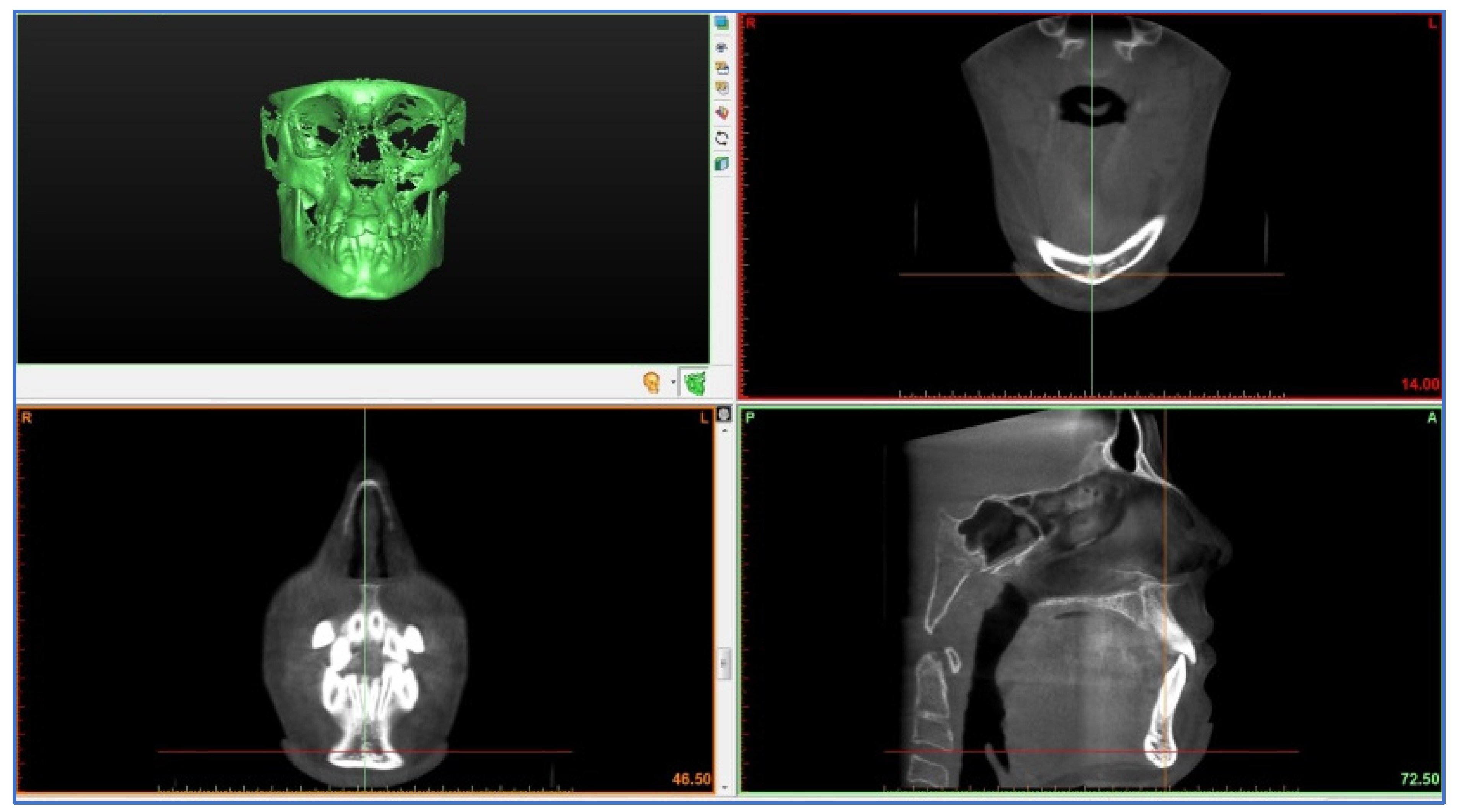
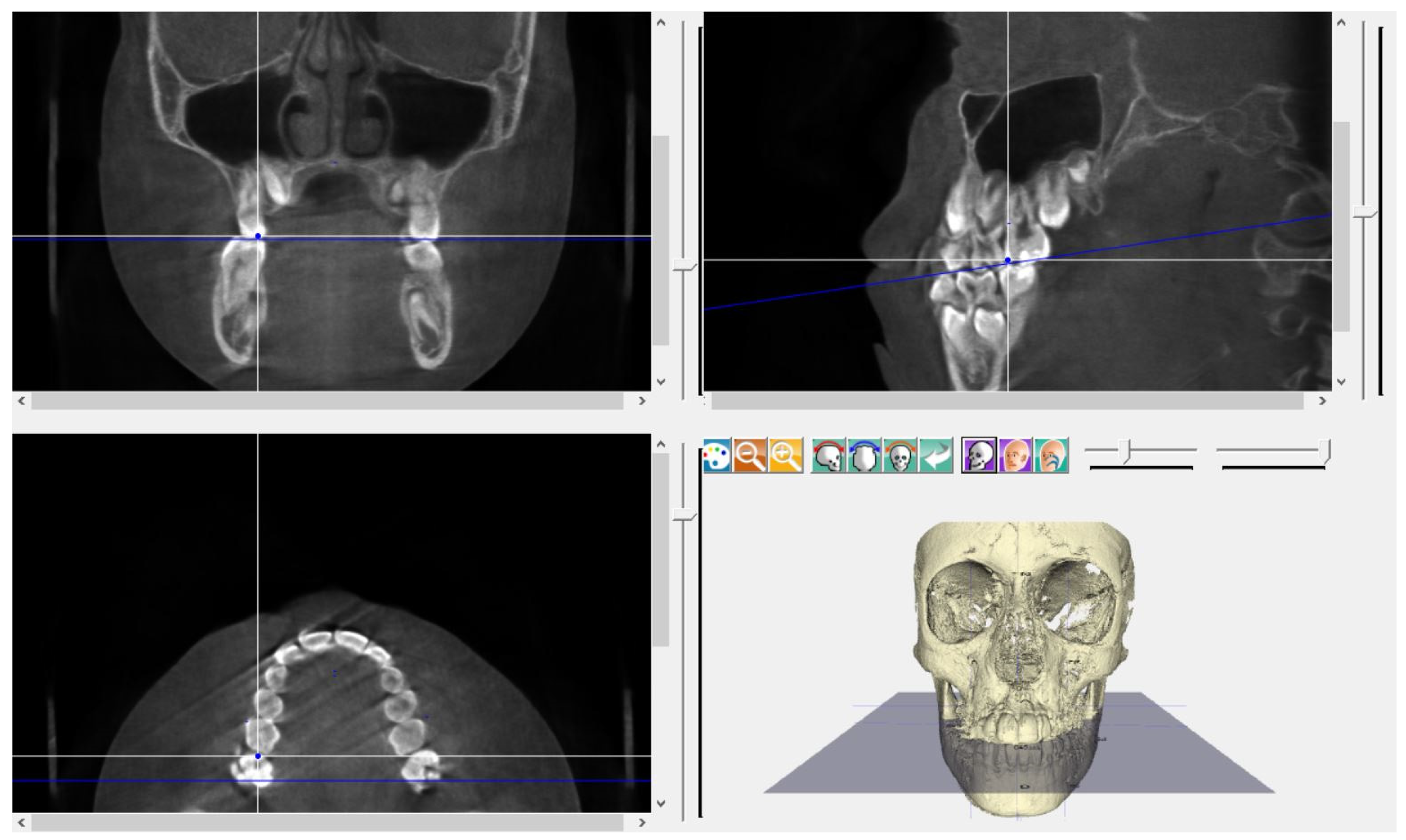
2. Materials and Methods
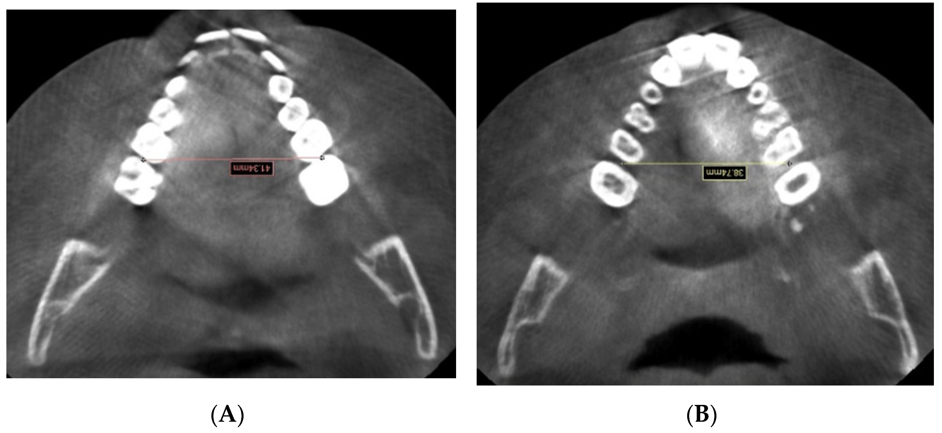
3. Results
4. Discussion
5. Conclusions
Author Contributions
Funding
Institutional Review Board Statement
Informed Consent Statement
Data Availability Statement
Conflicts of Interest
References
- Yitschaky, O.; Redlich, M.; Abed, Y.; Faerman, M.; Casap, N.; Hiller, N. Comparison of common hard tissue cephalometric measurements between computed tomography 3D reconstruction and conventional 2D cephalometric images. Angle Orthod. 2011, 81, 11–16. [Google Scholar] [CrossRef] [PubMed]
- Feragalli, B.; Rampado, O.; Abate, C.; Macrì, M.; Festa, F.; Stromei, F.; Caputi, S.; Guglielmi, G. Cone beam computed tomography for dental and maxillofacial imaging: Technique improvement and low-dose protocols. Radiol. Med. 2017, 122, 581–588. [Google Scholar] [CrossRef] [PubMed]
- Scarfe, W.C.; Azevedo, B.; Toghyani, S.; Farman, A.G. Cone Beam Computed Tomographic imaging in orthodontics. Aust Dent. J. 2017, 62 (Suppl. 1), 33–50. [Google Scholar] [CrossRef] [PubMed] [Green Version]
- Kapila, S. Contemporary concepts on cone-beam computed tomography in orthodontics. In Cone Beam Computed Tomography in Orthodontics: Indications, Insights and Innovations; Kapila, S., Ed.; Wiley-Blackwell: Hoboken, NJ, USA, 2014; pp. 5–42. [Google Scholar]
- Kapila, S.D.; Nervina, J.M. CBCT in orthodontics: Assessment of treatment outcomes and indications for its use. Dentomaxillofac. Radiol. 2015, 44, 20140282. [Google Scholar] [CrossRef] [Green Version]
- Salagnac, J.M. Proposition d’un indice définissant les indications médicales d’une disjonction intermaxillaire chez un sujet en période de croissance [Proposal for a clinical diagnosis index in rapid palatal expansion (RPE) in childhood]. Orthod. Fr. 2019, 90, 127–135. [Google Scholar] [CrossRef]
- Ghoneima, A.; Abdel-Fattah, E.; Hartsfield, J.; El-Bedwehi, A.; Kamel, A.; Kula, K. Effects of rapid maxillary expansion on the cranial and circummaxillary sutures. Am. J. Orthod. Dentofac. Orthop. 2011, 140, 510–519. [Google Scholar] [CrossRef] [Green Version]
- Haas, A.J. Rapid expansion of the maxillary dental arch and nasal cavity by opening the mid palatal suture. Angle Orthod. 1961, 31, 73–89. [Google Scholar]
- Starnbach, H.; Bayne, D.; Cleall, J.; Subtelny, J.D. Facioskeletal and dental changes resulting from rapid maxillary expansion. Angle Orthod. 1966, 36, 152–164. [Google Scholar]
- Babacan, H.; Sokucu, O.; Doruk, C.; Ay, S. Rapid maxillary expansion and surgically assisted rapid maxillary expansion effects on nasal volume. Angle Orthod. 2006, 76, 66–71. [Google Scholar]
- Christie, K.F.; Boucher, N.; Chung, C.H. Effects of bonded rapid palatal expansion on the transverse dimensions of the maxilla: A cone-beam computed tomography study. Am. J. Orthod. Dentofac. Orthop. 2010, 137 (Suppl. 4), S79–S85. [Google Scholar] [CrossRef]
- Pereira, J.D.S.; Jacob, H.B.; Locks, A.; Brunetto, M.; Ribeiro, G.L.U. Evaluation of the rapid and slow maxillary expansion using cone-beam computed tomography: A randomized clinical trial. Dent. Press J. Orthod. 2017, 22, 61–68. [Google Scholar] [CrossRef] [PubMed] [Green Version]
- International Commission on Radiological Protection. 1990 recommendations of the International Commission on Radiological Protection. ICRP publication 60. Ann. ICRP 1991, 21, 1–201. [Google Scholar]
- International Commission on Radiological Protection. The 2007 recommendations of the International Commission on Radiological Protection. ICRP publication 103. Ann. ICRP 2008, 37, 1–332. [Google Scholar]
- Durao, A.R.; Pittayapat, P.; Rockenbach, M.I.; Olszewski, R.; Ng, S.; Ferreira, A.P.; Jacobs, R. Validity of 2D lateral cephalometry in orthodontics: A systematic review. Prog. Orthod. 2013, 14, 31. [Google Scholar] [CrossRef] [Green Version]
- Rischen, R.J.; Breuning, K.H.; Bronkhorst, E.M.; Kuijpers-Jagtman, A.M. Records needed for orthodontic diagnosis and treatment planning: A systematic review. PLoS ONE 2013, 8, e74186. [Google Scholar] [CrossRef] [Green Version]
- Oz, U.; Orhan, K.; Abe, N. Comparison of linear and angular measurements using two-dimensional conventional methods and three-dimensional cone beam CT images reconstructed from a volumetric rendering program in vivo. Dentomaxillofac. Radiol. 2011, 40, 492–500. [Google Scholar] [CrossRef] [Green Version]
- Kumar, V.; Ludlow, J.; Soares Cevidanes, L.H.; Mol, A. In vivo comparison of conventional and cone beam CT synthesized cephalograms. Angle Orthod. 2008, 78, 873–879. [Google Scholar] [CrossRef]
- Park, J.H.; Hwang, C.J.; Choi, Y.J.; Houschyar, K.S.; Yu, J.H.; Bae, S.Y.; Cha, J.Y. Registration of digital dental models and cone-beam computed tomography images using 3-dimensional planning software: Comparison of the accuracy according to scanning methods and software. Am. J. Orthod. Dentofac. Orthop. 2020, 157, 843–851. [Google Scholar] [CrossRef]
- Sawchuk, D.; Alhadlaq, A.; Alkhadra, T.; Carlyle, T.D.; Kusnoto, B.; El-Bialy, T. Comparison of two three-dimensional cephalometric analysis computer software. J. Orthod. Sci. 2014, 3, 111–117. [Google Scholar]
- Farronato, G.; Garagiola, U.; Dominici, A.; Periti, G.; de Nardi, S.; Carletti, V.; Farronato, D. “Ten-point” 3D cephalometric analysis using low-dosage cone beam computed tomography. Prog. Orthod. 2010, 11, 2–12. [Google Scholar] [CrossRef]
- Burkhard, J.P.; Dietrich, A.D.; Jacobsen, C.; Roos, M.; Lübbers, H.T.; Obwegeser, J.A. Cephalometric and three-dimensional assessment of the posterior airway space and imaging software reliability analysis before and after orthognathic surgery. J. Craniomaxillofac. Surg. 2014, 42, 1428–1436. [Google Scholar] [CrossRef] [PubMed]
- Koretsi, V.; Tingelhoff, L.; Proff, P.; Kirschneck, C. Intra-observer reliability and agreement of manual and digital orthodontic model analysis. Eur. J. Orthod. 2018, 40, 52–57. [Google Scholar] [CrossRef] [PubMed]
- Joda, T.; Brägger, U. Digital vs. conventional implant prosthetic workflows: A cost/time analysis. Clin. Oral. Implant. Res. 2015, 26, 1430–1435. [Google Scholar] [CrossRef] [PubMed]
- Coachman, C.; Calamita, M.A.; Coachman, F.G.; Coachman, R.G.; Sesma, N. Facially generated and cephalometric guided 3D digital design for complete mouth implant rehabilitation: A clinical report. J. Prosthet. Dent. 2017, 117, 577–586. [Google Scholar] [CrossRef] [PubMed]
- Baan, F.; de Waard, O.; Bruggink, R.; Xi, T.; Ongkosuwito, E.M.; Maal, T.J.J. Virtual setup in orthodontics: Planning and evaluation. Clin. Oral. Investig. 2020, 24, 2385–2393. [Google Scholar] [CrossRef] [PubMed] [Green Version]
- Bruder, C.; Ortolani, C.L.F.; Lima, T.A.; Artese, F.; Faltin Junior, K. Evaluation of palate area before and after rapid maxillary expansion, using cone-beam computed tomography. Dent. Press J. Orthod. 2019, 24, 40–45. [Google Scholar] [CrossRef] [Green Version]
- Almuzian, M.; Ju, X.; Almukhtar, A.; Ayoub, A.; Al-Muzian, L.; McDonald, J.P. Does rapid maxillary expansion affect nasopharyngeal airway? A prospective Cone Beam Computerised Tomography (CBCT) based study. Surgeon 2018, 16, 1–11. [Google Scholar] [CrossRef]
- Fastuca, R.; Turiaco, H.; Assandri, F.; Zecca, P.A.; Levrini, L.; Caprioglio, A. Condylar Changes in Children with Posterior Crossbite after Maxillary Expansion: Tridimensional Evaluation. Children 2021, 8, 38. [Google Scholar] [CrossRef]
- Dindaroğlu, F.; Doğan, S. Evaluation and comparison of root resorption between tooth-borne and tooth-tissue borne rapid maxillary expansion appliances: A CBCT study. Angle Orthod. 2016, 86, 46–52. [Google Scholar] [CrossRef] [Green Version]
- Pangrazio-Kulbersh, V.; Jezdimir, B.; de Deus Haughey, M.; Kulbersh, R.; Wine, P.; Kaczynski, R. CBCT assessment of alveolar buccal bone level after RME. Angle Orthod. 2013, 83, 110–116. [Google Scholar] [CrossRef]
- Park, C.S.; Park, J.K.; Kim, H.; Han, S.S.; Jeong, H.G.; Park, H. Comparison of conventional lateral cephalograms with corresponding CBCT radiographs. Imaging Sci. Dent. 2012, 42, 201–205. [Google Scholar] [CrossRef] [PubMed] [Green Version]
- Luebbert, J.; Ghoneima, A.; Lagravère, M.O. Skeletal and dental effects of rapid maxillary expansion assessed through three-dimensional imaging: A multicenter study. Int. Orthod. 2016, 14, 15–31. [Google Scholar] [CrossRef] [PubMed]
- Sygouros, A.; Motro, M.; Ugurlu, F.; Acar, A. Surgically assisted rapid maxillary expansion: Cone-beam computed tomography evaluation of different surgical techniques and their effects on the maxillary dentoskeletal complex. Am. J. Orthod. Dentofac. Orthop. 2014, 146, 748–757. [Google Scholar] [CrossRef] [PubMed]
- Mummolo, S.; Marchetti, E.; Albani, F.; Campanella, V.; Pugliese, F.; Di Martino, S.; Tecco, S.; Marzo, G. Comparison between rapid and slow palatal expansion: Evaluation of selected periodontal indices. Head Face Med. 2014, 10, 30. [Google Scholar] [CrossRef] [PubMed] [Green Version]
- Ugolini, A.; Doldo, T.; Ghislanzoni, L.T.; Mapelli, A.; Giorgetti, R.; Sforza, C. Rapid palatal expansion effects on mandibular transverse dimensions in unilateral posterior crossbite patients: A three-dimensional digital imaging study. Prog. Orthod. 2016, 17, 1. [Google Scholar] [CrossRef] [Green Version]
- Lagravère, M.O.; Major, P.W. Proposed reference point for 3-dimensional cephalometric analysis with cone-beam computerized tomography. Am. J. Orthod. Dentofac. Orthop. 2005, 128, 657–660. [Google Scholar] [CrossRef]
- Zhang, K.; Xu, S.; Hu, C.; Zhang, H. Automatic classification of CT images of cerebral hemorrhage in dicom format based on BP neural network. J. Phys.Conf. Ser. 2020, 1629, 012009. [Google Scholar] [CrossRef]
- Lagravère, M.O.; Carey, J.; Heo, G.; Toogood, R.W.; Major, P.W. Transverse, vertical, and anteroposterior changes from bone-anchored maxillary expansion vs. traditional rapid maxillary expansion: A randomized clinical trial. Am. J. Orthod. Dentofac. Orthop. 2010, 137, 304.e1-12; discussion 304–305. [Google Scholar] [CrossRef]
- van Vlijmen, O.J.; Maal, T.; Bergé, S.J.; Bronkhorst, E.M.; Katsaros, C.; Kuijpers-Jagtman, A.M. A comparison between 2D and 3D cephalometry on CBCT scans of human skulls. Int. J. Oral. Maxillofac. Surg. 2010, 39, 156–160. [Google Scholar] [CrossRef]
- Baratieri, C.; Alves, M., Jr.; Bolognese, A.M.; Nojima, M.C.; Nojima, L.I. Changes in skeletal and dental relationship in Class II Division I malocclusion after rapid maxillary expansion: A prospective study. Dent. Press J. Orthod. 2014, 19, 75–81. [Google Scholar] [CrossRef] [Green Version]
- Chung, C.H.; Font, B. Skeletal and dental changes in the sagittal, vertical, and transverse dimensions after rapid palatal expansion. Am. J. Orthod. Dentofac. Orthop. 2004, 126, 569–575. [Google Scholar] [CrossRef] [PubMed]
- Habeeb, M.; Boucher, N.; Chung, C.H. Effects of rapid palatal expansion on the sagittal and vertical dimensions of the maxilla: A study on cephalograms derived from cone-beam computed tomography. Am. J. Orthod. Dentofac. Orthop. 2013, 144, 398–403. [Google Scholar] [CrossRef]
- Weissheimer, A.; de Menezes, L.M.; Mezomo, M.; Dias, D.M.; de Lima, E.M.; Rizzatto, S.M. Immediate effects of rapid maxillary expansion with Haas-type and hyrax-type expanders: A randomized clinical trial. Am. J. Orthod. Dentofac. Orthop. 2011, 140, 366–376. [Google Scholar] [CrossRef] [PubMed]
- Oliveira, N.L.; Da Silveira, A.C.; Kusnoto, B.; Viana, G. Three-dimensional assessment of morphologic changes of the maxilla: A comparison of 2 kinds of palatal expanders. Am. J. Orthod. Dentofac. Orthop. 2004, 126, 354–362. [Google Scholar] [CrossRef] [PubMed]
- Façanha, A.J.; Lara, T.S.; Garib, D.G.; da Silva Filho, O.G. Transverse effect of Haas and Hyrax appliances on the upper dental arch in patients with unilateral complete cleft lip and palate: A comparative study. Dental Press J. Orthod. 2014, 19, 39–45. [Google Scholar] [CrossRef]
- Cerruto, C.; Ugolini, A.; Di Vece, L.; Doldo, T.; Caprioglio, A.; Silvestrini-Biavati, A. Cephalometric and dental arch changes to Haas-type rapid maxillary expander anchored to deciduous vs permanent molars: A multicenter, randomized controlled trial. J. Orofac. Orthop. 2017, 78, 385–393. [Google Scholar] [CrossRef]
- Ugolini, A.; Cerruto, C.; Di Vece, L.; Ghislanzoni, L.H.; Sforza, C.; Doldo, T.; Silvestrini-Biavati, A.; Caprioglio, A. Dental arch response to Haas-type rapid maxillary expansion anchored to deciduous vs permanent molars: A multicentric randomized controlled trial. Angle Orthod. 2015, 85, 570–576. [Google Scholar] [CrossRef] [Green Version]
- Zimring, J.F.; Isaacson, R.J. Forces Produced by Rapid Maxillary Expansion. 3. FORCES Present during Retention. Angle Orthod. 1965, 35, 178–186. [Google Scholar]
- Geran, R.G.; McNamara, J.A.; Baccetti, T.; Franchi, L.; Shapiro, L.M. A prospective long-term study on the effects of rapid maxillary expansion in the early mixed dentition. Am. J. Orthod. Dentofac. Orthop. 2006, 129, 631–640. [Google Scholar] [CrossRef]
- Perillo, L.; De Rosa, A.; Iaselli, F.; D’Apuzzo, F.; Grassia, V.; Cappabianca, S. Comparison between rapid and mixed maxillary expansion through an assessment of dento-skeletal effects on posteroanterior cephalometry. Prog. Orthod. 2014, 15, 46. [Google Scholar] [CrossRef] [Green Version]
- Baldini, A.; Nota, A.; Santariello, C.; Assi, V.; Ballanti, F.; Cozza, P. A comparative as-sessment of changes in dental arches associated with different activation protocols of rapid maxillary expansion. Eur. J. Paediatr. Dent. 2018, 19, 35–39. [Google Scholar] [PubMed]
- Baldini, A.; Nota, A.; Santariello, C.; Caruso, S.; Assi, V.; Ballanti, F.; Gatto, R.; Cozza, P. Sagittal dentoskeletal modifications associated with different activation protocols of rapid maxillary expansion. Eur. J. Paediatr. Dent. 2018, 19, 151–155. [Google Scholar] [PubMed]
- Lagravère, M.O.; Ling, C.P.; Woo, J.; Harzer, W.; Major, P.W.; Carey, J.P. Transverse, vertical, and anterior-posterior changes between tooth-anchored versus Dresden bone-anchored rapid maxillary expansion 6 months post-expansion: A CBCT randomized controlled clinical trial. Int. Orthod. 2020, 18, 308–316. [Google Scholar] [CrossRef] [PubMed]
- Gürler, G.; Akar, N.K.; Delilbaşı, Ç.; Kaçar, İ. Skeletal changes following surgically assisted rapid maxillary expansion (SARME). Eur. Oral. Res. 2018, 52, 94–98. [Google Scholar]
- Dakhno, L.; Vyshemyrska, T.; Flis, P.; Burlakov, P. Comparative Transversal Evaluation of Upper Jaw following Rapid Maxillary Expansion in the Mixed Dentition Period. Cbct Analysis. Georgian Med. News. 2021, 316–317, 96–102. [Google Scholar]
- Bazargani, F.; Lund, H.; Magnuson, A.; Ludwig, B. Skeletal and dentoalveolar effects using tooth-borne and tooth-bone-borne RME appliances: A randomized controlled trial with 1-year follow-up. Eur. J. Orthod. 2021, 43, 245–253. [Google Scholar] [CrossRef]
- Scribante, A.; Gallo, S.; Pascadopoli, M.; Canzi, P.; Marconi, S.; Montasser, M.A.; Bressani, D.; Gandini, P.; Sfondrini, M.F. Properties of CAD/CAM 3D Printing Dental Materials and Their Clinical Applications in Orthodontics: Where Are We Now? Appl. Sci. 2022, 12, 551. [Google Scholar] [CrossRef]
- Cunha, T.M.A.D.; Barbosa, I.D.S.; Palma, K.K. Orthodontic digital workflow: Devices and clinical applications. Dent. Press J. Orthod. 2021, 26, e21spe6. [Google Scholar] [CrossRef]
- Elnagar, M.H.; Aronovich, S.; Kusnoto, B. Digital Workflow for Combined Orthodontics and Orthognathic Surgery. Oral Maxillofac. Surg. Clin. N. Am. 2020, 32, 1–14. [Google Scholar] [CrossRef]


| Skeletal Landmarks | ||
|---|---|---|
| Point (Abbr.) | Acronym | Explanation |
| ANS | Anterior Nasal Spine | Anterior point on maxillary bone |
| PNS | Posterior Nasal Spine | Posterior limit of bony palate or maxilla |
| S | Sella Turcica | Midpoint of sella turcica |
| N | Nasion | Most anterior point on frontonasal suture |
| Point A | Subspinale | Most concave point on anterior maxilla |
| Point B | Supramentale | Most concave point on mandibular symphysis |
| Me | Menton | Lowest point on mandibular symphysis |
| Go | Gonion | Most posterior inferior point on angle of mandible |
| It can also be constructed by bisecting the angle formed by | ||
| intersection of mandibular plane and ramus of mandible | ||
| MGo | Mid-Gonion | Middle point between right gonion and left gonion |
| If | Infraorbital Foramen | RIf (right); LIf (left) |
| Mf | Mental Foramen | RMf (right); LMf (left) |
| S-Go | Sella–Gonion | Posterior facial height |
| N-Me | Nasion–Menton | Anterior facial height |
| N-Ans | Nasion–Anterior | Upper anterior facial height |
| Nasal Spine | ||
| Ans-Me | Anterior Nasal Spine– | Lower anterior facial height |
| Menton | ||
| N-Ans+Ans-Me | Nasion–Anterior Nasal | Total anterior facial height |
| Spine+Anterior Nasal | ||
| Spine–Menton | ||
| SNA | Angle between Sella/ | Antero-position of maxilla relative to |
| Nasion plane and | upper cranial structures | |
| Nasion/A plane | ||
| SNB | Angle between Sella/ | Antero-position of mandible relative |
| Nasion plane and | to upper cranial structures | |
| Nasion/B plane | ||
| ANB | SNA—SNB | Anteroposterior relationship of the |
| mandible to the maxilla | ||
| Dental landmarks | ||
| 6RIM-6LIM | Distance between the interproximal contact point on the | |
| mesial surface of the first right molar and the interproximal | ||
| contact point on the mesial surface of the first left molar | ||
| 6RABM-6LABM | Distance between the mesial surface of the first right molar | |
| and the mesial surface of the first left molar at the | ||
| alveolar bone level | ||
| A0B0 | Orthogonal projections on the occlusal plane of points | |
| A and B | ||
| AOcl | Anterior occlusal point: middle point of the line that links | |
| upper and lower incisal points | ||
| MAOcl | Mid-anterior occlusal point: middle point between right | |
| AOcl and left AOcl | ||
| POcl | Posterior occlusal point: middle point of the occlusal surface of first permanent molars |
| Variables | Simplant | ‡ | Deltadent | ‡ | ¥ | ||
|---|---|---|---|---|---|---|---|
| Pre-Exp | Post-Exp | Pre-Exp | Post-Exp | ||||
| 6RABM-6LABM | 36.18 (2.88) a | 40.85 (2.59) b | p < 0.0001 | 36.47 (2.77) a | 41.25 (2.74) b | p < 0.0001 | NS |
| 6RIM-6LIM | 37.40 (2.79) a | 41.71 (2.85) b | p < 0.0001 | 39.49 (6.58) a | 43.97 (6.71) b | p < 0.0001 | NS |
| RIf-RMf | 54.63 (4.48) a | 56.24 (4.93) a | NS | 52.83 (5.29) a | 56.54 (4.84) b | p = 0.0211 | NS |
| LIf-LMf | 54.10 (4.63) a | 57.17 (4.96) a | NS | 52.73 (5.39) a | 56.28 (4.23) b | p = 0.0039 | NS |
| N-Ans+Ans-Me | 104.42 (11.09) a | 107.00 (10.29) b | p = 0.0436 | 103.94 (10.05) a | 107.50 (9.84) b | p = 0.0078 | NS |
| N-Ans | 46.39 (5.69) a | 48.62 (5.62) b | p = 0.0126 | 46.28 (5.83) a | 48.50 (5.94) b | p = 0.0015 | NS |
| Ans-Me | 58.03 (6.75) a | 58.38 (6.14) a | NS | 57.68 (5.84) a | 59.00 (5.54) a | NS | NS |
| Sup/inf facial H | 80.09 (9.85) a | 83.27 (8.78) a | NS | 80.36 (9.55) a | 81.64 (10.48) a | NS | NS |
| S-Go | 63.90 (6.16) a | 66.62 (6.31) b | p = 0.0024 | 62.72 (5.42) a | 65.05 (6.02) b | p = 0.0279 | NS |
| N-Me | 100.58 (10.35) a | 103.47 (9.92) a | NS | 101.07 (9.45) a | 104.64 (9.50) a | NS | NS |
| S-Go/N-Me (%) | 63.82 (4.98) a | 64.64 (5.30) a | NS | 62.28 (3.58) a | 62.36 (3.72) a | NS | NS |
| SNA | 83.12 (1.88) a | 83.02 (1.83) a | NS | 83.27 (1.64) a | 82.75 (1.73) a | NS | NS |
| SNB | 78.48 (2.93) a | 78.44 (1.91) a | NS | 78.53 (2.71) a | 78.08 (1.71) a | NS | NS |
| ANB | 4.71 (2.16) a | 4.67 (1.79) a | NS | 4.72 (2.22) a | 4.73 (1.75) a | NS | NS |
| WITS | 2.27 (0.85) a | 2.83 (1.28) a | NS | 3.21 (1.49) a | 2.53 (1.35) a | NS | NS |
Publisher’s Note: MDPI stays neutral with regard to jurisdictional claims in published maps and institutional affiliations. |
© 2022 by the authors. Licensee MDPI, Basel, Switzerland. This article is an open access article distributed under the terms and conditions of the Creative Commons Attribution (CC BY) license (https://creativecommons.org/licenses/by/4.0/).
Share and Cite
Sfondrini, M.F.; Pascadopoli, M.; Dicorato, S.; Todaro, C.; Nardi, M.G.; Gallo, S.; Gandini, P.; Scribante, A. Bone Modifications Induced by Rapid Maxillary Expander: A Three-Dimensional Cephalometric Pilot Study Comparing Two Different Cephalometric Software Programs. Appl. Sci. 2022, 12, 4313. https://doi.org/10.3390/app12094313
Sfondrini MF, Pascadopoli M, Dicorato S, Todaro C, Nardi MG, Gallo S, Gandini P, Scribante A. Bone Modifications Induced by Rapid Maxillary Expander: A Three-Dimensional Cephalometric Pilot Study Comparing Two Different Cephalometric Software Programs. Applied Sciences. 2022; 12(9):4313. https://doi.org/10.3390/app12094313
Chicago/Turabian StyleSfondrini, Maria Francesca, Maurizio Pascadopoli, Serena Dicorato, Claudia Todaro, Maria Gloria Nardi, Simone Gallo, Paola Gandini, and Andrea Scribante. 2022. "Bone Modifications Induced by Rapid Maxillary Expander: A Three-Dimensional Cephalometric Pilot Study Comparing Two Different Cephalometric Software Programs" Applied Sciences 12, no. 9: 4313. https://doi.org/10.3390/app12094313