Article

Robotic-Assisted Hand Therapy with Gloreha Sinfonia for the Improvement of Hand Function after Pediatric Stroke: A Case Report

1
Physical Medicine and Rehabilitation Unit, Campus Bio-Medico University of Rome, 00128 Rome, Italy
2
I.R.C.C.S. Santa Lucia Foundation, 00179 Rome, Italy
3
Unit of Advanced Robotics and Human-Centred Technologies, Campus Bio-Medico University of Rome, 00128 Rome, Italy
*
Author to whom correspondence should be addressed.
Appl. Sci. 2022, 12(9), 4206; https://doi.org/10.3390/app12094206
Submission received: 11 March 2022 / Revised: 8 April 2022 / Accepted: 18 April 2022 / Published: 21 April 2022
(This article belongs to the Special Issue Trends and Challenges in Robotic Applications)

Abstract

:
Background: Stroke in childhood presents a serious rehabilitation challenge since it leads to physical, cognitive and psychosocial disability. The objective of our study was to describe the effectiveness of robot-mediated therapy (RMT) with Gloreha Sinfonia in addition to a conventional treatment in the recovery of the sensory-motor capabilities of the paretic hand and the quality of life in a ten-year-old child after a stroke. Methods: The girl was enrolled to undergo 10 sessions of RMT with Gloreha Sinfonia. She was evaluated with functional scales and with upper limb kinematic analysis at pre-treatment (T0) and at the end of treatment (T1). Outcome measures were Fugl-Meyer Assessment-Upper Extremity (FMA-UE), Visual Analogic Scale (VAS) and Activities and Participation of Daily Life (ADL). In addition, a Force Assessment System based on Virtual Reality games was used to assess the force control and modulation capability at T0 and T1. Results: At the end of treatment, the patient improved in functional scales and in quality of life for greater involvement in some activity of daily living. Force control and modulation capability significantly increased after the treatment. Conclusions: This clinical case highlights possible positive effects of a combined (conventional plus robotic) rehabilitation treatment for the upper limb in pediatric stroke outcomes from both a sensorimotor and functional point of view, also improving the motivational and affective aspects of the patient and of family members. Further studies are needed to validate these results and to identify the most appropriate modalities and doses.

1. Introduction

Even though stroke has long been accepted as an adult health problem causing substantial morbidity and mortality, it is also an important cause of acquired brain injury in young patients, occurring most commonly in the neonate and during childhood. Pediatric stroke incidence rates, including both neonatal and childhood and both ischemic and hemorrhagic stroke, range from 3 to 25 per 100,000 children in developed countries. Newborns have the highest risk ratio: 1 in 4000 live births [1,2]. The incidence of arterial ischemic stroke is highest in children aged under 1 year [3]. Children also have a more diversified and larger number of risk factors for stroke that differ significantly from adults [1,4,5]. The risk factors for AIS in the pediatric population are arteriopathy, cardiac disease, cardiac surgery/interventions, sickle cell disease, infections, thrombophilia and perinatal factors [1,6]. The clinical presentation of childhood stroke varies depending on the age of the child, and the most common clinic presentations are functional neurological disorders (20.0%), transient neurological deficits (17.8%), migraine (15.6%) and seizure (11.1%) [6,7]. The recovery of the arm function is one of the main goals of rehabilitation attempts after childhood stroke; the upper limb function is essential in the performance of everyday activities and has a significant impact on independent functioning and the overall quality of life of the affected children. The aim of rehabilitation of the upper limbs is to prevent the disuse of the impaired side of the body. Many studies in the literature have shown that therapy involving sensorimotor exercises to simulate meaningful tasks used in daily life increases the functional recovery of the affected upper limb [8,9,10]. Realistic contexts of functional activities, such as reaching or pointing towards an everyday object, help patients to acquire control strategies to compensate muscle weakness and inaccuracies [11]. In order to rehabilitate the upper limbs, there are different treatments available. Constraint Induced Movement Therapy (CIMT) is a rehabilitative methodology widely used currently, even on infants under one year of age (named baby-CIMT); it is considered feasible and without adverse effects [12]. Kwakkel et al. demonstrated that high-intensity and task-specificity are two of the main features of any successful stroke rehabilitation program [13,14]. In the last few years, therapy aided by a robotic exoskeleton is noted for its capability of supporting repetitive and high-intensity training tasks [15]; a rehabilitation robot can assist the physiotherapist in administering programmable and customizable rehabilitation procedures according to the type of treatment required. When combined with interactive programs such as virtual reality (VR), robot-aided therapy can assign functional meaning to the therapy, creating a motivating environment [16,17]. Active and very inspiring practice, with intensive and oriented repetitions, is essential to induce changes in neuroplasticity within the sensory-motor system and improves performance in motor tasks [17]. The use of robotics and video gaming within a pediatric population can improve motivation and attention while focusing on the practice of specifically difficult motor tasks [18,19].
In the pediatric field, it is possible to combine rehabilitative treatments in any stroke phase, exploiting the potential of innovative technology for sensor-motor and cognitive recovery and for the continuum of care also at home [8]. In pediatric strokes, studies currently available are mainly aimed at the use of virtual reality and tele-rehabilitation. The use of robotic devices in the pediatric population is still under study. In fact, to date, no studies have investigated the efficacy of Gloreha in the motor recovery of hand functions in children after stroke. Therefore, this case report aims to describe the feasibility and the effectiveness of an RMT with Gloreha Sinfonia plus a conventional treatment in the recovery of the sensory-motor capabilities of the paretic hand and the quality of life in a ten-year-old child after stroke.
The paper structure is as follows: Section 2 describes the materials, i.e., Gloreha Sinfonia, the developed rehabilitation exercises, the experimental setup and protocol, the developed assessment system based on VR and force measurement, and the evaluated outcome measures; Section 3 reports the results of the experimental validation, which are discussed in Section 4. Conclusions and future work are provided in Section 5.

2. Materials and Methods

2.1. Trial Design

In this case report, a ten-year-old girl with right hemiparesis following ischemic stroke caused by probable primary central nervous system angioitis was enrolled to undergo 10 sessions of RMT with Gloreha Sinfonia.
The aim of this study was to provide an account of the efficacy of RMT using Gloreha Sinfonia in terms of improvements in the range of motion, spasticity, force modulation capability and functionality of the hemiplegic hand in pediatric stroke patients. This paper was organized according to CARE guidelines [20]. Figure 1 presents the timeline.

2.2. Participant

A nine-year and eleven-month-old girl on 24 March 2020, who apparently was in a state of full wellbeing, fell to the ground and lost consciousness after feeling a severe headache while she was playing with her brother; then, she had generalized hypertonus, trismus and emission of noises for about 10 min with apparent resolution. When the ambulance arrived, the little girl had regained consciousness and was able to carry out simple orders with her 4 limbs. When she arrived in the Umberto I ER, her speech appeared fluid and there were no evident cranial nerve deficits. Later, the girl complained of headaches again, and she had a reduced ability to move on the right side, deviation of the buccal rim and dysarthria. She underwent brain MRI in urgency, which highlighted “ischemic acute lesion in the left Rolandic area”. Her parents reported a history of mild and sporadic headache in the previous days. Transferred for competence to the ER of the Pediatric Hospital Bambino Gesù of Rome, the girl appeared alert, oriented and responsive to verbal stimulus. The right side was compromised, with deviation of the buccal rim and a deficit of tone and strength that was greater in the upper limb than in the lower limb; moreover, she had difficulty in trunk control in sitting positions and had no problems with dysphagia and desaturation. She started low molecular weight heparin, as per the hematological prescription and according to Pediatric Stroke Guidelines [8]. As soon as possible, the child was transferred to the Department of Neurology of the same hospital to complete the investigations planned for this case. During neurological hospitalization, the girl underwent another brain MRI, which confirmed recent ischemic lesions. In particular, they were in the territory of the left Middle Cerebral Artery (MCA) with greater extension compared to the first MRI examination, associated with a reduction in the left Internal Carotid Artery (ICA) caliber and left M1 and Al flow profile irregularities (left MCA and Anterior Cerebral Artery—ACA—arteritis). The baby was subjected to ultrasound of the neck vessels, evaluation and cardiological examinations, immuno-infectious examinations, thrombophilia screening, trace oligoclonal bands and platelet aggregation tests. In consideration of the exclusion of secondary causes and of vasculitis alterations in other parts of the body, the diagnostic suspicion of primary CNS angiitis was conducted, for which, to complete the diagnosis, and further in-depth examinations were carried out according to the national protocol for primary vasculitis [8]. By 27 March 2020 to 4 April 2020, the child had also started treatment with intravenous cortisone. In a short period of time, the child showed an improvement in clinical conditions, in particular in dysarthria and in the ability to make minimal movements with the right lower limbs. Upon discharge from neurology, she was prescribed aspirin, heparin suspension and cortisone scaling. On 4 April 2020, the child was transferred to the Pediatric Neurorehabilitation Department of the same hospital located in Palidoro (near Rome) to begin an intensive neuro-motor rehabilitative treatment. She was admitted to Bambino Gesù Rehab Department from 4 April 2020 to 12 June 2020.
Therefore, after 15 days from the stroke onset, she began intensive rehabilitation treatment consisting of 5 sessions/week of neuro-motor therapy, 2 sessions/week of speech therapy and 1 session/week of occupational therapy. In addition to conventional motor treatments, she performed robotic motor training for the upper limb (Mit Manus). The proposals were recognition activities of different textures, heights and shapes to stimulate the sensitivity of the right upper limb, activate the intrinsic muscles of the hand, and to improve the pronation–supination of the forearm and coordination of the two upper limbs in simple gestures of daily life. At the last MRI examinations on 10 April and 19 May 2020, respectively, there was a reduction in the known ischemic lesions in the left hemisphere and a reduction in the size of ICA, the M1/M2 sections of the MCA and the A1 section of the ACA. Upon discharge, the condition of the child improved overall, reached a moderate static and dynamic control of the trunk and started ambulatory training with the help of an ankle-foot orthosis, but right hemiparesis and dysarthria persisted.
Followed a period of non-in-patient rehabilitation at the Fondazione Santa Lucia in Rome, there she performed a neuro-motor rehabilitative treatment of 180 days, 4 times a week, including speech therapy and occupational therapy as well.
After 180 days of treatment and with respect for the recovery of the right upper limb, the girl was able to perform movements against gravity flexo-extension and abduction–adduction of the shoulder with extended elbow and spinal compensation. In addition, she was able to perform movements against gravity elbow flexo-extension, while hinting at muscle recruitment at the level of the wrist and hand extensor muscles and first finger opponent. However, the girl was able to find strategies to solve practical problems of daily life and involving the right upper limb in base activities.
In January 2021, the child began, in association with the conventional treatment and in progress at the Fondazione Santa Lucia, a treatment with the Gloreha robotic glove for the right upper limb at the Campus Bio-Medico University. The objective of the robotic treatment was to recover the movements of extension of the fingers and wrist and improve hand-wrist bending and manual dexterity movements. The use of Gloreha aimed also at recovering the sensory skills of the hand, improving its muscle tone and functionality. Moreover, the use of non-immersive virtual reality through interactive video games had the objective of stimulating cognitive abilities, such as attention, visual-spatial analysis, working memory and executive functions as well.

2.3. Gloreha Sinfonia

The Gloreha Sinfonia is a robotic device for the neuromotor rehabilitation of the upper limb, which can facilitate the patient in all phases of recovery. It can support the movement of the finger joints in passive, active-assisted and active modes. It consists of a complete set of gloves, braces and accessories for finger mobilization; a dynamic support to compensate the weight of the arm; a stimulating software equipped with 3D animation; a voice guide and audio video effects; a touchscreen PC and an ergonomic table for performing functional exercises and to allow the use with a wheelchair.
The device allows the execution of the following exercises:
  • Passive mobilization exercises (the movements are carried out entirely by the device);
  • Active-assisted exercises with graphic interface (the patient trains in flexion–extension of the fingers thanks to motivating games; the motors support and integrate the patient’s voluntary movements only to the extent necessary);
  • Active-assisted functional exercises with real objects (the patient trains fine grip);
  • Interactive games (the patient can improve dexterity);
  • All possible combinations of flexion–extension of the fingers;
  • Therapies based on the action–observation approach (the patient performs a task that he first observed in a preview video, supported by the robot);
  • Exercises with partial or total compensation of the weight of the upper limb, free to float in space interacting with real objects (the system is able to self-adjust based on the residual abilities of the patient).
Moreover, the robot allows the performance of therapies on patients who are bedridden or in an upright position or seated in a chair/wheelchair. It also allows a constant measurement of motor performance and condition of the patient’s hand (active/passive ROM, movement speed, coordination and improvement in the execution of the various tasks). The robotic glove records all data associated with each patient and allows the operator to monitor the performance of each subject treated. Graphs show the trend of the obtained results exercise by exercise and session by session so that the patient can have immediate feedback on the progress achieved.
Since this is a new technology, there are a limited number of studies in the literature on the use of Gloreha in pediatric and adult stroke patients, which show that the main clinical benefits related to its use are as follows: the maintenance and improvement of the joint range of the fingers of the hands; the prevention of adhesions, contractures and damage from immobilization; the reduction in pain, edema and hypertonia; proprioceptive stimulation, improvement of joint metabolism and lymphatic and blood circulation; the maintenance of functional afferences and the perception of the body; the increase in coordination, dexterity and functional independence; the increase in gripping and gripper strength; and the improvement of visual-spatial and attentional skills [21].

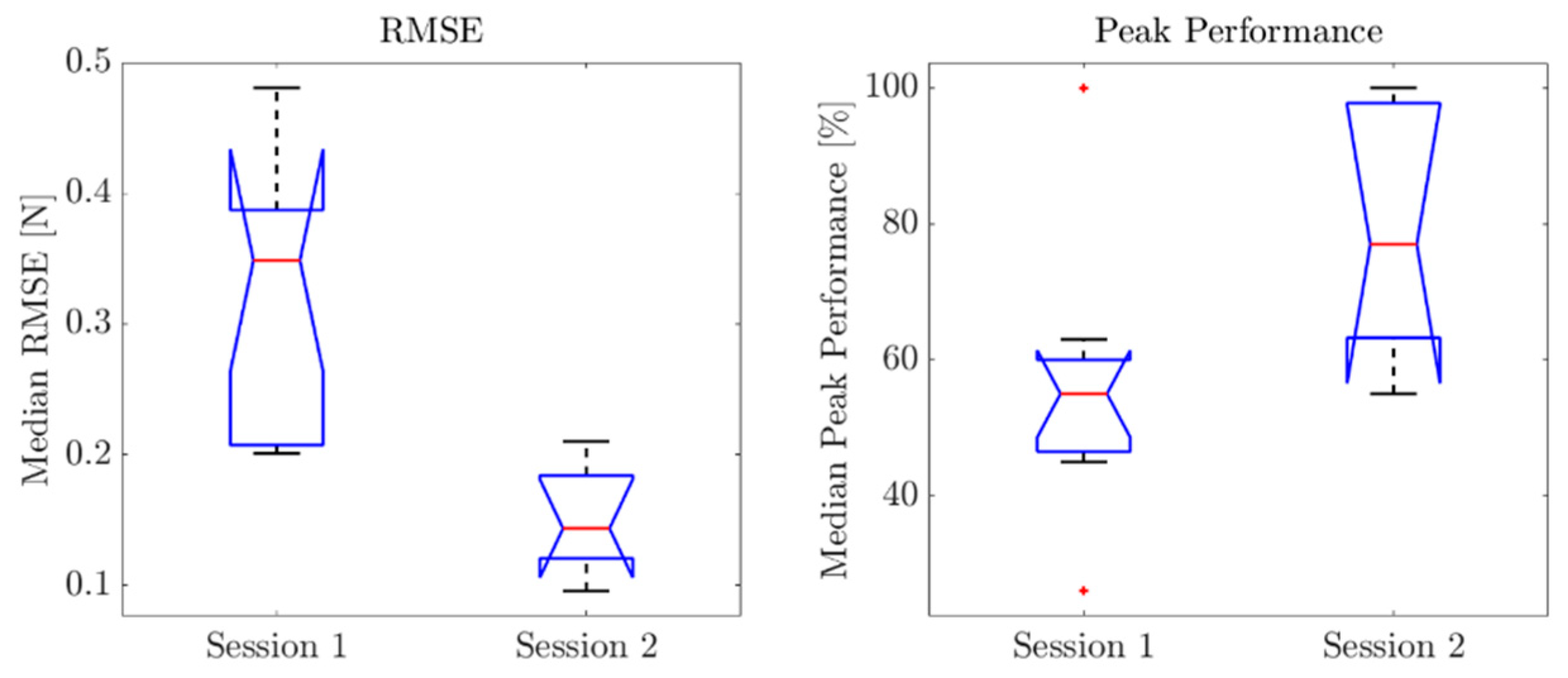
2.4. Force Assessment System Based on Virtual Reality Games

A Force Assessment System based on VR games was developed to assess the child’s improvements in grip force control and modulation. The system was composed of force sensors positioned at the fingertips of the Gloreha Sensor Glove and of custom-designed VR games in the form of tracking tasks purposely developed to assess different aspects of force control and modulation.
Previous works in the literature proposed tracking tasks for the assessment and training of grip force control in mildly to severely affected hemiparetic stroke patients. In the study of Kurillo et al. [22], the authors presented and tested a grip force training system that enabled the improvement of grip force control in 8 out of 10 post-stroke patients. In the study of Lindberg et al. of 2012 [23], the authors proved that the power grip force tracking tasks developed to assess grip force modulation capability were feasible to quantify the accuracy of grip force control.
The force sensors to be embedded in the Gloreha Sensor Glove were chosen according to the following technical specifications: (i) maximum dimension of 2 × 10 −2 m diameter; (ii) thickness lower than 5 × 10 −4 m; (iii) force range comparable to the one detected during hand rehabilitation training (0–25 N) [24]. The piezoresistive sensor FSR® Model 402 Short Tail (Interlink Electronics) was selected to meet the specifications. It has a diameter of 1.83 × 10 −2 m, a thickness of 4 × 10 −4 m, a force sensitivity range of 0.2–20 N and continuous force resolution. Piezoresistive sensors were selected because they are low cost, suitable for wearable applications, require a very simple conditioning electronics and have a good shock resistance.
Three sensors were embedded, respectively, in the fingertips of the first, second and third finger of the Gloreha Sensor Glove, i.e., the fingers mostly involved in daily life grasps, as shown in Figure 2. The interface between the sensor and the glove was modified by fixing a thin 3D printed PLA plate with double-sided tape (1 × 10 −3 m thickness and 2 × 10 2 m diameter) to uniformly distribute the force on the sensitive area of the sensor. A 3D-printed PLA support was positioned to interlock the plastic components of the glove that allow connecting Gloreha Sinfonia flexible transmissions to the glove itself. The sensors are secured on the fingertips by means of an elastic band fastened at the top of the PLA support.
The sensors with the developed PLA plate were calibrated using the Instron® testing machine to relate the force and output voltage.
The VR game was developed with Unity, using Microsoft Visual Studio as the pre-set editor and C# as the programming language. Attention was paid to provide an intuitive, clear and engaging visual feedback to the user. In fact, providing the patient with biofeedback can improve the outcome of the treatment and promote neuroplasticity [22].
The aim of the VR game in the form of tracking tasks was to assess the patient capability to finely control the sub-maximal forces exerted when grasping real objects, the ability to balance and release the grip and the general accuracy of force control.
The average force exerted by the three fingers was given in input to the VR game to move the avatar according to the force exerted on the object grasped by the participant (i.e., a wood parallelepiped 1.35 × 10−1 m × 6.5 × 10−2 m × 4 × 10−2 m). In the proposed VR game, shown in Figure 3, the patient was asked to move the avatar of the game vertically, according to the exerted force, in order to track the three proposed waveforms moving on the screen. The three waveforms were a “Ramp”, a “Square Wave” and a “Sinusoidal Wave” [25]. Each waveform was developed to assess different aspects of grip force control: the Ramp aimed at assessing the capability to gradually increase and decrease the grip force; the Square Wave aimed at assessing the capability to exert discrete force levels and stabilize the force; and the Sinusoidal Wave aimed at assessing the overall force modulation capability. “Ramp” and “Square Wave” were composed of 10 discrete force levels to be reached (and held for the “Square Wave”) and are uniformly distributed between the maximum and minimum forces recorded at the beginning of the trial, whereas the “Sinusoidal Wave” had a peak-to-peak amplitude that corresponded to the range between the minimum and maximum forces.
To assess the child’s improvements in grip force control and modulation, she was instructed to move the avatar (i.e., a turtle) and to follow the proposed waveform pattern in order to collect the maximum number of “bubbles” of the VR game with the avatar. In the first assessment session (T0), she performed 5 one-minute repetitions of the “Ramp” and 3 one-minute repetitions of the “Square Wave” and “Sinusoidal Wave” exercises, respectively. In the second assessment session (T1), she performed 3 one-minute repetitions of the “Ramp” and 2 one-minute repetitions of the “Square Wave” and “Sinusoidal Wave” exercises. Before each session, the maximum and minimum forces applicable by the child were recorded with a custom-made graphical user interface to set the force range in which the avatar could be moved. Then, the maximum force to be reached was set at 90% of the maximum recorded value.

2.5. Intervention

The Gloreha treatment lasted from January to April 2021, and the commitment was once a week for a total of 10 weeks; each session lasted 60 min. During each session, the child performed passive, active-assisted and active movements with gradually increasing complexity exercises supported and stimulated by sensory feedback. In addition, the child was also offered exercises in bi-dexterity to improve both the quality of the recovery of the paretic limb and its coordination and functionality in daily activities.

2.6. Outcome Measures

The child was evaluated with functional scales, with upper limb kinematic analysis and with an ad hoc developed force assessment tool (FSR model 402 Short Tail, Interlink Electronics) at pre-treatment (T0) and at the end of the treatment (T1).
She was evaluated with Fugl-Meyer Assessment-Upper Extremity (FMA-UE) for upper-extremity motor impairment and the Visual Analogic Scale (VAS) for pain intensity and activities and participation of daily life with ADL scale [26].
Improvements in force control and modulation capability were assessed by means of the Force Assessment System. Two main performance indicators were computed for each repetition of the three VR exercises: the Root Mean Square Error (RMSE) (N) between the target force pattern and the exerted force pattern and the Peak Performance (%), computed as the percentage of reached force peaks, for “Ramp” and “Sinusoid”, and the percentage of reached and held discrete force levels, for the “Square Wave”. A statistical analysis was performed to evaluate improvements of the child from one session to the other. The non-parametric one-way ANOVA test (i.e., Kruskal–Wallis test) was conducted between RMSE and Peak Performance indicators in the two sessions. The significance level was set at 5%.

3. Results

Results are reported in Table 1. At the end of treatment (T1), the patient improved in functional scales: FMA-UE had a percentage variation (∆%) of 44% from T0 (34/66) to T1 (49/66). Moreover, at the end of treatment, her quality of life was better for greater involvement in some little but significative activities of daily living such as food, dress and undress (ADL scale: 4/6 at T0 vs. 6/6 at T1) and had greater interest in the surrounding world with less fear of feeling different from others.
It is important to highlight that the child had never complained of pain (VAS scale 0/10) during both evaluations.
The Kruskal–Wallis test performed on RMSE and Peak Performance indicators between sessions at T0 and T1, allowed the assessment of improvements in force control and modulation. RMSE was significantly reduced from one session to the other (p value = 0.0018), meaning that training with the Gloreha improved the child’s capacity to follow target force patterns. Peak Performance significantly increased from the first to the second evaluation session (p value = 0.0120). Boxplots for the two performance indicators are shown in Figure 4, where the central mark is the median and the box edges are the 25th and 75th percentiles.
No adverse events occurred during the entire treatment.

4. Discussion

The aim of this study is to describe the effects on the range of motion, muscle tone and functionality of the paretic upper limb, particularly the hand, when Gloreha Sinfonia combined with conventional therapy was used for rehabilitation treatments in a pediatric stroke patient. The results revealed an improvement in FMA-UE, a significant reduction in RMSE and a significant improvement in Peak Performance.
To the best of our knowledge, this is the first study using Gloreha Sinfonia in pediatric strokes. Previous rehabilitation studies on pediatric stroke patients were mostly dedicated to the robotic treatment of the proximal portion of the upper limb, while there are still very few studies on the distal extremity, especially the hand. Gloreha Sinfonia allows the performance of tasks that combine the activity of the entire upper limb. In fact, unlike other robots designed exclusively for shoulder and elbow movements, Gloreha permits the improvement of distal control through the implementation of exercises focused on the use of hands combined with the involvement of the entire upper limb, reproducing activities of daily living. As other studies have previously mentioned, when a patient uses the distal part of the paretic upper limb, at the same time, the proximal segment is also trained, albeit the upper arm is supported or restrained in the distal group [27]. Furthermore, Gloreha Sinfonia is well suited for use in combination with other traditional rehabilitation activities because it can integrate rehabilitation treatments with highly stimulating and interactive exercises for patients.
Our results show an important enhancement in FMA-UE, with values that proceed from 34/66 to 49/66 between T0 and T1; they indicate an overall improvement of the upper limb due to a clinical advance both in the shoulder district, implemented thanks to reaching exercises, and to the hand control, achieved thanks to manipulation exercises. These results were significantly higher than the minimal clinically important difference (MCID) seen in adults by Page et al. [28], but it is not possible to make a comparison with a pediatric population, because their MCID is not present in the literature. In addition, FMA-UE results are in line with ADL records, which report improvements in activities of daily life due to a better use of the upper limb.
From Figure 4, it is evident that the child significantly improved her capacity of reaching force peaks and holding force levels. Furthermore, the results show that force control and modulation capability significantly increased after treatment.
We know from the girl’s parents that she has reported a psycho-emotional improvement and presented new interest in socializing with other children without fear of being judged and perceived less pronounced motor deficits and less disability at the end of therapy. This statement can be considered as another positive effect of the combined (robot + conventional rehab therapy) and continuative therapy. It is important to underline that the girl never complained of pain, demonstrating a good ad safe adaptability of the robot with respect to the child.
Another aspect that makes this robot an interesting tool in pediatric applications is the possibility of performing specific repetitive task-oriented exercises, placed in the form of playing a videogame; as already reported, robot-mediated therapy in children with acquired or congenital brain injury appears to be beneficial: enhancing motivation and improving perception, it incorporates the advantages of the enjoyable game-like experience [15,29]. Furthermore, the use of a virtual and motivational environment in which the patient has immerged has a positive effect to facilitate motor and sensory and cognitive relearning by stimulating the patient to challenge himself to perform better and to obtain a higher score in the game. Moreover, as recorded also by Mirkowski et al. in their systematic review [18], robotic therapy seems to significantly improve the upper extremity function and spasticity in children after strokes. This theory is also supported by the study of Wann et al., which considers motor learning theory and reports how the repetition of motor patterns is seen as a key factor in improving movement [30]. Moreover, Rizzo et al. reports how a rehabilitation treatment that uses feedback, either visual or auditory, contributes to gains made in motor learning [31]. It follows that Virtual Reality is a powerful medium for providing stimulation in the form of visual and auditory events to increase the motivation and desire to continue practicing [32].
To date, there are few studies in the literature on this specific population. Most pediatric upper extremity rehabilitation studies are aimed at patients with cerebral palsy, spinal cord injury or quadriplegia. As reported also in the case report of Čolović et al. [33], there is still no clear indication on how robotic therapy can be combined with conventional therapy and for how many times; however, combining robotic and traditional rehabilitation can improve the functional motor performance of the arm involved in the chronic recovery phase after a pediatric stroke.
Our results, being relative to a single case, cannot find a generalization. Further studies on larger samples, with control and randomization groups and with adequate follow-ups, are needed in order to reach meaningful conclusions. Furthermore, we have not assessed patient’s emotional changes and the level of social integration before and after the robotic treatment with specific and objective measures; the administration of scales that could quantify these changes could be useful to determine social aspects in further studies.

5. Conclusions

Given the results and given the literature evidence, Gloreha Sinfonia seems to be suitable for the treatment of post-stroke hand disabilities in the pediatric age, but further studies on larger populations, with stratification of the sample for clinical characteristics, as well as clinical scales that are more sensitive to any change are needed to support this hypothesis. It would be interesting in the future to consider how this type of technology can also support cognitive difficulties. In addition, this type of rehabilitative approach facilitates the need for personalized and easily monitored rehabilitation protocols. Clinical trials with follow-ups and on large populations could confirm the results obtained.

Author Contributions

Conceptualization, F.B., C.A. and D.M.; methodology, F.S. and F.C.; software, L.Z., F.C. and M.L.; formal analysis, F.S.; investigation, F.N.; data curation, L.Z. and F.C.; writing—original draft preparation, F.N., B.C. and L.C.; writing—review and editing, F.B. and L.C.; supervision, S.S. and M.B.; project administration, F.B. All authors have read and agreed to the published version of the manuscript.

Funding

This research received no external funding.

Institutional Review Board Statement

The study was conducted in accordance with the Declaration of Helsinki and approved by the Ethics Committee of Fondazione Policlinico Campus Bio-Medico of Rome (protocol code PT-O 20.22; date of approval: 29 March 2022).

Informed Consent Statement

Informed consent was obtained from all subjects involved in the study.

Data Availability Statement

Not applicable.

Conflicts of Interest

The authors declare no conflict of interest.

References

  1. Ferriero, D.M.; Fullerton, H.J.; Bernard, T.J.; Billinghurst, L.; Daniels, S.R.; DeBaun, M.R.; Deveber, G.; Ichord, R.N.; Jordan, L.C.; Massicotte, P.; et al. Management of Stroke in Neonates and Children: A Scientific Statement from the American Heart Association/American Stroke Association. Stroke 2019, 50, e51–e96. [Google Scholar] [CrossRef] [PubMed] [Green Version]
  2. Mallick, A.A.; Ganesan, V.; Kirkham, F.J.; Fallon, P.; Hedderly, T.; McShane, T.; Parker, A.P.; Wassmer, E.; Wraige, E.; Amin, S.; et al. Diagnostic Delays in Paediatric Stroke. J. Neurol. Neurosurg. Psychiatry 2015, 86, 917–921. [Google Scholar] [CrossRef] [PubMed]
  3. Mallick, A.A.; Ganesan, V.; Kirkham, F.J.; Fallon, P.; Hedderly, T.; McShane, T.; Parker, A.P.; Wassmer, E.; Wraige, E.; Amin, S.; et al. Childhood Arterial Ischaemic Stroke Incidence, Presenting Features, and Risk Factors: A Prospective Population-Based Study. Lancet Neurol. 2014, 13, 35–43. [Google Scholar] [CrossRef]
  4. Lanthier, S.; Carmant, L.; David, M.; Larbrisseau, A.; de Veber, G. Stroke in Children: The Coexistence of Multiple Risk Factors Predicts Poor Outcome. Neurology 2000, 54, 371–378. [Google Scholar] [CrossRef]
  5. Riela, A.R.; Roach, E.S. Etiology of Stroke in Children. J. Child Neurol. 1993, 8, 201–220. [Google Scholar] [CrossRef] [PubMed]
  6. Royal College of Paediatrics and Child Health Stroke in Childhood—Clinical Guideline for Diagnosis, Management and Rehabilitation|RCPCH. Available online: https://www.rcpch.ac.uk/resources/stroke-in-childhood-clinical-guideline (accessed on 10 March 2022).
  7. DeLaroche, A.M.; Sivaswamy, L.; Farooqi, A.; Kannikeswaran, N. Pediatric Stroke and Its Mimics: Limitations of a Pediatric Stroke Clinical Pathway. Pediatr. Neurol. 2018, 80, 35–41. [Google Scholar] [CrossRef]
  8. Ictus Cerebrale: Linee Guida Italiane Di Prevenzione e Trattamento Raccomandazioni e Sintesi. Available online: https://www.iso-stroke.it/wp-content/uploads/2017/02/LIBRO-SPREAD-VIII-ED-13-09-16.pdf (accessed on 10 March 2022).
  9. Bütefisch, C.; Hummelsheim, H.; Denzler, P.; Mauritz, K.H. Repetitive Training of Isolated Movements Improves the Outcome of Motor Rehabilitation of the Centrally Paretic Hand. J. Neurol. Sci. 1995, 130, 59–68. [Google Scholar] [CrossRef]
  10. Hendricks, H.T.; van Limbeek, J.; Geurts, A.C.; Zwarts, M.J. Motor Recovery after Stroke: A Systematic Review of the Literature. Arch. Phys. Med. Rehabil. 2002, 83, 1629–1637. [Google Scholar] [CrossRef]
  11. Thielman, G.; Kaminski, T.; Gentile, A.M. Rehabilitation of Reaching after Stroke: Comparing 2 Training Protocols Utilizing Trunk Restraint. Neurorehabil. Neural Repair 2008, 22, 697–705. [Google Scholar] [CrossRef]
  12. Eliasson, A.C.; Nordstrand, L.; Ek, L.; Lennartsson, F.; Sjöstrand, L.; Tedroff, K.; Krumlinde-Sundholm, L. The Effectiveness of Baby-CIMT in Infants Younger than 12 Months with Clinical Signs of Unilateral-Cerebral Palsy; an Explorative Study with Randomized Design. Res. Dev. Disabil. 2018, 72, 191–201. [Google Scholar] [CrossRef]
  13. Kwakkel, G.; Wagenaar, R.C.; Koelman, T.W.; Lankhorst, G.J.; Koetsier, J.C. Effects of Intensity of Rehabilitation after Stroke. A Research Synthesis. Stroke 1997, 28, 1550–1556. [Google Scholar] [CrossRef] [PubMed]
  14. Feys, H.M.; de Weerdt, W.J.; Selz, B.E.; Cox Steck, G.A.; Spichiger, R.; Vereeck, L.E.; Putman, K.D.; van Hoydonck, G.A. Effect of a Therapeutic Intervention for the Hemiplegic Upper Limb in the Acute Phase after Stroke: A Single-Blind, Randomized, Controlled Multicenter Trial. Stroke 1998, 29, 785–792. [Google Scholar] [CrossRef] [PubMed]
  15. Frascarelli, F.; Masia, L.; Di Rosa, G.; Cappa, P.; Petrarca, M.; Castelli, E.; Krebs, H.I. The Impact of Robotic Rehabilitation in Children with Acquired or Congenital Movement Disorders. Eur. J. Phys. Rehabil. Med. 2009, 45, 135–141. Available online: https://www.minervamedica.it/it/riviste/europa-medicophysica/articolo.php?cod=R33Y2009N01A0135 (accessed on 10 March 2022). [PubMed]
  16. Doerbeck, V.; Nallinger, A.; Burger, H.; Rosner, V.; Machuta, K.; Gruender, S.; Nastulla, T.; Schroeder, S.; Berweck, S. Integration of Armeo Spring Pediatrics in Inpatient Rehabilitation of Children and Adolescents with Hemiparesis. Neuropediatrics 2012, 43, PS14_11. [Google Scholar] [CrossRef]
  17. Friel, K.M.; Kuo, H.C.; Fuller, J.; Ferre, C.L.; Brandão, M.; Carmel, J.B.; Bleyenheuft, Y.; Gowatsky, J.L.; Stanford, A.D.; Rowny, S.B.; et al. Skilled Bimanual Training Drives Motor Cortex Plasticity in Children with Unilateral Cerebral Palsy. Neurorehabil. Neural Repair 2016, 30, 834–844. [Google Scholar] [CrossRef] [Green Version]
  18. Mirkowski, M.; McIntyre, A.; Faltynek, P.; Sequeira, N.; Cassidy, C.; Teasell, R. Nonpharmacological Rehabilitation Interventions for Motor and Cognitive Outcomes following Pediatric Stroke: A Systematic Review. Eur. J. Pediatr. 2019, 178, 433–454. [Google Scholar] [CrossRef]
  19. Gordon, A.M.; Okita, S.Y. Augmenting Pediatric Constraint-Induced Movement Therapy and Bimanual Training with Video Gaming Technology. Technol. Disabil. 2010, 22, 179–191. [Google Scholar] [CrossRef]
  20. Gagnier, J.J.; Kienle, G.; Altman, D.G.; Moher, D.; Sox, H.; Riley, D.; Group, C. Standards & Guidelines CARE: Linee Guida per Il Reporting Di Casi Clinici. Evidence 2016, 8, e1000147. [Google Scholar]
  21. Miccinilli, S.; Bravi, M.; Foti, C.; Morrone, M.; Maselli, M.; Santacaterina, F.; Scotto Di Luzio, F.; Zollo, L.; Sterzi, S.; Bressi, F. Robotic Hand Treatment of Patients Affected by Chronic Stroke: A Monocentric Longitudinal Pilot Study. J. Biol. Regul. Homeost. Agents 2020, 34, 79–86. [Google Scholar]
  22. Kurillo, G.; Gregoric, M.; Goljar, N.; Bajd, T. Grip Force Tracking System for Assessment and Rehabilitation of Hand Function. Technol. Health Care 2005, 13, 137–149. Available online: https://pubmed.ncbi.nlm.nih.gov/15990417/ (accessed on 6 April 2022). [CrossRef]
  23. Lindberg, P.G.; Roche, N.; Robertson, J.; Roby-Brami, A.; Bussel, B.; Maier, M.A. Affected and Unaffected Quantitative Aspects of Grip Force Control in Hemiparetic Patients after Stroke. Brain Res. 2012, 1452, 96–107. [Google Scholar] [CrossRef] [PubMed]
  24. de Pasquale, G.; Mastrototaro, L.; Pia, L.; Burin, D. Wearable System with Embedded Force Sensors for Neurologic Rehabilitation Trainings. In Proceedings of the Symposium on Design, Test, Integration and Packaging of MEMS/MOEMS, DTIP 2018, Rome, Italy, 22–25 May 2018; pp. 1–4. [Google Scholar] [CrossRef]
  25. Lapresa, M.; Tamantini, C.; Scotto Di Luzio, F.; Cordella, F.; Bravi, M.; Miccinilli, S.; Zollo, L. A Smart Solution for Proprioceptive Rehabilitation through M-IMU Sensors. In Proceedings of the 2020 IEEE International Workshop on Metrology for Industry 4.0 and IoT, MetroInd 4.0 and IoT 2020, Rome, Italy, 3–5 June 2020; pp. 591–595. [Google Scholar] [CrossRef]
  26. Katz, S.; Ford, A.B.; Moskowitz, R.W.; Jackson, B.A.; Jaffe, M.W. Studies of illness in the aged. The index of adl: A standardized measure of biological and psychosocial function. JAMA 1963, 185, 914–919. [Google Scholar] [CrossRef] [PubMed]
  27. Hsieh, Y.W.; Lin, K.C.; Wu, C.Y.; Shih, T.Y.; Li, M.W.; Chen, C.L. Comparison of Proximal versus Distal Upper-Limb Robotic Rehabilitation on Motor Performance after Stroke: A Cluster Controlled Trial. Sci. Rep. 2018, 8, 2091. [Google Scholar] [CrossRef] [PubMed] [Green Version]
  28. Page, S.J.; Fulk, G.D.; Boyne, P. Clinically Important Differences for the Upper-Extremity Fugl-Meyer Scale in People with Minimal to Moderate Impairment Due to Chronic Stroke. Phys. Ther. 2012, 92, 791–798. [Google Scholar] [CrossRef] [PubMed] [Green Version]
  29. Reid, D.T. Benefits of a Virtual Play Rehabilitation Environment for Children with Cerebral Palsy on Perceptions of Self-Efficacy: A Pilot Study. Pediatr. Rehabil. 2002, 5, 141–148. [Google Scholar] [CrossRef]
  30. Wann, J.P.; Rushton, S.K.; Smyth, M.; Jones, D. Virtual Environments for the Rehabilitation of Disorders of Attention and Movement. Stud. Health Technol. Inform. 1997, 44, 157–164. [Google Scholar] [CrossRef]
  31. Rizzo, A.A.; Buckwalter, J.G.; Neumann, U.; Kesselman, C.; Thiebaux, M. Basic Issues in the Application of Virtual Reality for the Assessment and Rehabilitation of Cognitive Impairments and Functional Disabilities. CyberPsychol. Behav. 2009, 1, 59–78. [Google Scholar] [CrossRef]
  32. Alt Murphy, M.; Häger, C.K. Kinematic Analysis of the Upper Extremity after Stroke—How Far Have We Reached and What Have We Grasped? Phys. Ther. Rev. 2015, 20, 137–155. [Google Scholar] [CrossRef]
  33. Čolović, H.; Dimitrijević, L.; Đurić, V.; Janković, S. Upper Limb Robotic Neurorehabilitation after Pediatric Stroke. Srp. Arh. Za Celok. Lek. 2020, 148, 368–376. [Google Scholar] [CrossRef] [Green Version]
Figure 1. Timeline.
Figure 1. Timeline.
Applsci 12 04206 g001
Figure 2. Force sensors positioned at the fingertips of Gloreha Sensor Glove.
Applsci 12 04206 g002
Figure 3. VR game for force assessment.
Applsci 12 04206 g003
Figure 4. Boxplots of RMSE and Peak Performance for session 1 (T0) and session 2 (T1).
Applsci 12 04206 g004
Table 1. Results of outcome measurement.
T0T1
ADL4/66/6
FMA-UE34/6649/66
VAS0/100/10
ADL: Activity of Daily Living; FMA-UE: Fugl-Meyer Assessment for Upper Limb; VAS: Visual Analogic Scale.
Publisher’s Note: MDPI stays neutral with regard to jurisdictional claims in published maps and institutional affiliations.

