Article

Comparative Studies on the Antioxidant, Antifungal, and Wound Healing Activities of Solenostemma arghel Ethyl Acetate and Methanolic Extracts

1
Botany Department, Faculty of Science, Aswan University, Aswan 81528, Egypt
2
Unit of Environmental Studies and Development, Aswan University, Aswan 81528, Egypt
3
Department of Pathology and Clinical Pathology, Faculty of Veterinary Medicine, South Valley University, Qena 83523, Egypt
4
Department of Clinical Sciences, College of Medicine, Princess Nourah Bint Abdulrahman University, P.O. Box 84428, Riyadh 11671, Saudi Arabia
5
Department of Pharmacology, Faculty of Veterinary Medicine, Menoufia University, Shebin Elkoum 32514, Egypt
6
Department of Anatomy and Embryology, Faculty of Medicine, Benha University, Benha 13518, Egypt
7
Department of Surgery, Anesthesiology, and Radiology, Faculty of Veterinary Medicine, Aswan University, Aswan 81528, Egypt
*
Authors to whom correspondence should be addressed.
Appl. Sci. 2022, 12(9), 4121; https://doi.org/10.3390/app12094121
Submission received: 21 February 2022 / Revised: 10 April 2022 / Accepted: 11 April 2022 / Published: 19 April 2022
(This article belongs to the Topic Compounds with Medicinal Value)

Abstract

:
Various herbal compounds are used for medical purposes due to their safety, as there are no or minimal side effects. This study was performed to assess the wound healing and antioxidant activities of ethyl acetate (EtOAc) and methanolic extract (MeoH) of Solenostemma arghel (S. arghel). Their antifungal activities were also evaluated against isolated swabs of equine wounds. They underwent GC-MS analysis for the characterization of both extracts. For wound healing evaluation, forty-five male albino rats were divided into three groups; the control group was treated with normal saline, and the other two groups were treated with S. arghel EtOAc and MeoH extract gels, respectively. The wounds were examined clinicopathologically and immunohistochemistry on the 3rd, 7th, and 14th days post-wounding. GC-Ms analysis of S. arghel recorded fifty-one volatile organic compounds (VOCs) within EtOAc extraction and thirty VOCs in MeoH extract. VOCs represented in EtOAc extract showed higher antioxidant activity and better and faster wound healing than VOCs of MeOH extract. The treated groups showed improved wound healing clinically and pathologically in comparison with the control group as they decreased the wound surface area (WSA) and percent (WSA%) and increased the wound contraction percent (WC%), epithelization, fibroblast proliferation with neovascularization, and reduced the inflammatory reaction. Moreover, the treated groups showed higher expression of vascular endothelial growth factor (VEGF) compared with the control. The EtOAc extract showed higher antifungal activity against Penicillium funiculosum, P. jensenii, M. cinctum, and Candida albicans, which were isolated from infected clinical equine wounds, than MeOH extract. The treated groups showed improved wound healing clinically and pathologically in comparison with the control group as they decreased the wound surface area (WSA) and percent (WSA%) and increased the wound contraction percent (WC%), epithelization, fibroblast proliferation with neovascularization, and reduced the inflammatory reaction. Moreover, the treated groups showed higher expression of vascular endothelial growth factor (VEGF) compared with the control. Additionally, the two extract gels showed promising healing of equine wounds. In conclusion, the study recommended the use of S. arghel EtOAc extract as it was proven to promote wound healing compared with MeoH extract.

1. Introduction

The wound is the disruption of cellular and anatomical continuity of living tissue produced by physical, chemical, electrical, or microbial insults to the tissue [1]. The subcutaneous tissue loses its integrity when wounded and colonized by microbes. These microorganisms involve potential pathogens, which interrupt the wound healing process in the injured tissues [2,3]. Wound infection is a major factor in the progression of chronicity, delaying wound healing [4].
The development of wound infection is an ongoing problem for many patients. Infected wounds can cause great distress in terms of associated morbidity and mortality, increased length of hospital admission, delayed wound healing, and increased discomfort, and have long been known to increase healthcare costs significantly, as the exudate levels are relatively higher in infected wounds. Pus and odor may indicate that the patient is pyrexial or has an increased neutrophil count [5].
Most chronic wound microbiome research is focused on bacteria, but contamination of the wound by fungi needs more elucidation. Ref. [6] classified seventeen fungal species from different wounds, which obstruct the healing of these wounds.
The yeasts of the genus Candida were the most numerous fungus. In particular, the high relative abundance of fungus within the polymicrobial ecology of wound infections, when present, shows that the fungus can be a major contributor to the biofilm of wounds. In addition, the diversity of fungal genera and species found in and among such wounds is more than previously thought or recorded [7]. The vulnerability to fungal wound infection is also increasing in geographical locations with a warm and humid climates [8].
The link between microbial aggregates and wound healing has been studied using a number of animal models. In order to imitate wound healing, most research used small rodents. However, the architecture of these species’ skin and healing patterns differ significantly from those of humans [9]. Horses have thick, tight skin, similar to humans [10]. In humans and horses, wound edge contraction has a minor role (20–25%) in extremity/limb healing, in contrast to the situation in loose-skinned animals such as rodents [11].
Horses appear to be valid model animals for wound healing in humans because of their similar skin architecture and the relative role of contraction and epithelialization in healing [12,13]. Because of their surroundings, horses’ wounds have a significant risk of infection, as the infected wounds house a variety of microorganisms. However, these germs can be challenging to identify and not react to antibiotic treatment, resulting in chronic non-healing wounds [14]. Therefore, in the study, we selected some cases of equine wounds for clinical application of the gel and to find a resolution for the problem of equine wounds through the investigation of new wound-healing agents.
Traditional medicine is widespread throughout the world, using natural medicinal plants. Solenostemma arghel is a desert medicinal plant indigenous to African countries (Egypt, Sudan, Algeria, Libya, and Chad) [15], which is considered folk medicine. African people use this plant as an anti-inflammatory, anti-rheumatic, antispasmodic, anti-diabetic, anti-nutrition, antimicrobial, and anticancer agent [16,17,18,19]. The medical effect of different parts of this plant comes especially from leaves because of phytochemicals richness, including various phenolic acids, flavones, glycosylated flavonoids, polyphenols, b-carotene, b-sitosterol, monoterpenes, pregnenes, and pregnane [17,20].
The present study was designed to compare the antifungal, antioxidant, and wound healing activities of EtOAc and MeoH S. arghel extracts in albino rats besides the clinical application of the extracts for investigation of wound healing in equines.

2. Materials and Methods

2.1. Chemical and Reagents

Methanol was purchased from (Lab-Scan Analytical Sciences, Gliwice, Poland). Ethyl acetate was purchased from ADWIC (Cairo, Egypt). XylaMed® (xylazine HCL, 100 mg, Bimeda, Inc., Oakbrook Terrace, IL, USA). Ketamax® (ketamine HCL, 50 mg) was obtained from (Troikaa Pharmaceuticals Ltd., Gujarat, India). Carbopol 940® was purchased from (Loba Chemie Company, Mumbai, India). All chemicals were of analytical grades.

2.2. Plant Materials

The vegetative part of S. arghel was collected from the extremely arid desert region of Southern Egypt, Aswan University campus, Aswan. The plant was washed with tap water, leaves and flowers were separated from the stem, air-dried at room temperature, followed by grinding into a powder with a grinder (mortar and pestle).

2.3. Preparation of Plant Extract

Powdered leaves and flowers (100 gm) were extracted with two organic solvents (EtOAc and MeoH (0.1 gm/mL)). Extraction was incubated overnight at room temperature and repeated using an ultrasonic homogenizer. The extracts were decanted, filtered, and the residue was re-extracted again under the same experimental conditions to ensure full extraction. The clear filtrates were air-dried using a rotary evaporator at 45 °C [21].

2.4. Collection and Preservation of Material from Injured Parts for Investigation of Yeast and Fungal Infections

By using sterile swabs, samples were collected from 12 equine species (a horse, a mare, 5 donkeys, and 5 female donkeys), which were admitted to the Brooke Hospital in Aswan, Egypt, suffered from traumatized wounds. Swabs were placed in a sterile cryotube and stored at −40 °C. These samples were soon cultured on Sabroud media for isolation of fungi and yeast, which were morphologically identified using a light microscope, and the isolated fungi were purified and kept in slants.

2.5. Antifungal Activity Assay

For screening of antifungal activity of Arghel plant extracts, a food technique was followed [22]. Sabroud medium was prepared and sterilized; then, the medium was supplemented with EtOAc and MeoH plant extracts at two concentrations (5 mg/mL and 10 mg/mL), mixed well, and kept at 4 °C overnight. The mycelial disc (0.5 cm in diameter) was deposited in the center of the plate (5.0 cm in diameter) according to the poisoned food method, which was used to evaluate the antifungal effect against pathogens [23]. After further incubation for 7 days at 28 ± 2 °C for the fungal strains tested, the diameters of fungal growth for control (fungus in media without any extraction and containing only solvent) and treated plates were measured with a calculation of the inhibition percentage [24].

2.6. DPPH Antioxidant Activity

The DPPH solution (0.5 mmol/L) was prepared in 95% methanol, and different concentrations (50, 100, and 150 µg) of Ethyl acetate and Methanol plant extracts were used. A total of 2 mL of DPPH solution (0.5 mnol/L) was added to (1 mL) of the test sample and incubated for 30 min at room temperature in darkness. After 30 min, the absorbance was measured at 517 nm. Ascorbic acid (AA) served as a positive control at a concentration of 10 µg. The percentage of the DPPH radical scavenging was calculated as:
Inhibition of DPPH radical (%) = [(control absorbance (Ai) − extract absorbance (At))/(control absorbance (Ai))] × 100
Inhibition (%) = (Ai − At)/Ai × 100%

2.7. GC-MS Analysis of Released VOCs from S. arghel EtOAc and MeoH Extracts

The extracted VOCs from the S. arghel were analyzed and identified by gas chromatography–mass spectrometry (GC/MS). The GC-MS analysis was carried out by using a TRACE GC Ultra Gas Chromatograph (THERMO Scientific Corp, Carlsbad, CA, USA), which connected with a Thermo mass spectrometer detector (ISQ Single Quadrupole Mass Spectrometer). The GC-MS system was equipped with a TR-5 MS column (30 m × 0.32 mm i.d., 0.25 μm film thickness). Analysis was achieved using helium gas at a flow rate of 1.0 mL/min and a split ratio of 1:10 using the following temperature program: 60 °C for 1 min; increasing 4.0 °C/min to 240 °C and held for 1 min. Both injector and detector were held at 210 °C, and diluted samples (1:10 hexane, v/v) of 1 μL of the mixtures were injected. Mass spectra were obtained by electron ionization (EI) at 70 eV, using a spectral range of m/z 40–450. The chemical constituents of the essential oil (Eos) identification was de-convoluted using (AMDIS version 2.70, http://www.amdis.net/, accessed on 11 April 2022) and identified by its retention indices (relative to n-alkanes C8–C22) mass spectrum matching to authentic standards (when available) or Wiley spectral library collection, and NSIT library database.

2.8. Preparation of S. arghel EtOAc and MeoH Extracts 2% Gels

The two gels were prepared according to [25]. In a mixture containing 100 mL D. W, 1.5 g of Carbopol 940® was added slowly and mixed well at a higher speed. Followed by the addition of extract until gel formation with 2% concentration.

2.9. Evaluation of In Vivo Wound Healing Activity

2.9.1. The Ethical Approval

All experimental procedures in this study were approved by the ethical committee of the Faculty of Veterinary Medicine, South Valley University, Qena, Egypt (VM-2022-0030). All animals were euthanized at the experimental end. The clinical study on equine was carried out according to the ethics of the Brooke Hospital in Aswan, Egypt.

2.9.2. Animals

Forty-five (45) male albino rats aged 2–3 months and with 150–200 g body weight were purchased from Abou-Rawah farm, Egypt. Rats were housed in plastic cages with balanced diet ad libitum, with clean water readily accessible to the caged rats in clean bottles under constant environmental conditions (temperature of 25 °C, normal daylight, and relative humidity of 45–55%). The rats were kept a week before the experiment for Acclimatization.

2.9.3. Excisional Wound Model

After the adaptive period, the rats were anesthetized with an intraperitoneal (IP) injection of ketamine (70 mg/kg) and xylazine (7 mg/kg) combination, and the hair on the back was shaved and disinfected with ethyl alcohol (70%). A 1.5 cm wound was created with the aid of toothed tissue forceps, surgical blades, and pointed scissors to keep the skin tension constant during wounding. The hemostasis was achieved by compression with sterile gauze, and wounds were left untreated for 24 h. After wound creation, the rats were housed individually, each rat in a separate cage. The wound site was cleaned with a tampon soaked in alcohol. The rats were randomized into 3 groups (15 rats each). The excisional cutaneous wounds of the control group (Group I) were treated topically with 0.9% normal saline once daily, and the excisional wounds of the other two groups (Group II, Group III) were treated topically with S. arghel EtOAc and MeoH extracts 2% gels, respectively. The two herbal formulations were applied to the wounds once daily for 14 days starting from the second day of wounding.

2.9.4. Assessment of Wound Diameter

The wound size was measured and photographed on 0, 3, 7, and 14 days post-treatment for visual comparison before and after clearance of the wound area. The wound surface area (WSA), wound surface area percent (WSA%), and wound contraction percent (WC%) were calculated according to the following formulae:
WSA = πr2 (cm2).
WSA % = WSA × 100 1.8 %
WC% = 100% − WSA%

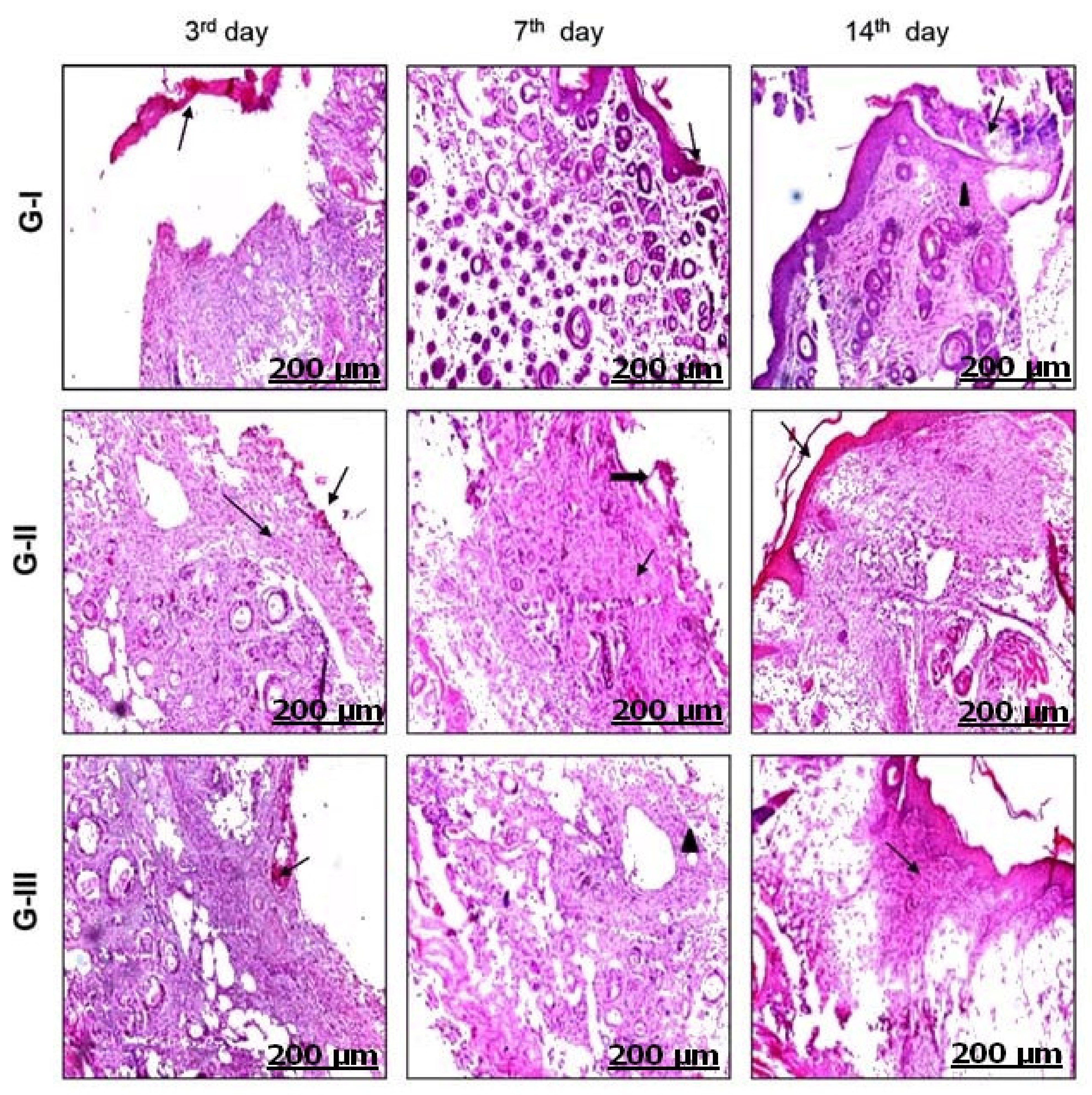
2.9.5. Histopathology

On the 3rd, 7th, and 14th days: 5 rats from each group were euthanized, and the wounds with part of normal skin were preserved in 10% neutral buffered formalin. The tissues were subjected to different concentrations of ethyl alcohol (70–80–90–100%), embedded in paraffin wax at 40–60 °C, sectioned into 5 μm thickness, and stained with hematoxylin–eosin stain [26].

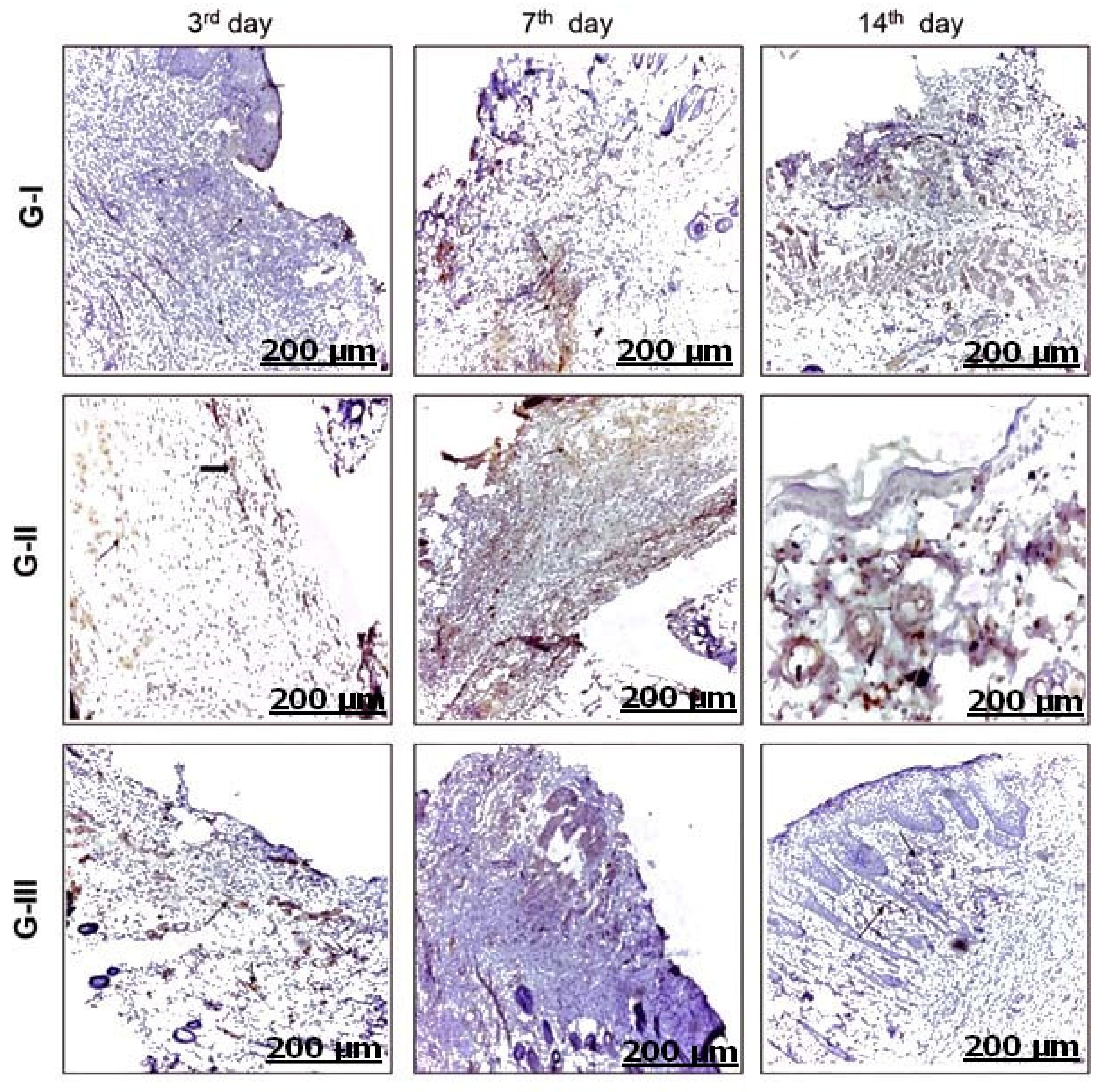
2.9.6. Immunohistochemistry

Immunohistochemistry assessment was performed according to [27]. Retrieval of antigen was performed by de-waxing and immersion of skin sections in a citrate buffer solution (0.05 M and pH 6.8). These sections were then treated with H2O2 (0.3%) and protein block. They were then incubated with polyclonal rabbit VEGF (BioGenix, 49026 Milmont Drive, Fremont, CA 94538 Emergo Europe, Molenstraat 15, NL-2513 BH The Hague, The Netherlands, cat. No. AR483-5R, dilution 1/100). The slides were washed up with PBS and then incubated with a goat anti-rabbit secondary antibody (cat. no. K4003, EnVision+™ System Horseradish Peroxidase Labelled Polymer; Dako, Glostrup, Denmark) for 30 min at room temperature. Eventually, slides were visualized with a DAB kit and stained with Mayer’s hematoxylin as a counterstain was evaluated under the light microscope. The staining intensity was assessed and reported as a percentage of positive cells in approximately 8 high power fields.

2.9.7. Clinical Study

The two prepared gels were topically applied on 5 equines (3 horses and 2 donkeys) that suffered from recent and old, traumatized contaminated wounds; some wounds were treated before without any tendency to wound healing.

2.9.8. Treatment Procedures

Before the gel application, the wound area was prepared according to the type of wound (old or recent); in a recent wound, hemorrhages were controlled by packing, and the wound area was aseptically prepared. In an old wound, unhealthy tissues were surgically debrided and freshened, and hemorrhages were controlled by packing.

2.10. Statistical Analysis

All statistical analyses were carried out by computer software (SPSS version 16.0, Chicago, IL, USA) using One-Way Analysis of Variance (ANOVA), and the data are presented as mean ± standard deviation (SD) values. The differences between mean values were considered significant at p < 0.05. The graphs were performed using Microsoft Excel 2010.3.

3. Results

3.1. GC-Ms Analysis of VOCs Produced by S. arghel in EtOAc and MeoH Extractions

Fifty-one VOCs were recorded in GC-Ms analysis of S. arghel EtOAc extraction (Table 1 and Figure 1a) while only thirty VOCs were recognized in plant MeoH extract (Table 2 and Figure 1b). The most abundant compounds in EtOAc extract (Table 1) were represented by Vitamin E (alpha-tocopherol) (9.99%); Diisooctyl phthalate (9.32%); Methyl hexadecadienoate and 5H-Cyclopropa[3,4]benz[1,2-e]azulen-5-one,9-(acetyloxy)-3-[(acetyloxy)methyl]-1,1a,1b,4,4a,7a,7b,8,9,9a-decahydro-4a,7b,9a-trihydroxy-1,1,6,8-tetramethyl-[1ar-(1aà,1bá,4aá,7aà,7bà,8à,9á,9aà)]-(7.36% each). Among the eight minor components of EtOAc extract, the most notable were 8-Dodecen-1-ol-acetate (Z)-; Undec-10-ynoic acid-heptadecylester; Undec-10-ynoic acid-octadecyl ester (0.62% each); Linoleoyl chloride; Digitoxin and Oxiranepentanoic acid-3-undecyl-, methyl ester, trans, (0.85% each) (Table 1).
The GC–MS analysis of MeoH extract predominantly revealed the presence of the main constituents as Hentriacontane; Sulfurous acid-hexyl octyl ester (11.34% each); Heneicosane; Heptadecane; Octadecane-1-iodo- (10.31% each) and Dibutyl phthalate (10.14%). While low VOCs percentage was recorded with Decane, 3,6-dimethyl-; 3-Ethyl-3-methylheptane (1.81% each); Octadecane, 2-methyl- (1.85%) and Dodecane; Tetradecane; 1-Iodo-2-methylnonane (1.88% each) (Table 2).

3.2. Antioxidant Activity of S. arghel

Both EtOAc and MeoH extracts showed recognized antioxidant activity. This activity increased with increasing the concentration of both extracts from 50 µg to 150 µg. While EtOAc extract showed higher antioxidant activity (19.21–58.23%) than methanol extract (13.71–44.82%) (Figure 2).

3.3. Contaminated Wounds by Fungi and Yeast

Exposed subcutaneous tissue provides a favorable substratum for fungi to contaminate and colonize the wounds. In this study, variable animals’ wounds were contaminated by three fungal species and one species of yeast. These contaminants were identified as P. funiculosum, P. jensenii, M. cinctum, and C. albicans. The most abundant species in wounds was C. albicans (18.0 colonies), which was isolated from all wounds of horses (7.0 colonies), mare (2.0 colonies), donkeys (3.0 colonies), and female donkeys (6.0 colonies). Myrothecium cinctum (12.0 colonies) and P. funiculosum (8.0 colonies) colonized horse (5.0 colonies and 3.0 colonies), mare (one colony and 2.0 colonies), three donkeys (3.0 colonies and 1.0 colony) and four female donkeys (3.0 colonies and 2.0 colonies), respectively. While, P. jensenii (5.0 colonies) was isolated from a horse (one colony), two female donkeys, and two donkeys (two colonies each).

3.4. Antifungal Activity of S. arghel VOCs

Generally, VOCs represented in S. arghel EtOAc extract showed higher antifungal activity than MeOH extract (Figure 3a,b). All fungal species were inhibited by S. arghel secondary metabolites extracted by MeoH and EtOAc in both concentrations (5 mg and 10 mg/mL). Penicillium funiculosum was the most sensitive to both extracts, with concentrations 5 mg and 10 mg/mL, where the inhibition percentage was high and ranged between 54.05 and 61.35%. Interestingly, the reaction of M. cinctum to S. arghel differed from other studied fungi, where this fungus recorded a higher inhibition percentage with MeoH extract (44.54% and 52.23%) than EtOAc (29.15% and 48.18%) with 5 mg and 10 mg/mL, respectively. Pencillium jensenii and C. albicans showed clear inhibition with 10 mg EtOAc (37.14% and 33.57%, respectively), while MeoH extract in both concentrations showed less inhibition percentage.

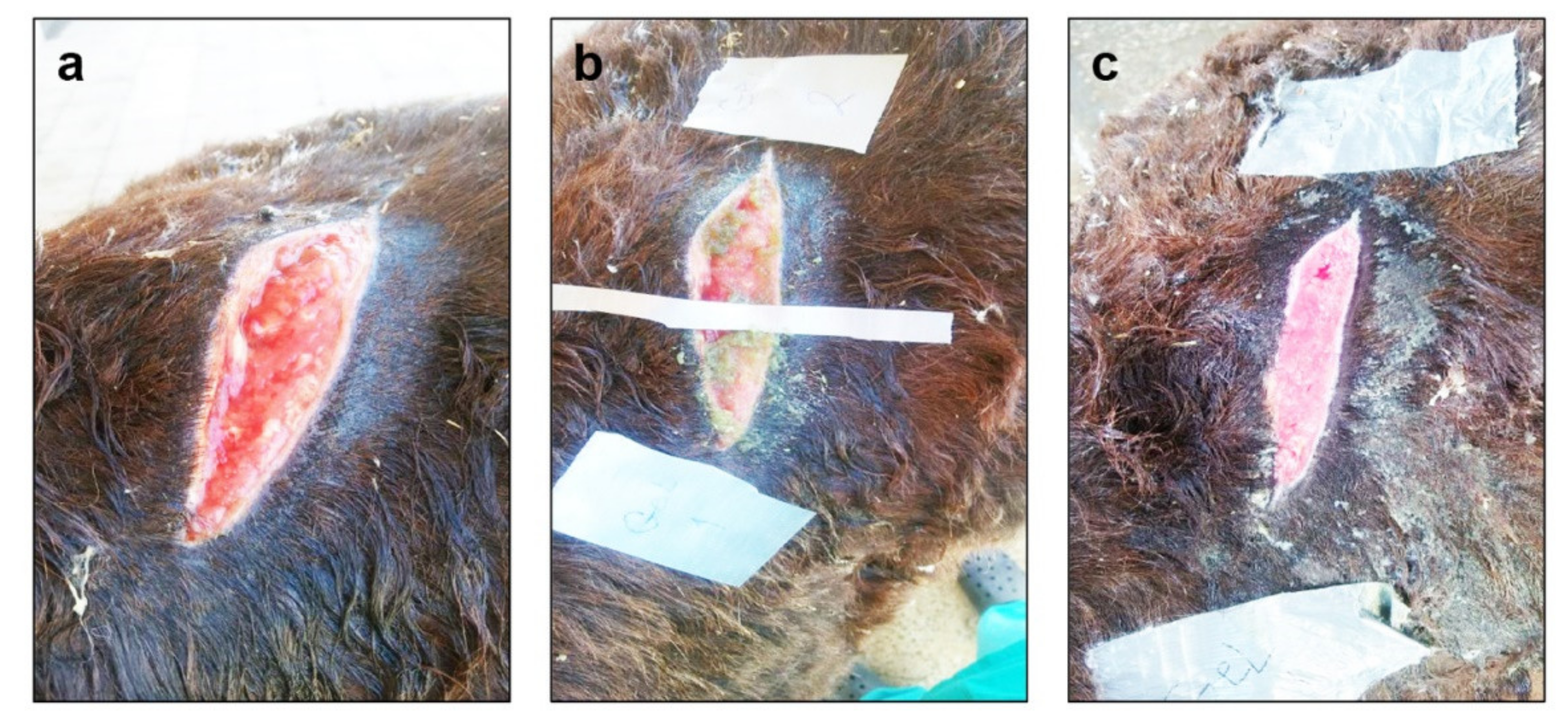
3.5. Clinical Observations

On the clinical assessment of wounds (Figure 4), it was observed that on the day of wounding, the wound edges showed hyperemia, edema, and swelling of the tissues around the wound margins. On the 3rd day post wounding, wounds of GI showed yellowish exudate with shrinkage of the wounds, which was accompanied by hyperemia and swelling of the wound edges. While in GII, the wounds were covered with brownish scabs with shrinkage and significantly decreased wounds size compared with the control. Additionally, the wounds of GIII were non-significantly narrower than the wounds size of GI, with brownish scab covering the wound surface, which sloughed from some of them. On the 7th day post-treatment, the wounds of GI were covered with thick moist scabs with significantly less wound contraction in comparison with GII and GIII. While the wounds size of GII was non-significantly narrower than that of GIII. On the 14th day post-treatment, the wounds contraction of GII and GIII were significantly higher than GI. The wounds of GII showed complete healing with scab formation in comparison with the wound of GIII. Meanwhile, the wounds of GI showed incomplete healing.
The WSA and WSA% (Table 3 and Table 4) on the 3rd and 7th day post-treatment significantly decreased in GII compared with GI, while the WC% (Table 5) significantly increased in GII compared with GI when p was ≤0.05. On the 14th day post-treatment, the WSA and WSA% significantly decreased in GII and GIII compared with GI; furthermore, the WC% significantly increased in GII and GIII compared with GI when p was ≤0.05.

3.6. Histopathological Findings

As shown in (Figure 5), on the 3rd day post-treatment, the microscopic examination of wounds in the GI revealed ulcerated and desquamated necrotic mass of tissue, inflammatory reaction with intense polymorph nuclear cells infiltration and dilated blood vessels with hemorrhage and edema of the underlying dermis. While in wounds of GII, the dermal blood vessels were congested in addition to fibroblast cells proliferation and with the presence of mononuclear cells infiltration. GIII showed congested blood vessels, hemorrhage, and increased inflammatory reaction. On the 7th day, GI showed fewer polymorph-nuclear cells in the dermis but more macrophages infiltration, less edema, and hemorrhages compared with the 3rd day of the experiment. While, GII showed moderate fibroblast proliferation, mild collagen deposition, and increased angiogenesis in the upper layer of the granulation tissue. The pathological examination of wounds in GIII revealed little fibroblast proliferation with increased inflammatory cells proliferation. On the 14th day, there was enhanced repair and remodeling of the wound area. The wounds of GI showed complete epithelization but with increased necrotic tissues and inflammatory cells. Moreover, GII revealed complete epithelization with few polymorph-nuclear cells in the dermal tissue with full skin thickness and healing near to the normal skin. Wounds of GIII were characterized by complete epithelization with intense polymorph-nuclear cells infiltration in the dermal tissue and with increased necrotic tissue.

3.7. Immunohistochemistry Findings

Microscopic examination of the wounds stained with vascular endothelial growth factor immune-staining on the 3rd, 7th, and 14th days post-treatment, where GI showed mild expression of VEGF within the endothelial cells of blood vessels and fibroblast at 3rd, 7th, and 14th days. While, GII showed mild VEGF on the 3rd day within endothelial cells of blood vessels, which increased on the 7th and 14th days within the fibroblast cells and endothelial cells of blood vessels besides epithelial cells of granulation tissue. In GIII, on the 3rd day of the experiment, VEGF was expressed mild reaction within the endothelial cells of blood vessels, and increased on the 7th and 14th days and appeared within the endothelial cells of blood vessels, fibroblast cells, and epithelial cells (Figure 6).

3.8. Clinical Study

Wound management strategies applied, such as the surgical debridement in the case of the old wounds, which were considered very important for the removal of dead and necrotized tissues that provide a suitable environment for the action of the gel for good and clean healing. Any hemorrhage that could have resulted from surgical debridement was arrested immediately and controlled by packing after clearance of the wound.
By application of the herbal formulations to the equines wounds, it was observed that wounds treated with S. arghel EtOAc gel showed rapid wound epithelization compared with S. arghel MeoH extract gel, and the S. arghel EtOAc gel was easily applied on wound surface compared with S. arghel MeoH extract gel (Figure 7, Figure 8 and Figure 9).

4. Discussion

The wound induces loss of the integrity of the skin resulting in functional imbalance, possibly accompanied by disability or even death [28]. Therefore, the wound healing process is one of the utmost medical issues in human and veterinary medicine, which explains the urgent need for developing new agents that possess wound healing activities [25].
Recently medicinal plants’ antioxidant and antimicrobial activity attracted many researchers. Almost all these phytochemicals activities are emulated with artificial antimicrobials and antioxidants [29]. The current study was conducted to evaluate the antifungal, antioxidant, and efficiency of topical S. arghel EtOAc and MeoH extracts for the cutaneous wounds treatment in experimentally induced wounds and equines suffered from traumatized wounds. Biomembrane-forming yeasts and cutaneous opportunistic pathogens were most strongly associated with wound necrosis and poor outcome [6]. VOCs extracted from S. arghel by EtOAc and MeoH in this study match VOCs identified by many previous studies with qualifications of biological and medical activities (Table 6). These activities were confirmed in this study by limiting the growth of fungal isolates from clinical cases of equines wounds due to active compounds of arghel EtOAc and MeoH extracts (Table 1 and Table 2) (Figure 3a,b). Both extracts inhibited the growth of the filamentous fungi (P. funiculosum, P. jensenii, and M. cinctum) and yeast (C. albicans) isolated from wounds. C. albicans is the most common human pathogen responsible for painful mucosal infection [30]. Therefore, inhibition of yeast cells is needed to limit the rapid reproduction of cells and reduce the pain. Interestingly, C. albicans colonized all studied wounds while filamentous fungi (P. funiculosum, P. jensenii, and M. cinctum) were irregularly distributed in the studied animals’ wounds. These results confirmed the hypothesis that infected skins serve as a portable for filamentous fungi to enter the host body [31]. Both extracts inhibited the sporulation of these fungi, which supports wound recovery. Fungal spores supported fungi in wound colonization and prolonged the wound’s inflammation [6]. Thus, limiting fungal contamination is an important step for wound treatment. Moreover, the antioxidant characteristics of S. arghel (Figure 2) encouraged the inhibition of fungi and wound healing.
It is known that S. arghel is used in traditional medicine as an antispasmodic, anti-inflammatory, and anti-rheumatic herbal [72]. The obtained results revealed that the surgically induced cutaneous rats’ wounds showed accelerated wound closure with minimal scare formation after treatment with S. arghel EtOAc extract gel in comparison with that treated with S. arghel MeoH extract gel. Ref. [73] reported that there are chemical compounds that occur naturally in plants and possess important wide applications in medicine, and so the wound healing activity of S. arghel is attributed to the phytochemical components of EtOAc extract, which have anti-inflammatory and antioxidant effects (Table 6).
The S. arghel EtOAc extract has good analgesic action due to its phytochemical ingredients as Neophytadiene, Digitoxin, and Vitamin E (alpha-tocopherol), which were recorded for its analgesic action (Table 6). As a result, topical use of S. arghel has a good effect in reducing the pain.
Equines are known for their tendency to wounding, probably due to their inquisitive nature, large size, and confining in areas with potential obstacles such as metal or wire and their known difficulties with healing. The present study showed the excellent beneficial use of S. arghel in the treatment of recent and old traumatized wounds in equines. Extract application resulted in relieving edema and inflammation around the wounds, disappearance of infection, and observable decreased wound surface. After extract application, the wounds were not covered with bandages due to the authors of [74] reporting that the bandaged wound took a longer time to heel than unbandaged wounds. The application of the extracts absorbing exudate, keeping the wound moist, which helps epithelization, prevent contamination of wound from flies due to presence of Oxiranepent Hexadecanoic Anoic Acid, 3-UndecyL-, Methyl Ester, Trans, which has pesticide activity as recorded by [44]. Ref. [75] reported that delayed healing associated with distal limb wounds is a particular problem in equine clinical practice. In this study, the topical extract of S. arghel, especially EtOAc extract, showed accelerated the wound healing of the distal limb wound.

5. Conclusions

From the aforementioned results, it can be concluded that S. argel possesses antifungal, antioxidant, and wound healing activities. These findings were well appreciated, with histopathological studies suggesting that S. argel can serve as a good therapeutic agent for the treatment of cutaneous wounds as well as beneficial effects for the treatment of equine wounds, especially those related to the distal limbs.

Author Contributions

F.F.A.-M., Z.M.M., A.E.-M., M.B. and A.A.M. conceived and designed the study. F.F.A.-M., isolated and identified the wound-associated fungi and screened the antifungal activity. F.F.A.-M. prepared the extracts, GCMS analysis, and identification of extract components. Z.M.M. and A.A.M. conducted the experiment and collected the data. Z.M.M. performed histopathological and immunohistochemistry studies. F.F.A.-M., S.F.I., Z.M.M., A.E.-M., M.B. and A.A.M. organized and analyzed the data. S.F.I. funded the acquisition. F.F.A.-M., Z.M.M., S.F.I., A.E.-M., M.B. and A.A.M. interpreted the data, wrote the paper, and revised the final draft. All authors have read and agreed to the published version of the manuscript.

Funding

This research was funded by Princess Nourah bint Abdulrahman University Researchers Supporting Project number (PNURSP2022R127), Princess Nourah bint Abdulrahman University, Riyadh, Saudi Arabia.

Institutional Review Board Statement

Not applicable.

Informed Consent Statement

Not applicable.

Data Availability Statement

The data presented in this study are available in this manuscript.

Acknowledgments

The authors appreciate the support provided by the Princess Nourah bint Abdulrahman University Researchers Supporting Project number (PNURSP2022R127), Princess Nourah bint Abdulrahman University, Riyadh, Saudi Arabia. Also, the authors would like to thank Magdi El-Sayed, Molecular Biotechnology Program, Biological Sciences Department, Faculty of Science, Galala University, New Galala City, Suez, Egypt, Ahmed Abdeen, Department of Forensic Medicine and Toxicology, Faculty of Veterinary Medicine, Benha University, Touch, Egypt, and AbdulRahman A. Saied, Researcher, National Food Safety Authority (NFSA), Aswan Branch, Egypt for their helpful efforts in revising and editing the manuscript, and preparing figures and graphs.

Conflicts of Interest

The authors declare no conflict of interest.

References

  1. Farahpour, M.R.; Habibi, M. Evaluation of the Wound Healing Activity of an Ethanolic Extract of Ceylon Cinnamon in Mice. Vet. Med. 2012, 57, 53–57. [Google Scholar]
  2. Mousa, H.-L. Aerobic, Anaerobic and Fungal Burn Wound Infections. J. Hosp. Infect. 1997, 37, 317–323. [Google Scholar] [CrossRef]
  3. Bowler, P.G. The Microbiology of Acute and Chronic Wounds. Wounds 1999, 11, 72–78. [Google Scholar]
  4. Bessa, L.J.; Fazii, P.; Di Giulio, M.; Cellini, L. Bacterial Isolates from Infected Wounds and Their Antibiotic Susceptibility Pattern: Some Remarks about Wound Infection. Int. Wound J. 2015, 12, 47–52. [Google Scholar] [CrossRef]
  5. Kingsley, A. A Proactive Approach to Wound Infection. Nurs. Stand. 2001, 15, 50. [Google Scholar] [CrossRef]
  6. Kalan, L.; Loesche, M.; Hodkinson, B.P.; Heilmann, K.; Ruthel, G.; Gardner, S.E.; Grice, E.A. Redefining the Chronic-Wound Microbiome: Fungal Communities Are Prevalent, Dynamic, and Associated with Delayed Healing. MBio 2016, 7, e01058-16. [Google Scholar] [CrossRef] [Green Version]
  7. Dowd, S.E.; Delton Hanson, J.; Rees, E.; Wolcott, R.D.; Zischau, A.M.; Sun, Y.; White, J.; Smith, D.M.; Kennedy, J.; Jones, C.E. Survey of Fungi and Yeast in Polymicrobial Infections in Chronic Wounds. J. Wound Care 2011, 20, 40–47. [Google Scholar]
  8. Gayas, M.A.; Ahmad, R.A.; Gugjoo, M.B.; Handoo, N. Fungal Wound Infections: Mini Review. Pharma Innov. 2018, 7, 295–298. [Google Scholar]
  9. Seth, A.K.; Geringer, M.R.; Hong, S.J.; Leung, K.P.; Mustoe, T.A.; Galiano, R.D. In Vivo Modeling of Biofilm-Infected Wounds: A Review. J. Surg. Res. 2012, 178, 330–338. [Google Scholar] [CrossRef]
  10. Jørgensen, E.; Lazzarini, G.; Pirone, A.; Jacobsen, S.; Miragliotta, V. Normal Microscopic Anatomy of Equine Body and Limb Skin: A Morphological and Immunohistochemical Study. Ann. Anat. Anat. Anz. 2018, 218, 205–212. [Google Scholar] [CrossRef]
  11. Sardari, K.; Kazemi, H.; Emami, M.R.; Movasaghi, A.R.; Goli, A.A. Role of Collagen Cross-Linking on Equine Wound Contraction and Healing. Comp. Clin. Path. 2009, 18, 239–247. [Google Scholar] [CrossRef]
  12. Theoret, C.L.; Wilmink, J.M. Aberrant Wound Healing in the Horse: Naturally Occurring Conditions Reminiscent of Those Observed in Man. Wound Repair Regen. 2013, 21, 365–371. [Google Scholar] [CrossRef]
  13. Jørgensen, E.; Bay, L.; Skovgaard, L.T.; Bjarnsholt, T.; Jacobsen, S. An Equine Wound Model to Study Effects of Bacterial Aggregates on Wound Healing. Adv. Wound Care 2019, 8, 487–498. [Google Scholar] [CrossRef] [Green Version]
  14. Westgate, S.J.; Percival, S.L.; Knottenbelt, D.C.; Clegg, P.D.; Cochrane, C.A. Microbiology of Equine Wounds and Evidence of Bacterial Biofilms. Vet. Microbiol. 2011, 150, 152–159. [Google Scholar] [CrossRef] [Green Version]
  15. El-Kamali, H.H.; Khalid, S.A. The Most Common Herbal Remedies in Dongola Province, Northern Sudan. Fitoter 1998, 69, 118–121. [Google Scholar]
  16. Kamel, M.S.; Ohtani, K.; Hasanain, H.A.; Mohamed, M.H.; Kasai, R.; Yamasaki, K. Monoterpene and Pregnane Glucosides from Solenostemma Argel. Phytochemistry 2000, 53, 937–940. [Google Scholar] [CrossRef]
  17. Hassan, H.A.; Hamed, A.I.; El-Emary, N.A.; Springuel, I.V.; Mitome, H.; Miyaoka, H. Pregnene Derivatives from Solenostemma Argel Leaves. Phytochemistry 2001, 57, 507–511. [Google Scholar] [CrossRef]
  18. El-Kheir, K.S.E.; Murwa, A.M. Chemical Composition, Minerals, Protein Fractionation, and Anti-Nutrition Factors in Leaf of Hargel Plant (Solenostemma argel). Eur. J. Sci. Res. 2010, 43, 430–434. [Google Scholar]
  19. Idris, T.I.M.; Ibrahim, A.M.A.; Mahdi, E.M.; Taha, A.K. Influence of Argel (Solenostemma argel Del. Hayne) Soil Applications on Flowering and Yield of Date Palm (Phoenix dactylifera L.). Agric. Biol. J. N. Am. 2011, 2, 538–542. [Google Scholar] [CrossRef]
  20. Ounaissia, K.; Pertuit, D.; Mitaine-Offer, A.-C.; Miyamoto, T.; Tanaka, C.; Delemasure, S.; Dutartre, P.; Smati, D.; Lacaille-Dubois, M.-A. New Pregnane and Phenolic Glycosides from Solenostemma Argel. Fitoterapia 2016, 114, 98–104. [Google Scholar]
  21. Kamel, N.M.; Abdel-Motaal, F.F.; El-Zayat, S.A. Endophytic Fungi from the Medicinal Herb Euphorbia Geniculata as a Potential Source for Bioactive Metabolites. Arch. Microbiol. 2020, 202, 247–255. [Google Scholar] [CrossRef] [PubMed]
  22. Sinha, G.K.; Gulati, B.C. Antibacterial and Antifungal Study of Some Essential Oils and Some of Their Constituents. Indian Perfum. 1990, 34, 126–129. [Google Scholar]
  23. Balouiri, M.; Sadiki, M.; Ibnsouda, S.K. Methods for in Vitro Evaluating Antimicrobial Activity: A Review. J. Pharm. Anal. 2016, 6, 71–79. [Google Scholar] [CrossRef] [PubMed] [Green Version]
  24. Singh, J.; Tripathi, N.N. Inhibition of Storage Fungi of Blackgram (Vigna mungo L.) by Some Essential Oils. Flavour Fragr. J. 1999, 14, 1–4. [Google Scholar] [CrossRef]
  25. Metwally, A.A.; Abdel-Hady, A.-N.A.A.; Haridy, M.A.M.; Ebnalwaled, K.; Saied, A.A.; Soliman, A.S. Wound-Healing Properties of Green (Using Lawsonia inermis Leaf Extract) and Chemically Synthesized Zno Nanoparticles in Albino Rats. Environ. Sci. Pollut. Res. 2021, 29, 23975–23987. [Google Scholar] [CrossRef]
  26. Sadaf, F.; Saleem, R.; Ahmed, M.; Ahmad, S.I. Healing Potential of Cream Containing Extract of Sphaeranthus Indicus on Dermal Wounds in Guinea Pigs. J. Ethnopharmacol. 2006, 107, 161–163. [Google Scholar] [CrossRef]
  27. Elbialy, Z.I.; Assar, D.H.; Abdelnaby, A.; Asa, S.A.; Abdelhiee, E.Y.; Ibrahim, S.S.; Abdel-Daim, M.M.; Almeer, R.; Atiba, A. Healing Potential of Spirulina Platensis for Skin Wounds by Modulating BFGF, VEGF, TGF-SS1 and α-SMA Genes Expression Targeting Angiogenesis and Scar Tissue Formation in the Rat Model. Biomed. Pharmacother. 2021, 137, 111349. [Google Scholar] [CrossRef]
  28. Theoret, C. Tissue Engineering in Wound Repair: The Three “R” s—Repair, Replace, Regenerate. Vet. Surg. 2009, 38, 905–913. [Google Scholar] [CrossRef]
  29. Santos, G.A.; Vila, M.M.D.C.; Chaud, M.V.; Silva, W.L.; de Castro, A.G.; de Oliveira, J.M., Jr.; Tubino, M.; Balcao, V.M. Antimicrobial and Antioxidant Screening of Curcumin and Pyrocatechol in the Prevention of Biodiesel Degradation: Oxidative Stability. Biofuels 2016, 7, 581–592. [Google Scholar] [CrossRef]
  30. Singh, D.K.; Tóth, R.; Gácser, A. Mechanisms of Pathogenic Candida Species to Evade the Host Complement Attack. Front. Cell. Infect. Microbiol. 2020, 10, 94. [Google Scholar] [CrossRef] [Green Version]
  31. Zakaria, A.; Abdel-Motaal, F.; Mahalel, U. Antifungal Activity of Ficus sycomorus L. Extracts against Dermatophytes and Other Associated Fungi Isolated from Camels Ringworm Lesions. J. Biol. Stud. 2018, 1, 116–132. [Google Scholar]
  32. Jerez, S.; Sierra, L.; de Bruno, M.P. 17-Octadecynoic Acid Improves Contractile Response to Angiotensin II by Releasing Vasocontrictor Prostaglandins. Prostaglandins Other Lipid Mediat. 2012, 97, 36–42. [Google Scholar] [CrossRef]
  33. Shehzad, A.; Qayyum, A.; Rehman, R.; Nadeem, F.; Raffi, M. A Review of Bioactivity Guided Medicinal Uses and Therapeutic Potentials of Noxious Weed (Alternanthera sessilis). Int. J. Chem. Biochem. Sci. 2018, 14, 95–103. [Google Scholar]
  34. Kadhim, M.J.; Al-Rubaye, A.F.; Hameed, I.H. Determination of Bioactive Compounds of Methanolic Extract of Vitis Vinifera Using GC-MS. Int. J. Toxicol. Pharmacol. Res. 2017, 9, 113–126. [Google Scholar] [CrossRef] [Green Version]
  35. Adegoke, A.S.; Jerry, O.V.; Ademola, O.G. GC-MS Analysis of Phytochemical Constituents in Methanol Extract of Wood Bark from Durio Zibethinus Murr. Int. J. Med. Plants Nat. Prod. 2019, 5, 1–11. [Google Scholar]
  36. Godwin, A.; Akinpelu, B.A.; Makinde, A.M.; Aderogba, M.A.; Oyedapo, O.O. Identification of N-Hexane Fraction Constituents of Archidium Ohioense (Schimp. Ex Mull) Extract Using GC-MS Technique. J. Pharm. Res. Int. 2015, 6, 366–375. [Google Scholar] [CrossRef]
  37. Varsha, K.K.; Devendra, L.; Shilpa, G.; Priya, S.; Pandey, A.; Nampoothiri, K.M. 2,4-Di-Tert-Butyl Phenol as the Antifungal, Antioxidant Bioactive Purified from a Newly Isolated Lactococcus sp. Int. J. Food Microbiol. 2015, 211, 44–50. [Google Scholar] [CrossRef]
  38. Lewis, T.; Macaulay, E.D.M. Design and Elevation of Sex-attractant Traps for Pea Moth, Cydia Nigricana (Steph.) and the Effect of Plume Shape on Catches. Ecol. Entomol. 1976, 1, 175–187. [Google Scholar] [CrossRef]
  39. Kim, G.; Kim, J.; Kang, M.; Jang, A.; Kim, Y.R.; Kim, S.; Chang, K.; Hong, J.J.; Park, J. Inhibitory Effect of 1-tetradecanol on Helicobacter Pylori-induced Production of Interleukin-8 and Vascular Endothelial Growth Factor in Gastric Epithelial Cells. Mol. Med. Rep. 2017, 16, 9573–9578. [Google Scholar] [CrossRef]
  40. Swamy, M.K.; Arumugam, G.; Kaur, R.; Ghasemzadeh, A.; Yusoff, M.M.; Sinniah, U.R. GC-MS Based Metabolite Profiling, Antioxidant and Antimicrobial Properties of Different Solvent Extracts of Malaysian Plectranthus Amboinicus Leaves. Evid. Based Complement. Altern. Med. 2017, 2017, 1517683. [Google Scholar] [CrossRef] [Green Version]
  41. Albergoni, V.; Piccinni, E.; Coppellotti, O. Response to Heavy Metals in Organisms—I. Excretion and Accumulation of Physiological and Non Physiological Metals in Euglena Gracilis. Comp. Biochem. Physiol. Part C Comp. Pharmacol. 1980, 67, 121–127. [Google Scholar] [CrossRef]
  42. Adnan, M.; Chy, N.U.; Mostafa Kamal, A.T.M.; Azad, M.O.K.; Paul, A.; Uddin, S.B.; Barlow, J.W.; Faruque, M.O.; Park, C.H.; Cho, D.H. Investigation of the Biological Activities and Characterization of Bioactive Constituents of Ophiorrhiza Rugosa Var. Prostrata (D. Don) & Mondal Leaves through in Vivo, in Vitro, and in Silico Approaches. Molecules 2019, 24, 1367. [Google Scholar]
  43. Brintha, S.; Rajesh, S.; Renuka, R.; Santhanakrishnan, V.P.; Gnanam, R. Phytochemical Analysis and Bioactivity Prediction of Compounds in Methanolic Extracts of Curculigo Orchioides Gaertn. J. Pharmacogn. Phytochem. 2017, 6, 192–197. [Google Scholar]
  44. Ogbuehi, G.U.I.; Echeme, J.B.O. Chemical Constituents of Methanol Leaf Extract of Aspilia Africana CD Adams by GC MS. Int. J. Adv. Res. Chem. Sci. 2018, 5, 21–29. [Google Scholar] [CrossRef]
  45. Perumal, G.M.; Prabhu, K.; Rao, M.R.K.; Janaki, C.S.; Kalaivannan, J.; Kavimani, M. The Gc Ms Analysis of Ethyl Acetate Extract of One Herbal Plant, ‘Jatrophacurcus’. NVEO-Nat. Volatiles Essent. OILS J. NVEO 2021, 8, 6347–6354. [Google Scholar]
  46. Harris, E.D. Copper as a Cofactor and Regulator of Copper, Zinc Superoxide Dismutase. J. Nutr. 1992, 122, 636–640. [Google Scholar]
  47. Parthipan, B.; Suky, M.G.T.; Mohan, V.R. GC-MS Analysis of Phytocomponents in Pleiospermium Alatum (Wall. Ex Wight & Arn.) Swingle, (Rutaceae). J. Pharmacogn. Phytochem. 2015, 4, 216–222. [Google Scholar]
  48. Sosa, A.A.; Bagi, S.H.; Hameed, I.H. Analysis of Bioactive Chemical Compounds of Euphorbia Lathyrus Using Gas Chromatography-Mass Spectrometry and Fourier-Transform Infrared Spectroscopy. J. Pharmacogn. Phyther. 2016, 8, 109–126. [Google Scholar]
  49. Kalaimagal, C. Identification of bioactive compounds in flower of Tabernaemontana divaricata (L.) using gas chromatography–mass spectrometry analysis. Asian J. Pharm. Clin. Res. 2019, 12, 129–132. [Google Scholar] [CrossRef]
  50. Amer, M.S.; Barakat, K.M.; Hassanein, A.E.A. Phthalate Derivatives from Marine Penicillium Decumbens and Its Synergetic Effect against Sepsis Bacteria. Biointerface Res. Appl. Chem 2019, 9, 4070–4076. [Google Scholar]
  51. Zeb, A.; Ahmad, S.; Ullah, F.; Ayaz, M.; Sadiq, A. Anti-Nociceptive Activity of Ethnomedicinally Important Analgesic Plant Isodon Rugosus Wall. Ex Benth: Mechanistic Study and Identifications of Bioactive Compounds. Front. Pharmacol. 2016, 7, 200. [Google Scholar] [CrossRef] [Green Version]
  52. Aziba, P.I.; Adedeji, A.; Ekor, M.; Adeyemi, O. Analgesic Activity of Peperomia Pellucida Aerial Parts in Mice. Fitoterapia 2001, 72, 57–58. [Google Scholar] [CrossRef]
  53. Narayanamoorthi, V.; Vasantha, K.; Maruthasalam, R. GC MS Determination of Bioactive Components of Peperomia pellucida (L.) Kunth. Biosci. Discov. 2015, 6, 83–88. [Google Scholar]
  54. Arrigoni-Blank, M.d.F.; Oliveira, R.L.B.; Mendes, S.S.; de Albuquerque Silva, P.; Antoniolli, Â.R.; Vilar, J.C.; de Holanda Cavalcanti, S.C.; Blank, A.F. Seed Germination, Phenology, and Antiedematogenic Activity of Peperomia pellucida (L.) HBK. BMC Pharmacol. 2002, 2, 12. [Google Scholar] [CrossRef] [Green Version]
  55. Ashmawy, N.A.; Al Farraj, D.A.; Salem, M.Z.M.; Elshikh, M.S.; Al-Kufaidy, R.; Alshammari, M.K.; Salem, A.Z.M. Potential Impacts of Pinus Halepensis Miller Trees as a Source of Phytochemical Compounds: Antibacterial Activity of the Cones Essential Oil and n-Butanol Extract. Agrofor. Syst. 2020, 94, 1403–1413. [Google Scholar] [CrossRef]
  56. Otieno, A.J. Antimicrobial Activity and Phytochemical Profiles of Warburgia Ugandensis Sprague (Canellaceae) Extracts from Different Populations across the Kenyan Rift Valley. Ph.D. Thesis, Kenyatta University, Nairobi, Kenya, 2016. [Google Scholar]
  57. Kim, D.H.; Park, M.H.; Choi, Y.J.; Chung, K.W.; Park, C.H.; Jang, E.J.; An, H.J.; Yu, B.P.; Chung, H.Y. Molecular Study of Dietary Heptadecane for the Anti-Inflammatory Modulation of NF-KB in the Aged Kidney. PLoS ONE 2013, 8, e59316. [Google Scholar] [CrossRef]
  58. Seong, R.K.; Kim, J.A.; Shin, O.S. Wogonin, a Flavonoid Isolated from Scutellaria Baicalensis, Has Anti-Viral Activities against Influenza Infection via Modulation of AMPK Pathways. Acta Virol. 2018, 62, 78–85. [Google Scholar] [CrossRef]
  59. Woźniak, D.; Dryś, A.; Matkowski, A. Antiradical and Antioxidant Activity of Flavones from Scutellariae Baicalensis Radix. Nat. Prod. Res. 2015, 29, 1567–1570. [Google Scholar] [CrossRef]
  60. Liu, Y.-F.; Gao, F.; Li, X.-W.; Jia, R.-H.; Meng, X.-D.; Zhao, R.; Jing, Y.-Y.; Wang, Y.; Jiang, W. The Anticonvulsant and Neuroprotective Effects of Baicalin on Pilocarpine-Induced Epileptic Model in Rats. Neurochem. Res. 2012, 37, 1670–1680. [Google Scholar] [CrossRef]
  61. Yang, Y.-Z.; Tang, Y.-Z.; Liu, Y.-H. Wogonoside Displays Anti-Inflammatory Effects through Modulating Inflammatory Mediator Expression Using RAW264. 7 Cells. J. Ethnopharmacol. 2013, 148, 271–276. [Google Scholar] [CrossRef]
  62. Dong, Q.; Chu, F.; Wu, C.; Huo, Q.; Gan, H.; Li, X.; Liu, H. Scutellaria Baicalensis Georgi Extract Protects against Alcohol-induced Acute Liver Injury in Mice and Affects the Mechanism of ER Stress. Mol. Med. Rep. 2016, 13, 3052–3062. [Google Scholar] [CrossRef] [PubMed] [Green Version]
  63. Zahra, G.; Khadijeh, B.; Hossein, D.; Ali, S. Essential Oil Composition of Two Scutellaria Species from Iran. J. Tradit. Chin. Med. Sci. 2019, 6, 244–253. [Google Scholar] [CrossRef]
  64. Takahashi, C.; Kikuchi, N.; Katou, N.; Miki, T.; Yanagida, F.; Umeda, M. Possible Anti-Tumor-Promoting Activity of Components in Japanese Soybean Fermented Food, Natto: Effect on Gap Junctional Intercellular Communication. Carcinogenesis 1995, 16, 471–476. [Google Scholar] [CrossRef] [PubMed]
  65. Quintanilla-Licea, R.; Morado-Castillo, R.; Gomez-Flores, R.; Laatsch, H.; Verde-Star, M.J.; Hernández-Martínez, H.; Tamez-Guerra, P.; Tamez-Guerra, R.; Rodríguez-Padilla, C. Bioassay-Guided Isolation and Identification of Cytotoxic Compounds from Gymnosperma Glutinosum Leaves. Molecules 2012, 17, 11229–11241. [Google Scholar] [CrossRef] [Green Version]
  66. Chakraborty, A.K.; Gaikwad, A.V.; Singh, K.B. Phytopharmacological Review on Acanthospermum Hispidum. J. Appl. Pharm. Sci. 2012, 2, 144–148. [Google Scholar]
  67. Kim, S.; Chung, W.; Kim, S.; Ko, S.; Um, J. Antiinflammatory Effect of Oldenlandia Diffusa and Its Constituent, Hentriacontane, through Suppression of Caspase-1 Activation in Mouse Peritoneal Macrophages. Phyther. Res. 2011, 25, 1537–1546. [Google Scholar] [CrossRef]
  68. Khajuria, V.; Gupta, S.; Sharma, N.; Kumar, A.; Lone, N.A.; Khullar, M.; Dutt, P.; Sharma, P.R.; Bhagat, A.; Ahmed, Z. Anti-Inflammatory Potential of Hentriacontane in LPS Stimulated RAW 264.7 Cells and Mice Model. Biomed. Pharmacother. 2017, 92, 175–186. [Google Scholar] [CrossRef]
  69. Dandekar, R.; Fegade, B.; Bhaskar, V.H. GC-MS Analysis of Phytoconstituents in Alcohol Extract of Epiphyllum Oxypetalum Leaves. J. Pharmacogn. Phytochem. 2015, 4, 149–154. [Google Scholar]
  70. Kumar, R.S.; Anburaj, G.; Subramanian, A.; Vasantha, S.; Selvam, A.P. Preliminary Phytochemical Investigation, Antimicrobial Activity and GC-MS Analysis of Leaf Extract of Capparis Zeylanica Linn. J. Pharmacogn. Phytochem. 2019, 8, 1399–1405. [Google Scholar]
  71. Roy, R.N.; Laskar, S.; Sen, S.K. Dibutyl Phthalate, the Bioactive Compound Produced by Streptomyces Albidoflavus 321.2. Microbiol. Res. 2006, 161, 121–126. [Google Scholar] [CrossRef]
  72. El-Shiekh, R.A.; Salama, A.; Al-Mokaddem, A.K.; Abdel-Sattar, E.A. Gastroprotective Effect of Mucilage Fraction from Solenostemma Argel via Cytoprotection and Attenuation of Oxidative Stress, Inflammation and Apoptosis. J. Herbmed Pharmacol. 2021, 10, 232–240. [Google Scholar] [CrossRef]
  73. Kumar, A.; Aravindhan, P.; Deecaraman, D.; Ilavarasan, R.; Padmanabhan, N. Neutral Components in the Leaves and Seeds of Syzygium Cumini. Afr. J. Pharm. Pharmacol. 2009, 3, 560–561. [Google Scholar]
  74. Dart, A.J.; Perkins, N.R.; Dart, C.M.; Jeffcott, L.B.; Canfield, P. Effect of Bandaging on Second Intention Healing of Wounds of the Distal Limb in Horses. Aust. Vet. J. 2009, 87, 215–218. [Google Scholar] [CrossRef]
  75. Carnwath, R.; Graham, E.M.; Reynolds, K.; Pollock, P.J. The Antimicrobial Activity of Honey against Common Equine Wound Bacterial Isolates. Vet. J. 2014, 199, 110–114. [Google Scholar] [CrossRef]
Figure 1. Gas chromatography-mass spectrometry (GC-MS) analysis charts of released volatile organic compounds emitted from S. arghel ethyl acetate (a) and methanol (b) extracts.
Applsci 12 04121 g001
Figure 2. Antioxidant activity calculated by DPPH inhibition percentage of S. arghel ethyl acetate and methanol extracts at 50,100 and 150 µg compared with control (Asorbic acid, AA).
Applsci 12 04121 g002
Figure 3. Screening of antifungal activity of Arghel plant extracts using food technique method. Inhibition of fungal growth in Sabroud medium supplemented with EtOAc and MeoH plant extracts at two concentrations (5 mg/mL and 10 mg/mL) compared with control (a). Histogram showed the inhibition percentage of pathogenic fungi isolated from contaminated wounds at 5 mg/mL and 10 mg/mL of both EtOAc and MeoH plant extracts (b).
Applsci 12 04121 g003
Figure 4. Photograph showing wound healing in GI, GII, and GIII at 3rd, 7th, and 14th days post-treatment, where the healing process accelerated in wounds treated with GI in comparison with GI and GIII groups.
Applsci 12 04121 g004
Figure 5. Photomicrograph of the control and treated groups on the 3rd, 7th, and 14th days post-treatment where GI at 3rd day showed ulcerated and desquamated necrotic tissue with inflammatory cells infiltration (arrows), while at 7th day, there were dilated blood vessels with epithelization. On 14th day, increased mononuclear inflammatory cells infiltration with necrotic tissue mass at the wound area was shown. While GII on the 3rd day showed inflammatory cells infiltration and mild angiogenesis, and on the 7th day, fibroblast proliferation with neovascularization and granulation tissue formation was shown. Moreover, on the 14th day, full skin thickness and healing near to normal skin were shown. GIII showed congested blood vessels with mild fibroblast proliferation on the 3rd day, while on the 7th day, increased fibroblast proliferation and neovascularization were shown. On the 14th day post-treatment, complete epithelization of wounds with mononuclear inflammatory cells infiltration was shown.
Applsci 12 04121 g005
Figure 6. Immunohistochemical expression of VEGF in control and treated wounds. GI showed mild expression of VEGF within the endothelial cells of blood vessels and fibroblast on the 3rd, 7th, and 14th days. GII showed mild VEGF on the 3rd day within endothelial cells of blood vessels, which then increased on the 7th and 14th days within the fibroblast cells and endothelial cells of blood vessels besides epithelial cells of granulation tissue. In GIII, on the 3rd day, VEGF was mild within the endothelial cells of blood vessels, and increased on the 7th and 14th days and appeared within the endothelial cells of blood vessels, fibroblast cells, and epithelial cells.
Applsci 12 04121 g006
Figure 7. (a) Four-year-old male donkey with an old lacerated accidental wound on the back. (b) The wound was divided into two parts and treated with gel (1) S. arghel EtOAc gel and gel (2) S. arghel MeoH extract gel. (c) The same animal, after 9 days of treatment, showed decreased wound size and complete epithelization of the wound.
Applsci 12 04121 g007
Figure 8. (a) Nine-year-old horse with recent wound at the left hind limb, the wound was divided into two parts and treated with and treated with gel (1) S. arghel EtOAc gel and gel (2) S. arghel MeoH extract gel. (b) The same animal, after 18 days of treatment, showed decreased wound size and complete epithelization with hypergranulation tissue due to the wound being near a movable part. (c) The same animal, after a month and a half, showed complete wound healing.
Applsci 12 04121 g008
Figure 9. (a) Eleven-year-old horse with an old accidental wound nearly at the level of elbow joint of the left forelimb (movable area) after different trials of treatment and suturing showed no tendency for healing with wound dehiscence. (b) The same wound after one week of treatment with S. arghel EtOAc gel showed complete epithelization with decreased wound size. (c) The wound, after 2 months of treatment, showed complete healing.
Applsci 12 04121 g009
Table 1. GC-MS analysis of VOCs produced by S. arghel leaves extracted by Ethyl acetate.
NOVOCs CompoundsMolecular FormulaRtMWArea%
117-Octadecynoic acidC18H32O24.442803.11
22,2-dimethyl-5-(3-methyloxiranyl)-cyclohexanoneC11H18O24.441823.11
3Linoleoyl chlorideC18H31ClO4.602980.85
42,2,3,3,4,4 Exadeutero,octadecanalC18H30D6O4.802741.13
51-Dodecanol, 3,7,11-trimethylC15H32O4.802281.13
61,3,5-Triazine-2,4-diamine,6-chloro-n-ethyl-C5H8ClN59.311731.49
71-ChlorooctadecaneC18H37Cl9.312881.49
82-Aminoethanethiol,hydrogen sulfate (Ester)C2H7NO3S210.301571.28
97-Hexadecenal, (Z)-C16H30O10.302381.28
109-Octadecenoic acid (Z)-C18H34O211.742821.53
111-DodeceneC12H2411.741681.53
123-TrifluoroacetoxydodecaneC14H25F3O211.742821.53
13DocosaneC22H4613.873101.60
14Farnesene epoxide, EC15H24O14.262201.49
1510,13-Octadecadiynoic acid, methyl esterC19H30O214.262901.49
162,4-Di-tert-butylphenolC14H22O14.362062.91
179H-FluoreneC13H1015.651664.94
18(E,E)-1,3,5-Tridecatriene-7,9,11-triyneC13H1015.651664.94
198-Dodecen-1-ol, acetate, (Z)-C14H26O215.812260.62
20Undec-10-ynoic acid, heptadecylesterC28H52O215.814200.62
21Undec-10-ynoic acid, octadecyl esterC29H54O215.814340.62
22Bisabolol oxide B (2-Furanmethanol, tetrahydro-à,à,5-trimethyl-5-(4-methyl-3-cyclohexen-1-yl),[2S-[2à,5á(R)]]-C15H26O217.082382.40
23Acetic acid, 10,11-dihydroxy-3,7,11 trimethyl-dodeca-2,6-dienyl esterC17H30O417.082982.40
242-Monooleoylglycerol trimethylsilyl etherC27H56O4Si217.255000.99
252,3-Bis[(trimethylsilyl)oxy]propyl (9z,12z)-9,12-octadecadienoateC27H54O4Si217.254980.99
26Benzoic acid, 2,4-bis(trimethylsiloxy)-, trimethylsilyl esterC16H30O4Si317.253700.99
271-TetradecanolC14H30O18.022141.27
28Loliolide,2(4H)-Benzofuranone, 5,6,7,7a-tetrahydro-6-hydroxy-4,4,7a-trimethylC11H16O319.201962.17
293′,4′,7-TrimethylquercetinC18H16O720.143441.27
30NeophytadieneC20H3820.362785.88
313-EicosyneC20H3820.362785.88
32DigitoxinC41H64O1320.587640.85
33Oxiranepentanoic acid, 3-undecyl-, methyl ester, transC19H36O320.583120.85
3401297107001 Tetraneurin—A-diolC15H20O520.622800.89
351-HeptatriacotanolC37H76O20.625380.89
36Phen-1,4-diol,2,3-dimethyl-5-trifluoromethylC9H9F3O220.872061.34
37Ethanol, 2-(9-octadecenyloxy)-, (Z)-C20H40O221.093121.83
3813-Heptadecyn-1-olC17H32O21.092521.83
397,9-Di-tert-butyl-1-oxaspiro(4,5)dec,a-6,9-diene-2,8-dioneC17H24O321.772764.97
40Isochiapin BC19H22O622.463461.45
41Isochiapin B %2<C19H26O622.463501.45
426,8-Di-c-á-glucosylluteolinC27H30O1622.736101.37
432,2-Dideutero octadecanalC18H34D2O24.392701.31
449-Hexadecenoic acidC16H30O224.392541.31
45PhytolC20H40O24.852965.16
469,12,15-Octadecatrienoic acid, 2,3-bis [(trimethylsilyl) oxy]propyl ester, (Z,Z,Z)C27H52O4Si225.104981.13
47Ethyl iso-allocholate or Ethyl 3,7,12-trihydroxycholan-24-oateC26H44O527.264361.21
48Diisooctyl phthalateC24H38O430.973909.32
49Vitamin E (alpha-tocopherol)C29H50O232.024309.99
505H-Cyclopropa[3,4]benz[1,2-e]azulen-5-one,9-(acetyloxy)-3-[(acetyloxy)methyl]-1,1a,1b,4,4a,7a,7b,8,9,9a-decahydro-4a,7b,9a-trihydroxy-1,1,6,8-tetramethyl-,[1ar-(1aà,1bá,4aá,7aà,7bà,8à,9á,9aà)]-C24H32O834.944487.36
51Methyl hexadecadienoateC17H30O234.942667.36
Table 2. GC-MS analysis of VOCs produced by S. arghel leaves extracted by methanol.
No.VOCs CompoundsFormulaRTMWArea%
1Heptane, 2,2,4,6,6-pentamethyl-C12H264.431704.083
2Tetradecane, 2,2-dimethyl-C16H344.432264.083
3Decane, 3,6-dimethyl-C12H265.221701.81
43-Ethyl-3-methylheptaneC10H225.221421.81
5EicosaneC20H427.382822.53
62-BromotetradecaneC14H29Br7.382762.53
7HexadecaneC16H347.382262.53
8DodecaneC12H267.7331701.883
9TetradecaneC14H307.7331981.883
101-Iodo-2-methylnonaneC10H21I7.7332681.883
11HeneicosaneC21H449.09029610.310
12HeptadecaneC17H369.09024010.310
13Octadecane, 1-iodo-C18H37I9.09038010.310
14Octadecane, 2-methyl-C19H409.1702681.835
15HexacosaneC26H549.4963668.846
16Heptadecane, 2-methyl-C18H389.4962548.846
17Heptadecane, 8-methyl-C18H389.4962548.846
18PentacosaneC25H529.5873522.645
19TriacontaneC30H629.5874222.645
20Heptadecane, 9-octyl-C25H529.6853522.395
21HentriacontaneC31H6411.37843611.344
22Sulfurous acid, hexyl octyl esterC14H30O3S11.37827811.344
23HeptacosaneC27H5611.4873802.169
24OctadecaneC18H3811.9392548.357
25Sulfurous acid, butyl heptadecyl esterC21H44O3S12.2713762.020
267,9-Di-tert-butyl-1-oxaspiro (4,5)deca-6,9diene-2,8-dioneC17H24O314.5712765.491
271,3-Pentadiene, 1,1-diphenyl-, (Z)C17H1614.5712205.491
28Dibutyl phthalateC16H2O415.24127810.14
29Heneicosane, 3-methyl-C22H4619.0923102.024
302-methyloctacosaneC29H6019.0924082.024
Table 3. Mean + SD of WSA in full-thickness skin wounds in control and treated groups.
Time in DaysGIGIIGIII
31.56 ± 0.3360. 80 ± 0.27 a0.97 ± 0.53
71.2 ± 0.350.47 ± 0.18 a0.7 ± 0.3
140.12 ± 0.60.002 ± 0.004 a0.02 ± 0.03 a
(a): Significant changes when compared with control group when p ≤ 0.05.
Table 4. Mean ± SD of WSA% in full-thickness skin wounds in control and treated groups.
Time in DaysGIGIIGIII
386.7±18.744.6 ± 14.94 a54 ± 29.4
766 ± 19.725.96 ± 9.8 a39.4 ± 16.7
146.4 ± 3.20.08 ± 0.2 a0.86 ± 1.7 a
(a): Significant changes when compared with control group when p ≤ 0.05.
Table 5. Mean ± SD of WCP in full-thickness skin wounds in control and treated groups.
Time in DaysGIGIIGIII
313.3 ± 18.755.4 ± 14.94 a30.3 ± 28.4
733.9 ± 19.774.04 ± 9.8 a60.4 ± 16.7
1493.6 ± 3.299.9 ± 0.2 a99.14 ± 1.7 a
(a): Significant changes when compared with control group when p ≤ 0.05.
Table 6. Activities of some VOCs emitted from S. arghel extractions (EtAoH and MeoH).
Compound NameActivity
17-Octadecynoic acidIncreased the efficacy of angiotensin II (17-ODYA-effect) as well as simultaneous incubation with miconazole (epoxygenase-inhibitor) and CAY 10434 (hydroxylaseinhibitor) [32]
1-ChlorooctadecaneHepatitis, bronchitis, tight chest, lung diseases and asthma treatment, anti-diabetic, anticancer, anti-ulcer, antioxidant, antimalarial, anti-diarrheal, prophylactic, antimicrobial, anti-inflammatory, antipyretic potentials, wound healing [33].
9-Octadecenoic acid (z)-Antimicrobial, anti-inflammatory [34], antioxidant activity, anticancer, anemiagenic, insectifuge, antiandrogenic, dermatitigenic [35]
DocosaneAntibacterial activity [36]
2,4-Di-tert-butylphenolFungicidal, antioxidant activity, anticancer [37]
8-Dodecen-1-ol, acetate, (Z)-Pesticide [38]
1-TetradecanolAnti-inflammatory effect and initiation of the reformation of the soft tissues [39]
NeophytadieneAnti-inflammatory, analgesic, antipyretic, antimicrobial, and antioxidant compound [40,41]; Carminative, Gastrin inhibitor Antiulcerative, Histamine release inhibitor, Antiprotozoal (Leishmania) Antiparasitic [42]
DigitoxinAnesthetic, proliferative diseases treatment, dementia treatment, Cardiotonic, Diuretic [43]
Oxiranepentanoic acid, 3-undecyl-, methyl ester, transAnti-inflammatory [40,44] antioxidant, hypocholesterolemic nematicide, pesticide, antiandrogenic flavor, hemolytic,5-Alpha reductase inhibitor, potent mosquito larvicide [44]
1-HeptatriacotanolAntibacterial, anticancer, antiprotozoal, chemo-preventive, anti-inflammatory, antimalarial, anti-flu, antiviral, enzyme inhibitor, anti-hyper-cholesterolemic [45].
13-Heptadecyn-1-olAnti-inflammatory, antifungal [34].
2,2-Dideutero OctadecanalAntimicrobial activity [46]
9-Hexadecenoic acidAnti-inflammatory [34]
PhytolAnti-inflammatory, anticancer, diuretic [47], antimicrobial and antioxidant, [48], [40]; Lipid metabolism regulator, antiparasitic, antihelmintic, antiprotozoal (Leishmania) Histamine release inhibitor, spasmolytic [42]
9,12,15-Octadecatrienoic acid, 2,3-Bis[(Trimethylsilyl) Oxy]propyl ester, (Z,Z,Z)Anti-inflammatory, hypocholesterolemic, anticancer, hepatoprotective, nematicide, antihistaminic, antieczemic, antiacne, antiarthritic, and antiandrogenic activities [49]
Diisooctyl phthalateAntimicrobial [50].
Vitamin E (alpha-tocopherol)Analgesic, neuropathic pain [51], lipid peroxidase inhibitor, anti-inflammatory, free radical scavenger, spasmolytic, histamine release inhibitor anti-infective [42]
5H-Cyclopropa[3,4]benz[1,2-e]azulen-5-one,9-(acetyloxy)-3-[(acetyloxy)methyl]-1,1a,1b,4,4a,7a,7b,8,9,9a-decahydro-4a,7b,9a-trihydroxy-1,1,6,8-tetramethyl-,[1ar-(1aà,1bá,4aá,7aà,7bà,8à,9á,9aà)]-Of a therapeutic value in the treatment of abscesses, boils, abdominal pain, acne, colic, fatigue, gout, headache, renal disorders, impotence, measles, mental disorders, wounds, and bleeding [52,53,54]
Methyl HexadecadienoateAntifungal activity [55].
HeneicosaneAntimicrobial activity [56].
HeptadecaneAnti-inflammatory and antioxidant [57].
Octadecane, 1-iodo-Antiviral [58] antioxidant [59], anticonvulsant [60], anti-inflammatory [61] and hepatoprotective [62], treatment of constipation, wounds, and stress [63].
HentriacontaneAntitumor activity [64]. Its cytotoxic effect on lymphoma cells was also reported [65]. Antimicrobial and anticancer activity [66]. Anti-inflammatory activity as it inhibits the inflammatory mediators [67] through the blockage of the NF-kB pathway via down-regulating pro-inflammatory mediators (NO, PGE2 and LTB4) and cytokines (TNF-a, IL-6, IL-1b and IL-10) [68].
HeptacosaneAnti-corrosive and antioxidant [69]
Sulfurous acid, butyl heptadecyl esterAntimicrobial [70]
Dibutyl phthalateAntimicrobial [71]
Publisher’s Note: MDPI stays neutral with regard to jurisdictional claims in published maps and institutional affiliations.

