Article

Orientin Alleviates Liver Inflammation via Downregulation of ZEB-2/PTEN Markers—Hepatic Stellate Cells Approach

by
Hany Ezzat Khalil
1,2,*,
Hairul-Islam Mohamed Ibrahim
3,4,
Kareem Ahmed El-Fass
5,
Sabah H. Akrawi
1 and
Mohamed A. Morsy
1,6
1
Department of Pharmaceutical Sciences, College of Clinical Pharmacy, King Faisal University, Al-Ahsa 31982, Saudi Arabia
2
Department of Pharmacognosy, Faculty of Pharmacy, Minia University, Minia 61519, Egypt
3
Department of Biological Sciences, College of Science, King Faisal University, Al-Ahsa 31982, Saudi Arabia
4
Department of System Biology, Pondicherry Center for Biological Science and Educational Trust, Kottakuppam 605104, India
5
Department of Pharmacy Practice, College of Clinical Pharmacy, King Faisal University, Al-Ahsa 31982, Saudi Arabia
6
Department of Pharmacology, Faculty of Medicine, Minia University, Minia 61511, Egypt
*
Author to whom correspondence should be addressed.
Appl. Sci. 2022, 12(5), 2725; https://doi.org/10.3390/app12052725
Submission received: 9 February 2022 / Revised: 28 February 2022 / Accepted: 4 March 2022 / Published: 6 March 2022
(This article belongs to the Special Issue Advances in Natural Bioactive Compounds and Biological Effects)

Abstract

:
Liver inflammation is associated with an increased risk of liver fibrosis that substantially progresses to cirrhosis. Recently, usage of the herbal supplement has been increased because of its emerging role to dominate oxidative stress in hepatic injury. Orientin is one of the bioactive flavonoids that possesses a diversity of curative activities. Therefore, the present study was conducted to evaluate the anti-inflammatory role of orientin (1 mg/kg) in vitro in lipopolysaccharide (LPS)-induced inflammation in hepatic stellate cells (HSCs) and in vivo in carbon tetrachloride (CCl4)-induced liver fibrosis in mice. Moreover, the current study was supported by in silico investigation. Orientin demonstrated protection against LPS-induced HSC inflammation as evidenced by a decrease in iNOS, NO, and TNF-α and inhibition of the fibrotic markers ZEB-2 and PTEN. In addition, orientin afforded protection against CCl4-induced liver fibrosis in mice as shown from decreased AST/ALT ratio, inhibition of the pro-inflammatory mediators TNF-α, IL-6, IL-8, and IFN-γ, reduction of fibrotic markers ZEB-2 and PTEN, and improvement of the histopathological changes. Furthermore, the docking study demonstrated virtual interactions of orientin with ZEB-2 and PTEN. Taken together, the current study suggested that the protective effects of orientin against LPS- and CCl4-induced liver inflammation are via inhibition of fibrotic markers and reduction of pro-inflammatory mediators.

1. Introduction

Liver inflammation is one common initiator of liver illness and is considered as the main contributor of hepatic tissue damage [1]. Consequently, inflammation leads to tissue modifications with the developing of fibrosis, cirrhosis, and finally, in many cases ending in primary liver cancer, and death [2,3]. Worldwide, more than 1 million deaths each year are due to the chronic liver inflammation associated with fibrosis and cirrhosis [4]. Inflammation of injured hepatocytes activates immune processes in liver. Inflammatory signaling of the liver is regulated by the action of immune cells, including Kupffer cells and inflammatory cells, such as infiltrating macrophages, T lymphocytes, neutrophils, and dendritic cells [5]. Consequently, such processes initiate and maintain the hepatic inflammation through the release of cytokines, such as zinc finger e-box binding protein 2 (ZEB-2), phosphatase and tensin homolog (PTEN), tumor necrosis factor (TNF-α), interleukins (IL-1 and IL-6), as well as interferon gamma (IFN-γ) [6,7,8,9,10]. These inflammatory mediators activate hepatic stellate cells (HSCs), which are the major source of the development of fibrosis in liver after interaction with HSCs via transforming growth factor (TGF-β) [1].
Flavonoids are a group of natural products which are important for human health [11]. Flavonoids exhibit broad therapeutic potential in cardiovascular, gastric, and liver diseases. Furthermore, flavonoids exhibited anti-inflammatory properties through regulation of inducible nitric oxide synthase (iNOS), cyclooxygenase-2 (COX-2), and other inflammatory-related factors [12,13,14,15,16,17,18]. Among these flavonoids, the C-glycosylated type including orientin, isoorientin, vitexin, and isovitexin are reported to possess anxiolytic, antioxidative activities [19,20]. Orientin is one in this bioactive category isolable from many medicinal plants such as basil, bamboo, passion flower, golden queen, flax, dayflower, and lavender. Orientin exhibits a diversity of medicinal properties, which comprise anti-inflammation, antioxidant, antimicrobial, neuroprotective, vasodilation, and cardio-protective effects [19,21]. Carbon tetrachloride (CCl4) is commonly used in industries as a solvent and in medicine as an anthelmintic. CCl4 is a well-known hepatotoxic in both humans and experimental animals. Previous studies reported its wide application in scientific research including flavonoids, as a model of hepatotoxicity associated with the release of TNF-α from activated hepatic macrophages [22,23,24]. Lipopolysaccharide (LPS) is an endotoxin made up of a polysaccharide and induces inflammation in various organs, including the liver, kidney, and brain, as well as murine macrophages and HSCs. Most of the toxicities observed in LPS-induced injury have been attributed to toxic mediators produced by activated cells, including TNF-α, iNOS, IL-1β, IL-6, ZEB-2, PTEN, and other pro-inflammatory molecules [25,26,27,28,29]. However, to the best of our knowledge, there was no previous detailed information about the effects of orientin on LPS-mediated pro-inflammatory responses on HSCs, as well as its anti-inflammatory activity towards CCl4-induced liver inflammation on experimental mice. Hence, the current study was designed to assess the effect of orientin on anti-inflammatory activity and its molecular mechanisms towards a CCl4-induced in vivo model and inflamed in vitro model including an in silico docking approach.

2. Materials and Methods

2.1. Animals and Experimental Design

Male C57Bl6j mice (at age of 6–8 weeks and weight range 20–25 g) were maintained in the College of Science, King Faisal University and accommodated in animal ethical conditions with a 12/12 h light/dark cycle and unrestricted access to nutrition conditions. Animal protocols were approved by the Deanship of Scientific Research (KFU-REC-2022-JAN-EA000401), King Faisal University, Saudi Arabia, and all animal experiments conformed to the appropriate standards. CCl4 was used to induce liver injury models. Mice were divided into four groups as follows: control group (corn oil was administered via oral gavage); inflammatory group (15% CCl4 (Merck-Sigma, Darmstadt, Germany) was used at a concentration of 5 mL/kg twice a week); inflammatory and orientin group (1 mg/Kg body weight of orientin (Carbosynth, Rietlistrasse, Staad, St. Gallen, Switzerland) was injected via intra peritoneal); and orientin alone group (1 mg/Kg was injected via intra peritoneal). HCSs were extracted in naïve mice and extracted cells used for ex vivo inflammatory studies. To assess the effect of orientin on liver protection and anti-inflammatory properties, the mice were sacrificed 3 weeks after injection.

2.2. Isolation and Culture of Primary Hepatic Stellate Cells

HSCs were isolated from male C57Bl6j mice as described previously [30]. Briefly, after in situ perfusion of the liver with 2-step proteinase–collagenase digestion, and HSCs were maintained in RPMI (Sigma-Aldrich, Taufkirchen, Germany) supplemented with 10% fetal bovine serum (Applied Biosystems, Waltham, MA, USA) and 1% anti-mycotic/antibacterial solution (Gibco, Waltham, MA, USA) at 37 °C in humidified conditions with 5% CO2. The purity of cells was quantified by a Bio-Rad cell counter. The media were changed after the 5th day of culture. The cultured cells were regarded to be HSCs and used to carry out experiments after 3 passages. Orientin- and CCl4-supplemented cell treatments were initiated 12 h after isolation.

2.3. Estimation of Cytokines for Liver Inflammation

HSCs were plated into 6-well plates, and when the cells were 80% confluent, 10 ng/mL LPS was added for 12 h. The cytokines from HSCs derived from liver tissue of naïve mice were quantified. Cayman scientific kits (Cayman Chemical Co., Ann Arbor, MI, USA) were used to quantify the TNF-α, IL-6, IL-8, and IFN-γ according to manufacturer instructions.

2.4. Nitrate Detection Assay

HSCs were cultured into 6-well plates, and at 80% of cells confluent, 10 ng/mL of LPS was inserted for 12 h. After that, 1 mL of fresh serum-free media was inserted into the culturing plate. Adjusted experimental media were cropped after 24 h. Nitric oxide in experimentally adjusted medium was evaluated by Griess assay (Beyotime Biotechnology, Shanghai, China) according to the manufacturer’s protocol [31].

2.5. RNA Extraction and RT-PCR Analysis

In brief, the cell culture dish was embedded on ice, TRIzol (Thermo Fisher, San Jose, California, USA) was added to lyse the cells for 5–10 min, and then followed by the method described in Khalil et al. [30]. Then, total cellular RNA was extracted according to the manufacturer’s protocol. One microgram of total RNA was reverse transcribed using the Prime Script TM RT-reagent kit (Takara, RR047A, Kusatsu, Shiga, Japan) [32].
The primer sequences used for amplification were as follows: GAPDH forward, 5′-GCAAGGATACTGAGAGCAAGAG-3′ and reverse, 5′-GGATGGAATTGTGAGGGAGATG-3′; ZEB-2 forward, 5′-AGCGACACGGCCATTATTTAC-3′ reverse, 5′-GTTGGGCAAAAGCATCTGGAG-3′; PTEN forward, 5′-TGTGGTCTGCCAGCTAAAGG-3′ and reverse, 5′-ACACACAGGTAACGGCTGAG-3′.

2.6. Western Blot

An overnight incubation of cells was performed prior to exposure to the specified treatments. The lysates of cultured cells were prepared using RIPA lysis buffer, then separated on an SDS-PAGE gel and transferred to a PVDF membrane. Western blot assays were performed with the aid of specified primary antibodies: ZEB-2 (mouse monoclonal antibody 1:1500) (Invitrogen, Waltham, MA, USA), PTEN (rabbit polyclonal antibody 1:1000) (Invitrogen, Waltham, MA, USA), iNOS (rabbit polyclonal antibody 1:1000) (Biorbyt, Cambridge, UK), β-actin (rabbit polyclonal antibody 1:2000) (Cell Signaling Technology, Beverly, MA, USA) and its HRP-labeled secondary antibodies [33], followed by detection with an enhanced chemiluminescence reagent. Chemiluminescence of expressed bands were examined to confirm the linear range of the chemiluminescence signals, and the quantifications were performed using the densitometry tool in ImageJ software v1.8.

2.7. Molecular Docking

2.7.1. Protein Preparation

Three-dimensional structures of proposed anti-apoptotic proteins, such as PTEN (PDB-ID-1D5R) and ZEB-2 (PDB-ID-2D8V), were retrieved from the RCSB PDB Protein Data Bank (https://www.rcsb.org/, accessed on 2 January 2022) to perform docking. To simulate, removal of confusing parameters such as water residue and free hydrogen atoms was undertaken. In addition, polar molecules were inserted to all protein structures applying the pymol tool and automated AutoDock tool. Autogrid was used to fit the protein molecule in gpf format and ligand protein complex in the center of the macromolecule with arrangement of 66% in each directional complex. Further active sites were examined in protein receptors using the web-based online tool Q-Site Finder. All ions, except for the binding site, and non-relevant crystallographic materials were detached. These active sites were chosen as the most favorable binding residues for the current docking simulation.

2.7.2. Ligand Preparation

The structure of orientin was retrieved from the PubChem compound database of the National Center for Biotechnology Information (PubChem ID: 5281675). The downloaded structure was converted into PDB format using a freely available open-source tool, using pymol and AutoDock docking tools. In the edit tool, the orientin structure in PDB format was converted to pdbqt and selected the root of the ligand using AutoDock followed by Autogrid tools [34].

2.8. Statistical Analysis

Data are expressed as mean ± SD. The significant difference was analyzed by the Student’s t-test. The multiple analyses were performed by one-way ANOVA using MS Office Excel. Comparisons between the means of various treatment groups were analyzed using the post hoc analysis of the Excel worksheet. A value of p < 0.05 was considered to be statistically significant. Data are represented from at least three independent experiments.

3. Results

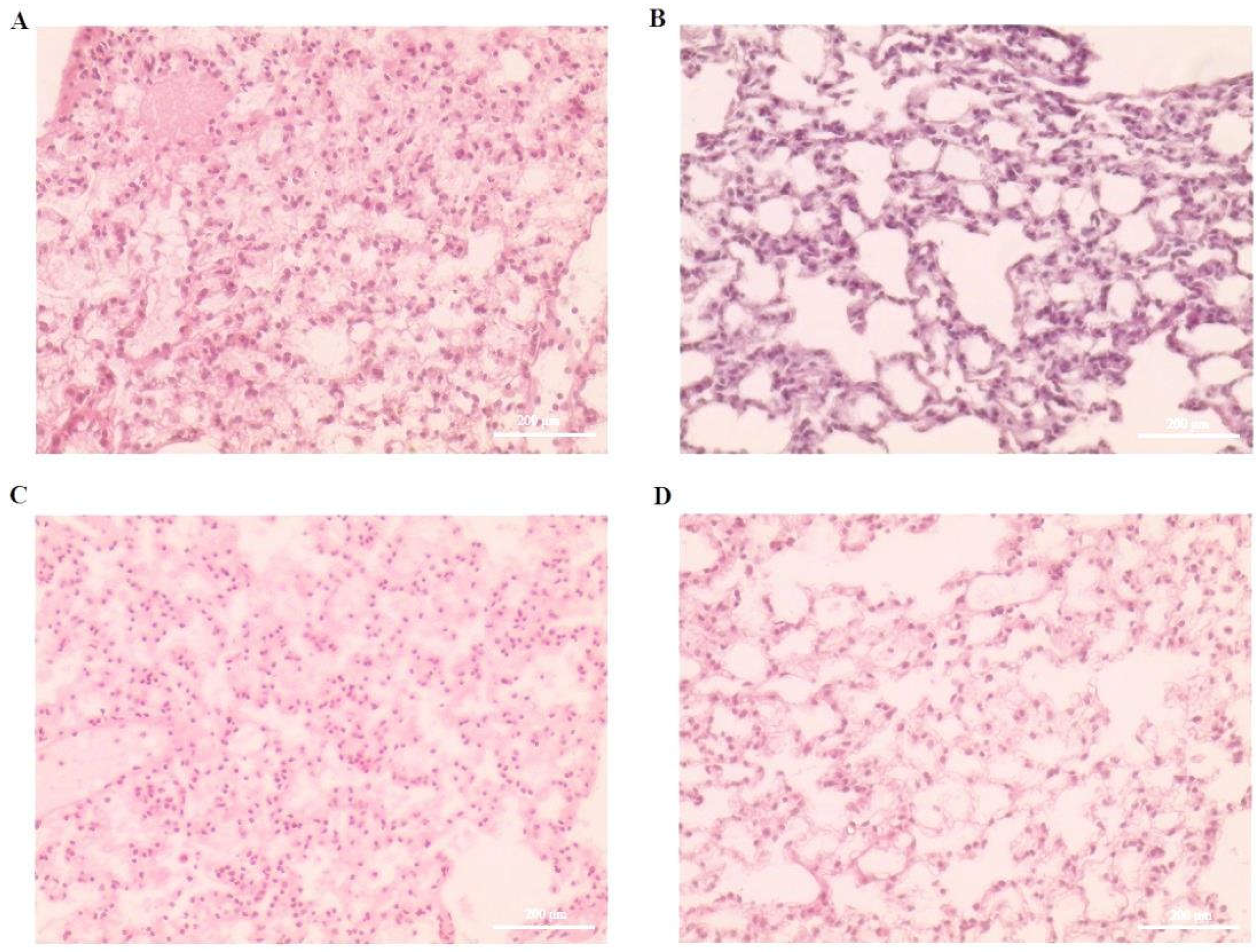
3.1. Effect of Orientin on CCl4-Induced Liver Inflammation and Immune Dysfunction

In this study, the protective effect of orientin on CCl4-induced liver inflammation in a mice model was investigated. As shown in Figure 1, induced liver inflammation was observed in disease groups, including the cirrhosis and inflammatory cellular infiltrations in the right lobe, and widespread hepatic fibrosis was observed as well. Moreover, the score of the liver injury was significantly higher in the CCl4 group compared to orientin alone and orientin with CCl4-treated groups. These orientin-treated disease mice distinctly alleviated the CCl4-induced liver damage and reduced infiltration of inflamed cells (Figure 2A). The serum liver functional markers ALT and AST of orientin-treated mice were quantified using the BioVision ELISA method [35]. Fibrosis and cirrhosis increased the aspartate aminotransferase (AST) and alanine aminotransferase (ALT) marker in mice (Figure 2B). Orientin attenuated the liver damage in CCl4-treated mice and evidently the ALT and AST markers were increased 1.91-fold in CCl4-treated mice, whereas the orientin-treated mice showed ALT/AST at a 1.23-fold ratio level compared to the control group (Figure 2B).

3.2. Effect of Orientin on Expression of Inflammatory Markers in CCl4-Induced Liver Tissues

Natural molecules play a major role in anti-inflammatory mechanisms in mice. As shown in Figure 3, orientin reduced pro-inflammatory markers such as TNF-α, IL-6, IL-8, and IFN-γ, which were quantified in CCl4-induced mice. The CCl4-treated mice showed an elevated level of these markers compared to the control group and orientin-treated group. Accordingly, the tissue lysate of orientin-treated mice showed significant reduction of TNF-α (from 421 to 241 pg/mL), IL-6 (from 214 to 133 pg/mL), IL-8 (from 800 to 580 pg/mL), and IFN-γ (from 228 to 175 pg/mL) compared to the CCl4 inflammatory group (Figure 3A–D). However, the orientin alone and orientin with CCl4-treated mice groups had strongly reduced inflammatory markers (p < 0.05). The fibrotic markers were potentially regulated by orientin, as it was regulated significantly in CCl4-induced liver tissues. The ZEB-2 mRNA was downregulated from 4.1-fold of the CCl4-induced condition to 2.2-fold of the orientin-treated condition. Similar findings were observed for PTEN mRNA and reduced from 9-fold to 4-fold (Figure 3E). The protein expression of ZEB-2 and PTEN were quantified in orientin-treated CCl4-induced mice and were downregulated from 2.7- and 2.5-fold of CCl4-induced inflammatory group to 1.6- and 1.9-fold in orientin-treated groups. respectively (Figure 3F,G). The orientin alone group showed insignificant expression of ZEB-2 and PTEN compared to the untreated control group. These results indicated that orientin significantly controlled the inflammatory and fibrotic markers in CCl4-induced mouse liver tissues.

3.3. Docking of Orientin with Mouse PTEN and ZEB-2

The docking study demonstrated the virtual interactions of orientin with phosphate membrane domains of mouse PTEN and stem cell membrane protein of mouse ZEB-2 using molecular docking software tools. The binding energy, ligand bonds, bond distance, and amino acid residue interaction were recorded in Table 1 and Table 2 and Figure 4A,B. Orientin interacted with PTEN (−6.44) amino acid residue of the chain A domain (Figure 4A), such as asparagine, threonine, glycine, and arginine forming a hydrogen bond. Interestingly, the strong binding interactions were observed as a carbon hydrogen bond in asparagine and arginine residue of the PTEN protein. The cation and hydrophobic bonds were observed at tyrosine, proline, and arginine positions of 163 to 170 amino acid residues. Totally, ten different bonds were formed between orientin with mouse PTEN protein. The ZEB-2 protein interacted with orientin with a binding energy of (−6.17) and, in total, six different amino acid residues in a position from 19 to 51 interacted with orientin chain B and C. Orientin formed a sigma bond between ZEB-2 at a position of arginine 166 and confirmed the formation of an overlapped covalent bond (Figure 4B). These interactions demonstrated the involvement of orientin in signaling of hepatic cellular fibrosis and inflammatory markers.

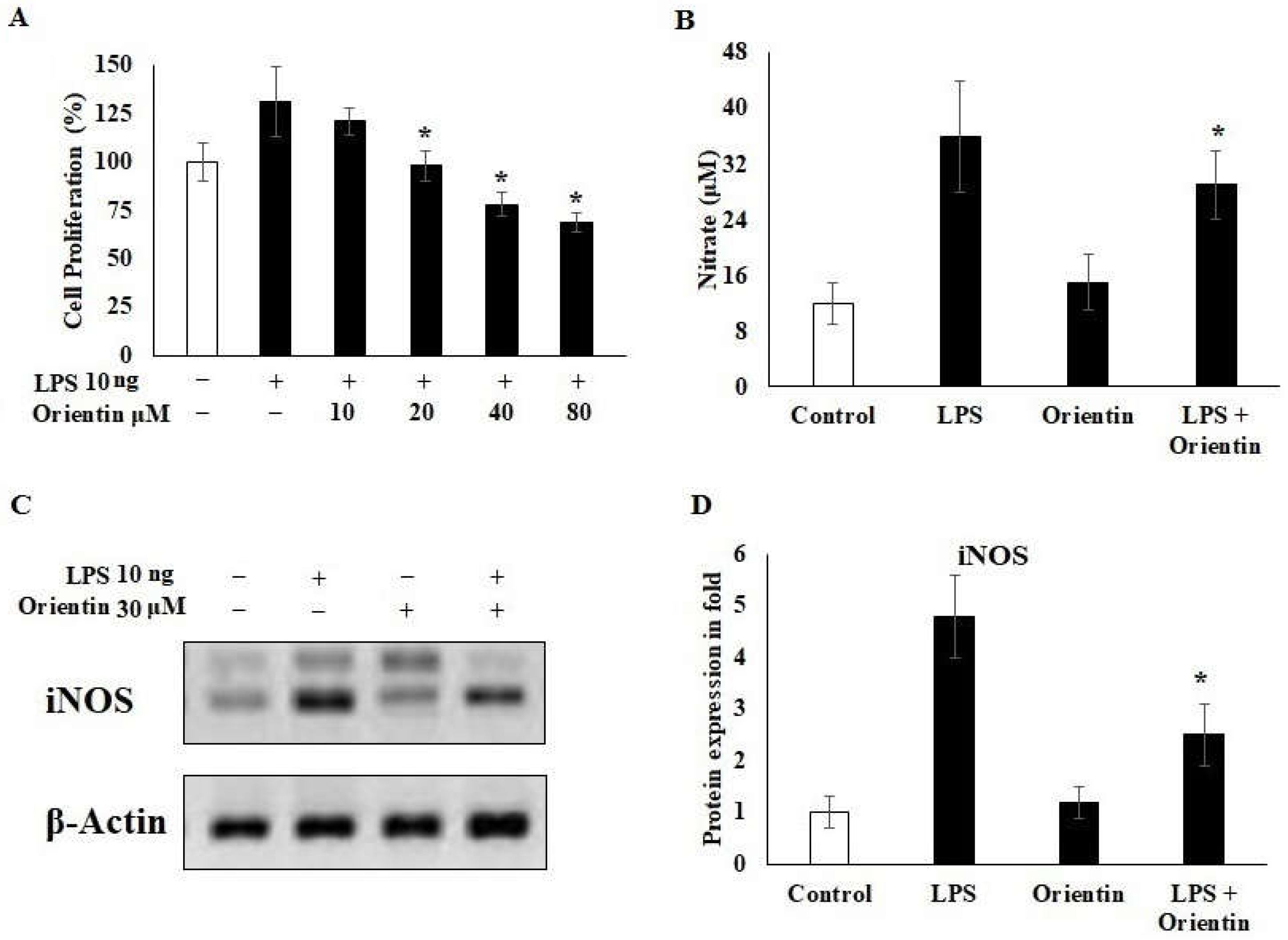
3.4. Effect of Orientin on LPS-Induced Inflammation in Hepatic Stellate Cells

Orientin controlled cell proliferation, nitric oxide, iNOS, and inflammatory structural stress in LPS-induced inflammatory mouse-derived HSCs (Figure 5A–D). As shown in Figure 5A, orientin significantly inhibited the LPS 10 ng/mL-induced proliferation in mice-derived HSCs. The LPS-challenged cells were exposed to orientin in concentration (10 to 80 μM) for 24 h and cell proliferation was quantified. The cell proliferation was inhibited gradually based on the dose of orientin concentration and it was found that 20 and 40 μM significantly controlled the proliferation and was non-toxic to murine hepatic cells. Thus, we selected an orientin concentration of 30 μM for our further ex vivo studies to evaluate the effect of orientin on LPS-induced HSC inflammation. LPS statistically stimulated HSC proliferation (Figure 5A), indicating that LPS could increase the functional metabolic and inflammatory markers of HSCs. The treatment of orientin reduces LPS-related cell functioning markers. The effect of orientin on anti-inflammatory markers in LPS-activated HSCs was evaluated; we treated murine HSCs with either LPS alone, or LPS with orientin concentrations of 30 μM. Nitric oxide (nitrate μM) was used to determine pro-inflammatory activity of the host cells. Orientin significantly decreased LPS-induced nitric oxide from 35 to 27 μM, whereas orientin alone showed a similar nitric oxide concentration compared to untreated control HSCs (Figure 5B). LPS-stimulated expression of iNOS was determined by immunoblot. Orientin alleviated iNOS expression in the LPS-induced group (2.2-fold) comparatively to the LPS alone group (4.3-fold) (p < 0.05) (Figure 5C,D). These results were co-related with HSC differentiation and maturation in LPS-induced conditions (Figure 6).

3.5. Effect of Orientin on Hepatic Fibrotic Signaling Markers ZEB-2, PTEN, and TNF-α

Orientin regulated the fibrotic markers in HSCs. The mRNA and protein expression of ZEB-2, PTEN, and TNF-α were quantified in orientin-treated LPS-challenged HSCs (Figure 7). The ZEB-2 mRNA was downregulated from 4.4-fold of the LPS-induced condition to 3.1-fold of the orientin-treated condition (Figure 7A). Similar findings were observed in protein expression of ZEB-2 and reduced from 6.2-fold to 2.4-fold (Figure 7B,C). This indicates orientin controlled the mesenchymal modification of HSCs and inhibited the hepatic inflammation. HSC activator PTEN was inhibited by orientin treatment. The mRNA and protein expression of PTEN was significantly downregulated in orientin-treated HSCs. Orientin inhibited the LPS-induced PTEN 3.4-fold of mRNA and 2.5-fold of protein expression into 2.6-fold and 1.9-fold of mRNA and protein, respectively (Figure 7A–C). The inflammatory marker TNF-α was significantly inhibited by orientin treatment and it was downregulated from 4.9-fold of LPS-induced mRNA to 3.1-fold of mRNA. Orientin treatment of LPS-treated HSC led to a decrease of TNFα protein levels (2.8-fold), when compared to HSCs treated only with LPS (4.5-fold) (Figure 7A–C). The ZEB-2 expression was increased in the orientin alone treated group compared to the untreated control group (Figure 7B,C).

4. Discussion

The need of for herbal supplements has been increased because of their merits to control oxidative hepatic damage [36]. Hepatotoxicity induced by CCl4 leads to liver injury, hepatic cell death, and activation of cell necrotic pathways [37]. The long-term exposure of CCl4 leads to chronic liver damage and hepatic carcinoma. Recent strategies focused on usage of plant-derived flavonoids, such as luteolin, as hepato-protective, anti-oxidative, and anti-inflammatory agents [38]. In this study, the effect of orientin on liver protection and anti-inflammatory mechanisms in liver tissues in mice were examined. The pathological score and disease index were significantly reduced by orientin treatment in a concentration of 1 mg/kg bw of mouse. The pathophysiology of liver tissue showed that orientin significantly reduced cirrhosis, necrotic cellular morphology, and inflamed infiltration in hepatic tissue (Figure 1). Some studies showed that the flavonoids improve the hepatic cellular architecture from CCl4-induced hepatic damage [39,40]. Liver metabolic enzymes play a major role in detoxification, fibrotic steatosis, and inflammatory modifications of the host. Several studies showed plant flavonoids potentially augmented the AST/ALT level in a CCl4-induced mouse model [41,42,43]. In this present study, AST/ALT was potentially reduced by orientin 1 mg/kg bw compared to the CCl4-induced mice group. The orientin alone treated mice showed similar levels compared to untreated groups (Figure 2A,B). These data are concurrent with the reported modulatory action of extracts of orientin using acetaminophen-induced liver damage in a mice model showing boosted ALT/AST levels, maintaining a normal liver structure, and moderate activity against the iNOS. The current study demonstrated detailed mechanisms of orientin towards reduction of TNF-α, IL-6, IL-8, and IFN-γ in a CCl4-induced mice model and downregulation of ZEB-2 and PTEN fibrotic markers in LPS-induced murine HSCs [44,45,46].
The inflammatory cytokines are key players of fibrotic mediators and are associated with worse prognosis of the inflammatory liver disease [26]. The CCl4 administration led to a rise in the level of pro-inflammatory mediators such as TNF-α, IL-6, IL-8, and IFN-γ (Figure 3A–D). However, the orientin treatment showed marked reduction in the inflammatory cytokines compared to the CCl4 control group. Some previous reports showed the similar ameliorative effect of naringenin and orientin on TNF-α, IL-6, and iNOS in mice models [15,44,45,46,47]. The liver fibrotic markers, ZEB-2 and PTEN, were upregulated in the CCl4-treated group, whereas orientin potentially reduced the expression of ZEB-2 and reached 1.7-fold from a 2.8-fold expression (Figure 3E–G). The PTEN expression was downregulated in the orientin treatment compared to the CCl4 treatment. Protein expression was reduced to 13% compared to the CCl4 group (Figure 3G). The virtual binding of orientin with ZEB-2 and PTEN was achieved and potent binding was observed (Figure 4A,B). The formation of pi-Alkyl hydrophobic bonds between orientin and PTEN changed the structural confirmation and led to less potent receptor molecules. The amino acid residues interacting with orientin from residue 160 to 167 played a major role in coupled complex regulations (Table 1). Regarding the ZEB-2 interaction with orientin, it formed a sigma bond between ZEB-2 at the position of residue 166 and confirmed the formation of an overlapped covalent bond. The coupled complex formations demonstrated the involvement of orientin, with ZEB-2 downregulating the signaling process of protein and controlling the fibrotic formation in intracellular regulatory elements (Table 2).
HSCs play major role in liver cirrhosis and lipid metabolism [48]. Previous studies evidenced that inhibition of HSC proliferation led to prevention of liver injury and fibrosis [49,50,51]. Therefore, natural molecules are focused on liver protection and host health. In this study, we first examined the effect of orientin on LPS-induced murine HSCs and estimated the ZEB-2 and PTEN fibrotic markers. The results indicated that orientin treatment caused cell protection from LPS-induced hepatotoxicity (Figure 5 and Figure 6A). The inflammatory nitric oxide marker was potentially reduced (from 36 to 27 μM) by orientin in LPS-induced HSCs (Figure 6B), indicating the negative progression of fibrosis in LPS-induced inflamed HSCs. The induced nitric oxide synthase is a key regulator of nitric oxide production and its signaling pathways [52]. The orientin treatment reduced the LPS-induced iNOS expression in HSCs and consequently confirmed the reduction of nitric oxide release (Figure 6C,D).
Fibrotic markers such as ZEB-2, PTEN, and TNF-α played a key role in hepatic fibrosis and mesenchymal transition of liver tissues [53,54,55]. In this study, transcripts and protein expressions of these markers were significantly reduced by orientin, which consequently control the transition of HSCs from mesenchymal to fibroblastic architecture. Significant downregulation of ZEB-2, PTEN, and TNF-α in the orientin treatment group confirmed that this phytochemical is able to inhibit the LPS-induced fibrotic process (Figure 7A–C).

5. Conclusions

The present study demonstrated that orientin significantly diminished liver cirrhosis, inflamed infiltration, and boosted AST/ALT levels in CCl4-induced mice. In addition, the levels of inflammatory markers (TNF-α, IL-6, IL-8, and IFN-γ) and fibrotic markers (ZEB-2 and PTEN) were decreased by orientin treatment in CCl4-induced liver toxicity. On the other hand, significant downregulation of ZEB-2, PTEN, and TNF-α was demonstrated in orientin treatment against LPS-induced HSC fibrotic processes. Orientin also showed reduction of iNOS expression and nitric oxide release in HSCs of the LPS-induced fibrotic cells. The results were supported with virtual binding studies. In conclusion, transcripts and protein expression of inflammatory, as well as fibrotic, markers were significantly attenuated by orientin in HSCs and subsequently control the transition of HSCs from mesenchymal to fibroblastic fashion.

Author Contributions

H.E.K.: conceptualization, funding acquisition, methodology, writing—review and editing, writing—original draft preparation and supervision; H.-I.M.I.: formal analysis, methodology, validation, software, writing—review and editing, writing—original draft preparation; K.A.E.-F.: writing—review and editing; S.H.A.: writing—review and editing; M.A.M.: validation, writing—review and editing. All authors have read and agreed to the published version of the manuscript.

Funding

This project was funded by the Deanship of Scientific Research (DSR), King Faisal University, Al-Ahsa, Saudi Arabia (Nasher Track, project No. 216092).

Institutional Review Board Statement

The study was conducted in accordance with the Declaration of Helsinki and approved by the Ethics Committee of King Faisal University (KFU-REC-2022-JAN-EA000401).

Informed Consent Statement

Not applicable.

Data Availability Statement

Not applicable.

Acknowledgments

The authors acknowledge with thanks the Deanship of Scientific Research (DSR), King Faisal University, Al-Ahsa, Saudi Arabia (Nasher Track, project No. 216092).

Conflicts of Interest

The authors declare no conflict of interest is associated with this work.

References

  1. Del Campo, J.A.; Gallego, P.; Grande, L. Role of inflammatory response in liver diseases: Therapeutic strategies. World J. Hepatol. 2018, 10, 1. [Google Scholar] [CrossRef] [PubMed]
  2. Chalasani, N.; Younossi, Z.; Lavine, J.E.; Diehl, A.M.; Brunt, E.M.; Cusi, K.; Charlton, M.; Sanyal, A.J. The diagnosis and management of non-alcoholic fatty liver disease: Practice Guideline by the American Association for the Study of Liver Diseases, American College of Gastroenterology, and the American Gastroenterological Association. Hepatology 2012, 55, 2005–2023. [Google Scholar] [CrossRef] [PubMed]
  3. Calvente, C.J.; Tameda, M.; Johnson, C.D.; Del Pilar, H.; Lin, Y.C.; Adronikou, N.; Du Jeu, X.D.M.; Llorente, C.; Boyer, J.; Feldstein, A.E. Neutrophils contribute to spontaneous resolution of liver inflammation and fibrosis via microRNA-223. J. Clin. Investig. 2019, 129, 4091–4109. [Google Scholar] [CrossRef] [PubMed] [Green Version]
  4. Koyama, Y.; Brenner, D.A. Liver inflammation and fibrosis. J. Clin. Investig. 2017, 127, 55–64. [Google Scholar] [PubMed]
  5. Nallagangula, K.S.; Nagaraj, S.K.; Venkataswamy, L.; Chandrappa, M. Liver fibrosis: A compilation on the biomarkers status and their significance during disease progression. Future Sci. OA 2017, 4, FSO250. [Google Scholar]
  6. De Haan, W.; Dheedene, W.; Apelt, K.; Décombas-Deschamps, S.; Vinckier, S.; Verhulst, S.; Conidi, A.; Deffieux, T.; Staring, M.W.; Vandervoort, P. Endothelial Zeb2 preserves the hepatic angioarchitecture and protects against liver fibrosis. Cardiovasc. Res. 2021, 1–14. [Google Scholar] [CrossRef]
  7. Jin, H.; Li, C.; Dong, P.; Huang, J.; Yu, J.; Zheng, J. Circular RNA cMTO1 promotes PTEN expression through sponging miR-181b-5p in liver fibrosis. Front. Cell. Dev. Biol. 2020, 8, 714. [Google Scholar] [CrossRef]
  8. Wandrer, F.; Liebig, S.; Marhenke, S.; Vogel, A.; John, K.; Manns, M.P.; Teufel, A.; Itzel, T.; Longerich, T.; Maier, O. TNF-Receptor-1 inhibition reduces liver steatosis, hepatocellular injury and fibrosis in NAFLD mice. Cell Death Dis. 2020, 11, 1–9. [Google Scholar] [CrossRef]
  9. Prystupa, A.; Kiciński, P.; Sak, J.; Boguszewska-Czubara, A.; Toruń-Jurkowska, A.; Załuska, W. Proinflammatory cytokines (IL-1α, IL-6) and hepatocyte growth factor in patients with alcoholic liver cirrhosis. Gastroenterol. Res. Pract. 2015, 2015, 1–7. [Google Scholar] [CrossRef] [Green Version]
  10. Attallah, A.M.; El-Far, M.; Zahran, F.; Shiha, G.E.; Farid, K.; Omran, M.M.; Abdelrazek, M.A.; Attallah, A.A.; El-Beh, A.A.; El-Hosiny, R.M. Interferon-gamma is associated with hepatic dysfunction in fibrosis, cirrhosis, and hepatocellular carcinoma. J. Immunoass. Immunochem. 2016, 37, 597–610. [Google Scholar] [CrossRef]
  11. Gonzalez-Gallego, J.; Sánchez-Campos, S.; Tunon, M. Anti-inflammatory properties of dietary flavonoids. Nutr. Hosp. 2007, 22, 287–293. [Google Scholar] [PubMed]
  12. Ullah, A.; Munir, S.; Badshah, S.L.; Khan, N.; Ghani, L.; Poulson, B.G.; Emwas, A.-H.; Jaremko, M. Important flavonoids and their role as a therapeutic agent. Molecules 2020, 25, 5243. [Google Scholar] [CrossRef] [PubMed]
  13. Ciumărnean, L.; Milaciu, M.V.; Runcan, O.; Vesa, Ș.C.; Răchișan, A.L.; Negrean, V.; Perné, M.-G.; Donca, V.I.; Alexescu, T.-G.; Para, I. The effects of flavonoids in cardiovascular diseases. Molecules 2020, 25, 4320. [Google Scholar] [CrossRef] [PubMed]
  14. Ginwala, R.; Bhavsar, R.; Chigbu, D.G.I.; Jain, P.; Khan, Z.K. Potential role of flavonoids in treating chronic inflammatory diseases with a special focus on the anti-inflammatory activity of apigenin. Antioxidants 2019, 8, 35. [Google Scholar] [CrossRef] [Green Version]
  15. Hernández-Aquino, E.; Muriel, P. Beneficial effects of naringenin in liver diseases: Molecular mechanisms. World J. Gastroenterol. 2018, 24, 1679. [Google Scholar] [CrossRef]
  16. Raso, G.M.; Meli, R.; Di Carlo, G.; Pacilio, M.; Di Carlo, R. Inhibition of inducible nitric oxide synthase and cyclooxygenase-2 expression by flavonoids in macrophage J774A. 1. Life Sci. 2001, 68, 921–931. [Google Scholar] [CrossRef]
  17. Gülçin, I. Antioxidant activity of food constituents: An overview. Arch. Toxicol. 2012, 86, 345–391. [Google Scholar] [CrossRef]
  18. Gulcin, İ. Antioxidants and antioxidant methods: An updated overview. Arch. Toxicol. 2020, 94, 651–715. [Google Scholar] [CrossRef] [Green Version]
  19. Lee, W.; Ku, S.-K.; Bae, J.-S. Vascular barrier protective effects of orientin and isoorientin in LPS-induced inflammation in vitro and in vivo. Vascul. Pharmacol. 2014, 62, 3–14. [Google Scholar] [CrossRef]
  20. Dantas-Medeiros, R.; Furtado, A.A.; Zanatta, A.C.; Torres-Rêgo, M.; Lourenço, E.M.G.; Alves, J.S.F.; Galinari, É.; de Oliveira Rocha, H.A.; Guerra, G.C.B.; Vilegas, W. Mass spectrometry characterization of Commiphora leptophloeos leaf extract and preclinical evaluation of toxicity and anti-inflammatory potential effect. J. Ethnopharmacol. 2021, 264, 113229. [Google Scholar] [CrossRef]
  21. Lam, K.Y.; Ling, A.P.K.; Koh, R.Y.; Wong, Y.P.; Say, Y.H. A review on medicinal properties of orientin. Adv. Pharmacol. Sci. 2016, 2016, 1–9. [Google Scholar] [CrossRef] [Green Version]
  22. Batool, R.; Kalsoom, A.; Akbar, I.; Arshad, N.; Jamil, N. Antilisterial Effect of Rosa damascena and Nymphaea alba in Mus musculus. Biomed. Res. Int. 2018, 2018, 1–9. [Google Scholar]
  23. Abtulov, M.; Valcheva-Kuzmanova, S. Flavonoids and the liver—A review. Scr. Sci. Pharm. 2021, 8, 7–16. [Google Scholar]
  24. Wu, Y.; He, Y.; Wang, R.; Zhao, X. Preventive Effect of Flavonoid Extract from the Peel of Gonggan (Citrus reticulata Blanco Var. Gonggan) on CCl4-Induced Acute Liver Injury in Mice. J. Inflamm. Res. 2021, 14, 5111. [Google Scholar] [CrossRef] [PubMed]
  25. Ajuwon, O.R.; Oguntibeju, O.O.; Marnewick, J.L. Amelioration of lipopolysaccharide-induced liver injury by aqueous rooibos (Aspalathus linearis) extract via inhibition of pro-inflammatory cytokines and oxidative stress. BMC Complement. Altern. Med. 2014, 14, 1–12. [Google Scholar] [CrossRef] [PubMed] [Green Version]
  26. Ho, C.-H.; Huang, J.-H.; Sun, M.-S.; Tzeng, I.; Hsu, Y.-C.; Kuo, C.-Y. Wild bitter melon extract regulates LPS-induced hepatic stellate cell activation, inflammation, endoplasmic reticulum stress, and ferroptosis. Evid. Based Complement. Alternat. Med. 2021, 2021, 1–11. [Google Scholar] [CrossRef] [PubMed]
  27. Rong, X.; Xu, J.; Jiang, Y.; Li, F.; Chen, Y.; Dou, Q.P.; Li, D. Citrus peel flavonoid nobiletin alleviates lipopolysaccharide-induced inflammation by activating IL-6/STAT3/FOXO3a-mediated autophagy. Food Funct. 2021, 12, 1305–1317. [Google Scholar] [CrossRef]
  28. Yan, G.; Chen, L.; Wang, H.; Wu, S.; Li, S.; Wang, X. Baicalin inhibits LPS-induced inflammation in RAW264. 7 cells through miR-181b/HMGB1/TRL4/NF-κB pathway. Am. J. Transl. Res. 2021, 13, 10127. [Google Scholar]
  29. Zamani-Garmsiri, F.; Emamgholipour, S.; Rahmani Fard, S.; Ghasempour, G.; Jahangard Ahvazi, R.; Meshkani, R. Polyphenols: Potential anti-inflammatory agents for treatment of metabolic disorders. Phytother. Res. 2021, 36, 415–432. [Google Scholar] [CrossRef]
  30. Mederacke, I.; Dapito, D.H.; Affò, S.; Uchinami, H.; Schwabe, R.F. High-yield and high-purity isolation of hepatic stellate cells from normal and fibrotic mouse livers. Nat. Protoc. 2015, 10, 305–315. [Google Scholar] [CrossRef] [Green Version]
  31. Goshi, E.; Zhou, G.; He, Q. Nitric oxide detection methods in vitro and in vivo. Med. Gas Res. 2019, 9, 192. [Google Scholar] [CrossRef]
  32. Khalil, H.E.; Ibrahim, H.-I.M.; Ahmed, E.A.; Emeka, P.M.; Alhaider, I.A. Orientin, a Bio-Flavonoid from Trigonella hamosa L., Regulates COX-2/PGE-2 in A549 Cell Lines via miR-26b and miR-146a. Pharmaceuticals 2022, 15, 154. [Google Scholar] [CrossRef] [PubMed]
  33. Khalil, H.E.; Ibrahim, H.-I.M.; Darrag, H.M.; Matsunami, K. Insight into Analysis of Essential Oil from Anisosciadium lanatum Boiss.—Chemical Composition, Molecular Docking, and Mitigation of Hepg2 Cancer Cells through Apoptotic Markers. Plants 2022, 11, 66. [Google Scholar] [CrossRef] [PubMed]
  34. Thirugnanasambantham, K.; Muralidaran, S.; Mandal, A.K.A. Molecular cloning, computational and expression analysis of anthocyanidin reductase in tea (Camellia sinensis). Appl. Biochem. Biotechnol. 2014, 174, 130–145. [Google Scholar] [CrossRef] [PubMed]
  35. Takasu, C.; Vaziri, N.D.; Li, S.; Robles, L.; Vo, K.; Takasu, M.; Pham, C.; Farzaneh, S.H.; Shimada, M.; Stamos, M.J. Treatment with dimethyl fumarate ameliorates liver ischemia/reperfusion injury. World J. Gastroenterol. 2017, 23, 4508. [Google Scholar] [CrossRef] [PubMed]
  36. Dutta, S.; Chakraborty, A.K.; Dey, P.; Kar, P.; Guha, P.; Sen, S.; Kumar, A.; Sen, A.; Chaudhuri, T.K. Amelioration of CCl4 induced liver injury in swiss albino mice by antioxidant rich leaf extract of Croton bonplandianus Baill. PLoS ONE 2018, 13, e0196411. [Google Scholar] [CrossRef] [PubMed] [Green Version]
  37. Chang, S.N.; Kim, S.H.; Dey, D.K.; Park, S.M.; Nasif, O.; Bajpai, V.K.; Kang, S.C.; Lee, J.; Park, J.G. 5-O-Demethylnobiletin alleviates CCl4-Induced acute liver injury by equilibrating ROS-mediated apoptosis and autophagy induction. Int. J. Mol. Sci. 2021, 22, 1083. [Google Scholar] [CrossRef]
  38. Akindele, A.J.; Oludadepo, G.O.; Amagon, K.I.; Singh, D.; Osiagwu, D.D. Protective effect of carvedilol alone and coadministered with diltiazem and prednisolone on doxorubicin and 5-fluorouracil-induced hepatotoxicity and nephrotoxicity in rats. Pharmacol. Res. Perspect. 2018, 6, e00381. [Google Scholar] [CrossRef] [Green Version]
  39. Gharbia, S.; Balta, C.; Herman, H.; Rosu, M.; Váradi, J.; Bácskay, I.; Vecsernyés, M.; Gyöngyösi, S.; Fenyvesi, F.; Voicu, S.N. Enhancement of Silymarin Anti-fibrotic Effects by Complexation With Hydroxypropyl (HPBCD) and Randomly Methylated (RAMEB) β-Cyclodextrins in a Mouse Model of Liver Fibrosis. Front. Pharmacol. 2018, 9, 883. [Google Scholar] [CrossRef] [Green Version]
  40. Owumi, S.; Lewu, D.; Arunsi, U.; Oyelere, A. Luteolin attenuates doxorubicin-induced derangements of liver and kidney by reducing oxidative and inflammatory stress to suppress apoptosis. Hum. Exp. Toxicol. 2021, 40, 1656–1672. [Google Scholar] [CrossRef]
  41. Okda, T.; Abd-Alhaseeb, M.; Barka, K.; Ragab, N. Ginger potentiates the effects of silymarin on liver fibrosis induced by CCl4: The role of galectin-8. Eur. Rev. Med. Pharmacol. Sci. 2019, 23, 885–891. [Google Scholar] [PubMed]
  42. Ullah, H.; Khan, A.; Baig, M.W.; Ullah, N.; Ahmed, N.; Tipu, M.K.; Ali, H.; Khan, S. Poncirin attenuates CCl4-induced liver injury through inhibition of oxidative stress and inflammatory cytokines in mice. BMC Complement. Med. Ther. 2020, 20, 1–14. [Google Scholar] [CrossRef] [PubMed] [Green Version]
  43. Li, H.; Yoon, J.-H.; Won, H.-J.; Ji, H.-S.; Yuk, H.J.; Park, K.H.; Park, H.-Y.; Jeong, T.-S. Isotrifoliol inhibits pro-inflammatory mediators by suppression of TLR/NF-κB and TLR/MAPK signaling in LPS-induced RAW264. 7 cells. Int. Immunopharmacol. 2017, 45, 110–119. [Google Scholar] [CrossRef] [PubMed]
  44. Xiao, Q.; Zhao, Y.; Ma, L.; Piao, R. Orientin reverses acetaminophen-induced acute liver failure by inhibiting oxidative stress and mitochondrial dysfunction. J. Pharmacol. Sci. 2022, in press. [CrossRef]
  45. Bello, O.M.; Ogbesejana, A.B.; Adetunji, C.O.; Oguntoye, S.O. Flavonoids isolated from Vitex grandifolia, an underutilized vegetable, exert monoamine A & B inhibitory and anti-inflammatory effects and their structure-activity relationship. Turk. J. Pharm. Sci. 2019, 16, 437. [Google Scholar]
  46. Mohamed, T.K.; Kamal, A.M.; Nassar, M.I.; Ahmed, M.A.; Haggag, M.G.; Ezzat, H.A. Phenolic contents of Gleditsia triacanthos leaves and evaluation of its analgesic, anti-inflammatory, hepatoprotective and antimicrobial activities. Life Sci. 2013, 10, 3445–3446. [Google Scholar]
  47. Jayaraman, J.; Jesudoss, V.A.S.; Menon, V.P.; Namasivayam, N. Anti-inflammatory role of naringenin in rats with ethanol induced liver injury. Toxicol. Mech. Methods 2012, 22, 568–576. [Google Scholar] [CrossRef]
  48. Qian, Y.; Han, J.; Zhou, L.; Yu, Q.; Xv, J.; Jin, Z.; Yang, Y.; Jiang, L.; Lou, D. Inhibition of epidermal growth factor receptor (EGFR) reduces lipopolysaccharide (LPS)-induced activation and inflammatory cytokines in hepatic stellate cells in vitro. Med. Sci. Monit. 2018, 24, 5533. [Google Scholar] [CrossRef]
  49. Yin, L.; Qi, Y.; Xu, Y.; Xu, L.; Han, X.; Tao, X.; Song, S.; Peng, J. Dioscin inhibits HSC-T6 cell migration via adjusting SDC-4 expression: Insights from iTRAQ-based quantitative proteomics. Front. Pharmacol. 2017, 8, 665. [Google Scholar] [CrossRef] [Green Version]
  50. Liu, M.; Xu, Y.; Han, X.; Yin, L.; Xu, L.; Qi, Y.; Zhao, Y.; Liu, K.; Peng, J. Dioscin alleviates alcoholic liver fibrosis by attenuating hepatic stellate cell activation via the TLR4/MyD88/NF-κB signaling pathway. Sci. Rep. 2015, 5, 1–13. [Google Scholar]
  51. Dey, P.; Panga, V.; Raghunathan, S. A cytokine signalling network for the regulation of inducible nitric oxide synthase expression in rheumatoid arthritis. PLoS ONE 2016, 11, e0161306. [Google Scholar] [CrossRef]
  52. Acharya, P.; Chouhan, K.; Weiskirchen, S.; Weiskirchen, R. Cellular mechanisms of liver fibrosis. Front. Pharmacol. 2021, 12, 1072. [Google Scholar] [CrossRef] [PubMed]
  53. Higashi, T.; Friedman, S.L.; Hoshida, Y. Hepatic stellate cells as key target in liver fibrosis. Adv. Drug Deliv. Rev. 2017, 121, 27–42. [Google Scholar] [CrossRef] [PubMed]
  54. Bian, E.-B.; Huang, C.; Ma, T.-T.; Tao, H.; Zhang, H.; Cheng, C.; Lv, X.-W.; Li, J. DNMT1-mediated PTEN hypermethylation confers hepatic stellate cell activation and liver fibrogenesis in rats. Toxicol. Appl. Pharmacol. 2012, 264, 13–22. [Google Scholar] [CrossRef] [PubMed]
  55. Di Gregorio, J.; Robuffo, I.; Spalletta, S.; Giambuzzi, G.; De Iuliis, V.; Toniato, E.; Martinotti, S.; Conti, P.; Flati, V. The epithelial-to-mesenchymal transition as a possible therapeutic target in fibrotic disorders. Front. Cell Dev. Biol. 2020, 8, 1–32. [Google Scholar] [CrossRef]
Figure 1. Morphological and histological evaluation of liver in mice. (A) Control group, (B) CCl4-treated group, (C) orientin (1 mg/kg)-treated group, and (D) CCl4 orientin (1 mg/kg)-treated group. Magnification ×200.
Applsci 12 02725 g001
Figure 2. Effect of orientin on liver. (A) The disease pathology scores for liver injury. (B) Effect of orientin on CCl4-induced changes in hepatic functional markers of ALT activity and AST activity. Orientin-treated liver tissues were excised after the 7th day of ip injection and homogenate using ice cold PBS. All values are expressed as mean ± SD. * Represents the significant comparison with the control. # Represents the significant comparison with control and CCl4-induced groups.
Applsci 12 02725 g002
Figure 3. Effect of orientin on liver inflammatory markers. The CCl4-induced mice were administered for orientin 1 mg/kg body weight of mice for 7 days and liver tissues were excised. Liver homogenate was prepared using ice cold cell lysis buffer with 0.1% DTT. (A) The TNF-α was estimated using a Cayman ELISA kit. (B) IL-6 was estimated in liver lysate using a mouse Cayman ELISA kit. (C) IFN-γ was estimated in liver lysate using a mouse Cayman ELISA kit. (D) IL-8 was estimated in liver lysate using a mouse Cayman ELISA kit. (E) mRNA expression of ZEB-2 and PTEN using real time PCR. (F,G) Protein quantification of ZEB-2 and PTEN in CCl4-induced and CCl4 with orientin treatment using immunoblot-based techniques. (The data are represented as the mean ± SD.) p < 0.05. * Represents the significant comparison with control and CCl4-induced groups.
Applsci 12 02725 g003
Figure 4. In silico docking of orientin (CID: 5281675) and (A) mouse PTEN (PDB-ID-1D5R) and (B) ZEB-2 (PDB-ID-2D8V) proteins binding analysis. Computational binding was performed using AutoDock software to demonstrate the illustration of interactions in the hydrophobic bond and other polar bonds of PTEN and ZEB-2. It shows the amino acid residue analysis of the interacted bond and its length, together with the binding pocket of ligand–receptor interactions.
Applsci 12 02725 g004
Figure 5. Effect of orientin on LPS-induced inflammatory HSCs. Naïve mouse-derived HSCs were isolated and cultured in an adherent-coated 12-well plate. Cultured cells were stimulated with LPS 10 ng for 2 h and orientin was treated for 12 h. Treated cells were evaluated for cell proliferation, nitrate quantification, and iNOS protein. (A) Cell proliferation of LPS-stimulated HSCs and LPS with orientin-treated cells were quantified using SRB assay. (B) Nitric oxide level as nitrate (μM) in LPS-stimulated and orientin-treated HSCs. (C,D) iNOS protein quantification using immunoblot. Values are expressed as three independent experiments and all values are expressed as mean ± SD. * Represents the significant comparison with control and LPS-induced groups.
Applsci 12 02725 g005
Figure 6. Effect of orientin on LPS-induced inflammatory HSCs. Naïve mouse-derived HSCs were isolated and cultured in an adherent-coated 12-well plate. Cultured cells were stimulated with LPS 10 ng for 2 h and treated with orientin (30 µM) for 12 h. (A) Control group, (B) LPS-treated group, (C) orientin (30 µM)-treated group, and (D) LPS and orientin (30 µM)-treated group.
Applsci 12 02725 g006
Figure 7. Effect of orientin on hepatic fibrotic signaling markers ZEB-2 and PTEN. Naïve mouse-derived HSCs were stimulated with LPS 10 ng for 2 h and treated for 12 h with orientin (30 µM). Treated cells were evaluated for ZEB-2, PTEN, and TNF-α mRNA and protein were quantified. (A) mRNA quantification of ZEB-2, PTEN, and TNF-α in LPS-stimulated HSCs and LPS with orientin-treated cells was undertaken using SYBR green-based real time PCR assay. (B,C) Protein quantification of ZEB-2, PTEN, and TNF-α in LPS-stimulated HSCs and LPS with orientin (30 µM)-treated cells using immunoblot-based techniques. Values are expressed as three independent experiments and all values are expressed as mean ± SD. * Represents the significant comparison with the control and LPS-induced groups.
Applsci 12 02725 g007
Table 1. Interactions of orientin and ZEB2 receptor residues of target protein.
Binding
Energy
Ligand
Efficiency
Intermole
Energy
Ligand Atoms (Ring)Docked Amino Acid Residue (Bond Length)
−6.17−0.19−6.38Conventional Hydrogen Bond:
Chain B—C3′-OChain A: ARG′36 ′HN′ (2.80 Å)
Chain B—C3′-OHChain A: ALA′32 ′O′ (1.80 Å)
Chain B—C4′-OHChain A: ALA′32 ′O′ (1.80 Å)
Chain C—C4-OChain A: THR′49 ′HG1′ (1.88 Å)
Chain A—C5-OHChain A: THR′49 ′OG1′ (1.93 Å)
C2-OHChain A: THR′19 ′O′ (2.20 Å)
C3-OHChain A: THR′19 ′OG1′ (2.18 Å)
C3-OHChain A: THR′19 ′O′ (2.61 Å)
Pi-Alkyl Hydrophobic bond:
OChain A: ALA′32′ (4.36 Å)
OChain A: LEU′20′ (5.00 Å)
OChain A: LEU′20′ (5.41 Å)
OChain A: PRO′51′ (5.18 Å)
Pi-sigma bond:
OChain A: LEU′20′ CD2′ (3.44 Å)
OChain A: ARG′166′ (4.77 Å)
ALA, alanine; ARG, arginine; LEU, leucine; ASN, asparagine; PRO, proline; THR, threonine.
Table 2. Interactions of orientin and PTEN receptor residues of target protein.
Binding
Energy
Ligand
Efficiency
Intermole
Energy
Ligand Atoms
(Ring)
Docked Amino Acid Residue
(Bond Length)
−6.44−0.2−6.44-Conventional Hydrogen Bond:
Chain B—C4′-OChain A: ASN′323 ′HD22′ (1.92 Å)
Chain B—C4′-OHChain A: THR′161 ′O′ (1.83 Å)
Chain B—C3′-OHChain A: THR′161 ′O′ (2.24 Å)
Chain C—C4′-OChain A: GLN′143 ′HE22′ (1.90 Å)
Chain C—C4′-OChain A: GLN′143 ′HE21′ (2.44 Å)
C4-OHChain A: ARG′167 ′HH12′ (2.35 Å)
-Carbon Hydrogen Bond
CChain A: ASP′318′OD2′ (3.19 Å)
CChain A: ARG′167′CD′ (2.90 Å)
-Pi-Alkyl Hydrophobic bond:
OChain A: TRY′170′ (5.65 Å)
OChain A: TRY′170′ (4.81 Å)
-Pi-Pi T-shaped bond:
OChain A: PRO′163′ (4.56 Å)
OChain A: ARG′166′ (4.77 Å)
-Pi-cation bond:
OChain A: ARG′166′ NH2′ (4.35 Å)
ARG, arginine; ASN, asparagine; ASP, aspartic acid; GLN, glutamine; PRO, proline; TRY, tyrosine.
Publisher’s Note: MDPI stays neutral with regard to jurisdictional claims in published maps and institutional affiliations.

