Article

Mesenchymal Stromal Cells (MSCs) Isolated from Various Tissues of the Human Arthritic Knee Joint Possess Similar Multipotent Differentiation Potential

1
Department of Orthopaedics and Trauma Surgery, Musculoskeletal University Center Munich (MUM), University Hospital, LMU Munich, Marchioninistraße 15, 81377 Munich, Germany
2
Department of General Surgery, Leopoldina Hospital of the City Schweinfurt, Gustav-Adolf-Str. 8, 97422 Schweinfurt, Germany
3
Department of General and Trauma Surgery, Main-Klinik Ochsenfurt, Am Greinberg 25, 97199 Ochsenfurt, Germany
4
Bernhard-Heine-Center for Locomotion Research, Department of Orthopedic, University of Würzburg, 97074 Würzburg, Germany
5
IZKF Research Group Tissue Regeneration in Musculoskeletal Diseases, University Hospital Würzburg, Röntgenring 11, 97070 Würzburg, Germany
6
Department of Orthopaedic Surgery, University of Würzburg, König-Ludwig-Haus, Brettreichstr. 11, 97074 Würzburg, Germany
7
Department of Orthopaedic, Trauma, Shoulder and Arthroplasty Surgery, Rhön-Klinikum Campus Bad Neustadt, Von-Guttenberg-Str. 11, 97616 Bad Neustadt, Germany
*
Author to whom correspondence should be addressed.
These authors contributed equally to this work.
Appl. Sci. 2022, 12(4), 2239; https://doi.org/10.3390/app12042239
Submission received: 13 December 2021 / Revised: 30 January 2022 / Accepted: 10 February 2022 / Published: 21 February 2022

Abstract

:
(1) Background: The mesenchymal stromal cells (MSCs) of different tissue origins are applied in cell-based chondrogenic regeneration. However, there is a lack of comparability determining the most suitable cell source for the tissue engineering (TE) of cartilage. The purpose of this study was to compare the in vitro chondrogenic potential of MSC-like cells from different tissue sources (bone marrow, meniscus, anterior cruciate ligament, synovial membrane, and the infrapatellar fat pad removed during total knee arthroplasty (TKA)) and define which cell source is best suited for cartilage regeneration. (2) Methods: MSC-like cells were isolated from five donors and expanded using adherent monolayer cultures. Differentiation was induced by culture media containing specific growth factors. Transforming growth factor (TGF)-ß1 was used as the growth factor for chondrogenic differentiation. Osteogenesis and adipogenesis were induced in monolayer cultures for 27 days, while pellet cell cultures were used for chondrogenesis for 21 days. Control cultures were maintained under the same conditions. After, the differentiation period samples were analyzed, using histological and immunohistochemical staining, as well as molecularbiological analysis by RT-PCR, to assess the expression of specific marker genes. (3) Results: Plastic-adherent growth and in vitro trilineage differentiation capacity of all isolated cells were proven. Flow cytometry revealed the clear co-expression of surface markers CD44, CD73, CD90, and CD105 on all isolated cells. Adipogenesis was validated through the formation of lipid droplets, while osteogenesis was proven by the formation of calcium deposits within differentiated cell cultures. The formation of proteoglycans was observed during chondrogenesis in pellet cultures, with immunohistochemical staining revealing an increased relative gene expression of collagen type II. RT-PCR proved an elevated expression of specific marker genes after successful differentiation, with no significant differences regarding different cell source of native tissue. (4) Conclusions: Irrespective of the cell source of native tissue, all MSC-like cells showed multipotent differentiation potential in vitro. The multipotent differentiation capacity did not differ significantly, and chondrogenic differentiation was proven in all pellet cultures. Therefore, cell suitability for cell-based cartilage therapies and tissue engineering is given for various tissue origins that are routinely removed during total knee arthroplasty (TKA). This study might provide essential information for the clinical tool of cell harvesting, leading to more flexibility in cell availability.

1. Introduction

Tissue engineering (TE) plays a major role in the future of tissue regeneration and restoration of functionality after degenerative tissue loss [1]. The triad of TE includes the use of biomaterials and growth factors, addressing specific signaling pathways to guide differentiation and development of living cells [1,2]. Currently, tissue loss and organ failure represent a growing threat to modern health and lead to significant expenses for healthcare systems worldwide [3,4]. One challenge is the steady increase in life expectancy and rising prevalence of risk factors, such as obesity, leading to increasing numbers of patients suffering from degenerative diseases, which could benefit from scientific progress in the field of regenerative medicine [4,5]. This problem is aggravated by the considerable lack of donor organs, as reported by the World Health Organization (WHO) [6].
Multiple disorders in the field of orthopaedic surgery, such as the degenerative loss of articular cartilage in osteoarthritis, skeletal diseases, or soft tissue defects, all substantially contribute to this problem [7]. Over 10% of men and 18% of women over the age of 60 are affected by symptoms of osteoarthritis [8]. Therefore, the number of joint replacement surgeries performed is expected to sharply rise over the next decades [8,9]. Although patient satisfaction after joint arthroplasty is high, survival rates of implants are limited by time, and revision surgery remains complicated, while being linked to higher complication rates [10,11,12,13].
These circumstances have promoted research, regarding TE and other cell-based approaches for articular cartilage repair, and treatment of other disorders of the locomotive system [7]. Mesenchymal stromal cells (MCSs) can be isolated from a variety of mature or neonatal tissues and offer extensive in vitro multipotent differentiation potential [1,14]. In 2006, the International Society for Cellular Therapy (ISCT) defined MSCs as plastic-adherent cells with osteogenic, adipogenic, and chondrogenic differentiation capacity in vitro [15]. In addition, MSCs express (≥95% positive) characteristic surface molecules CD73, CD90, and CD105 and lack the expression (≤2% positive) of hematopoietic cell markers [15]. Bone marrow-derived MSCs (BM-MSCs) are viewed as the gold standard and have shown promising results, when used for TE and regenerative therapy in vivo [16]. However, the isolation of BM-MSCs requires invasive measures and causes iatrogenic damage at the extraction site, while mostly only offering a small yield of cells [17]. Interestingly, earlier studies have identified multiple other tissue sources that offer vast amounts of easily accessible MSC-like cells, while minimizing donor site morbidity. MSC-like cells derived from some of these tissue sources showed greater proliferation capacity and even better multipotent differentiation potential in vitro, when compared to BM-MSCs [18,19,20].
While MSCs have been used for regenerative treatment of ligament, meniscal, or cartilage damage in the knee joint earlier, they have also been shown to reside in a variety of tissues, which form and surround the knee joint [18,21,22]. Therefore, the goal of our current in vitro study was to add further research on different sources of MSCs by proving the presence and comparing the characteristics of BM-MSCs and MSC-like cells isolated from arthritic hyaline cartilage, synovial tissue, the anterior cruciate ligament, meniscal tissue, and the infrapatellar fat pad from the human arthritic knee joint [19]. Our hypothesis was that BM-MSCs and MSC-like cells isolated from all six tissues from the arthritic knee joint possess similar in vitro multipotent differentiation potential. In contrast to earlier research, we simultaneously isolated MSC-like cells from all six tissues of all respective donors, in order to allow for an optimal comparison of all cell sources.

2. Materials and Methods

2.1. Isolation and Cultivation of Cells

Cells were isolated from six different tissues of the human arthritic knee joint. All tissues were extracted during total knee arthroplasty (TKA) in five female patients under the age of 65 (median age 59.6), with a mean body mass index of 25.08 kg/m2, suffering from moderate to severe osteoarthritis of the knee joint. Additional patient information and demographics are listed in Table 1. Bone marrow (BM) extracted from the femur, arthritic hyaline cartilage, anterior cruciate ligament (ACL), meniscus tissue, infrapatellar fat pad (IPF), and synovial membrane (SM) (see Table 2 for detailed information) were collected during primary TKA and served as donor tissues. Prior to sampling, written informed consent was obtained from every patient, in accordance with the local ethics committee of the University of Würzburg (186/18). All methods were carried out in accordance with the relevant guidelines and regulations.
Cells from the BM were isolated by manually processing the bone into little pieces, which were washed in Dulbecco’s modified eagle medium (DMEM)/Ham’s F12 (1:1; Life Technologies GmbH, Thermo Fisher Scientific, Dreieich, Germany) in a 50 mL test tube. The mixture was centrifuged for 5 min (min) to separate the cells from the marrow. Cell pellets at the bottom of the tube were resuspended in 10 mL of DMEM/Ham’s F12 and carefully poured over a cell strainer. To further purify the solution, it was once more centrifuged, and the pellets were resuspended in 10 mL of standard cell culture medium, consisting of DMEM/Ham’s F12, mixed with 10% foetal bovine serum (FBS, Bio&Sell, Feucht, Germany) and 1% penicillin/streptomycin (all Life Technologies GmbH, Thermo Fisher Scientific, Dreieich, Germany).
To isolate MSC-like cells from arthritic hyaline cartilage, the samples were carefully minced into small pieces and placed in a mix of 1 mL pronase E (0.2 mg/mL; Sigma-Aldrich, Schnelldorf, Germany) and 20 mL of standard cell culture medium. The solution was placed on a rotator for 1 h, at 37 °C, before being centrifuged. The pellet was then resuspended in a mix of 36 mL standard cell culture medium and 4 mL collagenase (0.175 U/mL; Serva Electrophoresis, Heidelberg, Germany) and incubated overnight at 37 °C.
The remaining tissues were shredded manually and incubated in a mix of 20 mL MSC media and 4 mL collagenase overnight. After the incubation period the mixture was poured over a cell strainer to separate the cells from the tissue debris. Cells were then centrifuged, resuspended in 10 mL of standard cell culture medium, counted using a Neubauer’s chamber, and transferred to 175 cm2 cell culture flasks (Greiner Bio-One GmbH, Frickenhausen, Germany). Once confluence was reached, all cells were counted, trypsinized, and cryopreserved in liquid nitrogen. Cryotubes (Greiner Bio-One GmbH) were used for storing the samples at −196 °C, in a medium consisting of 50% FBS, 40% DMEM/Ham’s F12, and 10% dimethyl sulfoxide (DMSO) (AppliChem GmbH, Darmstadt, Germany). To verify the origin of all native tissue samples collected during surgery, we retained samples for histological examination using H&E staining. Cumulative population doublings (CPD) between isolation of cells, and following primary passage, was calculated using following formula: CPD = log10/(N/N0) × 3.33. N was the number of cells at the end of passage one and N0 was the number of cells derived from primary isolation of native tissues.

2.2. Flow Cytometry Analysis of Surface Antigens

To verify that cells used for differentiation express typical MSC surface marker flow cytometry was performed for a representative donor after the first passage of cells to examine the expression of characteristic clusters of differentiation (CD). The four clusters tested for were CD44, CD73, CD90, and CD105. The co-expression of all four surface antigens is viewed as highly characteristic but not specific for MSCs [15]. Therefore, the co-expression of these four surface antigens is defined as a minimal criterion for declaring cells as MSC-like cells, as stated by the ISCT [15].
We examined the expression of the respective surface antigens for one donor to show that the isolated subpopulations of cells express MSC-like surface antigens. All five donors were anonymized before a single representative donor was chosen by blind drawing. Cells were washed in phosphate buffered saline (PBS), centrifuged, and resuspended in standard cell culture medium. Afterwards, the cells were counted using the Neubauer counting chamber and split into two 2.5 mL reaction tubes (Greiner Bio-One GmbH), with each tube containing at least 5 × 10⁵ cells per tissue sample. In the next step, 100 µL of an antibody-PBS mixture containing a PBS/1% FBS pre-mix, CD73 PerCP-eFluor710 antibody (catalogue no. 46-0739-41), CD44 eFluor 450 antibody (catalogue no. 48-0441-80), CD105 APC antibody (catalogue no. 17-1057-41), and CD90 FITC antibody (catalogue no. 11-0909-41) (all Thermo Fisher Scientific GmbH, Dreieich, Germany, used at the concentration recommended by the manufacturer) were added to one of the two tissue samples for flow cytometry. A total of 100 µL of a PBS/1% FBS pre-mix, lacking the antibodies, was used for negative controls. Once assembled, all samples were vortexed and stored in complete darkness, at 4 °C, for 30 min. Afterwards, all samples were washed in PBS/1% FBS pre-mix, centrifuged, resuspended in 2% paraformaldehyde in PBS, and, once again, stored in darkness at 4 °C. After 15 min, the cells were washed in PBS, centrifuged, resuspended in PBS/1% FBS pre-mix, and vortexed to create an even suspension and transferred to 12 × 75 mm round bottom tubes. For compensation, single antibody stainings were performed on compensation beads (catalogue no. 552843), according to the manufacturer’s protocol. Prepared samples were measured with an BD LSR II X flow cytometer, equipped with 405, 488, and 633 nm lasers (BD biosciences, Franklin Lakes, NJ, USA). Voltage settings were FSC = 368 and SSC = 242 V and fluorescence detection settings were FITC = 308, PerCP-eFluor 710 = 390, APC = 412, and eFluor 450 = 198 V, based on unstained controls. Results were analysed using the FlowJo 10.5.3 software (FlowJo LLC, Ashland, OR, USA). Compensation was calculated based on single stained compensation beads, and the applied settings are reported in Supplementary Table S1. Cells of interest were separated from cell debris, using forward scatter (FSC) and side scatter (SSC) signals, and the cell doublets were subsequently excluded. Afterwards, the percentage of cells staining positive for individual fluorescence dyes was determined where gates were set, using the respective unstained control as reference.

2.3. Cell Culture and Differentiation

The thawed cells were resuspended in standard cell culture medium in culture flasks to expand the cells in a monolayer culture. Once the cells reached confluency, they were collected, split, and partly redistributed to four six-well plates (Greiner Bio-One GmbH, Frickenhausen, Germany) for osteogenic and adipogenic, as well as eight 15 mL tubes for chondrogenic differentiation as pellet cultures. For adipogenic and osteogenic differentiation, we used standard adherent monolayer cultures and six-well plates. Differentiation was promoted by exposing cells to osteogenic and adipogenic differentiation media, as described in our earlier studies [22]. Osteogenic differentiation medium was supplemented with 100 nM dexamethasone, 50 µg/mL ascorbate, and 10 mM β-glycerophosphate (all Sigma-Aldrich, Schnelldorf, Germany), while the control cultures lacked osteogenic supplements. An adipogenic differentiation medium was supplemented with 1 µM dexamethasone, 1 µg/mL insulin, 0.5 mM 3-isobutyl-1-methylxanthine, and 100 µM indomethacin (all Sigma-Aldrich), while the control cultures lacked adipogenic supplements.
For chondrogenic differentiation, we used a pellet cell culture, according to our previous studies [23]. Briefly, to form the pellets, cells were suspended in serum-free DMEM, containing 1 mM pyruvate, 1% ITS + Premix (insulin, transferrin, and selenous acid containing culture supplement), 37.5 mg/mL ascorbate-2-phosphate, and 100 nM dexamethasone (all Sigma-Aldrich). Following this step, 200 µL aliquots (2 × 10⁵ cells) were placed in v-bottomed 96-well plates (Greiner Bio-One GmbH, Frickenhausen, Germany) to initiate aggregate formation. To induce chondrogenesis, pellet cultures were supplemented with TGF-β1 recombinant protein (Sigma-Aldrich) [23].
Half of the samples were exposed to the specific differentiation media, while the other half was used as a negative control group and, hence, only exposed to standard cell culture medium, as mentioned above. During the differentiation period, the cells were stored in an incubator to assure a permanent outer temperature of 37 °C and 5% CO2 level for optimal growth. The adipogenic and osteogenic media were changed every second day, while the chondrogenic media was changed every third day. We stopped the adipogenic and osteogenic differentiation after a differentiation period of 27 days (d). Pellets were harvested after 21 d of chondrogenic differentiation.

2.4. Histological Staining and Immunohistochemistry

Cells obtained from each donor and tissue were analysed using histological and immunohistochemical methods. In preparation, monolayer cultures were fixed in ice-cold methanol, while pellet cultures were placed in Tissue-Tek® Cryomold® Standard (Sakura Fintek, Torrance, CA, USA), before being shock frosted in liquid nitrogen. The frozen pellets were placed in a cryotome, sectioned, transferred to SuperFrost® cryosection slides (Thermo Fisher Scientific GmbH), and fixed using 3% ice-cold acetone.
Alizarin red S, alcian blue, and oil red O (all Sigma-Aldrich) stainings were used to showcase the extracellular matrix after osteogenic and chondrogenic differentiation and lipid droplet formation after adipogenic differentiation, as shown in our earlier studies [24,25,26]. In addition, chondrogenic differentiated pellets were stained using immunohistochemical methods to investigate the expression of collagen type II (COL II) and collagen type X (COL X), using polyclonal COL IIa1 (5 μg/mL; Acris Antibodies GmbH, Herford, Germany) and polyclonal COL X antibodies (5 μg/mL; Abcam, Berlin, Germany). To visualise the immunohistochemical, staining the avidin-biotin complex method was utilised, using the guidance manual, biotinylated antibodies, blocking serum, and peroxidase from the VECTASTAIN® Universal Elite® ABC and VECTOR® NovaRedTM peroxidase substrate kits (all Vector Laboratories, Burlingame, CA, USA). Primary antibodies were replaced by non-immune IgG antibodies (Sigma-Aldrich) for negative controls. All slides and wells were counterstained with hemalum (Sigma-Aldrich).

2.5. RNA Isolation and Semi-Quantitative RT-PCR

RNA was isolated from each of the samples and their corresponding negative controls. For this process, we used TRIzol reagent (Life Technologies, Thermo Fisher Scientific) and the Nucleo Spin® RNA II kit (Macherey-Nagel GmbH & Co. KG, Düren, Germany), in accordance with the user’s manual. After successfully extracting the RNA, 1 µg was reverse-transcribed by using random hexamer primers (Thermo Fisher Scientific) and Promega® M-MLV reverse transcriptase (Promega GmbH, Mannheim, Germany), according to the manufacturer’s instructions. Afterwards, 1 µL of cDNA was combined with GoTaq® DNA polymerase (Promega GmbH) and the gene-specific sense and anti-sense primers to combine for a 30 µL reaction mix. Different characteristic marker genes of the three differentiation lineages were quantified using semi-quantitative RT-PCR. Lipoprotein lipase (LPL) and proliferator-activated receptor γ 2 (PPARG2) were used as adipogenic marker genes. Osteocalcin (OC), alkaline phosphatase (ALP), and collagen type Ia2 (COL1A2) were used as osteogenic marker genes. Sex-determining regions Y-box 9 (SOX9), aggrecan (AGN), and COL IIa1 (COL2A1) served as marker genes for the chondrogenic differentiation of cells. All marker genes mentioned above represent the highly characteristic proteins and transcription factors secreted by adult or maturing osteocytes, adipocytes, and chondrocytes. Therefore, the expression of the examined marker genes, as well as possible differences, regarding changes in relative gene expression among MSC-subpopulations, act as a clear indicator for the qualitative and quantitative multipotent differentiation capacity of MSC-like cells [27]. All primers and their specific sequences, annealing temperatures, and RT-PCR cycle numbers were already established in the laboratory and are listed in Table 3 [19].
Afterwards, electrophoresis, using 2% agarose gels (Biozym Scientific GmbH, Hessisch Oldendorf, Germany) in Tris/Borate/EDTA buffer containing 5 µL per 100 mL GelRed® (Biotium, Fremont, CA, USA), was performed to visualise the results. For statistical analysis, the band densities for each primer pair were measured using the GelAnalyzer 2010a software, and the results were compared to the expression of the housekeeping gene eukaryotic elongation factor 1 α (EEF1A1). Changes in the relative gene expression of the respective differentiated cultures were calculated, in comparison to the respective undifferentiated negative controls, which were set as a baseline value of 1.

2.6. Statistical Analysis

To visualise the differences in the differentiation potential of the cells isolated from all examined tissues, semi-quantitative RT-PCR was performed on six different tissue types of the five examined donors (n = 5), with the experiments being performed in triplicate (n = 3). In addition, we compared cell numbers, after the primary isolation of the examined cells, from six different native tissues of five different patients (n = 5). Numeric data was visualised in the form of scattered box plots, featuring the median ± upper and lower quartile, as well as the highest and lowest values, pictured as error bars. Statistically significant differences in the semiquantitative RT-PCR were evaluated using the Kruskal–Wallis test, with subsequent post hoc testing using the Dunn–Bonferroni test. Due to a small sample size, the assumption of normally distributed data was rejected, and non-parametric testing was used. p-values < 0.05 were considered statistically significant. For the purpose of data analysis, statistical software was used (SPSS version 26, IBM Co., Armonk, NY, USA).

3. Results

3.1. Isolation of Cells from Respective Native Tissues

Cells isolated from all six respective native tissues were counted after primary isolation. They were counted again after primary passage and before being cultured for histological, immunohistochemical, and molecular biological experiments. The maximal possible amount of tissue was harvested and used for cell isolation for each of the six native tissues. CPD was calculated after isolation from native tissues and following primary passage, as mentioned in the method section.
CPD were highest for cells isolated from BM, followed by cells isolated from meniscus tissue and the SM (Figure 1). CPD was similarly lower in cells derived from cartilage, the ACL, and the IPF (Figure 1). CPD was significantly higher in cells derived from BM, when compared to cells derived from cartilage, the ACL, and the IPF (Figure 1).

3.2. Surface Markers of Isolated MSCs

To define the expression of the surface markers CD44, CD73, CD90, and CD105, flow cytometry was performed on cells derived from six different tissues of a single, representatively chosen donor (Figure 2). The co-expression of CD73, CD90, and CD105 is viewed as highly characteristic, but not specific, for MSCs.
CD44, CD90, and CD105 were present on more than 95% of all isolated cells. CD73 was expressed by over 95% of cells derived from BM, while cells derived from other tissues showed a lower expression of CD73, with none of the samples reaching more than 60% positivity for the surface marker.

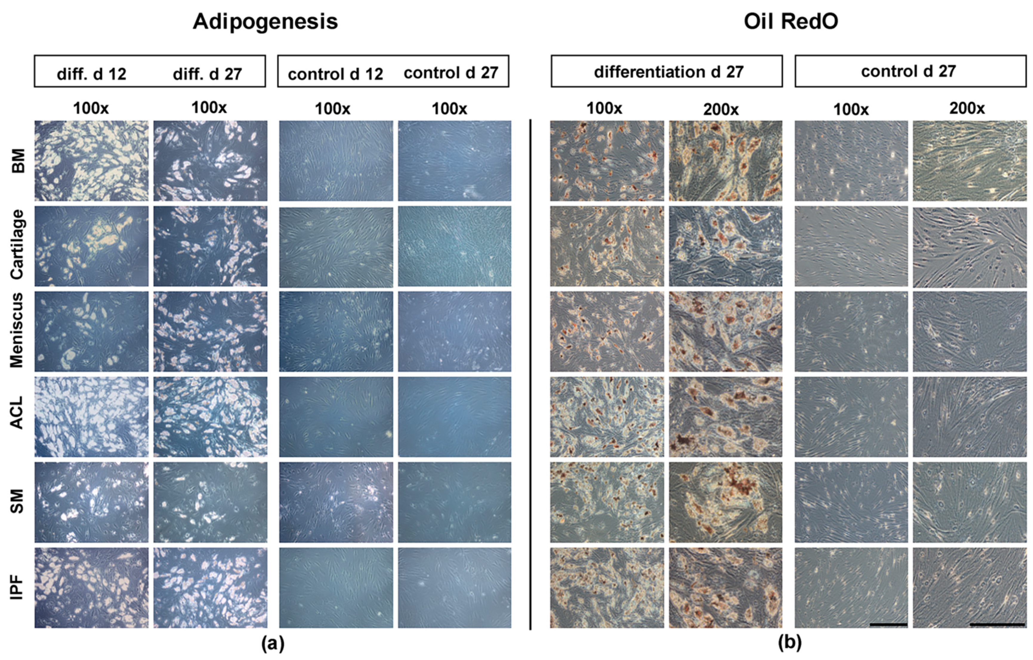
3.3. Histological Analysis of Adipogenesis

Following the adipogenic differentiation period of 27 d, all differentiated samples showed positive oil red O staining of vacuoles containing lipid droplets (Figure 3b), in comparison to the negative control groups. The comparison to negative control samples (Figure 3a) showed that the quantity of adipocyte-like cells increased in differentiated cultures over time. The negative control groups did not show any changes in cell structure or in the formation of lipid droplets. There were no qualitative differences regarding the oil red O staining of differentiated cell cultures from all six donor tissues.

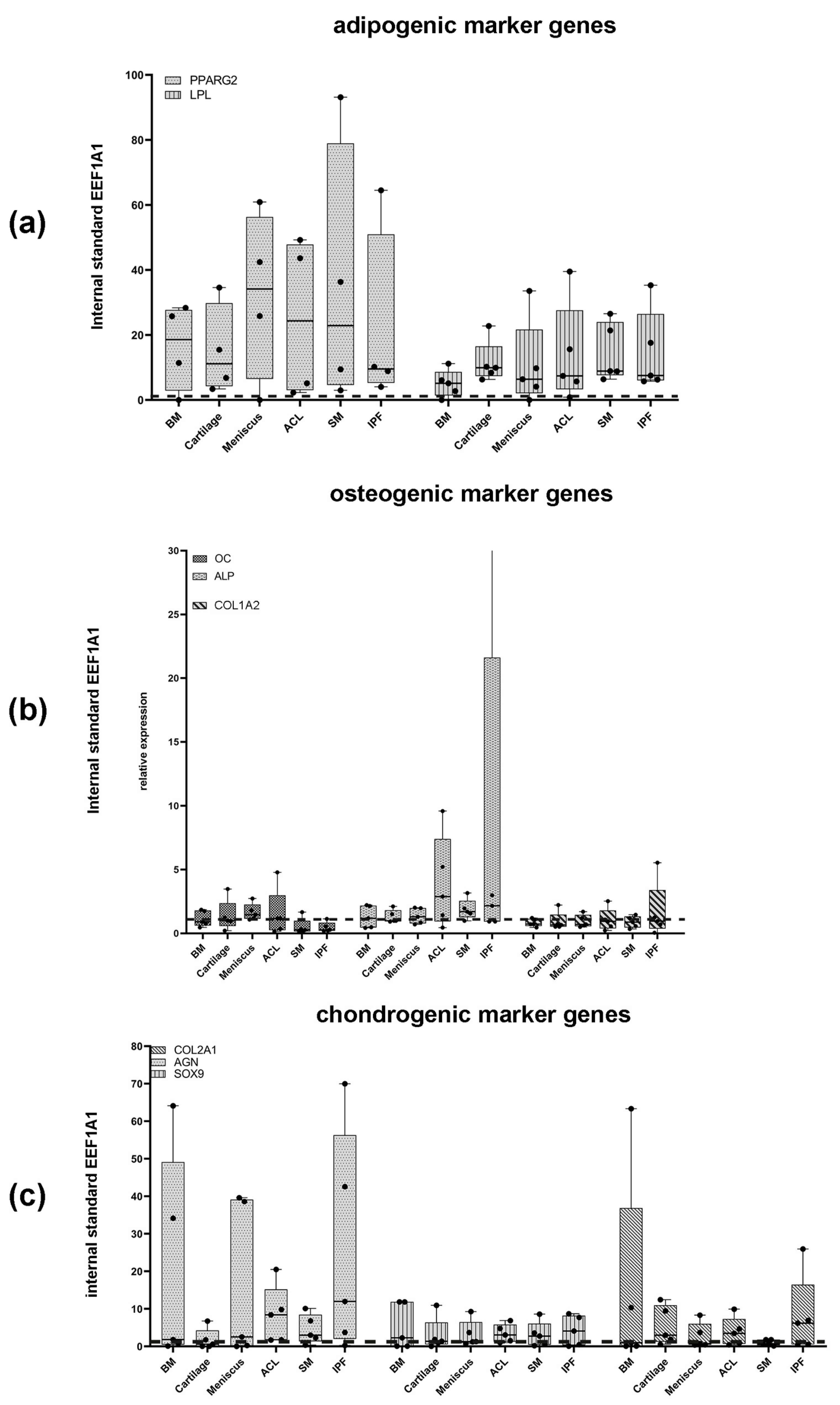
3.4. Expression of Adipogenic Marker Genes after 27 d

RT-PCR was used to examine the expression of adipogenic marker genes after 27 d of adipogenesis while undifferentiated negative controls served as controls. As shown in Figure 4a, we observed an increased expression of LPL, as well as PPARG2, in comparison to the negative controls. Samples derived from the meniscus and SM showed a particularly strong expression of the marker gene PPARG2. However, the relative gene expression did not differ significantly between the six tissue types for the PPRAG2 expression (p = 0.968), as well as for the LPL expression (p = 0.337). All findings presented broad scattering of single obtained values.

3.5. Histological and Immunohistochemical Analysis of Osteogenesis

Osteogenic differentiation was performed using monolayer cultures. Starting around 12 d, the differentiated cultures showed an increase in densely packed cells, when compared to the control cultures, exposed to standard cell culture medium (Figure 5a). By day 27, they had formed a spider-web-like structure, surrounded by extracellular dense deposits, resembling a mineralized extracellular matrix. Alizarin red S staining was performed to confirm the formation of calcium deposits within the ECM, as a sign of osteogenic differentiation. All differentiated cells showed a positive staining for alizarin red S (Figure 5b). When compared to other cell cultures cells isolated from BM, the SM, ACL, and meniscus tissue showed a more even distribution of stained mineralized matrix (Figure 5b, BM, Meniscus, ACL, and SM). Differentiated MSC-like cells, derived from arthritic hyaline cartilage and the IPF, showed a more punctual formation of calcium deposits, with fewer stained regions throughout the monolayer cultures, in general (Figure 5b, cartilage, IPF). Negative controls showed no positive staining.

3.6. Expression of Osteogenic Marker Genes after 27 d

RT-PCR was used to evaluate the expression of the three osteogenic marker genes, i.e., OC, ALP, and COL1A2. As shown in Figure 4b, differentiation led to a slight upregulation of OC and ALP in all of the differentiated cells. However, in cells isolated from the SM and IPF, the expression of OC remained rather low, while the differentiated cells, derived from the ACL and IPF, showed a high relative expression of ALP, with a broad scattering of single values. Finally, there was no clear upregulation of COL1A2 in differentiated cultures, in comparison to negative control groups (OC: p = 0.09, ALP: p = 0.519, COL1A2: p = 0.98).

3.7. Histological and Immunohistochemical Analysis of Chondrogenesis

After sectioning the chondrogenic differentiated pellets, alcian blue staining was performed to identify proteoglycans within the ECM. Differentiated pellets from all six examined native tissues showed positive staining, in comparison to negative controls (Figure 6a). Staining intensity in pellets, derived from BM, arthritic hyaline cartilage, and the SM, was particularly strong (Figure 6a, BM, cartilage, and SM). While all cultured pellets showed positive alcian blue staining, only those derived from BM and arthritic hyaline cartilage showed a macroscopic pattern resembling that of human hyaline cartilage, including chondrones and a cell-poor matrix.
In addition, immunohistochemical staining of COL II, which is the predominant type of collagen found in cartilage, were performed (Figure 6b). Staining revealed a rather uniform pattern of distribution of COL II throughout the differentiated pellet cultures of BM, arthritic hyaline cartilage, and the ACL, concentrated in the central areas of the pellet sections (Figure 6b, BM and cartilage). In comparison, the intensity of immunohistochemical Col II staining in the remaining four tissues was lower and rather concentrated around outer regions of the pellets (Figure 6b, Meniscus, ACL, and IPF). Negative controls did not show any positive staining for COL II.
Furthermore, immunohistochemical staining of COL X, which is viewed as a characteristic marker for chondrogenic hypertrophy, was performed (Figure 6c). All of the differentiated cultures showed slightly positive staining for COL X. Interestingly, positive areas were mostly present in the centre of pellet sections. The most intense staining for COL X could be observed in the pellets derived from BM, the ACL, and the IPF (Figure 6c). When comparing negative controls and differentiated pellet cultures, no clear increase in staining intensity was observed after 21 d of chondrogenic differentiation.

3.8. Expression of Chondrogenic Marker Genes after 21 d

RT-PCR was used to examine the expression of the chondrogenic marker genes AGN, SOX9, and COL2A1 (Figure 4c). In comparison to the negative controls, all samples showed an increased expression of the mentioned marker genes. Increase of the relative expression of the chondrogenic marker gene SOX9 was homogenous and independent of the cell source (Figure 4c, SOX9). Differentiated pellets derived from arthritic hyaline cartilage showed a homogenous expression of all three marker genes. All differentiated pellet cultures showed an increased expression of the chondrogenic marker gene AGN, with a broad scattering of single values. This was especially the case for pellet cultures derived from BM, meniscus tissue, and the IPF (Figure 4c). However, the differences of the relative gene expression between the tissue types turned out not be statistically significant (AGN: p = 0.677, COL2A1: p = 0.649, and SOX9: p = 0.984).

4. Discussion

MSCs have emerged as a promising part of cell-based regenerative treatment of traumatic or degenerative orthopaedic diseases, such as cartilage defects, osteoarthritis, or ligament damage [28,29]. Tissues other than BM have been shown to offer vast amounts of MSC-like cells, with multipotent differentiation potential, while requiring less invasive surgery for the isolation of cells [18,30].
In this present in vitro study, our goal was to detect and characterize less-well-known MSC-like cells, isolated from meniscal tissue, i.e., the ACL, IPF, and SM, during primary TKA of the arthritic knee joint. This study adds further research on the different MSC-subpopulations, mentioned above, by proving their presence and comparing their qualitative differentiation capacity in vitro. Our hypothesis was that BM-MSCs and MSC-like cells isolated from all other tissues of the arthritic knee joint possess multipotent differentiation potential, which we confirmed by our investigations.
Primarily, this study showed that multipotent and plastic-adherent cells could be isolated from all six examined tissues, i.e., BM, arthritic hyaline cartilage, meniscal tissue, the IPF, the SM, and the ACL, of the arthritic knee joint of five female patients undergoing primary TKA. Interestingly, CPD between isolation from native tissues and primary passage was significantly higher in cells derived from BM, when compared to cells derived from cartilage, the ACL, and the IPF. The presence and characteristics of BM-MSCs, which are viewed as the gold standard for TE and reside in the endosteal or perivascular niche, have been thoroughly examined in earlier studies [25,31,32]. In addition, Alsalameh et al. and Pretzel et al. isolated MSC-like cells from the superficial zone of arthritic or healthy hyaline cartilage [33,34]. Earlier research also proved the presence of MSC-like cells in the SM of the knee joint [35,36]. Inside the SM, MSC-like cells are believed to reside within the intimal layer and small blood vessels, located within the subintimal tissue [37]. Since synovial blood vessels act as a connection between the BM and synovial fluid, it is speculated that BM-MSCs may migrate from the BM to form MSC-like cells in the synovial fluid and SM of diarthrodial joints [37]. The IPF and the SM of the knee joint serve as a functional unit, which is why the IPF is often referred to as the adipose synovium [38]. Consequently, earlier studies proved the presence of MSC-like cells within the IPF [18,39]. We, and others, also showed that MSC-like cells can be isolated from the ACL, where they are believed to be located within small sinusoids, fascicles, or the endothelial lining of small blood vessels [22,40,41]. Despite the poor blood supply of meniscal tissue, there is also evidence that MSC-like cells or multipotent meniscal fibrochondrocytes (MFCs) can be isolated from meniscal debris [21]. Interestingly, all of the investigated native tissues rise from the lateral plate mesoderm (LPM) and are well-vascularized [42,43]. This further supports our hypothesis, when taking the embryological development of MSCs into account [43]. All of these findings support our hypothesis that multipotent and plastic-adherent cells expressing an MSC-characteristic set of surface antigens can be isolated from the six examined tissues of the arthritic knee joint.
Secondly, cells isolated from all six different tissues of an exemplary chosen donor sample co-expressed the surface antigens CD44, CD73, CD90, and CD105. The ISCT defined these surface antigens as highly characteristic, but not specific, for MSCs [15]. Our results showed that ≥95% of cells were positive for the surface markers CD44, CD90, and CD105, independent of the examined tissue source. Therefore, all cells showed an MSC-like pattern of surface antigens, separating them from tissue resident, plastic-adherent, and multipotent fibroblasts. Although all cells also expressed the surface antigen CD73, expression rates did not exceed 95% in cells, other than BM-MSCs, as defined by the minimal definition criteria for MSCs by the ISCT [15]. Earlier research has shown that different MSC-subpopulations may differ in their expression of surface antigens [20]. This has led to intense discussions regarding whether the characterization of unspecific, but characteristic, surface antigens, which may vary for different subpopulations of MSCs, should be concluded in the minimal criteria defining MSCs. In addition, it remains unclear if differences regarding the expression of characteristic surface antigens also effect the functional properties of different MSC-subpopulations. Additional research is necessary to further compare the possible differences in the expression of surface antigens and their effects on the therapeutic cell use of examined MSC-subpopulations.
However, our results, regarding the expression of characteristic surface antigens, are in line with multiple earlier studies, showing that BM-MSCs, derived from the knee joint, clearly co-expressed CD44, CD73, CD90, and CD105 [31,44]. A mixed population of chondrocytes and multipotent progenitor cells, isolated from both arthritic and hyaline cartilage of the knee joint, expressed CD105 and CD166 [33,34]. Interestingly, previous research showed that multipotent, plastic-adherent cells, isolated from the ACL, SM, or IFP of osteoarthritic and healthy knee joints, clearly co-expressed CD73, CD90, and CD105 [18,22,41,45]. Finally, Weili et al. and Segawa et al. showed that CD44, CD90, and CD105 positive cells could also be isolated from meniscal debris, which is in line with our current results [21,44].
MSC-like cells isolated from the BM, arthritic hyaline cartilage, the SM, the ACL, the IFP, and meniscal tissue showed clear trilineage differentiation potential in vitro, as defined in the ISCT‘s criteria for MSCs [15]. In this study, no qualitative differences were found, regarding the osteogenic and adipogenic differentiation capacity of BM-MSCs and MSC-like cells, isolated from the arthritic hyaline cartilage, ACL, the IPF, the SM, and meniscal tissue. Although, there were differences in the gene expression of specific marker genes, none of these reached statistical significance. In this context, it has to be added that no quantitative measurements of oil red O and alizarin red S stainings were performed. The same applied to the chondrogenic differentiation of MSC-like cells isolated from the six different native tissues, meaning that observed differences in histological and immunohistochemically stainings, as well as the relative expression of marker genes, are highly subjective. Differences regarding histological and immunohistochemically stainings, as well as relative expression of marker genes, could be due to phaseal upregulation. Interestingly, we observed a strong staining intensity and more chondrogenic-like phenotype, including chondrones and a cell-poor extracellular matrix (ECM), in the alcian blue staining of pellets, derived from arthritic MSC-like cells, which were derived from hyaline cartilage and BM-MSCs. At the same time, hypertrophic differentiation was least evident in cell pellets containing MSC-like cells from arthritic hyaline cartilage and the SM.
When examining the multipotent differentiation capacity of different MSC-subpopulations, BM-MSCs are often viewed as the gold standard, which is used for comparison. Matching our current findings, earlier studies showed that the qualitative multipotent differentiation potential of MSC-like cells isolated from arthritic hyaline cartilage and the IPF does not differ from that of BM-MSCs, and these subpopulations offer promising potential for clinical transition [33,34,39]. Primorac et al. and others showed that the treatment of patients with late-stage OA of the knee with adipose-derived MSCs led to improved clinical and functional outcome [46]. While the MSC-like cells isolated in our study showed limited proliferative capacity, they showed great chondrogenic differentiation potential, when analyzing the histological and immunohistochemical stainings, as well as the expression of chondrogenic marker genes.
In contrast, MSC-like cells isolated from the SM have been shown to possess a greater chondrogenic differentiation capacity than BM-MSCs [18,35]. Since chondrogenic differentiation potential was evaluated by measuring pellet dry weight and size, we could not directly compare the results observed in our current study, since these variables were not evaluated in our current study [18]. Few studies have also reported on the multipotent differentiation capacity of cells derived from different intraarticular ligaments [19,22,43]. When examining histological and immunohistochemical stainings, we found a slightly superior chondrogenic differentiation potential in MSC-like cells isolated from the ACL, when compared to BM-MSCs [22]. Surprisingly, Cheng et al. described a similar chondrogenic but an inferior osteogenic differentiation capacity when comparing ACL-derived MSC-like cells to BM-MSCs [41]. Finally, Weili et al. reported that MSC-like cells, isolated from meniscal tissue, present extensive multipotent differentiation capacity [21]. According to our findings, Segawa et al. previously showed that their qualitative adipogenic, osteogenic, and chondrogenic potential did not differ significantly from MSCs isolated from the SM, intraarticular ligaments, or BM of the same knee joints [44].
In summary, MSC-like cells were isolated from the BM, SM, IPF, ACL, and meniscal tissue of arthritic knee joints. Interestingly, all subpopulations of MSC-like cells, isolated from the same donor, did not differ, regarding their qualitative in vitro multipotent differentiation potential. Although cells isolated from hyaline cartilage showed the clear expression of MSC-like surface antigens, plastic-adherent growth, and great multipotent differentiation capacity, Pretzel et al. found that most of the isolated cells from hyaline cartilage are not progenitors after prolonged expansion in vitro but, rather, a mixed population of chondrocytes and multipotent progenitor cells [34]. All of the examined tissues are routinely removed during primary TKA. Interestingly, the SM, IPF, ACL, and meniscal tissue are also easily accessible via minimal-invasive surgery. The number of MSC-like cells that can be harvested during surgery often differs between native tissues and is larger for soft tissues, such as adipose and synovial tissue [37,47]. In contrast, BM, cartilage, and ligamentous tissues often only offer a limited number of cells, resulting in major challenges for clinical transition during both tissue harvesting and TE, since clinical cell-based therapy often requires large amounts of MSC-like cells for tissue repair [29,37,48]. In our current study, the number of primary isolated cells was largest in BM samples. In addition, the number of isolated cells was also high when using the two soft tissues SM or IPF. Interestingly, the number of primary isolated cells was higher in meniscus tissue than in arthritic hyaline cartilage. Both meniscus tissue and hyaline cartilage can be accessed via arthroscopic surgery, although the limited regeneration capacity of healthy hyaline cartilage also poses ethical issues, regarding the isolation of cartilage tissue. Up to date, there is only limited research regarding the characterization of MSC-like cells derived from intraarticular ligaments and meniscal tissue. However, their qualitative multipotent differentiation capacity does not seem to differ from that of the BM-MSCs or MSC-like cells derived from hyaline cartilage.
One central limitation to our study is that no experiments examining the quantitative differences of multipotent differentiation capacity were performed, meaning that all observed differences in histological and immunohistochemical stainings are highly subjective and, therefore, leave room for error. Hence, the following research should focus on examining quantitative differences, regarding multipotent differentiation of respective MSC-like cells to add to the qualitative differences presented in this study. Secondly, cumulative population doublings were highest in BM-MSCs and significantly lower in cells derived from arthritic hyaline cartilage, the ACL, and the IPF. These differences could pose a major obstacle for the clinical utilization of these MSC-like subpopulations, regarding the large amount of cells usually needed for therapeutic application. Thirdly, our current study only examined a small sample size of female patients. Earlier research has shown that differentiation capacity, proliferation capacity, and colony forming efficiency in MSCs may decline with donor age [49]. Besides, the loss of transplanted tissue and pro-inflammatory effects MSC-based cartilage repair may lead to osteophyte formation and tissue hypertrophy [17]. Further, the expression of surface antigens was only tested on cells derived from one of the five patients and did not include hematopoietic markers. However, the goal of our current study was only to show that examined cells express MSC-like surface antigens, not to compare the patterns of surface antigens between different MSC-subpopulations. Further the tissue-specific naïve MSC population, situated in the natural niche within the respective tissue, differs from the MSC population that is used in conventional cell culture experiments in vitro. Before setting up differentiation experiments, the heterogeneous MSC population is isolated by plastic adherence, cell expansion is performed in the presence of FCS, and, afterwards, cells are detached by trypsinization, in order to reseed them on new cell culture dishes. All these steps influence the resulting cell population, in such a way that MSC subpopulations are enriched, which differs from their naïve, original tissue-specific subpopulation. In addition, different subpopulations of MSC-like cells may require a specific set of growth factors and scaffolds to further optimize and fully examine their multipotent differentiation potential [43]. Future research should examine different growth factors and fitting biomaterials to optimize and maintain the multipotent differentiation potential of different subpopulations of MSC-like cells.

5. Conclusions

In conclusion, this work compared the adipogenic, osteogenic, and chondrogenic differentiation potential of BM-MSCs and MSC-like cells, isolated from various tissues of the human osteoarthritic knee joint. All MSC-subpopulations, isolated from a single donor during TKA, possessed similar in vitro multipotent differentiation potential. Interestingly, the qualitative differentiation capacity of MSC-like cells, isolated from less well-known sources, such as the ACL, IPF, and meniscal tissue, did not differ significantly from that of the BM-MSCs or MSC-like cells isolated form arthritic hyaline cartilage. Therefore, the isolation of the MSC-like cells offering extensive multipotent differentiation may not require maximal invasive surgery. Future studies should not only focus on different cell-sources but also the signaling pathways and scaffolds, in order to optimize all components of the triad of TE.

Supplementary Materials

The following supporting information can be downloaded at: https://www.mdpi.com/article/10.3390/app12042239/s1. Table S1: p-values for statistical difference of the relative gene marker expression between the six different tissue types.

Author Contributions

All authors have read and approved the final manuscript and contributed to the study design, data analysis, interpretation of data, drafting, and revision of the manuscript. K.P. performed the experiments. K.P., M.W. (Mike Wagenbrenner) and M.W. (Manuel Weißenberger) wrote the manuscript and designed the tables and figures. T.H. performed the statistical analysis of all experiments. All authors have read and agreed to the published version of the manuscript.

Funding

This publication was funded by the German Research Foundation (DFG) and University of Würzburg, in the funding programme Open Access Publishing. Marietta Herrmann is supported by the Interdisciplinary Center for Clinical Research (IZKF) at the University of Würzburg (Project D-361).

Institutional Review Board Statement

The study design and experiments were approved by University of Wuerzburg‘s institutional review board. In addition, participating patients agreed to the use of collected and examined surgical waste after undergoing total knee replacement surgery. Written informed consent was obtained from every patient, in accordance with the local Ethics committee of the University of Würzburg (186/18). All methods were carried out in accordance with relevant guidelines and regulations.

Informed Consent Statement

Informed consent was obtained from all subjects involved in the study.

Data Availability Statement

The datasets used and analyzed during the current study are available from the corresponding author on reasonable request.

Acknowledgments

We are grateful to Beate Geyer, Jutta Schneidereit, and Theresa Kreuzahler for their excellent technical assistance.

Conflicts of Interest

The authors declare no conflict of interest.

Abbreviations

ACL: anterior cruciate ligament; AGN: aggrecan; ALP: alkaline phosphatase; BM: bone marrow; CD: cluster of differentiation; cDNA: complementary DNA; COL I: collagen type I; COL1A2: collagen type Ia2; COL II: collagen type II; COL2A1: collagen type IIa1; COL X: collagen type X; CPD: cumulative population doublings; d: days; DMSO: dimethylsulfoxid; ECM: extracellular matrix; EEF1α: elongation factor 1α; FBS: fetal bovine serum; MSCs: mesenchymal stromal cells; H&E: hematoxylin-Eosin; IPF: infrapatellar fat pad; ISCT: International Society for Cellular Therapy; LPL: lipoproteinlipase; OC: osteocalcin; PBS: phosphate buffered saline; PPARG2: peroxisome proliferator-activated receptor γ; RT-PCR: reverse transcriptase polymerase chain reaction; SM: synovial membrane; SOX9: sex-determining region Y-box 9.

References

  1. Sharma, P.; Kumar, P.; Sharma, R.; Bhatt, V.D.; Dhot, P.S. Tissue Engineering; Current Status & Futuristic Scope. J. Med. Life 2019, 12, 225–229. [Google Scholar] [PubMed]
  2. Bauge, C.; Boumediene, K. Use of Adult Stem Cells for Cartilage Tissue Engineering: Current Status and Future Developments. Stem Cells Int. 2015, 2015, 438026. [Google Scholar] [CrossRef] [PubMed] [Green Version]
  3. Dzobo, K.; Thomford, N.; Senthebane, D.; Shipanga, H.; Rowe, A.; Dandara, C.; Pillay, M.; Motaung, K.S. Advances in Regenerative Medicine and Tissue Engineering: Innovation and Transformation of Medicine. Stem Cells Int. 2018, 2018, 2495848. [Google Scholar] [CrossRef] [PubMed] [Green Version]
  4. Jessop, Z.; Al-Sabah, A.; Francis, W.R.; Whitaker, I.S. Transforming healthcare through regenerative medicine. BMC Med. 2016, 14, 115. [Google Scholar] [CrossRef] [PubMed] [Green Version]
  5. Berebichez-Fridman, R.; Montero-Olvera, P.R. Sources and Clinical Applications of Mesenchymal Stem Cells: State-of-the-art review. Sultan Qaboos Univ. Med. J. 2018, 18, e264–e277. [Google Scholar] [CrossRef] [PubMed] [Green Version]
  6. Gonzalez, M.J. Keeping kidneys. Bull. World Health Organ. 2012, 90, 718–719. [Google Scholar]
  7. Żylińska, B.; Silmanowicz, P.; Sobczyńska-Rak, A.; Jarosz, Ł.; Szponder, T. Treatment of Articular Cartilage Defects: Focus on Tissue Engineering. In Vivo 2018, 32, 1289–1300. [Google Scholar] [CrossRef] [Green Version]
  8. Health at a Glance 2019 OECD Indicators: OECD Indicators; OECD Publishing: Berlin, Germany, 2019.
  9. Klug, A.; Gramlich, Y.; Rudert, M.; Drees, P.; Hoffmann, R.; Weißenberger, M.; Kutzner, K.P. The projected volume of primary and revision total knee arthroplasty will place an immense burden on future health care systems over the next 30 years. Knee Surg. Sports Traumatol. Arthrosc. 2020, 12, 3287–3298. [Google Scholar] [CrossRef]
  10. Karachalios, T.; Komnos, G.; Koutalos, A. Total hip arthroplasty: Survival and modes of failure. EFORT Open Rev. 2018, 3, 232–239. [Google Scholar] [CrossRef]
  11. Argenson, J.N.; Boisgard, S.; Parratte, S.; Descamps, S.; Bercovy, M.; Bonnevialle, P.; Briard, J.L.; Brilhault, J.; Chouteau, J.; Nizard, R.; et al. Survival analysis of total knee arthroplasty at a minimum 10 years’ follow-up: A multicenter French nationwide study including 846 cases. Orthop. Traumatol. Surg. Res. 2013, 99, 385–390. [Google Scholar] [CrossRef] [Green Version]
  12. Zhan, C.; Kaczmarek, R.; Loyo-Berrios, N.; Sangl, J.; Bright, R.A. Incidence and short-term outcomes of primary and revision hip replacement in the United States. J. Bone Joint Surg. Am. 2007, 89, 526–533. [Google Scholar] [CrossRef] [PubMed]
  13. Lee, D.H.; Lee, S.H.; Song, E.K.; Seon, J.K.; Lim, H.A.; Yang, H.Y. Causes and Clinical Outcomes of Revision Total Knee Arthroplasty. Knee Surg. Relat. Res. 2017, 29, 104–109. [Google Scholar] [CrossRef] [PubMed] [Green Version]
  14. Im, G.I. Tissue Engineering in Osteoarthritis: Current Status and Prospect of Mesenchymal Stem Cell Therapy. BioDrugs 2018, 32, 183–192. [Google Scholar] [CrossRef] [PubMed]
  15. Horwitz, E.M.; Blanc, K.L.; Dominici, M.; Mueller, I.; Slaper-Cortenbach, I.; Marini, F.C.; Deans, R.J.; Krause, D.S.; Keating, A. Clarification of the nomenclature for MSC: The International Society for Cellular Therapy position statement. Cytotherapy 2005, 7, 393–395. [Google Scholar] [CrossRef] [PubMed]
  16. Han, Y.; Li, X.; Zhang, Y.; Han, Y.; Chang, F.; Ding, J. Mesenchymal Stem Cells for Regenerative Medicine. Cells 2019, 8, 886. [Google Scholar] [CrossRef] [PubMed] [Green Version]
  17. Steinert, A.F.; Ghivizzani, S.C.; Rethwilm, A.; Tuan, R.S.; Evans, C.H.; Nöth, U. Major biological obstacles for persistent cell-based regeneration of articular cartilage. Arthritis Res. Ther. 2007, 9, 213. [Google Scholar] [CrossRef] [PubMed] [Green Version]
  18. Sakaguchi, Y.; Sekiya, I.; Yagishita, K.; Muneta, T. Comparison of human stem cells derived from various mesenchymal tissues: Superiority of synovium as a cell source. Arthritis Rheum. 2005, 52, 2521–2529. [Google Scholar] [CrossRef] [PubMed]
  19. Wagenbrenner, M.; Heinz, T.; Horas, K.; Jakuscheit, A.; Arnholdt, J.; Herrmann, M.; Rudert, M.; Holzapfel, B.M.; Steinert, A.F.; Weißenberger, M. The human arthritic hip joint is a source of mesenchymal stromal cells (MSCs) with extensive multipotent differentiation potential. BMC Musculoskelet Disord. 2020, 21, 297. [Google Scholar] [CrossRef]
  20. Elahi, K.C.; Klein, G.; Avci-Adali, M.; Sievert, K.D.; MacNeil, S.; Aicher, W.K. Human Mesenchymal Stromal Cells from Different Sources Diverge in Their Expression of Cell Surface Proteins and Display Distinct Differentiation Patterns. Stem Cells Int. 2016, 2016, 5646384. [Google Scholar] [CrossRef] [Green Version]
  21. Fu, W.; Xie, X.; Li, Q.; Chen, G.; Zhang, C.; Tang, X.; Li, J. Isolation, Characterization, and Multipotent Differentiation of Mesenchymal Stem Cells Derived from Meniscal Debris. Stem Cells Int. 2016, 2016, 5093725. [Google Scholar] [CrossRef] [PubMed]
  22. Steinert, A.F.; Kunz, M.; Prager, P.; Barthel, T.; Jakob, F.; Nöth, U.; Murray, M.M.; Evans, C.H.; Porter, R.M. Mesenchymal stem cell characteristics of human anterior cruciate ligament outgrowth cells. Tissue Eng. Part A 2011, 17, 1375–1388. [Google Scholar] [CrossRef] [PubMed]
  23. Steinert, A.F.; Proffen, B.; Kunz, M.; Hendrich, C.; Ghivizzani, S.C.; Nöth, U.; Rethwilm, A.; Eulert, J.; Evans, C.H. Hypertrophy is induced during the in vitro chondrogenic differentiation of human mesenchymal stem cells by bone morphogenetic protein-2 and bone morphogenetic protein-4 gene transfer. Arthritis Res. Ther. 2009, 11, R148. [Google Scholar] [CrossRef] [PubMed] [Green Version]
  24. Reichert, J.C.; Schmalzl, J.; Prager, P.; Gilbert, F.; McQuent, V.; Steinert, A.F.; Rudert, M.; Noth, U. Synergistic effect of Indian hedgehog and bone morphogenetic protein-2 gene transfer to increase the osteogenic potential of human mesenchymal stem cells. Stem Cell Res. Ther. 2013, 4, 105. [Google Scholar] [CrossRef] [PubMed] [Green Version]
  25. Nöth, U.; Osyczka, A.M.; Tuli, R.; Hickok, N.J.; Danielson, K.G.; Tuan, R.S. Multilineage mesenchymal differentiation potential of human trabecular bone-derived cells. J. Orthop. Res. 2002, 20, 1060–1069. [Google Scholar] [CrossRef]
  26. Steinert, A.F.K.M.; Prager, P.; Gobel, S.; Klein-Hitpass, L.; Ebert, R.; Noth, U.; Jakob, F.; Gohlke, F. Characterization of bursa subacromialis-derived mesenchymal stem cells. Stem Cell Res. Ther. 2015, 6, 114. [Google Scholar] [CrossRef] [PubMed] [Green Version]
  27. Zołocińska, A. The expression of marker genes during the differentiation of mesenchymal stromal cells. Adv. Clin. Exp. Med. 2018, 27, 717–723. [Google Scholar] [CrossRef] [PubMed]
  28. Park, Y.B.; Ha, C.W.; Rhim, J.H.; Lee, H.J. Stem Cell Therapy for Articular Cartilage Repair: Review of the Entity of Cell Populations Used and the Result of the Clinical Application of Each Entity. Am. J. Sports Med. 2018, 46, 2540–2552. [Google Scholar] [CrossRef]
  29. Berebichez-Fridman, R.; Gómez-García, R.; Granados-Montiel, J.; Berebichez-Fastlicht, E.; Olivos-Meza, A.; Granados, J.; Velasquillo, C.; Ibarra, C. The Holy Grail of Orthopedic Surgery: Mesenchymal Stem Cells-Their Current Uses and Potential Applications. Stem Cells Int. 2017, 2017, 2638305. [Google Scholar] [CrossRef] [PubMed]
  30. Mafi, R.; Hindocha, S.; Mafi, P.; Griffin, M.; Khan, W.S. Sources of adult mesenchymal stem cells applicable for musculoskeletal applications—A systematic review of the literature. Open Orthop. J. 2011, 5 (Suppl. 2), 242–248. [Google Scholar] [CrossRef] [PubMed]
  31. Pittenger, M.F.; Mackay, A.M.; Beck, S.C.; Jaiswal, R.K.; Douglas, R.; Mosca, J.D.; Moorman, M.A.; Simonetti, D.W.; Craig, S.; Marshak, D.R. Multilineage potential of adult human mesenchymal stem cells. Science 1999, 284, 143–147. [Google Scholar] [CrossRef] [Green Version]
  32. Charbord, P. Bone marrow mesenchymal stem cells: Historical overview and concepts. Hum. Gene. Ther. 2010, 21, 1045–1056. [Google Scholar] [CrossRef] [PubMed] [Green Version]
  33. Pretzel, D.; Linss, S.; Rochler, S.; Endres, M.; Kaps, C.; Alsalameh, S.; Kinne, R.W. Relative percentage and zonal distribution of mesenchymal progenitor cells in human osteoarthritic and normal cartilage. Arthritis Res. Ther. 2011, 13, R64. [Google Scholar] [CrossRef] [PubMed] [Green Version]
  34. Alsalameh, S.; Amin, R.; Gemba, T.; Lotz, M. Identification of mesenchymal progenitor cells in normal and osteoarthritic human articular cartilage. Arthritis Rheum. 2004, 50, 1522–1532. [Google Scholar] [CrossRef] [PubMed]
  35. Hatakeyama, A.; Uchida, S.; Utsunomiya, H.; Tsukamoto, M.; Nakashima, H.; Nakamura, E.; Pascual-Garrido, C.; Sekiya, I.; Sakai, A. Isolation and Characterization of Synovial Mesenchymal Stem Cell Derived from Hip Joints: A Comparative Analysis with a Matched Control Knee Group. Stem Cells Int. 2017, 2017, 9312329. [Google Scholar] [CrossRef] [PubMed]
  36. De Bari, C.; Dell’Accio, F.; Tylzanowski, P.; Luyten, F.P. Multipotent mesenchymal stem cells from adult human synovial membrane. Arthritis Rheum. 2001, 44, 1928–1942. [Google Scholar] [CrossRef]
  37. Li, N.; Gao, J.; Mi, L.; Zhang, G.; Zhang, L.; Zhang, N.; Huo, R.; Hu, J.; Xu, K. Synovial membrane mesenchymal stem cells: Past life, current situation, and application in bone and joint diseases. Stem Cell Res. Ther. 2020, 11, 381. [Google Scholar] [CrossRef] [PubMed]
  38. Macchi, V.; Stocco, E.; Stecco, C.; Belluzzi, E.; Favero, M.; Porzionato, A.; De Caro, R. The infrapatellar fat pad and the synovial membrane: An anatomo-functional unit. J. Anat. 2018, 233, 146–154. [Google Scholar] [CrossRef] [Green Version]
  39. Huri, P.Y.; Hamsici, S.; Ergene, E.; Huri, G.; Doral, M.N. Infrapatellar Fat Pad-Derived Stem Cell-Based Regenerative Strategies in Orthopedic Surgery. Knee Surg. Relat. Res. 2018, 30, 179–186. [Google Scholar] [CrossRef] [PubMed] [Green Version]
  40. Huang, T.F.; Chen, Y.T.; Yang, T.H.; Chen, L.L.; Chiou, S.H.; Tsai, T.H.; Tsai, C.C.; Chen, M.H.; Ma, H.L.; Hung, S.C. Isolation and characterization of mesenchymal stromal cells from human anterior cruciate ligament. Cytotherapy 2008, 10, 806–814. [Google Scholar] [CrossRef] [PubMed]
  41. Cheng, M.T.; Yang, H.W.; Chen, T.H.; Lee, O.K. Isolation and characterization of multipotent stem cells from human cruciate ligaments. Cell Prolif. 2009, 42, 448–460. [Google Scholar] [CrossRef] [PubMed]
  42. Sheng, G. The developmental basis of mesenchymal stem/stromal cells (MSCs). BMC Dev. Biol. 2015, 15, 44. [Google Scholar] [CrossRef] [PubMed] [Green Version]
  43. Caplan, A.I. Mesenchymal Stem Cells: Time to Change the Name! Stem Cells Transl. Med. 2017, 6, 1445–1451. [Google Scholar] [CrossRef] [PubMed] [Green Version]
  44. Segawa, Y.; Muneta, T.; Makino, H.; Nimura, A.; Mochizuki, T.; Ju, Y.J.; Ezura, Y.; Umezawa, A.; Sekiya, I. Mesenchymal stem cells derived from synovium, meniscus, anterior cruciate ligament, and articular chondrocytes share similar gene expression profiles. J. Orthop. Res. 2009, 27, 435–441. [Google Scholar] [CrossRef] [PubMed]
  45. Garcia, J.; Wright, K.; Roberts, S.; Kuiper, J.H.; Mangham, C.; Richardson, J.; Mennan, C. Characterisation of synovial fluid and infrapatellar fat pad derived mesenchymal stromal cells: The influence of tissue source and inflammatory stimulus. Sci. Rep. 2016, 6, 24295. [Google Scholar] [CrossRef] [Green Version]
  46. Hudetz, D.; Borić, I.; Rod, E.; Jeleč, Ž.; Kunovac, B.; Polašek, O.; Vrdoljak, T.; Plečko, M.; Skelin, A.; Polančec, D.; et al. Early results of intra-articular micro-fragmented lipoaspirate treatment in patients with late stages knee osteoarthritis: A prospective study. Croat. Med. J. 2019, 60, 227–236. [Google Scholar] [CrossRef] [PubMed]
  47. Veronesi, F.; Maglio, M.; Tschon, M.; Aldini, N.N.; Fini, M. Adipose-derived mesenchymal stem cells for cartilage tissue engineering: State-of-the-art in in vivo studies. J. Biomed. Mater. Res. A 2014, 102, 2448–2466. [Google Scholar] [CrossRef] [PubMed]
  48. Mobasheri, A.; Csaki, C.; Clutterbuck, A.L.; Rahmanzadeh, M.; Shakibaei, M. Mesenchymal stem cells in connective tissue engineering and regenerative medicine: Applications in cartilage repair and osteoarthritis therapy. Histol. Histopathol. 2009, 24, 347–366. [Google Scholar] [PubMed]
  49. Yang, Y.K. Aging of mesenchymal stem cells: Implication in regenerative medicine. Regen. Ther. 2018, 9, 120–122. [Google Scholar] [CrossRef] [PubMed]
Figure 1. Cumulative population doublings of cells derived from all native tissues, between isolation and after primary passage, pictured as median ± standard deviation (error bars). Cells derived from bone marrow (BM), hyaline cartilage, the meniscus, the anterior cruciate ligament (ACL), the synovial membrane (SM), and the infrapatellar fat pad (IPF) were counted, following primary isolation from all six native tissues. After primary passage and before initiating the following experiments, cells were harvested and counted again. Cumulative population doubling (CPD) was calculated using the formula CPD = log10/(N/N0) × 3.33. N was the number of cells at the end of passage one, and N0 was the number of cells derived from primary isolation of native tissues. The median of CPD is pictured, respectively. Error bars picture the standard deviation.
Applsci 12 02239 g001
Figure 2. Flow cytometry analysis of the expression of surface antigens on cells isolated from all six tissues of one single patient. Cells from bone marrow (a), hyaline cartilage (b), the meniscus (c), the anterior cruciate ligament (ACL) (d), the synovial membrane (SM) (e), and the infrapatellar fat pad (IPF) (f) were examined for the co-expression of surface antigens cluster of differentiation (CD)44, CD73 (CD44/CD73), CD90, and CD105 (CD90/CD105). The results were pictured using the FlowJo 10.5.3 software by FlowJo, LLC. While almost all cells (≥95%) were positive for the surface markers CD44, CD90, and CD105, the percentage of CD73+ cells ranged from about 13–99%, depending on the donor tissue.
Applsci 12 02239 g002
Figure 3. Histological assay of adipogenesis in mesenchymal stromal cells after 27 days in adherent monolayer cell cultures. For inducing adipogenesis monolayer cultures containing cells from bone marrow, hyaline cartilage, the meniscus, the anterior cruciate ligament (ACL), the synovial membrane (SM), and the infrapatellar fat pad (IPF) were incubated with adipogenic differentiation medium for 27 days (d). Controls were maintained in cell culture medium under the same conditions. Both native, unstained tissue samples (a) and oil red O stainings (b) from control and differentiated samples were compared. Adipogenic assays were performed with all samples (five donors, six different MSC population each), and we show one representative donor of each staining. Representative samples were captured at low (100×; black bar = 200 μm) and high (200×; black bar = 150 μm) magnification.
Applsci 12 02239 g003
Figure 4. Scattered dot plots picturing the changes in relative gene expression of specific marker genes as median ± upper and lower quartile and highest and lowest values (error bars), as measured by semiquantitative RT-PCR in mesenchymal progenitor cells at the end of the respective differentiation period. Cells derived from bone marrow, hyaline cartilage, the meniscus, the anterior cruciate ligament (ACL), the synovial membrane (SM), and the infrapatellar fat pad (IFP) were incubated in adipogenic, osteogenic (27 days (d)) and chondrogenic (21 d) differentiation medium. The median of the changes of the relative expression of the adipogenic (a) marker genes—lipoproteinlipase (encoded by LPL) and proliferator-activated receptor γ (encoded by PPARG2)—the osteogenic (b) marker genes—collagen type Ia2 (encoded by COL1A2), alkaline phosphatase (encoded by ALP), and osteocalcin (encoded by OC)—as well as the chondrogenic (c) marker genes—aggrecan (encoded by AGN), collagen type IIa1 (encoded by COL2A1), and sex-determining region Y-box 9 (encoded by SOX9)—are pictured, respectively. Box plots picture the upper and lower quartiles and error bars picture the respective highest and lowest values of changes in the relative expression of specific marker genes. Cell cultures treated with standard cell culture medium served as negative controls. Elongation factor 1α (encoded by EEF1A1) was used as the housekeeping gene and internal controls. The expression of respective marker genes in undifferentiated control cultures served as a baseline value, which differentiated cultures were compared against, and was pictured as a dashed line (value = 1). Differentiation assays were performed with all samples (five donors, six different MSC population each), and we show one representative donor of each staining.
Applsci 12 02239 g004
Figure 5. Histological assay of osteogenesis in cells after 21 days in adherent monolayer cell cultures. Induced osteogenesis monolayer cultures, containing cells from bone marrow (BM), hyaline cartilage, the meniscus, the anterior cruciate ligament (ACL), the synovial membrane (SM), and the infrapatellar fat pad (IFP) were incubated with osteogenic differentiation medium for 27 days (d). Controls were maintained in cell culture medium under the same conditions. Both native, unstained tissue samples (a) and alizarin red S stainings (b) from control and differentiated samples were compared. Osteogenic assays were performed with all samples (five donors, six different MSC population each), and we show one representative donor of each staining. Representative samples were captured at low (100×; black bar = 200 μm) and high (200×; black bar = 150 μm) magnification.
Applsci 12 02239 g005
Figure 6. Histological and immunohistochemical analysis of chondrogenesis in cell-pellets after 21 d of pellet culture. Induced chondrogenesis pellets, containing cells from bone marrow (BM), hyaline cartilage, the meniscus, the anterior cruciate ligament (ACL), the synovial membrane (SM) and the infrapatellar fat pad (IFP), were incubated with chondrogenic differentiation medium for 21 days (d). Controls were maintained in cell culture medium under the same conditions. After 21 d, alcian blue staining (a) was performed for the detection of proteoglycans. Immunohistochemical stainings of collagen type II (COL II) (b) and collagen type X (COL X) (c) were performed on pellet sections cells from BM (1), hyaline cartilage (2), the meniscus (3), the ACL (4), the SM (5), and the IPF (6) after incubation in chondrogenic differentiation medium for 21 d. Positive staining for COL II (b) and COL X (c) appeared brown. Chondrogenic assays were performed with all samples (five donors, six different MSC population each), and we show one representative donor of each staining. Representative samples were captured at low (50×; black bar = 300 μm) and high (200×; black bar = 300 μm) magnification.
Applsci 12 02239 g006
Table 1. Patient demographics and detailed information regarding the five donors undergoing total knee arthroplasty. Precise description of the five different patients that underwent total knee arthroplasty and served as donors for the isolation of mesenchymal stromal cell (MSC)-like cells.
Patient NumberAge (Years)BMI (kg/m2)Secondary
Diagnosis
Osteoarthritis Grade
(Kellgren & Lawrence)
Smoking
16123.62hypothyreodism3no
25529.07hypertension, breast cancer4no
36118.22-3yes
46228.05-3no
55926.44hypertension4yes
Table 2. Detailed description of the six different tissues, derived from five individual donors, used for the isolation of BM-MSCs and MSC-like cells. Precise description of the six different donor tissues that were harvested during TKA and later used for the isolation of bone-marrow derived mesenchymal stromal cells (BM-MSCs) and mesenchymal stromal cell (MSC)-like cells from the arthritic hyaline cartilage, meniscus, anterior cruciate ligament (ACL), synovial membrane (SM), and infrapatellar fat pad (IPF).
NomenclatureDonor TissueSurgical Extraction
BM-MSCsBone marrow (BM) of the proximal tibia and the femoral condylesThe femoral condyles and the proximal tibia are removed with a bone saw during surgery to replace the damaged knee joint with the endoprosthesis. Bone marrow was extracted from the bone using a bone curette.
MSC-like cellsArthritic hyaline cartilage of the tibia and the femoral condylesMacroscopically eroded hyaline cartilage was scraped off the femoral condyles with a scalpel.
Inner and outer meniscusThe inner and outer meniscus are removed during the procedure and secured.
Anterior cruciate ligament (ACL)The ACL was removed during the procedure and secured.
Synovial membrane (SM)To access the knee-joint the synovial membrane was removed, and a sample was secured.
Infrapatellar fat pad (IPF)The infrapatellar fat pad is removed during surgery and was secured.
Table 3. Primer details for semiquantitative RT-PCR.
GenePrimer Sequence (5′-3′)Anneal. Temp. (°C)Product Size (bp)Cycles
Markers of adipogenesis
LPLSense: GAGATTTCTCTGTATGGCACC
Antisense: CTGCAAATGAGACACTTTCTC
51.025030
PPARG2Sense: GCTGTTATGGGTGAAACTCTG
Antisense: ATAAGGTGGAGATGCAGGCTC
61.038035
Markers of chondrogenesis
COL2A1Sense: TTTCCCAGGTCAAGATGGTC
Antisense: CTTCAGCACCTGTCCACCA
58.037435
AGNSense: TGAGGAGGGCTGGAACAAGTACC
Antisense: GGAGGTGGTAATTGCAGGGAACA
54.039230
SOX9Sense: ATCTGAAGAAGGAGAGCGAG
Antisense: TCAGAAGTCTCCAGAGCTTG
58.026335
Markers of osteogenesis
COL1A2Sense: GGACACAATGGATTGCAAGG
Antisense: TAACCACTGCTCCACTCTGG
54.046132
OCSense: ATGAGAGCCCTCACACTCCTC
Antisense: GCCGTAGAAGCGCCGATAGGC
59.038735
ALPSense: TGGAGCTTCAGAAGCTCAACACCA
Antisense: TCTCGTTGTCTGAGTACCAGTCC
51.045435
Internal control
EEF1A1Sense: AGGTGATTATCCTGAACCATCC
Antisense: AAAGGTGGATAGTCTGAGAAGC
54.023425
Publisher’s Note: MDPI stays neutral with regard to jurisdictional claims in published maps and institutional affiliations.

