1. Introduction
Bacterial infections happen when microorganisms exhibit its pathogenicity thus inducing disease and causing damage of the host area. Whilst various drugs were developed to prevent and to treat infections, drug resistance and drug side effects still remain an important global health problem. Although many procedures for prevention and treatment of several infections already exist, there is an essential need for improved or novel manners for bacterial annihilation. An insufficient therapy could be due to the inefficient drug delivery, despite a drug’s therapeutic efficacy having been demonstrated for years. The occurrence of drug resistance caused by the excess and misuse of the antimicrobial agents, thus limiting their efficacy, represents a major interest for public health today. This issue determined multidisciplinary research to find improved therapies to defeat antibiotic resistance. An important approach to prevent and to treat several infections caused by drug resisting pathogens is the use of drug-loaded nanoparticles in order to deliver the antimicrobial agent to the targeted area. The advantages of the nano-medicine include the improvement of drug efficiency and stability, decrease of drug toxicity and prolongation of drug release, and precise-targeting to in infected area [
1,
2]. The development of polymeric nanoparticles loaded with antimicrobial drugs enhanced the therapeutic action and the pharmacokinetics of antimicrobials. Several drugs can be encapsulated into the submicron-sized polymeric matrix or absorbed on the surface [
3,
4,
5,
6].
Erythromycin (
Figure 1), a macrolide antibiotic, is currently used for several bacterial infections treatment, often prescribed as an alternative to penicillin allergic patients. It is an effective drug administered as a prophylactic treatment or for various bacterial infections in several medical specialties, such as cardiology, neurosurgery, pediatrics, pediatric surgery, pulmonology, orthopedics, and obstetrics/gynecology. Its therapeutic actions include respiratory tracts infections, chlamydia infections, syphilis, pelvic inflammatory disease, skin infections, and digestive infections, as well as prevention of bacterial endocarditis and relapses of acute rheumatoid arthritis in patients allergic to beta-lactams, chlamydial conjunctivitis, listeriosis, legionella pneumophila infections, and whooping cough [
7,
8].
Erythromycin limits the development of bacteria by interfering with protein synthesis. Erythromycin binds irreversibly to the 50S ribosomal subunit (23S sequence), thus preventing the synthesis of bacterial proteins. The action spectrum of erythromycin is similar to that of penicillin, which is also active against some strains of penicillin-resistant staphylococci, being generally bacteriostatic or bactericidal, depending on the dose. It acts, especially, on gram-positive germs (streptococci, pneumococci), as well as on gram-negative germs (neisserii, haemophili), spirochetes, rickettsia, and mycoplasmas. Adverse reactions include gastrointestinal disorders (nausea, vomiting, diarrhea), which disappear after discontinuation of treatment or after dose reduction, or as hepatotoxic phenomena with long-term treatment [
9,
10,
11,
12,
13].
Nanotechnology implied in medical field brings out considerable therapeutic benefits, especially in public health. An excellent approach in treating and prevention infections consist of using intercalated nanomaterials loaded with specific drugs as carriers to deliver the API at the affected site. Targeted nanoparticles transport has the potential to carry various drugs to diagnose, to treat, and to prevent diseases at cellular, and even molecular, level [
14,
15,
16].
This study refers to synthesis and physico-chemical characterization of erythromycin-loaded poly (lactic acid) (PLA—
Figure 2) and poly (lactic-co-glycolytic acid) (PLGA—
Figure 3), as polymers chains are partitioned by hydrolysis into lactic and glycolytic acid natural metabolites, being then eliminated by citric acid cycle metabolic pathway. In the last few decades, both PLA and PLGA biopolymers have been studied for various medical and pharmaceutical applications, especially as drug delivery systems, as they proved high safety and excellent biocompatibility. An important issue of this work is the encapsulation of erythromycin active molecules into PLA and PLGA nanoparticles and their evaluation for further delivery to targeted site of infections [
17,
18,
19,
20,
21,
22,
23,
24]. These antibiotic-loaded polymeric nanoparticle delivery systems improve the prevention and/or treatment of severe infections caused by antibiotic pathogen resistance by improving preparation methods, thus ensuring a drug release profile according to its action. Moreover, this approach includes erythromycin as anti-infective agent knowing that, today, the medical field around the world is facing drug-resistant microorganisms. Prolonged and controlled release of encapsulated erythromycin consists of longer contact time in the circulatory system to prevent the multiplication of bacteria, especially multi-drug resistant ones, and to limit the side effects of the drug.
The characterization of as synthesized samples was performed in this study to provide useful formulate for a prolonged and controlled drug release system in prevention and treatment of several infections.
In addition to the obtained samples type drug delivery systems used in medicine, medical teams must have key communication skills effective in improving patient care. An extended approach for standardizing and improving communication, especially between disciplines, is the “SBAR” method (Situation, Background, Assessment, and Recommendation) [
25,
26].
Interdisciplinary cooperation is important for obtaining new drug formulations in the context of reducing morbidity and mortality in healthcare facilities. An important goal of this research is to obtain a new polymer nanostructure loaded with antibiotics for the prevention and treatment of healthcare-associated infections. PLA/PLGA polymers are good candidates for drug carriers due to their biodegradability [
27,
28]. A significant feature of these delivery systems is the disintegration kinetics, which can be prolonged for the sustained release of therapeutically agents, thus ensuring enough time for the drug to cause its effect [
29,
30,
31,
32]. An important aspect in nanomedicine is the incorporation of the antibiotic in the polymeric structure of PLA and PLGA to limit the action of infectious agents. This work focused on preparing erythromycin-loaded poly (lactic acid) (PLA) and poly (lactic-co-glycolic acid) (PLGA) and further evaluation of this broad spectrum curative agent encapsulated into polymer matrix.
2. Materials and Methods
2.1. Materials
PLA and PLGA polymers 50:50 H with an average molecular weight of 40,000–65,000 Da and 40,000–75,000 Da, respectively, were purchased from Sigma Aldrich (Darmstadt, Germany). PVA, as a surface active agent, and the other reagents used for samples preparation were also purchased from Sigma Aldrich, Germany. Ultrapure water from a Milli-Q water equipment was used.
2.2. Preparation Methods
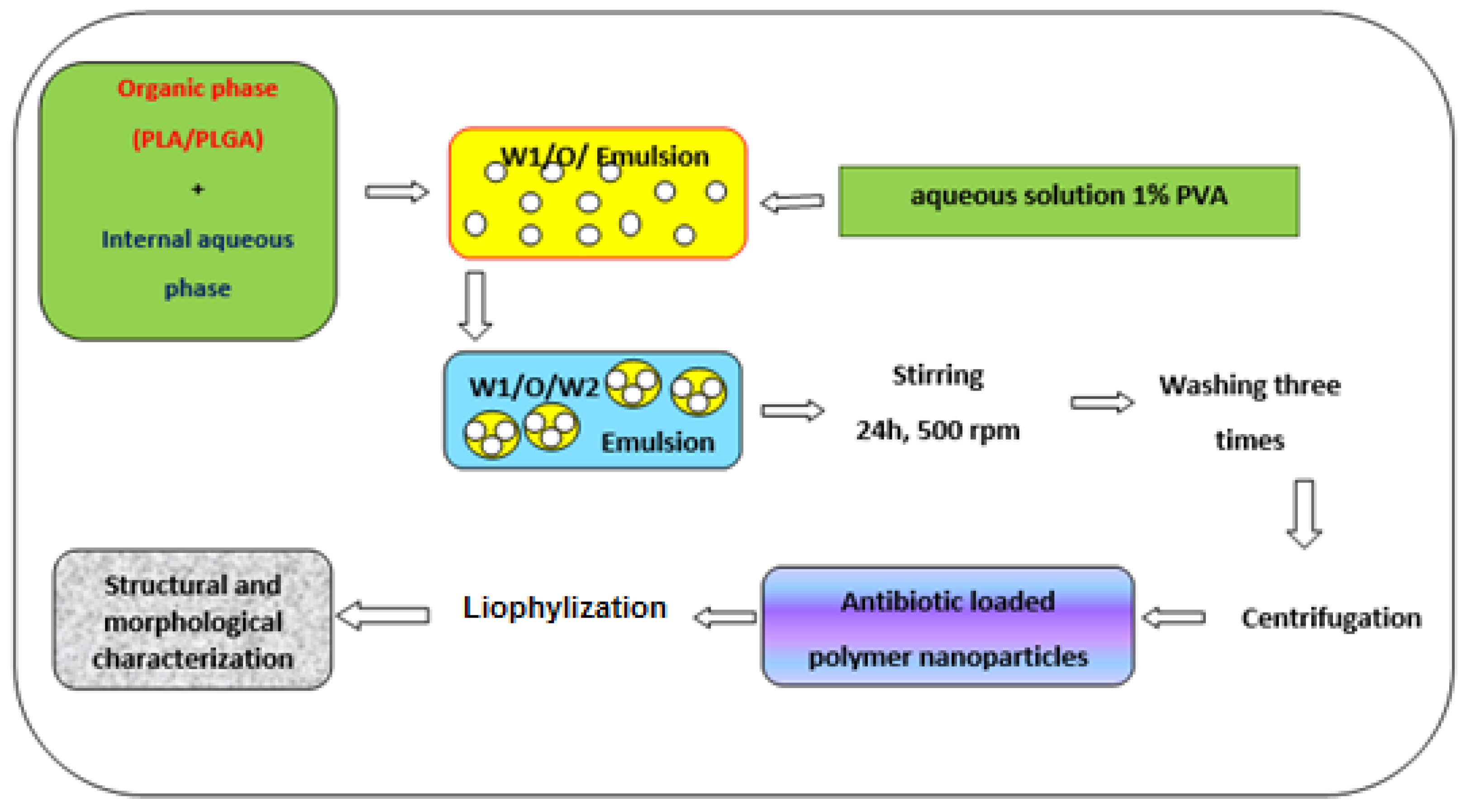
Double emulsion solvent evaporation technique was used to prepare erythromycin-PLA and erythromycin-PLGA nanoparticles. The organic phase contained 0.5 g of PLA and PLGA, each dissolved in 10 mL mixture of dichloromethane and acetone in a volume ratio of 85:15. The internal aqueous phase contained 40 mg erythromycin (in a weight ratio of 1:10 drug-polymer) dissolved in phosphate buffer solution of pH 6.0. Afterward, organic phase and internal aqueous phase were mixed together by ultra-sonication for half a minute under cooling atmosphere to form W1/O emulsion, which was slowly added to an aqueous solution containing 100 mL of 1% PVA under vigorous stirring at 7000 rpm about 10 min. Then, the obtained W1/O/W2 mixture was agitated at 500 rpm for 24 h, for almost entirely organic solvent evaporation. The resulting products were vigorously cleaned using ultrapure water and centrifuged for 20 min at 10,000 rpm, until the nanoparticles formed. Finally, the antibiotic-polymer nanohybrids were lyophilized for further analysis, and the supernatant was stored for assessment of drug content.
The as-prepared erythromycin-polymer nano-scaled particles-type drug delivery systems (
Figure 4) were denoted as Ery-PLA and Ery-PLGA, respectively.
2.3. Characterization Methods
ZetaSizer Nano ZS was used for the detection of aggregates and measurement of small samples permitting the evaluation of particle and molecule size, translational diffusion, electrophoretic mobility, and zeta potential of particles at high and low concentrations. The zeta potential analyzer uses electrophoretic light scattering for particles, molecules and surfaces, measuring range for sizes from 0.3 nm to 10 µm.
The HPLC (high pressure liquid chromatography) method was used in order to determine the amount of the erythromycin from the obtained samples. The stationary phase used a column at 70 °C, and the mobile phase consisted of a mixture of acetonitrile-0.2 M ammonium acetate-methanol-water, in a 450:100:100:350 volume ratio, adjusted at pH 7.0. The UV detection of column effluent was registered at λ = 215 nm. The injected volume was 100 µL for the test solutions consisting of erythromycin propionate-loaded polymers, the reference solution containing standard erythromycin A, and the reference solution containing erythromycin B CRS and erythromycin C CRS, in equal amounts, diluted to 50 mL with hydrolysis solution of dibasic potassium hydrogen phosphate (pH 8.0).
In vitro release study of erythromycin from PLA/PLGA nanoparticles was carried out using a Cary 60 UV-Vis—Agilent spectrometer (Agilent, Santa Clara, CA, USA), into a phosphate buffer solution at a pH value of 7.5. A well-established amount of each sample was suspended in 5 mL PBS in a centrifuge tube and ultrasound for a short time. The samples were then incubated at 37 °C, and, at set times, the samples were centrifuged. The supernatant was then collected, and the nano-powders were reconstituted by the addition of fresh PBS. The percentage of antibiotic release was represented as a function of contact time.
Morphological features of erythromycin-loaded both PLA and PLGA nanoparticles were analyzed by using a TESCAN scanning electron microscope (Brno, Czech Republic).
Another important characterization of drug-loaded polymer is represented by thermal analytical study. The Thermogravimetric Analyzer, Discovery TGA 5500 (TAInstruments, New Castle, DE, USA), monitors the stability of a sample (polymers, organic and inorganic compounds) in terms of weight variation versus time/temperature, in a controlled atmosphere, giving information about temperature (range: room temperature ÷ 1000 °C), evaluation of the degradation temperatures, steps of degradation, and determination of weight loss at a certain temperature under specific conditions.
An Alpha Bruker FT-IR spectrometer (Bremen, Germany), spectral range 4000–400 cm−1, and 4 cm−1 resolution, permits evaluation of interaction ways between the antibiotic and the two polymers.
3. Results
Physical properties analysis was performed by determination of particle size distribution and zeta potential, as shown in
Figure 5 and
Figure 6.
Particle size distribution of PLA and PLGA nanoparticles indicated a mean particle size of approximately 44 nm and −25.2 mv zeta potential, and mean particle size of 180 nm and −33.1 mv for PLGA, respectively.
After loading the antibiotic onto PLA/PLGA matrix, erythromycin-loaded PLA nanoparticles were recorded around 325 nm in size, while drug-loaded PLGA nanoparticles were recorded approximately 258 nm in size, depending on the initial antibiotic concentration. Electro-kinetic potential measured at the exterior side of the colloidal nanostructures indicated zeta potential values of drug-loaded polymer nanoparticles of −11.3 mv for PLA and −17.3mv for PLGA, respectively, which denoted the degree of rejection of similarly charged particles in dispersion. A higher zeta potential for nanoparticles indicates the stability of colloidal dispersions resisting nanoparticles aggregation.
HPLC chromatograms are presented in
Figure 7. It can be seen from the chromatogram of the samples that the retention times of erythromycin are approximately the same as the retention times of erythromycin in the reference chromatogram.
The percentage content of erythromycin A was calculated using the chromatogram shown in
Figure 7a, as a reference, and the percentage content of erythromycin B using the chromatogram shown in
Figure 7b, as a reference. The amount of erythromycin propionate in both samples was expressed as the sum of erythromycin A and erythromycin B and was 99.7% of the initial amount used to prepare antibiotic-loaded nanoparticles for both samples.
We observed (
Figure 8) that drug release from Ery-PLA was slowly more increased than from Ery-PLGA nanostructures. In the case of Ery-PLA, almost 55% of the antibiotic was controlled released within 4 h, and 75% was released within 2 days. In the case of erythromycin-loaded PLGA nanoparticles, approximately 55% of drug was released within 4 h, while 75% was released within 3 days. These results evidenced that the antibiotic encapsulation into PLGA nanoparticles was a little bit slower than into PLA nanoparticles.
Surface morphologies (
Figure 9) exhibited spherical, rigid, and ring-shaped nanoparticles. This panel revealed regular and isolated nanoparticles attributed to lactide content, which prevent aggregates formation.
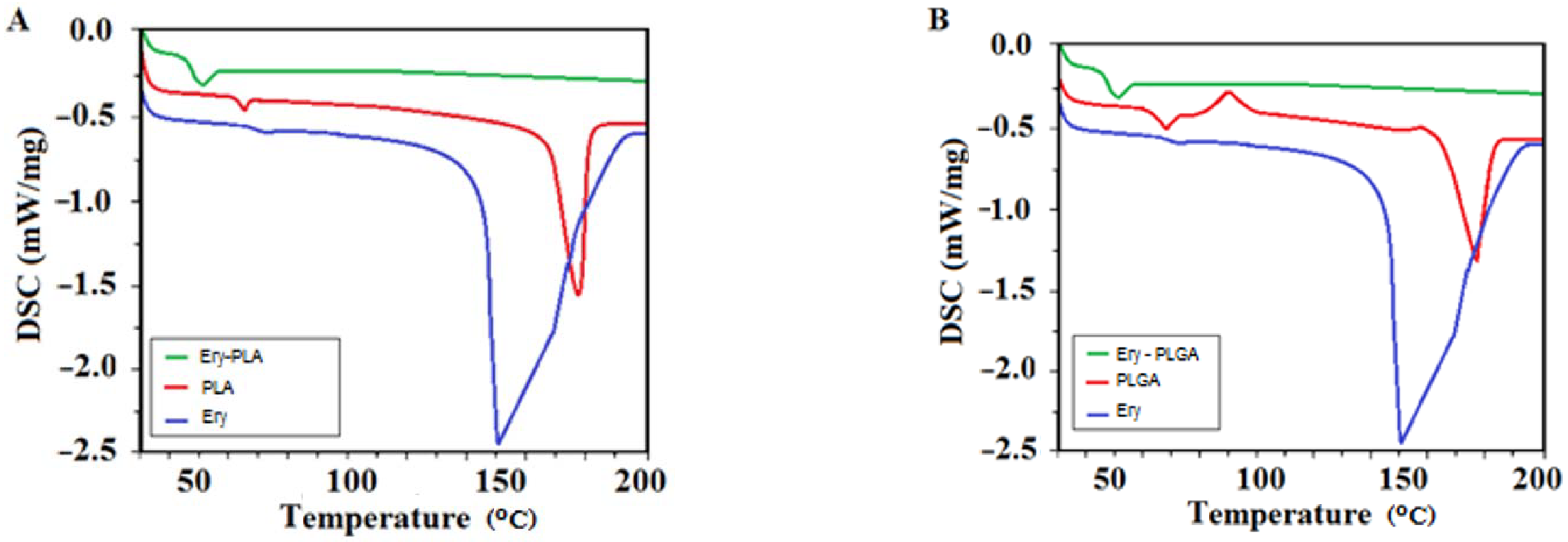
The result of our investigation showed (
Figure 10), in the case of Ery-PLA and Ery-PLGA samples, that the pure drug has an endothermic peak at around 150 °C, assigned to a melting point. The antibiotic melting peak disappeared for both antibiotic encapsulated PLA/PLGA nanoparticles thermographs, denoting the existence of erythromycin and indicating a possible uniform distribution of the drug throughout the host polymer at the molecular stage.
Furthermore, the thermographs also revealed a reduction in both PLA and PLGA transition temperature concerning nano-encapsulation of erythromycin due to increase of particles size, in the case of which glass transition temperature is inversely proportional to the amorphous polymers particle size.
FTIR spectra (
Figure 11) of Ery-PLA and Ery-PLGA nano-architectures revealed chemical structure of drug-loaded polymer nanoparticles and peaks were almost similar.
FTIR data revealed that Ery-PLGA nanoparticles’ peaks were around 3650 cm−1 and 3500 cm−1 due to O-H stretching vibrations, at near 2950 cm−1 and 2990 cm−1 assigned to C-H stretch vibrations, 1760 cm−1 to C=O, 1460 cm−1 attributed to C-H binding, 1390 cm−1 to N=O link, 1180 cm−1 correlated to C-H stretching, 1080 cm−1 to C-O stretch vibrations, and 850 cm−1 and 760 cm−1 attributed to N-H shiver.
Furthermore, characteristic peaks of polymer were noticed, and there was no modification of characteristic peaks, denoting the lack of chemical interaction between erythromycin and polymer nanoparticles.
FTIR data revealed that Ery-PLGA nanoparticles’ peaks at around 3650 cm−1 and 3000 cm−1, attributed to O-H stretching vibrations, at 1760 cm−1 to C=O stretch, at 1460 cm−1 assigned to C-H link, at 1400 cm−1 to N=O binding, 1180 cm−1 and 1090 cm−1 to C-N stretch, and at 860 cm−1 and 750 cm−1 to N-H shiver. Some functional groups were less in number for simple erythromycin, confirming that the drug was not responsible for any interactions with the two polymers.
4. Discussion
In recent years, the administration of the drug after treatment of the infected area has become of major interest [
33,
34] because infections can recur. Recent studies show that poly (lactic acid) (PLA) and poly (lactic-co-glycolic acid) (PLGA) are biocompatible and biodegradable polymers, being a good host for the incorporation of a wide variety of antimicrobial drugs. Antibiotic-loaded chitosan nanoparticles obtained by ionic gelation technique using TPP as a crosslinking factor are of durable stability. Entrapment of erythromycin active molecules within the network of the polymer matrix implies more stable pharmaceutical formulation. This work was concerned with preparing a novel antibiotic carrier using PLA/PLGA nanoparticles loaded with erythromycin using the W/O/W double emulsion solvent evaporation method [
35]. Erythromycin-loaded PLA exhibited 340 nm in size, while drug-loaded PLGA nanoparticles revealed approximately 270 nm in size. Particle size distribution results indicated that the nanoparticle sizes are related to the concentration of the antibiotic used to load PLA/PLGA polymer. Electro-kinetic potential (zeta potential) refers to the difference in electric potential between the electric charge at the surface of a solid nanoparticle in the dispersion and the charge of the diffuse electric layer, denoting the degree of rejection of similarly charged nanoparticles in dispersion. A higher zeta potential for the obtained nano-composites disclosed the stability of colloidal dispersions resisting nanoparticles aggregation. The amount of the erythromycin determined using HPLC analysis was expressed as sum of erythromycin A, erythromycin B, and erythromycin C, resulting in 99.7% of the initial amount used to prepare the nanocomposites [
36]. Antibiotic release profile revealed that erythromycin release from Ery-PLA is faster than from Ery-PLGA nanocomposites. For the Ery-PLA sample, almost 50% of the antibiotic was rereleased within 4 h, and 75% was released within 2 days. For the Ery-PLGA sample, approximately 50% of drug was controlled released within 8 h, while 75% was released within 3 days. As a consequence of these results, antibiotic binding to PLGA was stronger than to PLA polymer, indicating that PLGA-drug can be a better delivery system. Morphological characterization of erythromycin encapsulated PLA and PLGA nanoparticles by scanning electron microscopy displayed spherical, rigid, and ring-shaped nanoparticles, as well as regular and isolated nanoparticles assigned to lactide content, which prevent aggregates formation. A correlation between the smooth surface and the lactide content which confer hydrophobicity to the polymer can be done. Moreover, due to the presence of lactide, the contact between particles becomes weaker, preventing agglomeration. A thermal analytical study indicated, for both samples, that pure drug has an endothermic peak at around 150 °C assigned to a melting point. The erythromycin melting peak disappeared for both antibiotic encapsulated PLA/PLGA nanoparticles thermographs. This evidenced the existence of erythromycin, showing a uniformly distribution of the drug throughout the host polymer. The thermographs also revealed a reduction in both polymers’ transition temperature concerning nano-encapsulation of erythromycin. That was owed to the increase of particles size, in the case of which glass transition temperature is inversely proportional to amorphous polymer’s particles’ size. Thermal analysis of antibiotic-loaded polymer as a drug delivery system is of importance since the processes used to samples preparation are able to modify the organization of the polymer chains [
21]. Vibration spectrum of encapsulated antibiotic involves the type of interaction occurring between the active molecule and polymer due to the vibrations of the atoms’ interaction, which can be modified in frequency and intensity. From FTIR analysis, characteristic peaks of polymer was noticed, and there was no alteration of characteristic peaks by comparison with the spectra of erythromycin and the two neat polymers procured from an electronic spectra library (not shown, being used in another reference by some of the authors), demonstrating the absence of chemical interaction between erythromycin and PLA/PLGA nanoparticles. It can be concluded that the antibiotic is present with no alteration of its structure, without binding other functional groups by hydrogen bond. The two polymers and the antibiotic structure contain a C-O bond. FTIR spectra confirm the lack of interaction of the drug with the PLA and PLGA polymers [
37]. These results suggest that the bactericidal effect of erythromycin-loaded PLA and PLGA nanoparticles would be significantly stronger than the antibiotic-active substance. Obtaining and characterizing antibiotic-loaded polymer nanoparticles was a first step in the development of a new drug-type formulations—PLA/PLGA nanoparticles. The authors also consider the stability study for one year, which will be performed every 3 months, as well as the microbial activity of the samples.


 ,
, .jpg)


