Article

Assessment of Spinal Stability after Discectomy Followed by Annulus Fibrosus Repair and Augmentation of the Nucleus Pulposus: A Finite Element Study

1
Department of Orthopaedics, Shuang Ho Hospital, Taipei Medical University, New Taipei City 23561, Taiwan
2
Department of Orthopaedics, School of Medicine, College of Medicine, Taipei Medical University, Taipei 110, Taiwan
3
Division of Neurosurgery, Department of Surgery, National Taiwan University Hospital, Taipei 10022, Taiwan
4
Department of Orthopaedics, Taichung Tzu Chi Hospital, Taichung 427, Taiwan
5
Graduate Institute of Biomedical Materials and Tissue Engineering, College of Biomedical Engineering, Taipei Medical University, Taipei 110, Taiwan
*
Author to whom correspondence should be addressed.
Appl. Sci. 2022, 12(23), 11906; https://doi.org/10.3390/app122311906
Submission received: 25 October 2022 / Revised: 16 November 2022 / Accepted: 18 November 2022 / Published: 22 November 2022
(This article belongs to the Special Issue Frontiers in Orthopedic Surgery)

Abstract

:
Lumbar disc herniation (LDH) is a common condition which can lead to back pain. Although surgical treatments for LDH are well established, complications such as spinal instability and narrowing of adjacent facet joints are still frequently reported. The purpose of this study was to use finite element models to evaluate the stability of the L3–L4 segment after conservative or aggressive percutaneous transforaminal endoscopic discectomy (PTED) with and without an artificial material filler to correct LDH. Compared to the intact model, aggressive PTED reduced the stability of the segment (increased ROM) and narrowed the space between facet joints in the medial/lateral (ML) direction during flexion (maximum 6.7 degrees change in ROM and 90.5% spacing between facet joints), extension (maximum 2.1 degrees and 38.6%), and axial rotation (maximum 4.2 degrees and 90.1%). Aggressive PTED had a similar effect in the anterior/posterior (AP) direction during lateral bending (maximum 2.0 degrees and 44.2%). Augmenting the nucleus pulposus with a polyurethane filler after aggressive PTED improved spinal stability in both the ML and AP directions in all simulated motions, with results similar to the intact model. However, using a hydrogel filler did little to stabilize the spine, likely because the material is too soft to support the heavy, sustained loading. In conclusion, this study found that if an aggressive discectomy is required, augmenting the nucleus pulposus with a PU filler provides sufficient support to stabilize the spine, while hydrogel fillers offer little support.

1. Introduction

Herniated intervertebral discs are a common cause of pain and restricted mobility in the legs and back, particularly in the lumbar spine. Lumbar disc herniation is most prevalent between the ages of 30 and 60 years old [1]. A herniation occurs when the nucleus pulposus tissue inside the disc pushes on the outer annular fibrosus layer, creating a bulge or tear in the annular layer. The protrusion can press on nerves around the spine and cause considerable pain for the sufferer or a loss of sensation in the lower limbs. Physical therapy, oral steroids, or non-steroidal anti-inflammatory drugs and infiltrations are common methods for the conservative management of symptomatic patients [2,3,4]. However, when such conservative methods are ineffective, surgery is usually required to alleviate pain and other symptoms [5].
Typically, surgery for a herniated disc requires a laminectomy with a discectomy or, in more severe cases, interbody fusion or an artificial disc replacement may be required. Re-herniation of a previously treated disc is not rare after lumbar discectomy [6], especially in patients who were treated with conservative methods, but higher Oswestry disability scores and more severe lower back pain have been reported in patients treated through aggressive resection of the disc [6]. Some clinical studies found that removing more nuclear material from the disc leads to a greater loss in disc height [7,8,9]. In a finite element study, Prado et al. [10] reported increased ROM during rotation movements and a narrowing of adjacent facet joints after discectomy, which could be expected to increase the risk of facet degeneration. Similarly, in a radiological evaluation study, Skomorac et al. [11] found a significant increase in translation at the operated level after discectomy. Such instability of the motion segment may be a consequence of structural changes in the spine from the discectomy procedure, according to clinical and in vitro studies [12,13]. Hence, devices have been introduced that allow a ruptured annulus to be repaired to prevent further degeneration; for cases where the nucleus pulposus has already been ejected from the disc, biomaterials have been developed that can be injected into the disc to fill the vacant space [14,15,16,17]. However, in practice, most of these repair devices do not offer significant benefits to the patient, and many have been withdrawn from the market after reported complications [18].
Filling the cavity in the nucleus pulposus with an artificial material has the potential to restore stability to the affected segments and limit the progressive loss in disc height. There is a variety of biocompatible artificial material fillers commercially available, ranging from soft hydrogels to more rigid PU fillers, with no general consensus on which material type offers the best restorative effect. Hydrogels are quite inert materials that rarely cause immune reactions and have a controllable degradation rate while emitting nontoxic degradation products or final metabolites [19]. On the other hand, PU has a longer history as an effective biomaterial and has been used in nucleus replacement devices with a demonstrated ability to restore multidirectional segmental flexibility similar to the intact condition [20,21]. The nucleus pulposus is composed of a jelly-like material that consists of water and collagen fibers. It is non-homogeneous, so replacing the nucleus pulposus with artificial fillers can have unintended consequences. The structural integrity of the annulus also plays an important role in maintaining the stability of the segment and disc height. The purpose of this study is to evaluate segmental stability in the lumbar spine after limited and aggressive discectomy using finite element analysis. We aimed to test different biomaterials injected into a ruptured intervertebral disc to identify a suitable filler material for restoring segmental stability.

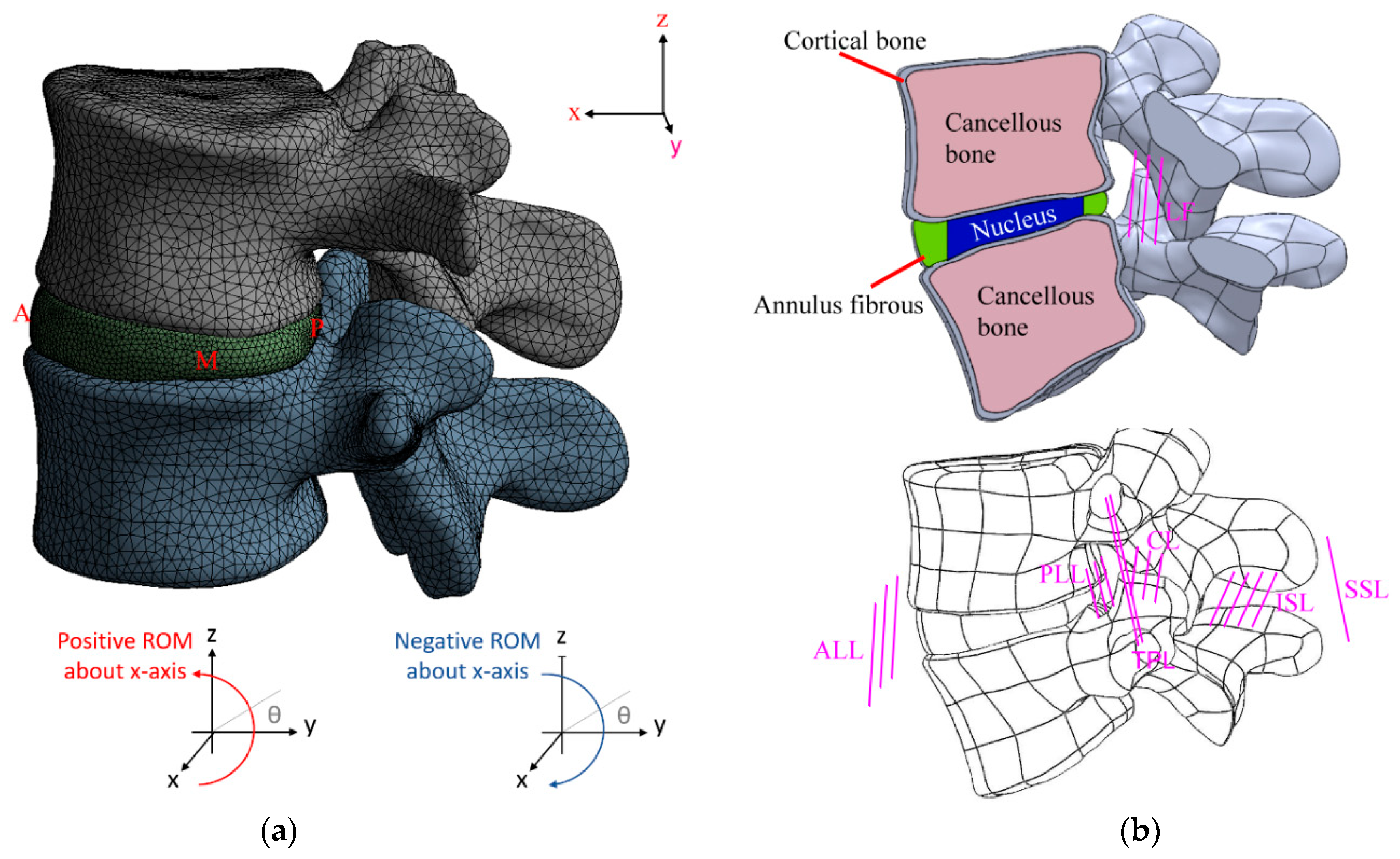
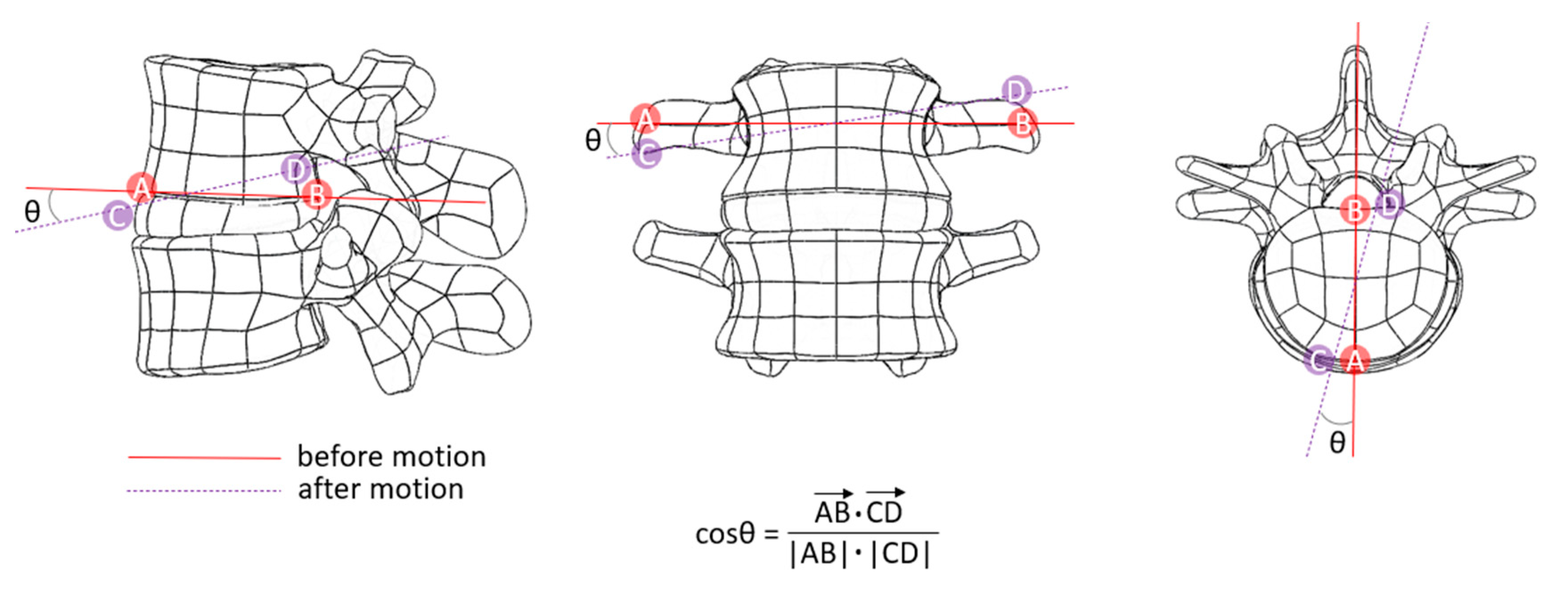
2. Materials and Methods
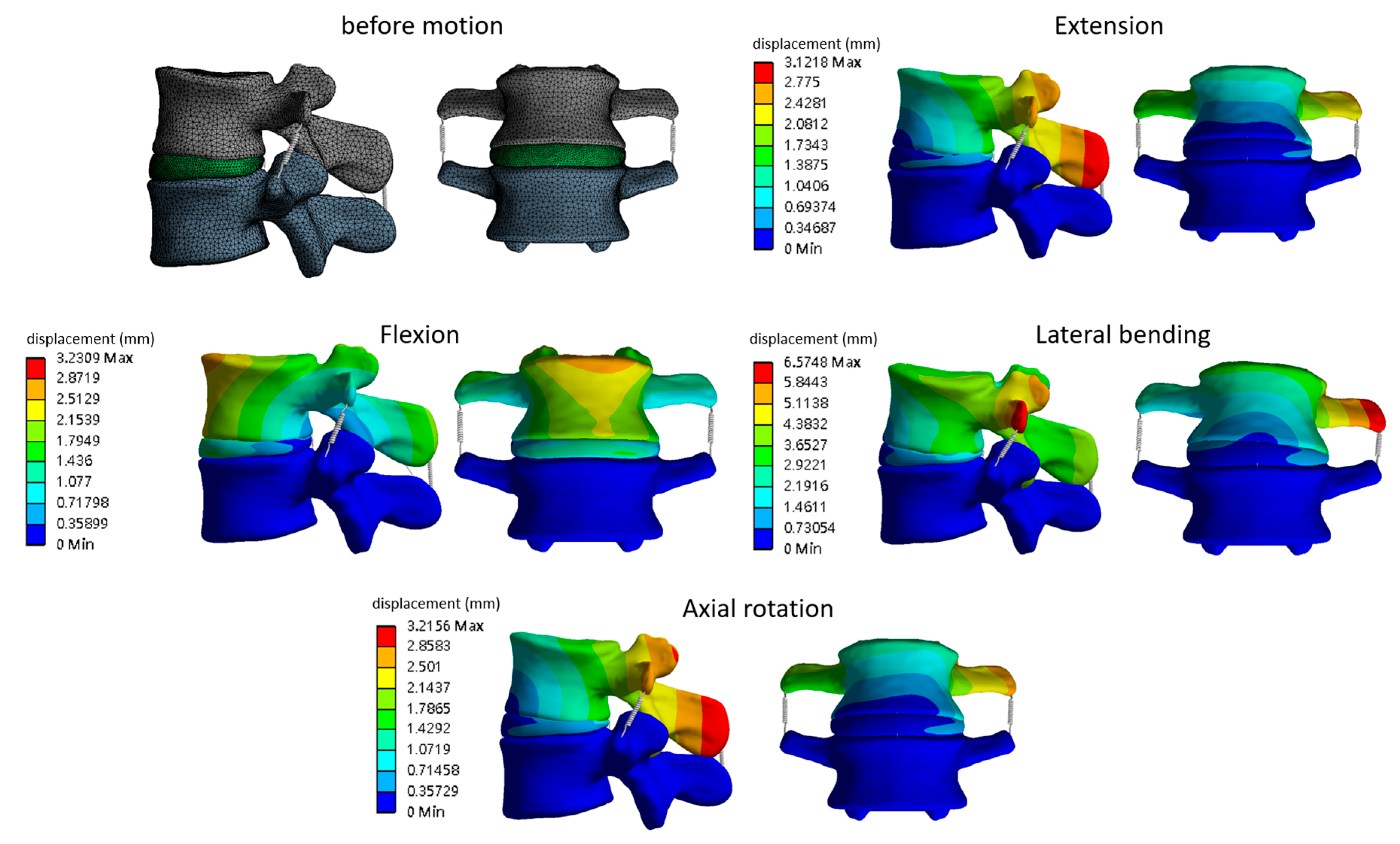
A finite element model of the L3–L4 lumbar spine segment was created using the software ANSYS 14 (ANSYS Inc., Canonsburg, PA, USA). The model (Figure 1a) was developed using geometry from a physiologically accurate spinal model that included intervertebral discs and vertebrae (Zygote Media Group, Inc., American Fork, UT, USA). The material properties of the L3–L4 model shown in Table 1 were sourced from the literature [22,23,24,25]. The model included six ligaments which were represented as 2-node tension-only spring elements (Figure 1b) and were approximated as nonlinear with insertion points approximated to typical anatomy [22]. The model was validated by comparing the range of motion against experimental data from a published in vitro study and finite element analysis results from the literature [25]. An unconstrained pure moment of 7.5 Nm was applied in four directions (flexion, lateral bending, extension, and axial rotation) to the superior endplate of T7, and the inferior endplate of L5 was fixed. The range of motion of the model was recorded and demonstrated to be within the published ranges in the literature [25].
Five finite element models of the L3–L4 segment were developed in this study: (a) healthy condition (Figure 2a); (b) after conservative percutaneous transforaminal endoscopic discectomy (PTED) to correct lumbar disc herniation (LDH) (Figure 2b), with a volume of 581 mm3 removed from the intervertebral disc; (c) after aggressive PTED to correct LDH (Figure 2c), with 2172 mm3 removed from the disc; (d) after aggressive PTED to correct LDH with annulus fibrosus repair (Figure 2d), with 2172 mm3 removed from the disc; and (e) after aggressive PTED with nucleus pulposus augmentation (2172 mm3 removed) to correct LDH (Figure 2e), where the vacated space was filled with a polyurethane elastomer or hydrogel [28].
The material properties of the implants are shown in Table 1. The cortical bone, cancellous bone, and disc were modeled using 8-node solid elements. For the disc, 12 double cross-linked fibrous layers were embedded in the ground substance, and the fiber stiffness increased proportionally from the outermost layer to the innermost layer. The nucleus pulposus was modeled as an incompressible fluid using 8-node fluid elements. Mesh convergence was achieved when an unconstrained pure moment of 7.5 Nm applied to the superior endplate of L3 resulted in a change in displacement of the disc of less than 2%. The resulting model consisted of approximately 290,400 elements and 616,500 nodes.
The interfaces between facet articular surfaces were treated as standard contact pairs at all levels. An unconstrained pure moment was applied to the superior endplate of L3 to simulate the physiological motions of a lumbar spine, which were reported as the maximum voluntary motions in an in vivo study by Schmidt et al. [25] (Table 2). The distal vertebra was restricted from all motion by rigidly anchoring the inferior endplate of L4.
This study assessed spinal stability (range of motion, Figure 3) and changes in disc height and distance between facet joints (FJs) [10] during physiological motions of the lumbar spine. Simulations were conducted on a healthy spine model, after conservative and aggressive PTED to correct LDH, and after aggressive PTED with nucleus pulposus augmentation.

3. Results

The range of motion (ROM) of the L3–L4 lumbar model was obtained for all models when moved in flexion, extension, axial rotation, and lateral bending (Table 3). The distance between facet joints (FJ gap) was calculated as a percentage change from the intact model. The results from models c and d show that aggressive PTED reduced the stability of the spine and narrowed the FJ gap in the medial/lateral (ML) direction during flexion (maximum 6.7 degrees and 90.5%), extension (maximum 2.1 degrees and 38.6%), and axial rotation (maximum 4.2 degrees and 90.1%). Aggressive PTED also affected the stability and reduced the FJ gap in the anterior/posterior (AP) direction during lateral bending (maximum 2.0 degrees and 44.2%). Conservative PTED (model b) had a lesser effect on the range of motion and spacing between facet joints.
Following PU augmentation after aggressive PTED (models d and e), the spine was more stable in the ML and AP directions than in models c and d, with the results being close to the intact model. However, augmenting the nucleus with hydrogel did not have the same effect, and there was little difference from the PTED-only models.
Figure 4 shows the change in height of the intervertebral disc as a percentage of the intact model (model a, INT) at the anterior (A), lateral medial (M), and posterior (P) regions of the disc. Both conservative and aggressive PTED reduced the disc height on the lateral medial side of the disc in all motions. In flexion, PU augmentation increased the disc height on the anterior and lateral medial side (increased 27.7% and 97.4% as compared to INT), but this result was not replicated with the hydrogel, which caused an almost fivefold reduction in disc height from the intact model. A similar result was achieved in extension, with hydrogel augmentation resulting in a 2.3-fold reduction in disc height.
In axial rotation and lateral bending, although PU augmentation showed an increase in disc height at the anterior (A), medial (M), and posterior (P) sites, the values were still less than those of the intact state without loadings. Hydrogel augmentation did not restore the disc height but caused less of a reduction than aggressive PTED alone. Figure 5 shows the displacement on the finite element model (model b) when subjected to different motion conditions. The lateral bending condition resulted in the greatest displacement on the model.

4. Discussion

Discectomy is a standard surgical procedure for treating herniated intervertebral discs. However, studies have shown that discectomy can compromise the integrity of the disc and increase the risk of degenerative changes [30,31]. Intervertebral discs are a key component of the spine that allow for movement between adjacent vertebrae while also acting as shock absorbers for transmitting loads. Hence, structural changes in a disc can disrupt its normal functionality and accelerate degeneration, leading to secondary back pain. Segmental instability after discectomy has been attributed to damage to the annulus that has not been adequately repaired [12,13]. Previous studies on the lumbar spine, including clinical assessments and finite element analysis [10,11,23], only recorded the segmental ROM in one direction (x-axis, y-axis, or z-axis). Furthermore, in cases of discectomy, no studies to date have evaluated differences in the stability of the operated segment after the nucleus was partially replaced with a PU or hydrogel filler. The purpose of this study was to assess changes in the stability of the lumbar spine after limited and aggressive discectomy that was repaired using different methods and filler materials.
The results showed increased lateral bending in all discectomy models for each of the simulated movements, which could cause nerve impingement or narrowing of the foramen. The vacated space in the nucleus pulposus after discectomy causes the superior vertebra to sink under axial loading, leading to a slight leaning towards the site of the injury. The aggressive discectomy models without material filler (groups c and d) showed greater lateral bending than the conservative discectomy model (group b). Filling the vacated space with either PU or hydrogel reduced the severity of the lateral bending, with the PU material showing the greatest reduction in asymmetrical bending of all aggressive surgical models.
In flexion/extension, the ROM and FJ gap after aggressive discectomy (group c) and aggressive discectomy with annular repair (group d) were approximately 10% greater than in the intact model. Groups c and d also showed a greater ROM than other groups, but with a narrower FJ gap. With aggressive discectomy, about 40% of the whole nucleus was removed. The remaining tissue could not provide enough resistance to maintain a normal disc height, which led to excessive flexion/extension and a narrowing of the FJ gap. In an in vitro experiment and FEM study by Stadelmann et al. [32], the ROM of the spinal segment was found to increase after the nucleus pulposus was removed. Similarly, Wilke et al. [33] reported a reduction in disc height and an 18% increase in ROM in flexion/extension after the nucleus pulposus was removed. After aggressive discectomy, replenishing the empty space with an artificial filler stabilized the spinal segment and reduced the flexion/extension angle. However, the PU material (group e) was better able to restore the FJ gap that the softer hydrogel filler (group f). For group d, repairing the injury to the annulus after aggressive discectomy only offered a slight improvement in extension motion control, which was not sufficient to compensate for the reduction in initial stiffness after aggressive discectomy. Interestingly, conservative discectomy (group b) and aggressive discectomy with PU material (group e) reduced the ROM in flexion to less than in the intact model, which is likely due to the change in disc height. The results for groups b and e showed less sinking of the anterior disc and less rising of the posterior disc than in the intact group during flexion, which resulted in a lower flexion angle. A potential cause is that the posterior part of the disc (empty space or filler material) could not create an effective rising force on the posterior region to counteract the push force from the anterior region of the disc, which limited the flexion angle.
There was little difference in the ROM between all groups when a rotation moment was applied, but the ROM did increase for the bending motion in groups b and c. When the disc was augmented with PU or hydrogel, the bending motion was still greater, but to a lesser degree. In an in vitro study, Goel et al. [34] reported little change in the rotational ROM after conservative discectomy or aggressive discectomy, but also did not record multi-axial translations under an axial rotation moment. Goel found that the rotational ROM only increased after total discectomy, which is supported by Huang et al. [35], who used finite element analysis to show that the range of axial rotation similarly increased after total discectomy. The results for rotational ROM in this current study were similar to Goel et al. [34] but had an additional increased bending motion after aggressive discectomy. In the lateral bending condition, groups b, c, d, and f showed a higher bending ROM and greater extension than the intact model. Filling the cavity in the nucleus with PU acted to reduce bending and extension movements and stabilize the spine. In contrast, using hydrogel only offered a slight improvement in bending and extension control. Previous studies have indicated that the softer nature of hydrogel fillings mean that the augmented disc cannot support as much load, limiting the stability of the spinal segment [36].
There are some limitations to this study. First, the material characteristics of the entire model were assumed to be isotropic and homogenous, and the effects of muscle contraction were not considered. This is not truly representative of the physiological state of the human spine. Second, only the L3–L4 spinal segment was simulated, so the effect of adjacent vertebrae was not captured. Also, the discectomy type and properties of the material filler were simplified in each group. This study is limited by the finite element models not being capable of evaluating the time-dependent behavior of the materials, but instead the models were built to accurately estimate reactions to multi-axial motions.

5. Conclusions

This study found that discectomy can increase the ROM of the operative segment in lateral bending, and this change was shown to be partially reversed when the nucleus pulposus was augmented with a PU filler. Inserting a PU filler into the vacated space in the disc can improve the stability of the spine after discectomy and reduce the risk of developing degenerative disc disease. Finite element simulations of such procedures and biomaterials allow for optimal approaches to be developed to improve clinical outcomes after implantation.

Author Contributions

Conceptualization, L.-C.W., Y.-Y.H. and C.-J.C.; methodology, C.-J.C., Y.-F.C. and Y.-Y.H.; project administration, L.-C.W.; resources, F.-Y.T. and L.-C.W.; validation, F.-Y.T. and Y.-F.C.; writing—original draft, C.-J.C.; writing—review and editing, L.-C.W., Y.-Y.H. and C.-J.C. All authors have read and agreed to the published version of the manuscript.

Funding

This research received no external funding.

Institutional Review Board Statement

Not applicable.

Informed Consent Statement

Not applicable.

Conflicts of Interest

The authors declare no conflict of interest.

References

  1. Fjeld, O.R.; Grøvle, L.; Helgeland, J.; Småstuen, M.C.; Solberg, T.K.; Zwart, J.A.; Grotle, M. Complications, Reoperations, Readmissions, and Length of Hospital Stay in 34,639 Surgical Cases of Lumbar Disc Herniation. Bone Joint J. 2019, 101-B, 470–477. [Google Scholar] [CrossRef] [PubMed]
  2. Gugliotta, M.; da Costa, B.R.; Dabis, E.; Theiler, R.; Jüni, P.; Reichenbach, S.; Landolt, H.; Hasler, P. Surgical versus Conservative Treatment for Lumbar Disc Herniation: A Prospective Cohort Study. BMJ Open 2016, 6, e012938. [Google Scholar] [CrossRef] [Green Version]
  3. Demirel, A.; Yorubulut, M.; Ergun, N. Regression of Lumbar Disc Herniation by Physiotherapy. Does Non-Surgical Spinal Decompression Therapy Make a Difference? Double-Blind Randomized Controlled Trial. J. Back Musculoskelet Rehabil. 2017, 30, 1015–1022. [Google Scholar] [CrossRef] [PubMed]
  4. Kim, C.H.; Choi, Y.; Chung, C.K.; Kim, K.J.; Shin, D.A.; Park, Y.K.; Kwon, W.K.; Yang, S.H.; Lee, C.H.; Park, S.B.; et al. Nonsurgical Treatment Outcomes for Surgical Candidates with Lumbar Disc Herniation: A Comprehensive Cohort Study. Sci. Rep. 2021, 11, 3931. [Google Scholar] [CrossRef] [PubMed]
  5. Harper, R.; Klineberg, E. The Evidence-Based Approach for Surgical Complications in the Treatment of Lumbar Disc Herniation. Int. Orthop. 2019, 43, 975–980. [Google Scholar] [CrossRef]
  6. Carragee, E.J.; Spinnickie, A.O.; Alamin, T.F.; Paragioudakis, S. A Prospective Controlled Study of Limited versus Subtotal Posterior Discectomy: Short-Term Outcomes in Patients with Herniated Lumbar Intervertebral Discs and Large Posterior Anular Defect. Spine 2006, 31, 653–657. [Google Scholar] [CrossRef] [PubMed]
  7. McGirt, M.J.; Eustacchio, S.; Varga, P.; Vilendecic, M.; Trummer, M.; Gorensek, M.; Ledic, D.; Carragee, E.J. A Prospective Cohort Study of Close Interval Computed Tomography and Magnetic Resonance Imaging after Primary Lumbar Discectomy: Factors Associated with Recurrent Disc Herniation and Disc Height Loss. Spine 2009, 34, 2044–2051. [Google Scholar] [CrossRef] [PubMed]
  8. Lequin, M.B.; Barth, M.; Thomė, C.; Bouma, G.J. Primary Limited Lumbar Discectomy with an Annulus Closure Device: One-Year Clinical and Radiographic Results from a Prospective, Multi-Center Study. Korean J. Spine 2012, 9, 340. [Google Scholar] [CrossRef] [PubMed]
  9. Ledic, D.; Vukas, D.; Grahovac, G.; Barth, M.; Bouma, G.J.; Vilendecic, M. Effect of Anular Closure on Disk Height Maintenance and Reoperated Recurrent Herniation Following Lumbar Diskectomy: Two-Year Data. J. Neurol. Surg. A Cent Eur. Neurosurg. 2015, 76, 211–218. [Google Scholar] [CrossRef] [PubMed]
  10. Prado, M.; Mascoli, C.; Giambini, H. Discectomy Decreases Facet Joint Distance and Increases the Instability of the Spine: A Finite Element Study. Comput. Biol. Med. 2022, 143, 105278. [Google Scholar] [CrossRef] [PubMed]
  11. Skomorac, R.; Delić, J.; Bečulić, H.; Jusić, A. Radiological evaluation of lumbosacral spine for post discectomy segmental instability. Medicinski Glasnik 2016, 13, 142–147. [Google Scholar] [PubMed]
  12. Tibrewal, S.; Pearcy, M.; Portek, I.; Spivey, J. A Prospective Study of Lumbar Spinal Movements before and after Discectomy Using Biplanar Radiography. Correlation of Clinical and Radiographic Findings. Spine 1985, 10, 455–460. [Google Scholar] [CrossRef] [PubMed]
  13. Hou, S.; Tu, K.; Xu, Y.; Zhang, W.; Wang, H.; Wang, D. Effect of Partial Discectomy on the Stability of the Lumbar Spine. A Study of Kinematics. Chin. Med. J. 1990, 103, 396–399. [Google Scholar]
  14. Guardado, A.A.; Baker, A.; Weightman, A.; Hoyland, J.A.; Cooper, G. Lumbar Intervertebral Disc Herniation: Annular Closure Devices and Key Design Requirements. Bioengineering 2022, 9, 47. [Google Scholar] [CrossRef] [PubMed]
  15. Cho, P.G.; Shin, D.A.; Park, S.H.; Ji, G.Y. Efficacy of a Novel Annular Closure Device after Lumbar Discectomy in Korean Patients: A 24-Month Follow-Up of a Randomized Controlled Trial. J. Korean Neurosurg. Soc. 2019, 62, 691–699. [Google Scholar] [CrossRef] [Green Version]
  16. Bailey, A.; Araghi, A.; Blumenthal, S.; Huffmon, G.v. Prospective, Multicenter, Randomized, Controlled Study of Anular Repair in Lumbar Discectomy: Two-Year Follow-Up. Spine 2013, 38, 1161–1169. [Google Scholar] [CrossRef]
  17. Berlemann, U.; Schwarzenbach, O. An Injectable Nucleus Replacement as an Adjunct to Microdiscectomy: 2 Year Follow-up in a Pilot Clinical Study. Eur. Spine J. 2009, 18, 1706–1712. [Google Scholar] [CrossRef] [PubMed] [Green Version]
  18. Bartlett, A.; Wales, L.; Houfburg, R.; Durfee, W.K.; Griffith, S.L.; Bentley, I. Optimizing the Effectiveness of a Mechanical Suture-Based Anulus Fibrosus Repair Construct in an Acute Failure Laboratory Simulation. J. Spinal Disord. Tech. 2013, 26, 393–399. [Google Scholar] [CrossRef] [PubMed]
  19. Qi, C.; Yan, X.; Huang, C.; Melerzanov, A.; Du, Y. Biomaterials as carrier, barrier and reactor for cell-based regenerative medicine. Protein Cell 2015, 6, 638–653. [Google Scholar] [CrossRef] [PubMed] [Green Version]
  20. Tsantrizos, A.; Ordway, N.R.; Myint, K.; Martz, E.; Yuan, H.A. Mechanical and biomechanical characterization of a polyurethane nucleus replacement device injected and cured in situ within a balloon. SAS J. 2008, 2, 28–39. [Google Scholar] [CrossRef] [Green Version]
  21. Vanaclocha-Saiz, A.; Vanaclocha, V.; Atienza, C.M.; Clavel, P.; Jorda-Gomez, P.; Barrios, C.; Vanaclocha, L. Finite Element Analysis of a Bionate Ring-Shaped Customized Lumbar Disc Nucleus Prosthesis. ACS Appl. Bio Mater. 2022, 5, 172–182. [Google Scholar] [CrossRef] [PubMed]
  22. Wu, Y.; Chen, C.H.; Tsuang, F.Y.; Lin, Y.C.; Chiang, C.J.; Kuo, Y.J. The Stability of Long-Segment and Short-Segment Fixation for Treating Severe Burst Fractures at the Thoracolumbar Junction in Osteoporotic Bone: A Finite Element Analysis. PLoS ONE 2019, 14, e0211676. [Google Scholar] [CrossRef] [Green Version]
  23. Goel, V.K.; Monroe, B.T.; Gilbertson, L.G.; Brinckmann, P. Interlaminar Shear Stresses and Laminae Separation in a Disc: Finite Element Analysis of the L3-L4 Motion Segment Subjected to Axial Compressive Loads. Spine 1995, 20, 689–698. [Google Scholar] [CrossRef] [PubMed]
  24. Morgan, E.F.; Bayraktar, H.H.; Keaveny, T.M. Trabecular Bone Modulus-Density Relationships Depend on Anatomic Site. J. Biomech. 2003, 36, 897–904. [Google Scholar] [CrossRef]
  25. Schmidt, H.; Heuer, F.; Drumm, J.; Klezl, Z.; Claes, L.; Wilke, H.J. Application of a Calibration Method Provides More Realistic Results for a Finite Element Model of a Lumbar Spinal Segment. Clin. Biomech. 2007, 22, 377–384. [Google Scholar] [CrossRef]
  26. Kanyanta, V.; Ivankovic, A. Mechanical Characterisation of Polyurethane Elastomer for Biomedical Applications. J. Mech. Behav. Biomed. Mater. 2010, 3, 51–62. [Google Scholar] [CrossRef]
  27. Silva, P.; Crozier, S.; Veidt, M.; Pearcy, M.J. An Experimental and Finite Element Poroelastic Creep Response Analysis of an Intervertebral Hydrogel Disc Model in Axial Compression. J. Mater. Sci. Mater. Med. 2005, 16, 663–669. [Google Scholar] [CrossRef] [PubMed]
  28. Chiang, C.J.; Cheng, C.K.; Sun, J.S.; Liao, C.J.; Wang, Y.H.; Tsuang, Y.H. The Effect of a New Anular Repair after Discectomy in Intervertebral Disc Degeneration: An Experimental Study Using a Porcine Spine Model. Spine 2011, 36, 761–769. [Google Scholar] [CrossRef] [PubMed]
  29. Dreischarf, M.; Zander, T.; Shirazi-Adl, A.; Puttlitz, C.M.; Adam, C.J.; Chen, C.S.; Goel, V.K.; Kiapour, A.; Kim, Y.H.; Labus, K.M.; et al. Comparison of Eight Published Static Finite Element Models of the Intact Lumbar Spine: Predictive Power of Models Improves When Combined Together. J. Biomech. 2014, 47, 1757–1766. [Google Scholar] [CrossRef] [PubMed] [Green Version]
  30. Illien-Jünger, S.; Lu, Y.; Purmessur, D.; Mayer, J.E.; Walter, B.A.; Roughley, P.J.; Qureshi, S.A.; Hecht, A.C.; Iatridis, J.C. Detrimental Effects of Discectomy on Intervertebral Disc Biology Can Be Decelerated by Growth Factor Treatment during Surgery: A Large Animal Organ Culture Model. Spine J. 2014, 14, 2724–2732. [Google Scholar] [CrossRef] [PubMed] [Green Version]
  31. Mariconda, M.; Galasso, O.; Attingenti, P.; Federico, G.; Milano, C. Frequency and Clinical Meaning of Long-Term Degenerative Changes after Lumbar Discectomy Visualized on Imaging Tests. Eur. Spine J. 2009, 19, 136–143. [Google Scholar] [CrossRef] [PubMed]
  32. Stadelmann, M.A.; Stocker, R.; Maquer, G.; Hoppe, S.; Vermathen, P.; Alkalay, R.N.; Zysset, P.K. Finite Element Models Can Reproduce the Effect of Nucleotomy on the Multi-Axial Compliance of Human Intervertebral Discs. Comput. Methods Biomech. Biomed. Eng. 2020, 23, 934–944. [Google Scholar] [CrossRef]
  33. Wilke, H.J.; Kavanagh, S.; Neller, S.; Haid, C.; Claes, L.E. Effect of a Prosthetic Disc Nucleus on the Mobility and Disc Height of the L4–5 Intervertebral Disc Postnucleotomy. J. Neurosurg. Spine 2001, 95, 208–214. [Google Scholar] [CrossRef] [PubMed]
  34. Goel, V.; Goyal, S.; Clark, C.; Nishiyama, K.; Nye, T. Kinematics of the Whole Lumbar Spine: Effect of Discectomy. Spine 1985, 10, 543–554. [Google Scholar] [CrossRef] [PubMed]
  35. Huang, J.; Yan, H.; Jian, F.; Wang, X.; Li, H. Numerical Analysis of the Influence of Nucleus Pulposus Removal on the Biomechanical Behavior of a Lumbar Motion Segment. Comput. Methods Biomech. Biomed. Eng. 2015, 18, 1516–1524. [Google Scholar] [CrossRef]
  36. Cannella, M.; Isaacs, J.L.; Allen, S.; Orana, A.; Vresilovic, E.; Marcolongo, M. Nucleus Implantation: The Biomechanics of Augmentation versus Replacement with Varying Degrees of Nucleotomy. J. Biomech. Eng. 2014, 136, 051001. [Google Scholar] [CrossRef]
Figure 1. L3–L4 spine segment model: (a) finite element mesh; (b) model containing supraspinous ligament (SSL), interspinous ligament (ISL), capsular ligaments (CL), transverse process ligaments (TPL), posterior longitudinal ligament (PLL), anterior longitudinal ligament (ALL), and ligamentum flavum (LF).
Applsci 12 11906 g001
Figure 2. Five finite element models developed in this study: (a) healthy spinal segment, (b) IV disc after conservative PTED, (c) after aggressive PTED, (d) after aggressive PTED with annular repair, (e) after aggressive PTED with nucleus pulposus augmentation.
Applsci 12 11906 g002
Figure 3. Method to determining the ROM (θ represents the ROM).
Applsci 12 11906 g003
Figure 4. Change in intervertebral disc height at the anterior (A), posterior (P), and medial (M) sides of the disc as a percentage of the intact model (model a, INT) for (a) flexion, (b) extension, (c) axial rotation, and (d) lateral bending motions.
Applsci 12 11906 g004
Figure 5. Displacement on the finite element model (model b) during different motions.
Applsci 12 11906 g005
Table 1. Material properties of lumbar spine model.
PropertyModulus (MPa)νReferences
Cortical bone12,0000.2Goel et al., 1995 [23]
Cancellous bone3000.2Morgan et al., 2003 [24]
Annulus fibrosusMooney-Rivlin
c1 = 0.18, c2 = 0.045
NASchmidt et al., 2007 [25]
Nucleus pulposusMooney-Rivlin
c1 = 0.12, c2 = 0.03
NA
LigamentsHyperelasticNA
Polyurethane elastomer4.7 MPa0.499Kanyanta et al. [26]
Hydrogel0.45 MPa0.2Silva et al. [27]
NA = not applicable.
Table 2. Loading conditions for simulated motions of a lumbar spine [29].
Body PositionCompressive Force (N)Moment (Nm)
Flexion11757.5
Extension5007.5
Lateral bending7007.8
Axial rotation7205.5
Table 3. ROM and FJ gap in each model when moved through different motions.
Body PositionFlexion
ROM about the
x-Axis (Degree), ML Direction [% #]y-Axis (Degree), AP Direction [% #]z-Axis (Degree) [% #]FJ Gap *
model a (intact)−0.7
[undefined]
3.6
[−21.7%]
0.0
[0%]
-
model b (conservative)−6.7
[undefined]
5.1
[10.9%]
0.0
[0%]
−11.5%
model c (aggressive)−6.7
[undefined]
5.1
[10.9%]
0.0
[0%]
−90.5%
model d (aggressive with annular repair)1.1
[undefined]
3.4
[−26.1%]
0.2
[undefined]
−89.4%
model e (PU)−4.5
[undefined]
4.6
[0%]
0.3
[undefined]
−40.5%
model e (Hydrogel)−0.7
[undefined]
3.6
[−21.7%]
0.0
[0%]
−73.1%
Body PositionExtension
x-Axis (Degree), ML Direction [% #]y-Axis (Degree), AP Direction [% #]z-Axis (Degree)
[% #]
FJ Gap *
model a (intact)0.0−2.40.0-
model b (conservative)−0.5
[undefined]
−1.3
[45.8%]
0.0
[0%]
−13.1%
model c (aggressive)−2.1
[undefined]
−2.1
[12.5%]
0.0
[0%]
−38.6%
model d (aggressive with annular repair)−2.1
[undefined]
−2.0
[16.7%]
0.0
[0%]
−37.8%
model e (PU)0.9
[undefined]
−1.4
[41.7%]
0.2
[undefined]
−48.0%
model e (Hydrogel)−1.5
[undefined]
−1.8
[25%]
0.3
[undefined]
−50.5%
Body PositionAxial Rotation
x-Axis (Degree), ML Direction
[% #]
y-Axis (Degree), AP Direction
[% #]
z-Axis (Degree)
[% #]
FJ Gap *
model a (intact)0.00.1−1.5-
model b (conservative)−0.7
[undefined]
−1.3
[1400%]
−1.6
[−6.7%]
−12.3%
model c (aggressive)−4.2
[undefined]
−0.8
[−900%]
−1.6
[−6.7%]
−90.1%
model d (aggressive with annular repair)−4.2
[undefined]
−0.8
[−900%]
−1.6
[−6.7%]
−89.0%
model e (PU)0.8
[undefined]
0.0
[−100%]
−1.4
[6.7%]
−84.5%
model e (Hydrogel)−2.6
[undefined]
−1.0
[−1100%]
−1.2
[20%]
−88.9%
Body PositionLateral Bending
x-Axis (Degree), ML Direction
[% #]
y-Axis (Degree), AP Direction
[% #]
z-Axis (Degree)
[% #]
FJ Gap *
model a (intact)−3.3−0.2−1.2-
model b (conservative)−4.6
[−39.4%]
−1.7
[−750%]
−1.2
[0%]
−13.1%
model c (aggressive)−6.4
[−93.9%]
−2.0
[−900%]
−1.2
[0%]
−44.2%
model d (aggressive with annular repair)−6.4
[−93.9%]
−2.0
[−900%]
−1.2
[0%]
−43.3%
model e (PU)−1.2
[63.6%]
0.2
[200%]
−1.0
[16.7%]
−27.7%
model e (Hydrogel)−5.4
[−63.6%]
−1.7
[−750%]
−0.9
[25%]
−42.6%
* FJ gap: as compared to INT. # ROM: as compared to intact.
Publisher’s Note: MDPI stays neutral with regard to jurisdictional claims in published maps and institutional affiliations.

