Article

Reciprocating System for Secondary Root Canal Treatment of Oval Canals: CBCT, X-rays for Remnant Detection and Their Identification with ESEM and EDX

1
Endodontic Clinical Section, School of Dentistry, DIBINEM, University of Bologna, 40125 Bologna, Italy
2
Laboratory of Green Biomaterials and Oral Pathology, School of Dentistry, DIBINEM, University of Bologna, 40125 Bologna, Italy
3
Laboratory of Dental Radiology, School of Dentistry, DIBINEM, University of Bologna, 40125 Bologna, Italy
*
Author to whom correspondence should be addressed.
Appl. Sci. 2022, 12(22), 11671; https://doi.org/10.3390/app122211671
Submission received: 14 October 2022 / Revised: 11 November 2022 / Accepted: 12 November 2022 / Published: 17 November 2022
(This article belongs to the Special Issue Innovative Techniques in Endodontics)

Abstract

:
Aim of the study: to evaluate root filling remnants after secondary root canal treatments (SRCTs) of oval-shaped canals with X-rays and cone beam computed tomography (CBCT). The SRCTs were performed using reciprocating NiTi instruments. Methods: Single-rooted teeth (N = 64) were randomly treated with Reciproc Blue (RB) and filled with AH Plus/single cone (SC group) or AH Plus/Guttafusion (GF group). After seven days of storage in HBSS (Hanks balanced salt solution), Gates Glidden burs #2/3 and RB #25 and #40 were used for the SRCTs. The time to complete the procedure was measured. X-rays and CBCT were used to calculate, respectively, the area and the volume occupied by the remnants in the coronal, middle, and apical thirds of each canal. Environmental scanning electron microscopy (ESEM) and energy dispersive X-ray spectroscopy (EDX) were used for qualitative evaluation and morphology composition of the remnants in sectioned roots. A statistical analysis was performed using Sigma Plot (version 13, IBM, Armonk, NY, USA). The study was designed according to PRILE guidelines. Results: After the SRCTs, the middle thirds of the root canals showed the presence of remnants in both groups, as demonstrated by X-rays and CBCT. The GF group showed a statistically significant higher volume of remnants than the SC Group only in the middle third. The ESEM supported by the EDX revealed the remnant composition by the detection of trace elements of sealer and gutta-percha in all root canals. Conclusion: The study demonstrated that the middle third of root canals is a critical region where remnants were packed and spread in the buccal-lingual sides of canals. ESEM-EDX detected a fine layer of filling remnants in all root thirds, suggesting a larger canal contamination than the X-rays and CBCT examinations revealed.

1. Introduction

The main goal of secondary root canal treatments is the removal of contaminated remnants and microorganisms that are responsible of periapical disease and microbial infection [1,2,3]. Unshaped and uncleaned root canal areas after SRCTs potentially hide residual bacterial biofilm and could be the cause of persistent endodontic infections [4,5,6].
Over the last few years, several techniques and instruments specifically designed for gutta-percha removal have been proposed, but none were able to completely remove all remnants [7,8,9,10].
The shaping and cleaning ability of the endodontic instruments can also be directly influenced by anatomical complexities [11]. The oval configuration of the canal still represents a clinical challenge [12,13]. The buccal-lingual areas of oval root canals are difficult to shape with flexible NiTi instruments in continuous rotation. An ineffective instrumentation leads to the incomplete removal of the smear layer and root canal remnants of previous treatments. As a consequence, insufficient root canal disinfection and a low-quality obturation are obtained, hindering long-term root canal success [11].
Reciprocating motion was introduced some years ago for root canal treatments. This movement allows us to overcome some of the limits of traditional NiTi instruments and improve their mechanical properties [14].
The clinical criteria to determine the quality of SRCTs are the absence of materials along the visible root canal walls and along the spires of the instrument, the absence of remnants during the irrigation phases, and the tactile perception of smooth canal walls. Periapical X-rays are the most common method to assess the presence of remnants [15], but they potentially might not identify all remnants in retreatment procedures [16]. More recently, CBCT has also been used to allow an accurate 3D evaluation of the remnants [17]. Environmental scanning electron microscopy connected to energy dispersive X-ray spectroscopy (ESEM-EDX) allows us to observe wet samples without any preparation or metallic coating [10].
New carrier-based systems are more frequently used to fill the root canal in association with epoxy-based sealers [18]. These systems are faster and easier to use for filling root canals [19]. In this in vitro study, the efficacy of Reciproc Blue to remove remnants from oval-shaped canals filled with two different systems was evaluated with phosphor plate X-rays and CBCT. ESEM-EDX was used to analyze the morphology of the remnants and their positions in the inner side of the root canals after the SRCTs.
The null hypothesis is that the reciprocating NiTi technique could effectively remove the remnants along all of the oval root canal.

2. Materials and Methods

2.1. Sample Selection and Preparation

This study was approved by the Ethics committee (protocol no. 602-2020-OSS-AUSLBO-20032) and was conducted in accordance with the Declaration of Helsinki. The manuscript of this laboratory study has been written according to Preferred Reporting Items for Laboratory studies in Endodontology (PRILE) 2021 guidelines [20]. Figure 1 is a visual representation of the study design and its outcomes.
Oval-shaped single-rooted teeth (N = 64) extracted for periodontal reasons were selected for the study. Teeth with root caries, multiple root canals, immature apex, and/or previous endodontic treatments were previously excluded. A preoperative X-ray (Dürr Dental VistaScan Plus, Bietigheim-Bissingen, Germany) was performed to assess the root canal anatomy. Additionally, a preoperative CBCT (NewTom VGi, Verona, Italy) was obtained to acquire information on morphology of root canals. Only oval-shaped configuration was included. Mesiodistal and buccolingual diameters of each root canal at 7 mm from the apex were measured using a dedicated software (Horos Project, Geneva, Switzerland) (Figure 2).
Teeth were stored at room temperature in sterile water with 1% NaOCl (Niclor 5 Ogna, Muggiò, Italy) until use. To ensure uniformity, crowns were removed using a high-speed water-cooled turbine and a diamond-coated bur (Intensiv SA, Montagnola, Switzerland) that was 12 ± 1 mm from the apex. To evaluate root canal patency and standardize the working length, a 25 mm #10 stainless K-File (Dentsply Maillefer, Ballaigues, Switzerland) was used as a scouting instrument until its tip was visible at the apical foramen (WL).
Each specimen was then placed and fixed in a customized mold with a polyvinylsiloxane impression material (Optosil, Kulzer GmbH, Munich, Germany) to clinically simulate the alveolar position. All specimens remained in the same position for the entire study to allow the root canal instrumentation, filling procedure, and radiographic examination.

2.2. First Root Canal Treatment

After scouting with K-file #10, each root canal was shaped with RB (#25.8 regressive taper) (VDW GmbH, Munich, Germany) reciprocating NiTi instruments activated by a silver Reciproc endomotor (VDW, GmbH, Munich, Germany) with “Reciproc All” setting.
Solutions of 5 mL of 5.25% NaOCl (Niclor 5 Ogna, Muggi, Italy) and 3 mL of 10% EDTA (Tubuliclean Ogna, Muggi, Italy) were applied to each canal for 5 min. After the instrumentation, each canal had additional irrigation, consisting of 1.0 mL of 5.25% NaOCl for 3 min, 0.5 mL of 10% EDTA for 1 min, and 1.0 mL of NaOCl for 3 min. To dry each canal, sterile paper points were used.

2.3. Root Canal Filling Procedures

An epoxy resin-based sealer (AH Plus, Dentsply DeTrey GmbH, Konstanz, Germany) was mixed, immediately inserted, and gently spread into the root canal with a K-file #10.
In the SC group (N = 32), a predetermined single cone of gutta-percha (#25.04 taper) (Dentsply Maillefer, Ballaigues, Switzerland) was slowly placed to the working length. Excess gutta-percha was removed using a heated instrument and the remaining gutta-percha was vertically packed with a plugger for 5 s.
In the GF group (N = 32), carrier-based obturators (#25.04 taper) (Guttafusion system, VDW, GmbH, Munich, Germany) were inserted with a slow and constant movement to the WL.
After root canal filling procedures, a periapical X-ray was acquired. A temporary restoration (Coltosol, Coltene, Switzerland) was used to fill the root canals coronally before storage in 15 mL Hanks balanced salt solution (HBSS) used as simulated body solution for 7 days at 37 °C.

2.4. Secondary Root Canal Treatment

After one week, both SC and GF groups were subjected to the SRCTs in the same way. Temporary restoration was removed using a high-speed water-cooled handpiece with a diamond bur. An initial pathway was created with Gates-Glidden burs #2 and #3 (Dentsply Maillefer, Ballaigues, Switzerland) to approximately 4 mm depth in the gutta-percha. RB #25 activated with silver Reciproc endomotor with “Reciproc All” setting was gently inserted into the pathway and pushed into the canal to remove the coronal part of gutta-percha. The instrument was then removed, and the material entrapped among the spires of the instrument was removed using a sterile sponge. The procedure was repeated three to four times, each time applying a little pressure in the apical direction, until the WL was attained. Time was recorded and reported.
A Phosphor plate X-ray was taken once the WL was reached. An apical enlargement was then performed with RB #40 (#40.06 taper) that was gently forced to the apex, avoiding excessive pressure on the root canal wall. The irrigation was made at each step with a total amount of 3.0 mL of 5% NaOCl and 3.0 mL of 10% EDTA. Each canal was subjected to further irrigation with 1.0 mL NaOCl and 0.5 mL EDTA and final irrigation with 0.5 mL saline solution for 3 min. A canal was considered “clinically re-treated” when the RB #40 reached the WL and remnants were no longer observed on the canal walls and between the spires of the instrument. The total working time was recorded. No gutta-percha solvents were used during the treatment.

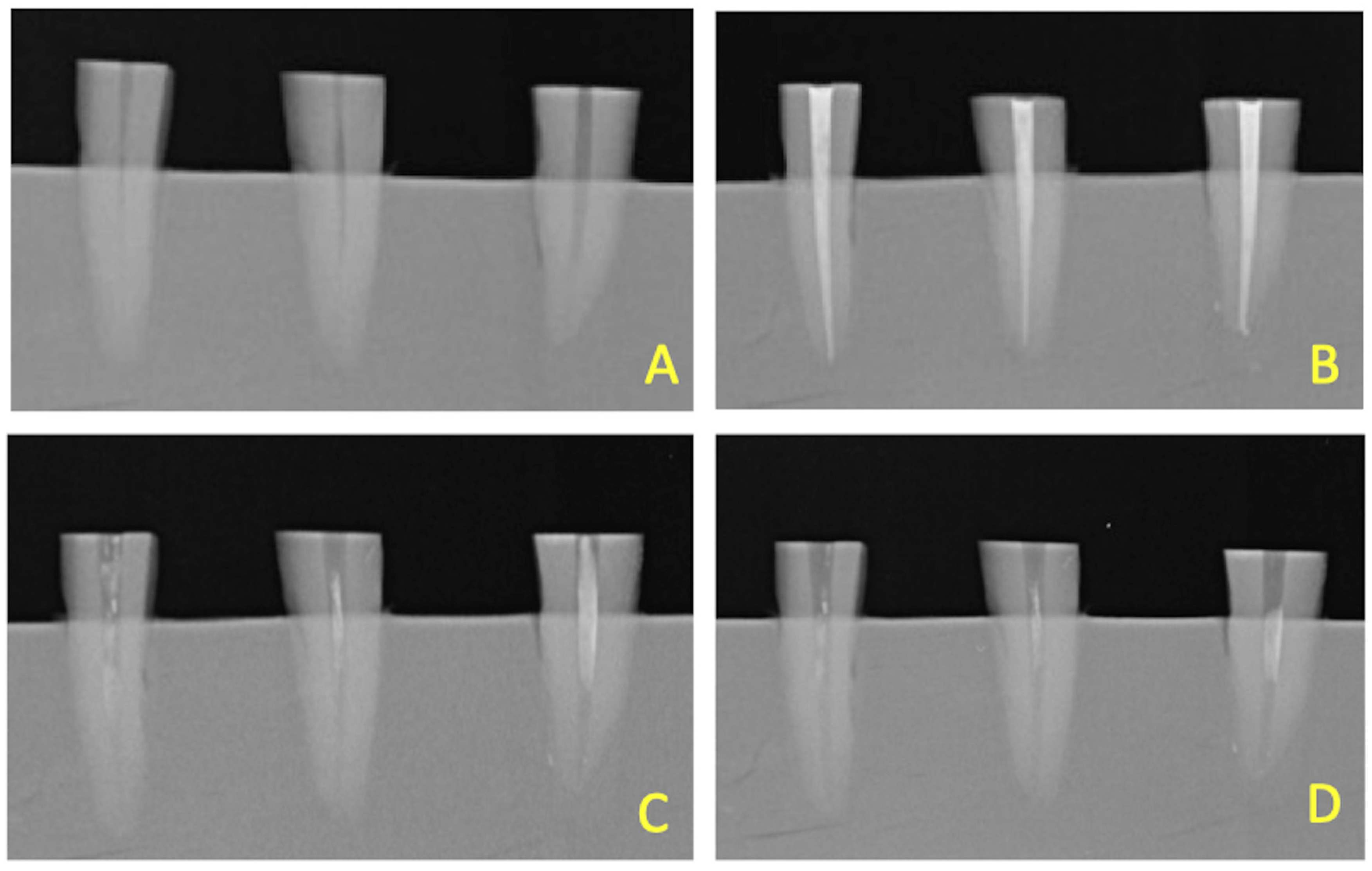
2.5. Radiographic Evaluation

Periapical phosphor plate X-rays were performed with parallel technique and a film-focus distance of 30 cm, using a customized container (Figure 3). The following parameters were used: exposure time of 0.125 s, 65 kV, and 10 mA. These plates were scanned using the default setting in a calibrated Dürr scanner and the acquired images were saved uncompressed for NNT Viewer.
The entire root canal area (mm2) of each sample was measured before the first treatment, after the root canal filling, after the retreatment with RB #25, and after the use of RB #40. Root canal area with unremoved remnants (mm2) still present at apical, middle, and coronal thirds was also measured. An open-source software (ImageJ, NIH software, Bethesda, MD, USA) was used for all the evaluations. One blinded operator evaluated the X-rays.

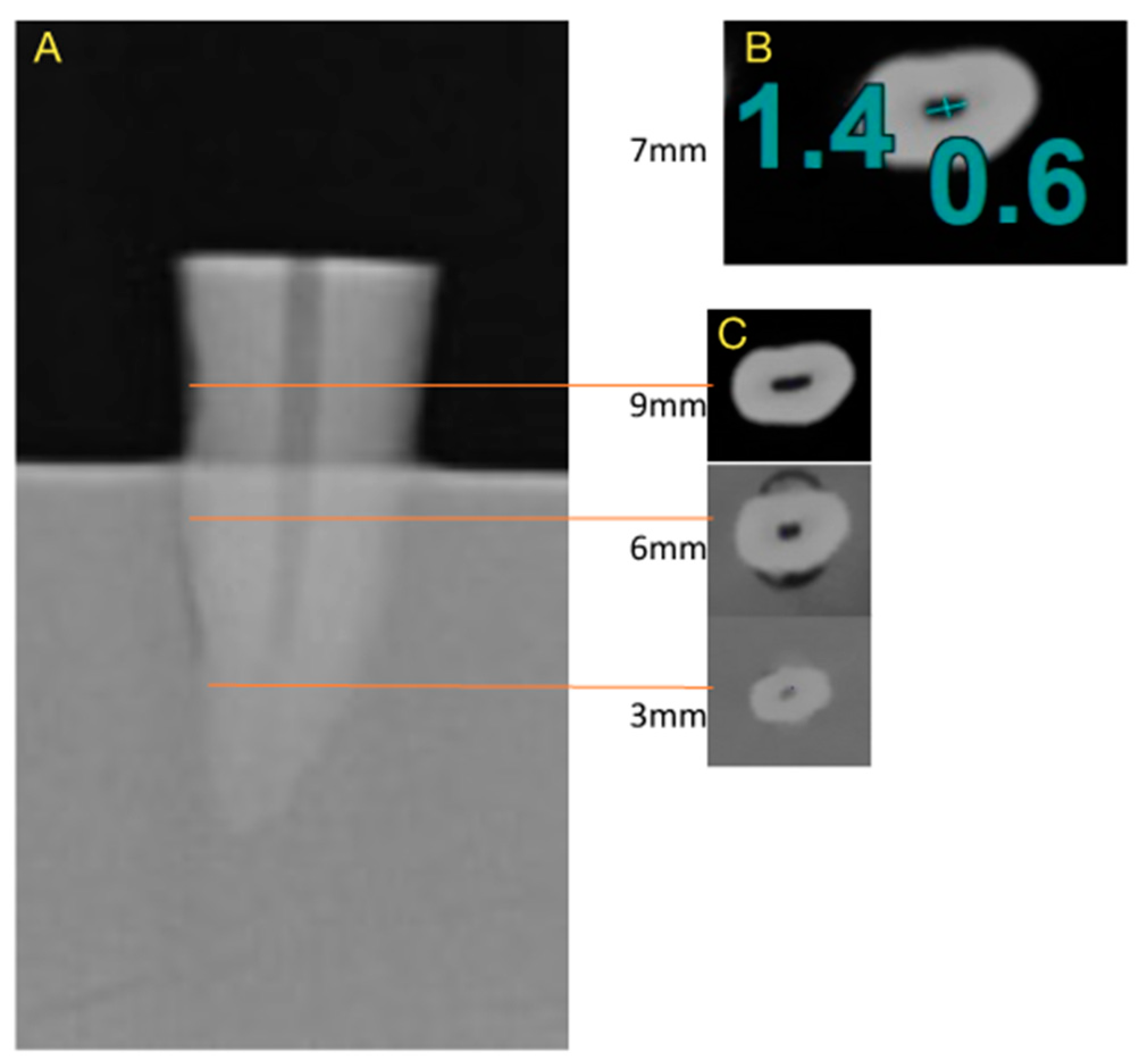
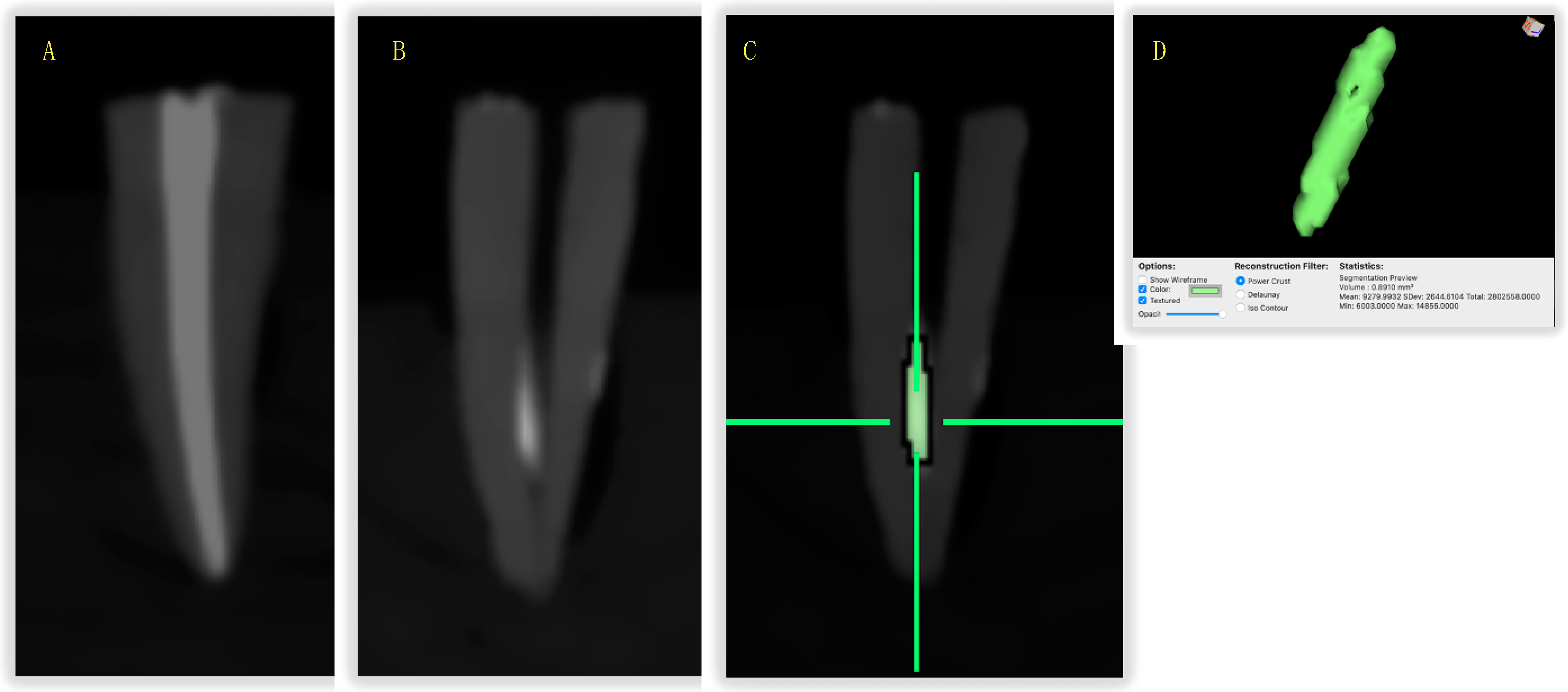
2.6. CBCT Evaluation

CBCT was performed for each sample immediately after the X-rays. All CBCTs were performed in the HiRes Zoom mode under the following settings: a field of view (FOV) of 5 mm × 8 mm, a tube voltage of 110 kVp and 6.53 mA, a resolution of 0.282 mm voxels, a 0.125 mm axial thickness, an exposure time of 4.3 s, and 395.9 mGy cm2. Remnant volume (mm3) was measured into the root canal after SRCTs at the apical, middle, and coronal thirds. Remnant volume was identified by the use a threshold-based semiautomatic segmentation on the greyscale values (grey voxels for dentine, black values for void canal and white for filling material). ROI (region of interest) was determined for each sample and a 3D model of remnants inside root canal was reconstructed to calculate the volume of the remaining material (Figure 4). HOROS software was used for all the evaluations. One blinded operator evaluated the CBCT.

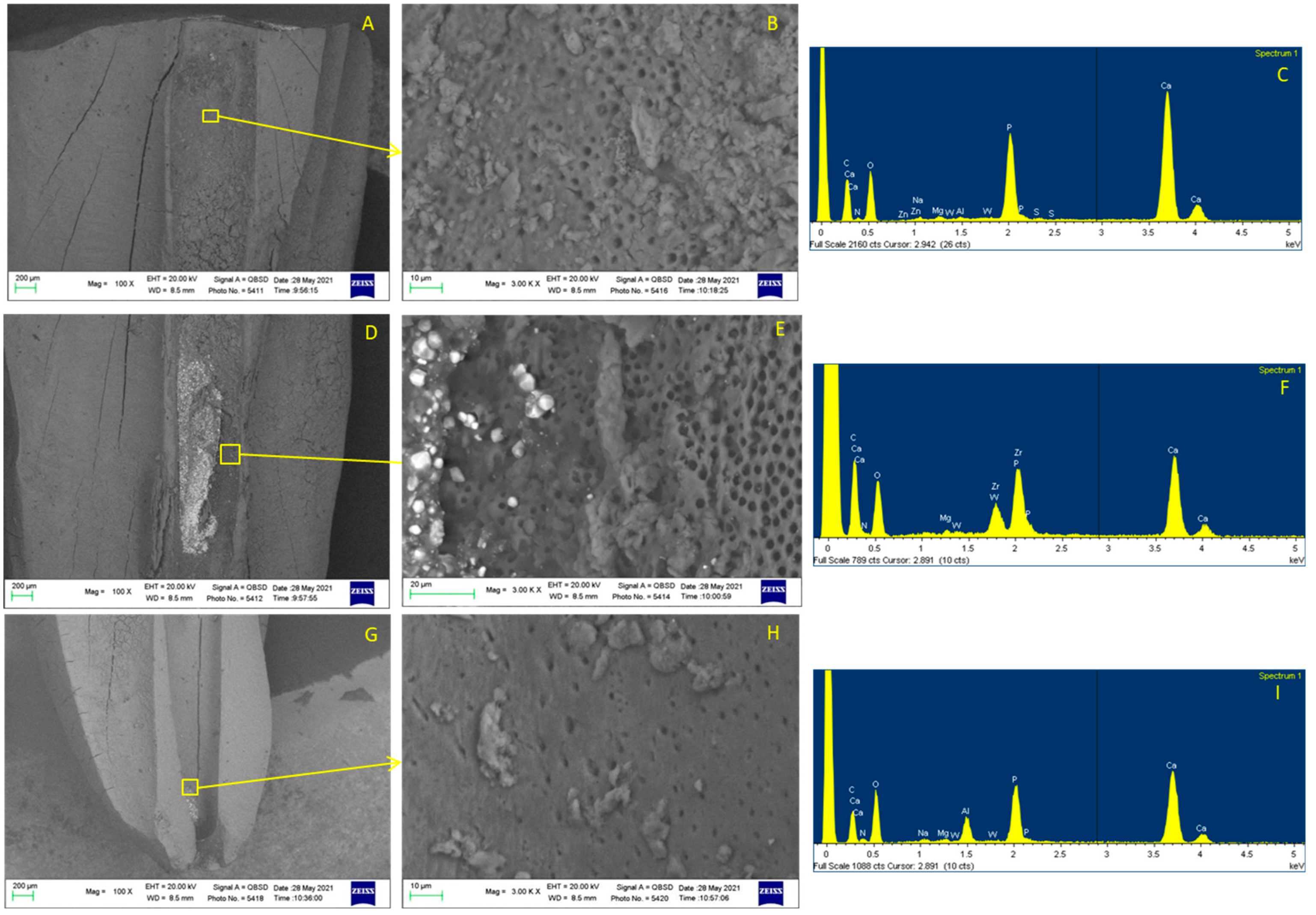
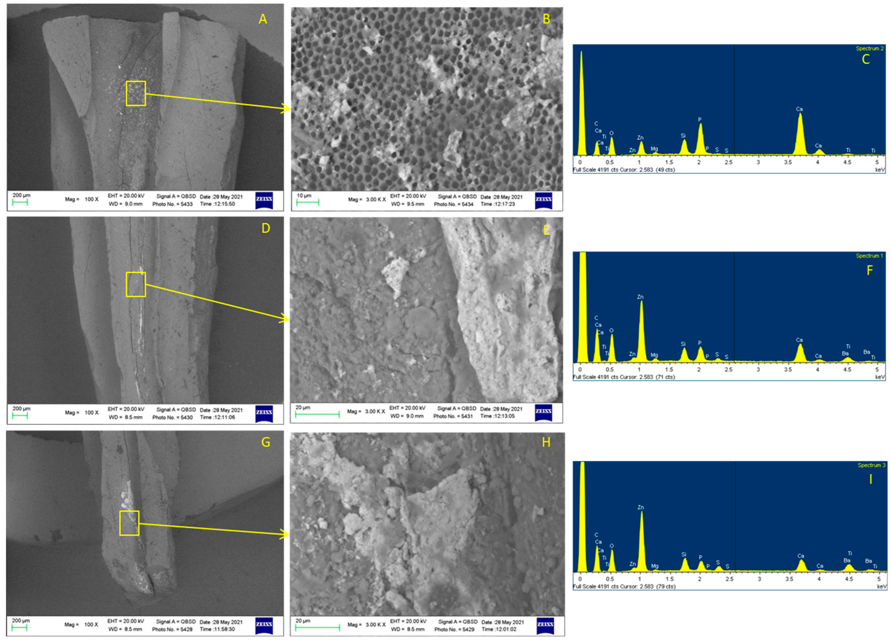
2.7. ESEM-EDX Evaluation

A high-speed, water-cooled handpiece with a diamond cylindrical bur was used to cut two longitudinal grooves in each root from the apex to the coronal section. To divide the root into two longitudinal parts, grooves were cut with a chisel. To conduct the ESEM-EDX analysis, one of the two halves was randomly chosen.
ESEM analysis was carried out at 100× and 3000× magnification to observe macro- and micro-morphology of root canals dentin surface and the location of remnants.
Without any prior preparation, the specimens were placed straight on the ESEM stub and analyzed (uncoated samples). Operative parameters were as follows: low vacuum (100 pascal), accelerating voltage of 20–25 kV, working distance 8.5 mm, and 133 eV resolution in Quadrant Back-Scattering Detector (QBSD) mode (0.5 wt% detection level, amplification time of 100 μs, and measuring time of 60 s).
EDX spectroscopy was used to identify the elemental composition of remnants. The ZAF correction approach, which involves calculating independent corrections for the atomic number effect (Z), absorbance (A), and fluorescence (F), was used to investigate the qualitative and semiquantitative element (weight% and atomic%) content [10]. The compositions of sealer and gutta-percha (i.e., Zr, W, Zn, etc.) were used to identify the type of remnants (sealer, gutta-percha, or dentin smear layer). Three measurements were taken for each sample.

2.8. Statistical Analysis
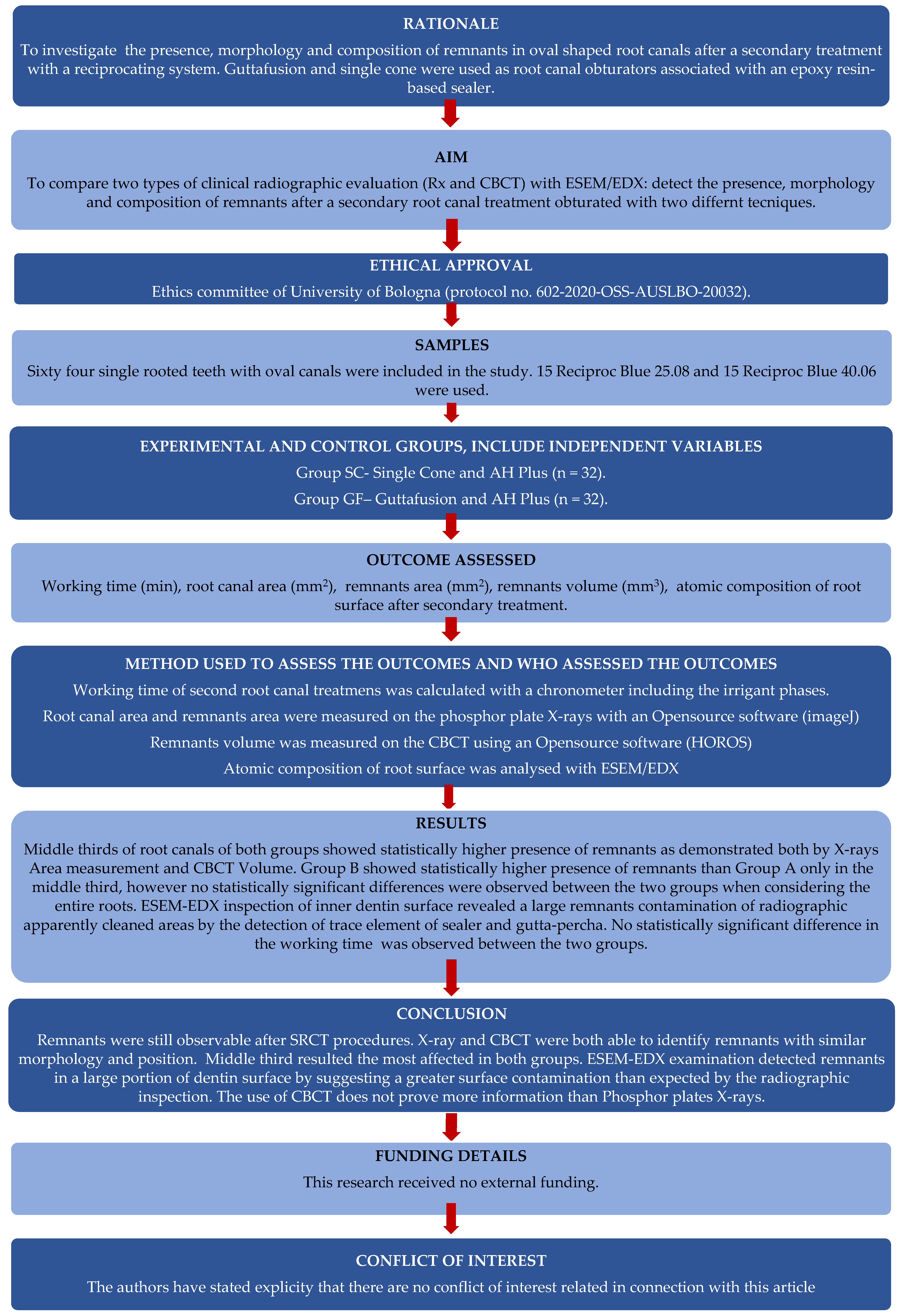
Statistical analysis was performed using Sigma Plot (version 13, IBM, USA). Data were analyzed using one-way analysis of variance (ANOVA) followed by Student–Newman–Keuls test. The statistical significance was set at p < 0.05.
Pearson correlation was performed to detect any statistical correlation between the area of smear layer residuals detected by X-ray (area expressed in mm2) and volume of smear layer detected by CBCT (volume expressed in mm3).

3. Results

3.1. Time to Complete SRCTs

The working time that was calculated for the SRCTs is reported in Table 1. No significant differences were observed in the retreatment times of the two filling groups. To complete the SRCTs.

3.2. Assessment of Intracanal Remnants: X-rays and CBCT

The pre-operative root canal area was calculated using X-rays. No differences were observed between the two groups (p > 0.05). The root canal areas were then calculated on the X-rays, respectively, after the completion of the first endodontic treatment, after the completion of the SRCTs procedures with the RB #25, and after the use of the RB #40 as reported in Table 2.
SRCTs with RB #25 increased root canal area by 12% for SC group and 14% for GF group compared to the pre-operative root canal area. The use of RB #40 further increased the area of root canal by 8% in both groups compared to the previous treatment with RB #25.
Table 3 reports the mean values of the remnants areas (mm2) and volumes (mm3) at the coronal, middle, and apical thirds.
In both filling groups, the remnants occupied a large portion of the middle third of the canals. The coronal and apical thirds showed lower levels of remnants.
The area of the remnants in the middle thirds was statistically significantly higher than that measured in the coronal and apical thirds.
Table 4 and Table 5 report the correlation of the remnants areas measured by X-ray and the remnants volumes measured by CBCT. Both radiological examinations were able to detect the same remnants in the canal third with the exception of the apical third of the GF group, in which discrepancies were found.

3.3. ESEM-EDX Analysis

In the SC group, the coronal third examination revealed few electron-dense granules on the dentinal surface (Figure 5). The dentinal tubules were partially occluded by the smear layer. The EDX microanalysis showed the presence of tracer elements that can be attributed to the sealer (W) and gutta-percha cone (Zn).
The dentine surface at the middle third was characterized by a thick layer of remnants. The images at 100× showed an irregular layer, with some areas filled by electron-dense remnants and a few areas with limited remnants. The images at 3000× showed dentinal tubules completely filled by electron-dense granules. The EDX revealed high peaks of elements that can be attributed to the AH Plus (Wolframium, W) and gutta-percha cone (Zinc, Zn, and Silicon, Si). Nitrogen (N), an index of the organic components of dentine (collagen), was not detected in this area. The dentine surface at the apical third was partially free from remnants. At 100×, the analysis displayed a few thin layers of remnants close to the anatomical apex. At 3000×, it revealed the dentine surface with a few remnants. In this area, the tubules were mostly open and well visible. The EDX microanalysis revealed a low presence of sealer tracer elements (W) and no gutta-percha tracing elements.
In the GF group, the ESEM images at the coronal and at the middle third displayed a high presence of electron-dense remnants (Figure 6). The dentine surface was characterized by open dentinal tubules and partially covered by electron-dense remnants. These remnants were not detected in the periapical X-ray and CBCT examinations. At the middle third, the ESEM images (100×) revealed a large remnant layer.
At a high magnification (3000×), the remnants completely covered the dentinal structures, with no signs of tubules. The EDX examination of the remnants revealed a high presence of elements from Guttafusion, namely Zn, Barium (Ba), and Titanium (Ti). Some remnants that were not observable in radiographical examinations were detected in the apical portion. The root canal dentine surface was observed at high magnifications (3000×). The EDX images revealed the presence of elements attributable to gutta-percha (Zn and Si). The remnants were observed in correspondence to the radiopaque materials revealed by the X-rays and CBCT. In the SC group, the remnants were mostly composed of AH Plus and some gutta-percha, evidenced by the high presence of electron-dense granules (high presence of W from the EDX analysis). In the GF group, the element constitution was mostly characterized by Zn and Ba (the tracer elements of Guttafusion/gutta-percha) with a uniform thick layer and a lower presence of electron-dense granules.

4. Discussion

In the present study, SRCTs with two reciprocating instruments, RB #25 and RB #40, were performed on root canals previously filled with two different techniques (SC and GF). The principal aim of the study was to evaluate the effectiveness of the reciprocating NiTi instrument in removing root-filling materials and their remnants from root canal systems characterized by an oval-shaped configuration.
The persistence of remnants inside the root canal was detected by two radiographic methods commonly used in clinical activities and by one in vitro system to obtain more information on the ultra-morphology and composition of the remnants after the SRCTs. The presence of remnants and their positions in the root canal has been proposed [21,22,23] to be an index of the effectiveness of the SRCTs.
Phosphor plate X-rays were used to measure the area occupied by the remnants. In addition, the volume of the remnants was measured using CBCT. In this way the volume and the position of the remnants were detected in a 3D configuration. Therefore, the X-rays offered a 2D evaluation of the presence/absence of remnants, but no information on the 3D positions in the canal. Thus, information on the area and volume should be combined and taken in account by clinicians during SRCTs.
In this study, a greater amount of remnants was observed on the middle third of the canals. Both the X-rays and CBCT confirmed the presence of dense remnants solidly packed into the middle thirds. The CBCT evaluation demonstrated that the presence of the remnants is predominantly localized into the lingual and buccal sides of the root canals. Previous studies have demonstrated the presence of a large amount of remnants corresponding to the roots apical third [24,25], suggesting that the conventional manual or rotary systems used to remove filling materials may induce the formation of an apical critical area [25]. In a similar way, remnants may still also be detectable using a self-adjusting file (SAF) and a hollow, compressible NiTi instrument, through which a continuous flow of an irrigant can be provided [26].
These data suggest some further considerations. The conicity, the specific design of the RB, and the reciprocating movement may have contributed to the removal of remnants from the apical third, which resulted cleaner and free from remnants compared to the middle third. Thus, it is probable that RBs may be valid instruments in removing apical debris.
RB #25 and RB #40 instruments have similar sizes and taper in the middle third because of their regressive variable taper. So, despite its greater apical diameter, the RB #40 is not able to remove more remnants from the middle thirds. Obviously, the use of Gates Glidden burs in the first 4 mm of the root canals—the coronal thirds—was helpful in removing all the remnants and keep these parts of the root canals extremely clean and free from any deposits or residues. Despite a previous study reported a higher removal effect of the gutta-percha using solvents, such as orange oil, chloroform, and eucalyptol [27], no solvents have been used in the present study. The use of solvents may induce a greater spreading of softened gutta-percha in the buccal-vestibular portions of the oval root canal. Moreover, the new improvements in the mechanical properties of NiTi have led clinicians to use gutta-percha solvents less [28].
In this study, we included the use of ESEM connected with EDX. ESEM is a new method using a microscopic device able to inspect humid dentine surfaces and to reduce artefacts from the dehydration of the sample. ESEM-EDX allows the microscopic evaluation of the inner surfaces of root canals to detect the presence, morphology, and atomic composition of remnants and dentin surfaces, in accordance with previous studies [10,29,30].
Our ESEM observations demonstrated that the mesial and distal surfaces of the root canal had a uniform dentin morphology free from remnants, but with a thin smear layer and with visible dentinal tubules. In contrast, the lingual and the buccal surfaces were partially covered by an easily detectable remnant layer. In the EDX analysis, these remnants resulted in a mixture of dentin chips, gutta-percha fragments (Si and Ba tracer elements), and small scraped pieces of sealer (W and Ba tracers). Interestingly, some root samples that appeared free of remnants in the radiographic evaluation were contaminated in the ESEM-EDX investigation. This finding could have an interesting implication in clinical practice suggesting the use of NiTi with an increased taper as last shaping file even when X-rays show no signs of remnants. The evaluation of remnants with conventional periapical X-rays was affected by the 2D nature of the exam. In several samples, a large area of the canal appeared in the 2D X-ray inspection completely covered and contaminated by remnants, but the CBCT evaluation showed only the lingual portion of the dentin surface as covered.
Notoriously, bacteria are present in persistent infections and must be removed. Probably they remain trapped between the porosity and empty spaces of the filling material and can rapidly re-contaminate the entire root canal space. Consequently, the infected root canal filling remnants must be completely removed as well as some portions of the infected dentinal tubules [31,32].
The use of X-rays and CBCT to detect the presence of remnants has clinical importance and deserves further consideration. Both techniques are well known and are usually performed in clinical settings. Some authors suggest that the use of CBCT could improve the success of SRCTs procedures in difficult cases [33]. 3D analyses can detect with a greater accuracy the possible causes of treatment failure, the complexity of the apical anatomy, and untreated or at least unfilled root canal areas [34,35].
Our data are confirmed by previous studies in which micro-CT scans were used [36,37]. Micro-CT scans are a gold-standard reference for evaluating remnants and 3D canal configuration [38]. Unfortunately, the use of micro-CT is not possible in clinical practice as it may be used only for laboratory studies.
Limitations of the present study include the following: all the teeth were treated after the removal of the crowns; the blooming effect (artefact induced by CBCT) may partially affect the image of the root remnants; the evaluated parameters need to be further validated in an in vivo clinical study.
The present study supports the following concepts: (1) The use of the RB #40 in oval-shaped canals showed a sub-optimal removal of remnants from apical thirds and an evident persistence of remnants only in the medium thirds. (2) Reciprocating instruments seem effective in removing remnants from apical thirds, the critical root canal area. (3) The 2D X-ray analysis with periapical radiographies was able to detect the persistence and position of remnants, which was confirmed by ESEM-EDX. (4) The ESEM-EDX examination detected microscopic remnants in a large portion of the dentin surface, suggesting a more diffuse surface contamination than was evaluated by the radiographic analysis. (5) The EDX analysis was able to detect the composition of filling materials and identify material remnants versus dentin fragments.

Author Contributions

Conceptualization, C.P.; methodology, C.P.; software, A.S. and P.P.; validation, A.S. and F.Z.; formal analysis, A.S. and F.Z.; investigation, A.S.; resources, C.P. and P.P.; data curation, A.S. and F.Z.; writing—original draft preparation, C.P., M.G.G. and A.S.; writing—review and editing, M.G.G. and A.B.; visualization, A.S. and F.Z.; supervision, C.P. and M.G.G. All authors have read and agreed to the published version of the manuscript.

Funding

This research received no external funding.

Institutional Review Board Statement

The study was conducted in accordance with the Declaration of Helsinki. This study was approved by the Ethics committee (protocol no. 602-2020-OSS-AUSLBO-20032).

Informed Consent Statement

Not applicable.

Data Availability Statement

Not applicable.

Conflicts of Interest

The authors have stated explicitly that there is no conflict of interest in connection with this article.

References

  1. Nair, P.N.R. On the causes of persistent apical periodontitis: A review. Int. Endod. J. 2006, 39, 249–281. [Google Scholar] [CrossRef]
  2. Rôças, I.N.; Hülsmann, M.; Siqueira, J.F., Jr. Microorganisms in Root Canal-treated Teeth from a German Population. J. Endod. 2008, 34, 926–931. [Google Scholar] [CrossRef] [PubMed]
  3. Ricucci, D.; Siqueira, J.F., Jr. Biofilms and apical periodontitis: Study of prevalence and association with clinical and histopathologic findings. J. Endod. 2010, 36, 1277–1288. [Google Scholar] [CrossRef] [PubMed]
  4. Siqueira, J.F., Jr. Aetiology of root canal treatment failure: Why well-treated teeth can fail. Int. Endod. J. 2001, 34, 1–10. [Google Scholar] [CrossRef] [PubMed] [Green Version]
  5. Ng, Y.L.; Mann, V.; Gulabivala, K. Outcome of secondary root canal treatment: A systematic review of the literature. Int. Endod. J. 2008, 41, 1026–1046. [Google Scholar] [CrossRef]
  6. Siqueira, J.F., Jr.; Alves, F.R.; Almeida, B.M.; de Oliveira, J.C.M.; Rôças, I.N. Ability of chemomechanical preparation with either rotary instruments or self-adjusting file to disinfect oval-shaped root canals. J. Endod. 2010, 36, 1860–1865. [Google Scholar] [CrossRef]
  7. Hülsmann, M.; Bluhm, V. Efficacy, cleaning ability and safety of different rotary NiTi instruments in root canal retreatment. Int. Endod. J. 2004, 37, 468–476. [Google Scholar] [CrossRef]
  8. Somma, F.; Cammarota, G.; Plotino, G.; Grande, N.M.; Pameijer, C.H. The Effectiveness of Manual and Mechanical Instrumentation for the Retreatment of Three Different Root Canal Filling Materials. J. Endod. 2008, 34, 466–469. [Google Scholar] [CrossRef]
  9. Kfir, A.; Tsesis, I.; Yakirevich, E.; Matalon, S.; Abramovitz, I. The efficacy of five techniques for removing root filling material: Microscopic versus radiographic evaluation. Int. Endod. J. 2012, 45, 35–41. [Google Scholar] [CrossRef]
  10. Prati, C.; Zamparini, F.; Spinelli, A.; Pelliccioni, G.A.; Pirani, C.; Gandolfi, M.G. Secondary Root Canal Treatment with Reciproc Blue and K-File: Radiographic and ESEM-EDX Analysis of Dentin and Root Canal Filling Remnants. J. Clin. Med. 2020, 9, 1902. [Google Scholar] [CrossRef]
  11. Peters, O.A. Current challenges and concepts in the preparation of root canal systems: A review. J. Endod. 2004, 30, 559–567. [Google Scholar] [CrossRef] [PubMed] [Green Version]
  12. Martins, M.P.; Duarte, M.A.; Cavenago, B.C.; Kato, A.S.; da Silveira Bueno, C.E. Effectiveness of the ProTaper Next and Reciproc Systems in Removing Root Canal Filling Material with Sonic or Ultrasonic Irrigation: A Micro-computed Tomographic Study. J. Endod. 2017, 43, 467–471. [Google Scholar] [CrossRef] [PubMed]
  13. Guimarães, L.S.; Gomes, C.C.; Marceliano-Alves, M.F.; Cunha, R.S.; Provenzano, J.C.; Siqueira, J.F., Jr. Preparation of Oval-shaped Canals with TRUShape and Reciproc Systems: A Micro-Computed Tomography Study Using Contralateral Premolars. J. Endod. 2017, 43, 1018–1022. [Google Scholar] [CrossRef]
  14. Ferreira, F.; Adeodato, C.; Barbosa, I.; Aboud, L.; Scelza, P.; Zaccaro Scelza, M. Movement kinematics and cyclic fatigue of NiTi rotary instruments: A systematic review. Int. Endod. J. 2017, 50, 143–152. [Google Scholar] [CrossRef]
  15. Ball, R.L.; Barbizam, J.V.; Cohenca, N. Intraoperative endodontic applications of cone beam computed tomography. J. Endod. 2013, 39, 548–557. [Google Scholar] [CrossRef]
  16. Schropp, L.; Kirkevang, L.L. Accuracy and Reliability of Intraoral Radiographs in Determining the Cleanliness of Root Canals after Endodontic Retreatment. Eur. Endod. J. 2017, 2, 1. [Google Scholar] [CrossRef] [Green Version]
  17. Baxter, S.; Schöler, C.; Dullin, C.; Hülsmann, M. Sensitivity of conventional radiographs and cone-beam computed tomography in detecting the remaining root-canal filling material. J. Oral. Sci. 2020, 62, 271–274. [Google Scholar] [CrossRef] [PubMed]
  18. Schäfer, E.; Schrenker, C.; Zupanc, J.; Bürklein, S. Percentage of Gutta-percha Filled Areas in Canals Obturated with Cross-linked Gutta-percha Core-carrier Systems, Single-Cone and Lateral Compaction Technique. J. Endod. 2016, 42, 294–298. [Google Scholar] [CrossRef]
  19. Pérez-Alfayate, R.; Mercade, M.; Algar-Pinilla, J.; Cisneros-Cabello, R.; Foschi, F.; Cohen, S. Root Canal Filling Quality Comparison of a Premixed Calcium Silicate Endodontic Sealer and Different Carrier-Based Obturation Systems. J. Clin. Med. 2021, 10, 1271. [Google Scholar] [CrossRef]
  20. Nagendrababu, V.; Murray, P.E.; Ordinola-Zapata, R.; Peters, O.A.; Rôças, I.N.; Siqueira, J.F., Jr.; Priya, E.; Jayaraman, J.; Pulikkotil, S.J.; Camilleri, J.; et al. PRILE 2021 guidelines for reporting laboratory studies in Endodontology: A consensus-based development. Int. Endod. J. 2021, 54, 1482–1490. [Google Scholar] [CrossRef]
  21. Pirani, C.; Pelliccioni, G.A.; Marchionni, S.; Montebugnoli, L.; Piana, G.; Prati, C. Effectiveness of Three Different Retreatment Techniques in Canals Filled With Compacted Gutta-Percha or Thermafil: A Scanning Electron Microscope Study. J. Endod. 2009, 35, 1433–1440. [Google Scholar] [CrossRef] [PubMed]
  22. Rödig, T.; Reicherts, P.; Konietschke, F.; Dullin, C.; Hahn, W.; Hülsmann, M. Efficacy of reciprocating and rotary NiTi instruments for retreatment of curved root canals assessed by micro-CT. Int. Endod. J. 2014, 47, 942–948. [Google Scholar] [CrossRef]
  23. Bago, I.; Plotino, G.; Katić, M.; Ročan, M.; Batinić, M.; Anić, I. Evaluation of filling material remnants after basic preparation, apical enlargement and final irrigation in retreatment of severely curved root canals in extracted teeth. Int. Endod. J 2020, 53, 962–973. [Google Scholar] [CrossRef] [PubMed]
  24. Alves, F.R.F.; Rôças, I.N.; Provenzano, J.C.; Siqueira, J.F., Jr. Removal of the Previous Root Canal Filling Material for Retreatment: Implications and Techniques. Appl. Sci. 2022, 12, 10217. [Google Scholar] [CrossRef]
  25. Pirani, C.; Iacono, F.; Chersoni, S.; Sword, J.; Pashley, D.H.; Tay, F.R.; Looney, S.; Gandolfi, M.G.; Prati, C. The effect of ultrasonic removal of various root-end filling materials. Int. Endod. J. 2009, 42, 1015–1025. [Google Scholar] [CrossRef]
  26. Keleş, A.; Şimşek, N.; Alçin, H.; Ahmetoglu, F.; Yologlu, S. Retreatment of flat-oval root canals with a self-adjusting file: An SEM study. Dent. Mater J. 2014, 33, 786–791. [Google Scholar] [CrossRef] [PubMed] [Green Version]
  27. Scelza, M.F.; Coil, J.M.; Maciel, A.C.; Oliveira, L.R.; Scelza, P. Comparative SEM evaluation of three solvents used in endodontic retreatment: An ex vivo study. J. Appl. Oral. Sci. 2008, 16, 24–29. [Google Scholar] [CrossRef] [Green Version]
  28. Ferreira, I.; Pina-Vaz, I. The Novel Role of Solvents in Non-Surgical Endodontic Retreatment. Appl. Sci. 2022, 12, 5492. [Google Scholar] [CrossRef]
  29. Hülsmann, M.; Rümmelin, C.; Schäfers, F. Root canal cleanliness after preparation with different endodontic handpieces and hand instruments: A comparative SEM investigation. J. Endod. 1997, 23, 301–306. [Google Scholar] [CrossRef]
  30. Prati, C.; Foschi, F.; Nucci, C.; Montebugnoli, L.; Marchionni, S. Appearance of the root canal walls after preparation with NiTi rotary instruments: A comparative SEM investigation. Clin. Oral. Investig. 2004, 8, 102–110. [Google Scholar] [CrossRef]
  31. Love, R.M. Bacterial adhesins--their role in tubule invasion and endodontic disease. Austral. Endod. J. 2002, 28, 25–28. [Google Scholar] [CrossRef] [PubMed]
  32. Vieira, A.R.; Siqueira, J.F., Jr.; Ricucci, D.; Lopes, W.S. Dentinal tubule infection as the cause of recurrent disease and late endodontic treatment failure: A case report. J. Endod. 2012, 38, 250–254. [Google Scholar] [CrossRef] [PubMed]
  33. Patel, S.; Brown, J.; Semper, M.; Abella, F.; Mannocci, F. European Society of Endodontology position statement: Use of cone beam computed tomography in Endodontics: European Society of Endodontology (ESE) developed by. Int. Endod. J. 2019, 52, 1675–1678. [Google Scholar] [CrossRef] [PubMed] [Green Version]
  34. Cohenca, N.; Shemesh, H. Clinical applications of cone beam computed tomography in endodontics: A comprehensive review. Quintessence Int. 2015, 46, 657–668. [Google Scholar] [PubMed]
  35. Di Nardo, D.; Zanza, A.; Pagnoni, F.; Xhajanka, E.; Testarelli, L. An Update on Advanced Diagnostic Imaging in Dentistry. Diagnostics 2022, 12, 1041. [Google Scholar] [CrossRef]
  36. Bago, I.; Suk, M.; Katic, M.; Gabric, D.; Anic, I. Comparison of the effectiveness of various rotary and reciprocating systems with different surface treatments to remove gutta-percha and an epoxy resin-based sealer from straight root canals. Int. Endod. J. 2019, 52, 105–113. [Google Scholar] [CrossRef] [Green Version]
  37. Keleş, A.; Arslan, H.; Kamalak, A.; Akçay, M.; Sousa-Neto, M.D.; Versiani, M.A. Removal of filling materials from oval-shaped canals using laser irradiation: A micro-computed tomographic study. J. Endod. 2015, 41, 219–224. [Google Scholar] [CrossRef]
  38. Ajina, M.A.; Shah, P.K.; Chong, B.S. Critical analysis of research methods and experimental models to study removal of root filling materials. Int. Endod. J. 2022, 55, 119–152. [Google Scholar] [CrossRef]
Figure 1. PRILE 2021 Flowchart.
Applsci 12 11671 g001
Figure 2. Sample selection. Preoperative X-ray (A). Preoperative CBCT: mesiodistal and buccolingual diameters of each root canal were measured at 7 mm from the apex to ensure the oval shape of the root canal (B). Axial section at 3, 6, and 9 mm from the apex (C).
Applsci 12 11671 g002
Figure 3. Radiographic examination of customized mold with three teeth before treatment (A), after root canal filling (B), after retreatment with RB #25, (C) and after retreatment with RB #40 (D).
Applsci 12 11671 g003
Figure 4. CBCT representation after root canal filling (A) and after SRCTs with RB #40 (B). Remnants are still present in the middle third of the root canal after SRCTs. Each CBCT representation was examined with HOROS software to measure the volume of filling material after SRCTs (C). The volume of remnants was identified using a semiautomatic segmentation based on the values of the grayscale (gray values for dentin, black values for the root canal, and white for filling material). The region of interest (ROI) was then determined and reconstructed in 3D to calculate the volume of remnants (D).
Applsci 12 11671 g004
Figure 5. Root canal obturated with SC technique, sectioned and examined with ESEM/EDX. Coronal third examination at 100× (A) revealed a few electron-dense granules on the dentinal surface. Dentinal tubules at 3000× were partially occluded by smear layer (B). EDX microanalysis showed the presence of tracer elements that can be attributed to the sealer (W) and gutta-percha cone (Zn) (C). Dentine surface at the middle third at 100× (D) was characterized by a thick layer of remnants. Image at 3000× (E) shows an irregular layer with some areas filled by electron dense remnants, and few areas with limited remnants. EDX (F) revealed high peaks of elements attributable to AH plus (W) and gutta-percha cone (Zn, and Si). N, index of collagen was not detected in this area. Dentine surface at the apical third (G) was partially free from remnants. At 100× displayed few thin layers of remnants close to the anatomical apex. At 3000× revealed dentine surface with a few remnants (H). In this area, the tubules were mostly open and well visible. EDX microanalysis revealed low presence of sealer tracer elements (W) and no gutta-percha tracing elements (I).
Applsci 12 11671 g005
Figure 6. Root canal obturated with GF technique, sectioned, and examined with ESEM/EDX. Coronal third examination at 100× (A) revealed a few electron-dense granules on the dentinal surface. Dentine surface at 3000× (B) was characterized by open dentinal tubules and partially covered by the electron-dense remnants. EDX microanalysis (C) showed the presence of tracer elements attributable gutta-percha (Zn). Middle third at 100× (D) displayed a high presence of electron-dense remnants. At high magnification (3000×), (E) the remnants completely covered the dentinal structure, with no signs of tubules. Some remnants were detected in the apical portion at 100× magnification (G). At high magnification (3000×), (H) the dentinal tubules were completely covered by remnants. EDX on the remnants revealed high presence of elements from Guttafusion, namely Zn, Ba and Ti, and Si (F,I).
Applsci 12 11671 g006
Table 1. Working time (min) calculated to perform the secondary treatment with RB #25 and completed with RB #40. Mean ± Standard Deviations.
GroupSRCT #25 TimeSRCT #40 Time
SC Group (n = 32)7.02 ± 1.101.18 ± 0.53
GF Group (n = 32)8.22 ± 1.202.00 ± 1.10
Table 2. Root canal Area (mm2) calculated on radiography: preoperative, post-treatment, after secondary treatments with RB #25 and with RB #40. Percentage of increase after each treatment is reported. Mean ± Standard Deviations.
GroupPreoperativePost-TreatmentSRCT #25SRCT #40
AHPlus/SingleCone (n = 32)4.87 ± 0.657.11 ± 0.78 (+46%)7.94 ± 0.63 (+12%)8.53 ± 0.65 (+8%)
AHPlus/GuttaFusion (n = 32)4.82 ± 0.616.97 ± 0.81 (+45%)7.98 ± 0.60 (+14%)8.61 ± 0.61 (+8%)
Table 3. Area (mm2) of root canal remnants calculated on radiography and the Volume (mm3) calculated by the use of CBCT and visible into the root canal after the secondary treatments with RB #25 and with RB #40. Area and Volume were measured at coronal, middle and apical thirds. Mean ± Standard Deviations.
SC Group (n = 32)GF Group (n = 32)
Canal ThirdArea (mm2)Volume (mm3)Area (mm2)Volume (mm3)
Coronal0.17 ± 0.29 a0.21 ± 0.25 a0.14 ± 0.21 a0.17 ± 0.35 a
Middle0.44 ± 0.38 b0.46 ± 0.61 b0.62 ± 0.49 b0.56 ± 0.78 b
Apical0.23 ± 0.20 a0.17 ± 0.28 a0.28 ± 0.25 a0.23 ± 0.36 a
Total0.83 ± 0.660.85 ± 0.851.03 ± 0.700.97 ± 1.19
Different letters (vertical column) represent statistically significant differences among the root canal thirds (p < 0.05).
Table 4. Pearson correlation of areas and volumes of SC group (p value was set at 0.05).
Apical Third
Area
Middle Third
Area
Coronal Third
Area
Total
Area
Apical Third
Volume
Pearson correlation0.5160.4040.1370.493
p value0.00010.0020.1720.0001
N32323232
Middle Third
Volume
Pearson correlation0.2700.5860.3220.594
p value0.0280.00010.0140.0001
N32323232
Coronal Third
Volume
Pearson correlation−0.0870.1330.6510.262
p value0.2770.1820.00010.034
N32323232
Total
Volume
Pearson correlation0.2900.5750.4730.678
p value0.0180.00010.00010.0001
N32323232
Table 5. Pearson correlation of areas and volumes of GF group (p value was set at 0.05).
Apical Third
Area
Middle Third
Area
Coronal Third
Area
Total
Area
Apical Third
Volume
Pearson correlation0.1450.4200.3520.458
p value0.2260.0120.0300.006
N32323232
Middle Third
Volume
Pearson correlation0.0210.6190.5970.629
p value0.4570.00010.00010.0001
N32323232
Coronal Third
Volume
Pearson correlation−0.1250.1990.5950.280
p value0.2600.1500.00010.071
N32323232
Total
(Volume)
Pearson correlation0.0220.5910.6700.631
p value0.4560.0000.0000.0001
N32323232
Publisher’s Note: MDPI stays neutral with regard to jurisdictional claims in published maps and institutional affiliations.

