Article

Investigation of Adenosine Precursors and Biologically Active Peptides in Cultured Fresh Mycelium of Wild Medicinal Mushrooms

by
Nikolay Solakov
1,†,
Milena Kostova
2,†,
Kamelia Loginovska
1,
Zlati Markov
1,
Ana Caroline de Oliveira
3 and
Yordan Muhovski
3,*
1
Institute of Cryobiology and Food Technologies, 53 Cherni Vrah Blvd., 1407 Sofia, Bulgaria
2
Department of Plant Physiology, Biochemistry and Genetics, Faculty of Agronomy, Agricultural University Plovdiv, 12 Mendeleev Blvd., 4000 Plovdiv, Bulgaria
3
Biological Engineering Unit, Department of Life Sciences, Walloon Agricultural Research Centre, Chaussée de Charleroi, BP 234, 5030 Gembloux, Belgium
*
Author to whom correspondence should be addressed.
These authors contributed equally to this work.
Appl. Sci. 2022, 12(20), 10618; https://doi.org/10.3390/app122010618
Submission received: 19 September 2022 / Revised: 11 October 2022 / Accepted: 18 October 2022 / Published: 20 October 2022
(This article belongs to the Special Issue Advances in Natural Bioactive Compounds and Biological Effects II)

Abstract

:
Adenosine, known as an endogenous neuroprotective agent and acting as a cytoprotective modulator in organisms, is the subject of considerable scientific interest. Medicinal mushrooms represent a good source of bioactive compounds due to their composition and potent adaptogenic action, affecting more than 300 biochemical processes and functions in organisms. The aim of the present study was to investigate adenosine precursors and biologically active peptides in cultured fresh mycelium with focus on Ganoderma lucidum (Reishi) and Cordyceps sinensis as best studied species. Biologically active extracts are derived by fermentation with Bacillus subtilis NBIMCC 2353 strain. Enhancement of the total proteolytic activity of Bacillus subtilis culture medium enriched with native bovine collagen protein and mycelium was recorded. The results demonstrate a clear trend of increasing cordycepin and adenosine content in the Cordyceps militaris sample grown in culture medium with optimized composition, with the presence of cordycepin being 2.22% and adenosine being 0.64%. The obtained increase was 1.9% for cordycepin and 0.24% for adenosine, respectively. The application of combined biotechnological approaches in the use of biologically active components from natural organic sources and the resulting final product with high biological activity determine the present study as relevant and significant for its practical application.

1. Introduction

Adenosine is essential for the proper functioning of every cell in the body. As a component or precursor for RNA and DNA synthesis, and in its native form, adenosine acts as a neurotransmitter at the P1 and P2 purine receptors [1]. Cordycepin from Cordyceps sinensis is the main active constituent which is most widely studied for its medicinal value along with its nutraceutical potential [2,3]. Thousands of wild mushrooms are known in nature, of which about 600 species have been tested for their effects on human health [4,5]. Medicinal mushrooms produce valuable enzymes and bioactive molecules with different therapeutic effects [6,7]. Therefore, they are considered as flourishing organisms to develop different healthcare and biotechnological products [8]. All have been found to exhibit immunomodulatory properties [9,10]. It was indicated that peptides from Ganoderma lucidum blocked the soybean lipoxygenase activity and was analyzed as the major antioxidant agent, particularly in inhibiting lipid peroxidation of biological systems [11]. The mushroom matrix contains several major groups of biologically active substances, such as terpenes, sterols, functional polysaccharides, potent antioxidants, and inhibitors. Depending on the composition of the biological matrix, additional health effects are also found, which determines the scientific interest in their use as a substrate for the preparation of bioproducts with diverse functional activities.
Currently, no more than 10% of known fungal organisms are well studied [12], providing a tremendous opportunity to conduct research in this area, as well as to obtain bioactive extracts for use in agriculture, the food industry, and pharmaceutics. According to the published studies, the medicinal mushrooms appear to facilitate improvements in different directions, e.g., quality of life, a reduction of side effects by conventional therapies, hematologic parameters, antitumor activity, and immunomodulation, and furthermore medicinal mushrooms seem to be safe [13]. The development of methods for the cultivation of medicinal mushrooms in a controlled laboratory environment reveals a number of opportunities for achieving good results in the cultivation of different fungal strains [14]. Most research related to the study of organisms of the fungal kingdom is aimed at controlled cultivation, increasing the valuable biologically active substances in the biological matrix, and applying the methods for isolation and incorporation into various formulations in the production of products from the agricultural, food, and pharmaceutical industries. Notably, cordycepin (30-deoxyadenosine, COR), discovered in the broth of Cordyceps militaris, has received a large amount of attention due to its therapeutic potential, as well as its effects on intracellular signal transduction and cell adhesion [15]. Successfully isolated, cordycepin from Cordyceps militaris is used as a highly potent plant growth inhibitor [16]. The present study is aimed at the development of technology for the preparation of biologically active complexes of wild higher fungi and microbial cultures and their incorporation into a functional product with targeted health effects.

2. Materials and Methods

2.1. Experimental Material

The following higher medicinal fungi were used as a sources for biologically active compounds: Ganoderma Lucidum, Zhejiang Kangzhou Biotechnology Co., Ltd., Jinhua, China; Ganoderma neo-japonicum OGGNJ100 Out-Grow USA, Cordyceps sinensis OGCS100 Out-Grow USA, Cordyceps militaris OG1000CM Out-Grow USA, and Cordyceps militaris, Shanghai- producer strain (used in the semi-industrial method), Zhejiang Kangzhou Biotechnology Co., Ltd., Jinhua, China; Bacillus subtilis NBIMCC 2353 producer strain provided by NBPMCC were used in fungal fermentation.

2.2. Cultivation Condition and Starter Cultures

The starter cultures were propagated in a standardized medium purchased from Out-Grow USA provided in powdered form. Liquid medium was prepared by dissolving 6 g in 500 mL of distilled water heated to boiling while slowly homogenizing for 2–3 min. The solution was filtered through 40-micron filter paper, and the filtrate was sterilized. Liquid cultures were kept at 28 °C for 7 days with no light induction and 7 days on a light. Culture growth is visually monitored from day 4 to day 14. In order to obtain a high number of viable cells, the resulting inoculum was cultured in liquid culture medium M0 and then seeded in liquid and solid culture media. Four variants of enriched liquid nutrient media (M1, M2, M3, and M4) were developed based on the M0 (control) nutrient medium with the composition listed in Table 1.
For the optimal content of nutrients and inorganic elements of fresh mycelium fermentation culture medium with different content was prepared. As carbon source, glucose and dextrose were used. Mycelium of C. militaris, C. sinensis, G. lucidum, and G. neo-japonicum was activated on a sterilized PDA medium plate and incubated at 25 °C in the darkness for 7 days. The shake flask culture was run for 5–7 days and the fungal mycelium was harvested from the flasks by filtration, and then dried to constant dry weight (dw). One hundred milliliters of the sample with 10% of mycelium liquid seed was placed in a 500-mL flask and cultured at 25 °C for 5 days in a shaking incubator at 110 rpm. The pH was not adjusted, followed by autoclaving for 30 min at 121 °C. The culture experiments were performed in 500-mL glass jars containing basal medium after inoculation with 10% (v/v; the biomass of fresh mycelium).
The cell culture of Ganoderma lucidum, Ganoderma neo-japonicum, Cordyceps sinensis, and Cordyceps militaris was cultured in the culture media (M0, M1, M2, M3, and M4) for 72 h at 28 rpm, shaking amplitude of 100 rpm with no light induction, then cultured in a photoperiod of 16 h light (300 mL) to 8 h dark for a period of 12 days. In order to determine the mycelium growth, an analytical method was additionally developed using the ratio of the volume fractions occupied by the spheres of fresh mycelium to the remaining fraction of the total volume occupied by the culture medium (Vm = Vp-p − VLiquid), where Vm is volume of mycelial spheres, Vp-p the total volume of the solution together with the mycelium, and VLiquid the volume of the decanted culture medium.

2.3. Mycelial Cultivation on Solid Culture Media

The basic solid medium (S1) was formed by adding agar-agar to the M0. Enriched nutrient media were obtained by adding additional organic sources, whole grain rice (S2), and old aged oak chips (S3) (Table 2). The dry ingredients were dissolved in 1 L of distilled water, heated in a water bath to 60 °C until a gelatinous mass is obtained. The prepared solution is dispensed into a 270 mL glass jar at a dose of 100 g. The jars are fitted with an inoculation opening and a bactericidal filter with a pore size of 220 nm. The prepared medium was inoculated with 5 mL of the liquid inoculum from the cell cultures (Ganoderma lucidum KBGL001, Ganoderma neo-japonicum GGNJ100, Cordiceps sinensis OGCS100, and Cordiceps militaris OG1000CM), of which 25 doses were loaded for each individual culture. The inoculated media were arranged in an incubator for controlling cultivation at 28 °C and access to natural light.

2.4. Cultivation of Bacteria

Optimizations of Bacillus subtilis NBIMCC 2353 culture medium composition were performed in order to enhance the total enzyme activity by addition of native bovine collagen protein and mycelium of Ganoderma neo-japonicum, Ganoderma lucidum, Cordyceps sinensis, and Cordyceps militaris. Different concentrations of native bovine collagen were added to the control medium (K) at pH = 8. The composition of the media (P1–P3) is listed in Table 3.
One milliliter of Bacillus subtilis NBIMCC 2353 culture was inoculated into 100 mL of culture medium. The process of colony formation and growth was carried out at 30 °C and shaking amplitude of 100 rpm. Aliquots of each culture medium were separated to determine the proteolytic activity immediately before the initiation of the process, after 24, 48, and 72 h, respectively.

2.5. Quantification of Adenosine and Cordycepin Content in Fungal Mycelium and Fruiting Body Crude by High-Performance Liquid Chromatography (HPLC)

HPLC analysis for adenosine and cordycepin quantification was performed using an HP Agilent 1260 Infinity Quaternary LC instrument, DAD (λ = 260 nm), Agilent Poroshell 120 HPH-C18, (4.6 × 100 mm, 2.7 μm), elution mode isocratic mode with eluent rate 0.7 mL/min, Run (15 min) and liquid phase with composition: (A) H2O: MeOH [92:8], (B) H2O:MeOH [85:15], A:B (98:2). Aliquots of the extracts were diluted with MeOH (50%) at a concentration of 100 mg/mL. The samples were sonicated until complete dissolution and filtered with a 220 nm pore size cartridge filter before injection. Reference standards of adenosine (A9521) and cordycepin (C3394) from Sigma Aldrich were used. A standard solution containing a mixture of the two standards at a concentration of 100 µg/mL was prepared and used to generate a calibration curve. The calibration solutions were diluted with MeOH (50%) over the linearity range, with concentrations of 25 µg/mL, 50 µg/mL, and 100 µg/mL, respectively.

2.6. Determination of Antioxidant Activity

A standard DPPH-radical neutralizing tocopherol substance and a calibration curve were used, considering the results for the presence of antioxidant activity. For the determination of antioxidant activity, to 0.3 mL of DPPH solution (0.2 mmol) was added 0.5 mL of methanol and 0.5 mL of a 1 mg/mL solution of the medicinal mushrooms extract. The prepared solution was well mixed, homogenized, and allowed to stand in the dark at room temperature for 120 min. The absorbance was measured at 517 nm against methanol as standard. Trolox solutions with concentrations from 0.50 to 3.0 µg/mL are used for the standard straight. For the spectrophotometric analysis UV-Vis spectrophotometer, model Biochrom Libra S20 was used.

2.7. Sodium Dodecyl Sulfate–Polyacrylamide Gel Electrophoresis (SDS-PAGE)

In order to determine protein fractions, an SDS-PAGE was performed. A 6.0% pooling gel and a 12% separating gel were prepared. The lyophilized mycelial samples of Ganoderma lucidum, Ganoderma neo-japonicum, Cordyceps sinensis, and Cordyceps millitaris were rehydrated in deionized water at a concentration of the resulting solution of 200 mg/mL and centrifuged for 10 min at 4 °C and at speed of 8000 rpm on a Beckman centrifuge. For final sample preparation, 1 part of the supernatant was mixed with 1 part of sample buffer containing 0.2 M Tris HCL (pH 6.8). A protein electrophoresis system, Pharmacia LCB MultiDrive XL equipped with OmniPAGE WAVE Electrophoresis System, was used. Electrophoresis was performed at a running condition of 30 mA and an initial voltage of 105 V for 200 min. The gel was stained with a Coomassie brilliant blue G—250 stain solution for 30 min. The protein purity was calculated densitometrically on an “ERI-10”. Gel reading and molecular mass calculations were performed using Gel Analyzer-19 software. The following markers were used: Sigma marker, Mix 5, STD (BSA, Lc, Lg, La), and BSA.

3. Results

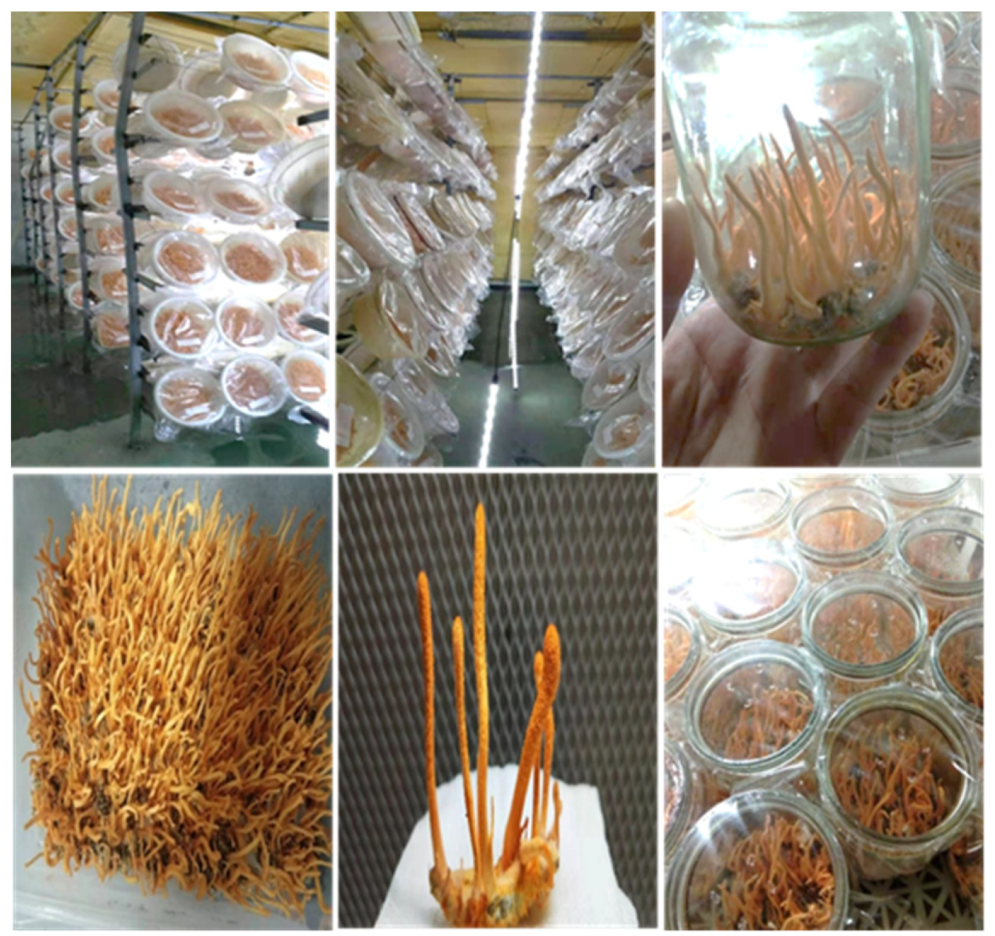
3.1. Frooting Body Cultivation

It was found experimentally that the best mycelial growth occurred in medium M2 containing glucose as carbon source, followed by M4 and M3, M1, and the lowest growth was observed in control medium M0. From the evaluation, the culture medium M2 was selected for subsequent mycelial cultivation. During mycelial development, connected cellular structures in the form of hyphae are formed by the multiplication of the fungal cell culture, representing channels for the transport of nutrients and water for nutrition. Thus, part of the solution from the nutrient medium passes into the hyphae. By measuring the volume of unused nutrient medium, the growth of the mycelium is determined. The resulting mycelial culture is subjected to fermentation with fresh Bacillus subtilis cell culture in order to produce the biologically active extract and, further, a functional product.
A total of 5000 L of working media were prepared, 2500 L of IM1 media and 2500 L of IM2 media, respectively. The resulting working media were poured into 12,500 × 750 mL jars, filled to a volume of 200 mL for IM1 medium and 12,500 jars for IM2 medium. After sterilization, the media were inoculated with a liquid cell culture of Cordyceps militaris—Shanghai producer strain (Figure 1).
The analysis of the data obtained shows that in IM1 medium, the amount of fresh fruit body obtained represents 35% relative to the feedstock, the mycelial culture represents 92.5% relative to the feedstock, and the total amount of feedstock for the production of extracts represents 127.5% relative to the feedstock.
In medium IM2, the amount of fruit body obtained represents 55% relative to the starting feedstock, the mycelial culture represents 96% relative to the starting feedstock, and the total amount of raw material for the production of extracts represents 151% relative to the starting feedstock.
This result indicates that under these culture conditions, the total amount of substrates in the system in IM1 medium increased by 27.5% and in IM2 medium increased by 51%, which is probably due to the nitrogen, carbon dioxide, and water absorbed from the air. The total amount of raw materials obtained from conducting the experiment was 937.5 kg of fresh mycelial culture and 450 kg of fresh fruiting body. The raw materials were subjected to extraction by fermentation and lyophilized.

3.2. Enhancement of Total Proteolytic Activity

Total proteolytic enzyme activity was measured as proteolytic units per milliliter and found to be highest at 48 h post inoculation (Table 4). Compared to the control, higher proteolytic activity was found when protein sources were added. Higher proteolytic activity was found in the variants with added fungal substrate compared to the variant with added collagen. This is probably due to the richer composition of the fungal mycelium relative to collagen. This warrants the use of this approach in the development of the new product technology.
Enhancement of chitinase enzyme activity of Bacillus subtilis culture medium via the addition of non-hydrolyzed live mycelial fragments of four medicinal mushroom species, i.e., Ganoderma lucidum, Ganoderma neo-japonicum, Cordyceps sinensis, Cordyceps militaris, and shrimp shell chitin. There was a trend of increase in chitinase enzyme activity on addition of components containing chitin from shrimp and fresh mycelium. The reported chitinase enzyme activity of the media with added fresh mycelium of fungi was higher compared to the variant with added shrimp chitin and the control media (Table 5). Based on the results, it can be assumed that Bacillus subtilis is a suitable producer strain for use in a biotechnological process for the enzymatic digestion of mushroom substrate from fresh mycelium and can be successfully applied in the production of the functional product under development. Considering the highest reported result at the 48th hour, a two-day processing cycle can be envisaged in the production technology.

3.3. Analysing of Antioxidant Activity

Table 6 presents the result of a spectrophotometric assay for the determination of antioxidant activity in mushroom mycelium extracts. A calibration curve was developed to calculate the results obtained using a standard DPPH radical neutralizing tocopherol substance. The results are presented as tocopherol antioxidant equivalent capacity (TEAC).
The results obtained from the Cordyceps militaris extract cultured under industrial conditions by the optimized method demonstrated higher antioxidant activity against the DPPH- radical, which correlated with the results obtained for the total phenolic content. Also, a difference between the fresh mycelium samples versus the other samples of the adult fungal cultures is clearly visible. This is probably due to the fact that at the initial stage of cultivation, the accumulation of substances with antioxidant activity is low, and it is more likely at this stage that enzymes involved in fungal metabolism are present in the mycelial cultures. This highlights the possibility of targeting the cultivation model according to the need to obtain a biologically active extract. In cases where a high presence of phenolic compounds with high antioxidant activity is required, the optimized cultivation method should be applied up to the fruiting body stage, and in cases where the production of a biologically active extract with high enzymatic activity is required, the fresh mycelial culture method should be applied.

3.4. Determination of Cordycepin and Adenosine Derivative Content
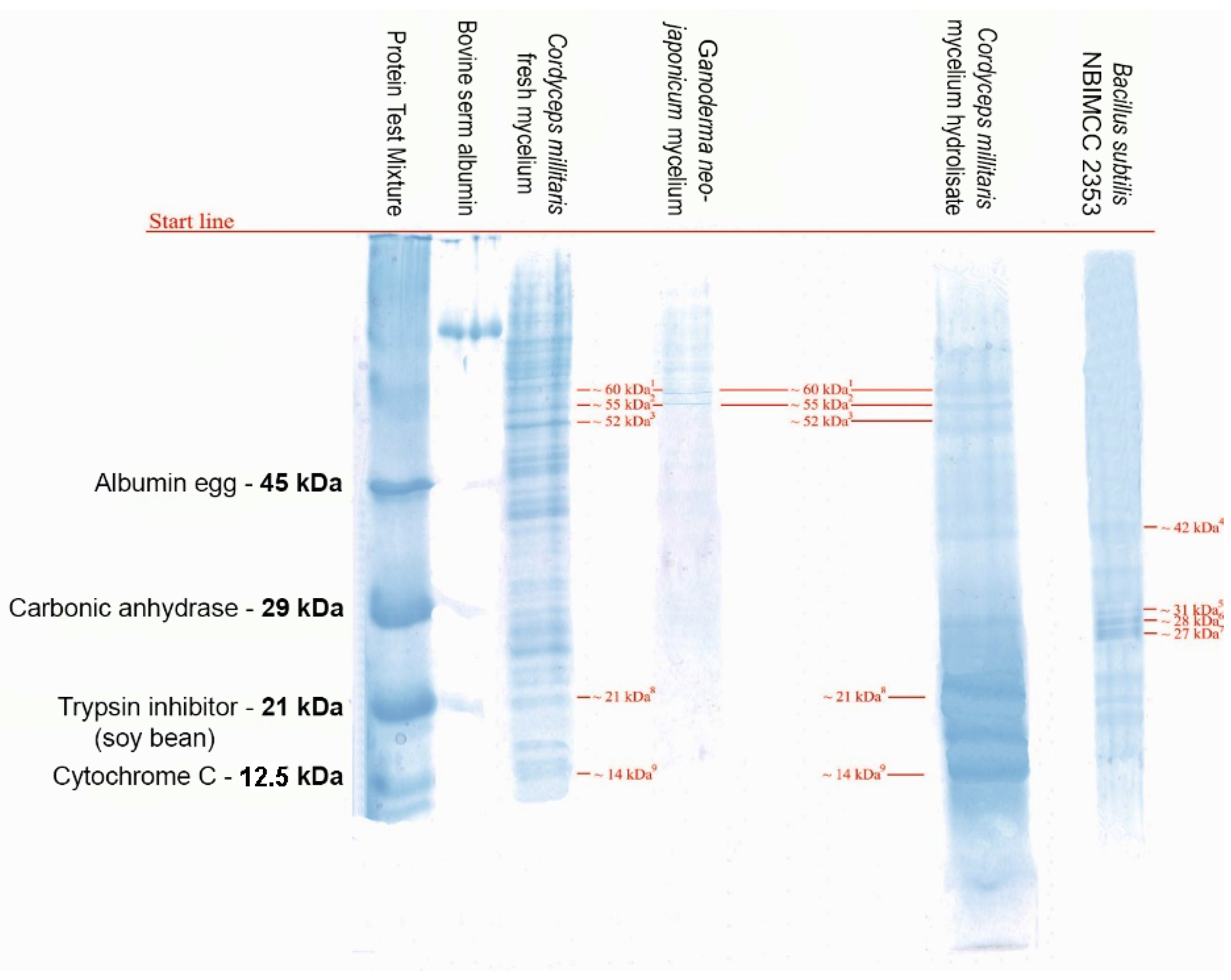
HPLC analysis was performed for the determination of cordycepin and adenosine derivatives. The results of the study are presented in Table 7.
The results presented in Table 7, demonstrate a clear trend of increasing cordycepin and adenosine content in the Cordyceps militaris sample cultured in the culture medium with optimized composition, with the presence of cordycepin being 2.22% and adenosine 0.64%, while in the Cordyceps militaris sample cultured in the culture medium with standard composition it was 0.32% and 0.4% for cordycepin and adenosine, respectively. The realized increase was 1.9% for cordycepin and 0.24% for adenosine, respectively. Representative HPLC chromatograms of the adenosine and cordycepin derivatives before and after the optimization of the method for cultivation of Cordyceps militaris and the corresponding standards have been added as Supplementary Materials (Figure S1). This result shows that the culture medium with the optimized composition in terms of increasing the cordycepin content can be successfully applied for laboratory and industrial production in small and large volumes. Additionally, a comparison of cordycepin and adenosine content data in mycelium samples cultured for nine months versus fresh mycelium samples laboratory grown in liquid medium for a period of 72 h were made. The results showed lower contents of the target substances in samples of fresh mycelium cultured in the laboratory compared to samples of mycelium grown industrially over a period of nine months. This is an expected result due to the fact that, during the initial phase of cultivation, during the first 72 h, intense enzymatic reactions take place in the mycelial culture leading to the utilization of a greater amount of adenosine and consequently a lower synthesis of its derivative substance cordycepin. This result demonstrates the fact that the use of mycelial culture for enzyme production is most productive in the interval up to 72 h.

3.5. SDS-PAGE

In order to determine protein fractions, an SDS-PAGE was performed [17]. Experimental demonstration of the presence of biologically active components in mycelial extracts of Ganoderma neo-japonicum, Ganoderma lucidum, Cordyceps sinensis, and fruiting body of Cordyceps militaris by the detection of proteins of different molecular mass was performed (Figure 2). A biochemical study was carried out on the protein spectrum of experimental samples of medicinal mushrooms and a culture sample of Bacillus subtilis NBIMCC 2353 according to the method of Laemmli [18].
The analysis of the protein spectrum of the culture fluid reported the presence of protein fractions with molecular masses ranging from 21 to 45 kDa. The molecule weights of major proteins in Cordyceps samples were from 6.5 to 97.2 kDa. The fractions of molecular mass of 27.7 kDa correspond to the enzyme nattokinase. Nattokinase, an enzyme secreted by B. subtilis, has been proven to exhibit fibrinolytic activity, in addition to its other beneficial effects, such as anti-coagulation, anti-atherosclerosis, anti-hypertension, and neuroprotection [19,20,21,22].

4. Discussion

For more than three decades, medicinal mushrooms have been approved in east Asian countries as an adjunct to standard cancer treatments and have an extensive clinical history of safe use as a single agent or in combination with chemotherapy [13,23].
Cultivation methods were refined with time, with respect to the knowledge regarding suitable substrates to grow for certain mushroom species, optimal temperature, initial pH requirement, white light, influence of heavy metal, aseptic techniques, and ways to curb diseases and pests to yield good harvest [17,24,25]. Water and ethanol mixture, used for the separation of cordycepin from Cordyceps militaris by a one-step extraction process, could be successfully applied as cost effective with high yield and purity cordycepin production [26]. Methods of near-infrared (NIRS) spectroscopy, compared with the high-performance liquid chromatography, used to predict adenosine and cordycepin concentrations in fruiting bodies of medicinal mushrooms provided excellent accuracy with minimal sample preparation [27].
Comparing the antimicrobial [28], antioxidant, and cytotoxic properties of the methanol extracts from fruiting bodies and fermented mycelia of C. militaris, the results revealed the stronger DPPH radical scavenging activity, while the fermented mycelia had stronger total antioxidant capacity, chelating ability, and reducing power, from which it could be suggested that they had their own role and worked in different ways [29].
The bioactive compounds presented in G. lucidum which can have immune system enhancement properties make it very special and classified under Host Defense Potentiators (HDP) [30,31]. Ganoderma research, specifically regarding its clinical aspects, requires more supportive clarifications for the dosage and side effects in human beings. The mechanism of action of different bioactive molecules isolated from G. lucidum is yet to be elucidated.
A technological model for the creation of a biologically active product based on organic sources has been developed. Extracts obtained as a result of the cultivation of medicinal fungi Ganoderma lucidum, Ganoderma neo-japonicum, Cordyceps sinensis, Cordyceps militaris, and the bacterial producer strain Bacillus subtilis were selected as the starting material for the production of the functional product.
The results obtained from the Cordyceps militaris extract cultured under industrial conditions by the optimized method demonstrated higher antioxidant activity against the DPPH- radical, which correlated with the results obtained for the total phenolic content. Also, a difference between the fresh mycelium samples versus the other samples of the adult fungal cultures is clearly visible. This is probably due to the fact that at the initial stage of cultivation, the accumulation of substances with antioxidant activity is low, and it is more likely at this stage that enzymes involved in fungal metabolism are present in the mycelial cultures. This highlights the possibility of targeting the cultivation model according to the need to obtain a biologically active extract. In cases where a high presence of phenolic compounds with high antioxidant activity is required, the optimized cultivation method should be applied up to the fruiting body stage, and in cases where the production of a biologically active extract with high enzymatic activity is required, the fresh mycelial culture method should be applied. Many of the enzymes produced by medicinal mushrooms are themselves biologically active substances. This is the case of metalloproteases, which, in addition to their proteolytic activity, also exhibit fibrinolytic action. The substance with the strongest antioxidant properties found in nature, namely the enzyme superoxide dismutase (SOD), classified with the enzyme code EC 1.15.1.1, is found in a variety of organisms, but in greater quantities in medicinal mushrooms [11]. Now that the medical community recognizes the stimulating properties of mushrooms, the combined market for gourmet products and medicinal foods is growing rapidly. By optimizing cultivation methods, it is possible to grow wild species under controlled condition that cannot be cultivated in an open place other than their natural habitat. As the example case is the wild strain of Cordyceps sinensis, which grows naturally at altitudes above 4000 m above sea level, under laboratory conditions, the DNA code of the wild strain of Cordyceps sinensis has been manipulated using snake venom protein to produce a hybrid, thus producing the best-known strain for laboratory cultivation, CS-4 [32]. CS-4 has been cultivated massively to produce a mycelial culture in liquid reactors and to obtain a rich biologically active extract with a composition very similar to that of wild Cordyceps sinensis, which is found in the mountainous region of Tibet, at altitudes above 4000 m above sea level.
Ganoderma (approximately seven species, including Ganoderma lucidum (Red Reishi), Ganoderma neo-japonicum (Black Reishi), and Ganoderma aplantu) is the most applied mushroom for medicinal purposes, possessing various therapeutic properties, such as anticancer activity, antioxidant, anti-inflammatory, antiallergic, and neuroprotective properties, and has a beneficial effect on the liver and kidneys, showing antiviral and antibacterial activity. Extracts with a high content of biologically active substances are used to strengthen the immune system, for prophylaxis in cardiovascular diseases, hypertension, diabetes, respiratory and gastrointestinal disorders, immunodeficiency states, etc. [7,33].
Bacillus subtilis has established itself in industry as a producer strain of various biologically active substances and enzymes leading to high total proteolytic, fibrinolytic, cellulase, phytinase, lipase, amylase, betaglucanase, and chitinase activities [34].
At the present report a fresh mycelium in spherical form was obtained which could be easily separated from the liquid phase of the culture medium, improving the speed of the production cycle. By adding native (non-hydrolyzed fragments) biopolymers, including specific proteins, chitin, etc., to the culture medium composition, the culturing process was successfully directed towards a higher production of peptides with targeted enzymatic activity (general proteolytic, fibrinolytic, cellulase, betaglucanase, chitinase). For this, the composition of the culture media was optimized by adding an industrially obtained extract of Ganoderma lucidum and Cordyceps sinensis with a high content (40%, [wt] of β-glucans).
Of immense interest for the production of biologically active extracts of medicinal mushrooms is freeze-drying, in which the solvent is sublimated under vacuum conditions and separated from the processed extract [35], and the solutes remain in the dried raw material.
The biological power embodied in the mycelium remains a largely untapped resource. Experiments and developments with mushroom mycelium will enable any farm, recycling centers, businesses, and individuals with a direct economic and environmental interest to achieve good results [36]. Prospects will be opened for better exploitation of all the advantages derived from the cultivation and processing of mushrooms to obtain valuable products for medicinal and prophylactic use. There were reports that with the extraction rate of adenosine as an index, the critical factors were extracting temperature, extracting time, and solid-liquid ratio [37].
Several pharmacologically active compounds have been reported from Cordyceps militaris, and among those, cordycepin has gained additional attention due to its wide spectrum of cellular and biological action [5,38]. Cordycepin and its related analogues have remarkable clinical health effects [3], including action on hepatic, renal, cardiovascular, respiratory, nervous, sexual, and immunological systems, besides having anti-cancer, antiviral, antioxidant, anti-inflammatory, and anti-microbial activities [5]. Mycelial extracts from Ganoderma lucidum and Cordyceps sinsnsis exhibited an antiproliferative action on highly invasive MDAMB-231 human breast cancer cells and arrested the cells at the G2/M phase of the cell cycle according to a report [39]. Cordyceps sinsnsis affords cardio protection possibly through enhanced adenosine receptor activation [40]. The cytotoxicity of partially purified peptide extracts from L. squarrosulus was indicated in lung cancer H460, H292, and H23 cells [21]. An investigation based on mutated Cordyceps militaris strains by UV irradiation showed approximately 1.5-fold increased productivity compared to wild type [41].

5. Conclusions

A method for the production of fresh mycelium of medicinal mushrooms in a liquid culture medium in order to obtain a high content of biologically active peptides with enzymatic activity was optimized. In order to increase the number of viable cells in a shorter period of time, a culturing method for Bacillus subtilis strain was developed. The application of combined biotechnological approaches in the use of biologically active components from natural organic sources and the resulting final product with high biological activity determine the present study as relevant and significant for its practical application.

Supplementary Materials

The following supporting information can be downloaded at: https://www.mdpi.com/article/10.3390/app122010618/s1, Figure S1: Chromatographic profile of the adenosine and cordycepin derivatives before (a), and after the optimization (b) of the semi-industrial method for cultivation and the production of biologically active substances from Cordyceps millitaris and standard mixture (c).

Author Contributions

Conceptualization, N.S.; methodology, N.S.; validation, N.S., A.C.d.O. and M.K.; formal analysis, N.S. and M.K.; investigation, N.S., M.K. and Z.M.; resources, N.S., M.K., Y.M. and K.L.; data curation, N.S., M.K. and Y.M.; writing—original draft preparation, M.K.; writing—review and editing, M.K., A.C.d.O. and Y.M.; visualization, N.S.; supervision, N.S. and M.K. All authors have read and agreed to the published version of the manuscript.

Funding

This research received no external funding.

Institutional Review Board Statement

Not applicable.

Informed Consent Statement

Not applicable.

Data Availability Statement

Not applicable.

Conflicts of Interest

The authors declare no conflict of interest.

References

  1. DeNinno, M.P. Chapter 11—Adenosine. In Annual Reports in Medicinal Chemistry; Bristol, J.A., Ed.; Academic Press: Cambridge, MA, USA, 1998; Volume 33, pp. 111–120. [Google Scholar]
  2. Ashraf, S.A.; Elkhalifa, A.E.O.; Siddiqui, A.J.; Patel, M.; Awadelkareem, A.M.; Snoussi, M.; Ashraf, M.S.; Adnan, M.; Hadi, S. Cordycepin for health and wellbeing: A potent bioactive metabolite of an entomopathogenic medicinal fungus Cordyceps with its nutraceutical and therapeutic potential. Molecules 2020, 25, 2735. [Google Scholar] [CrossRef] [PubMed]
  3. Yue, K.; Ye, M.; Zhou, Z.; Sun, W.; Lin, X. The genus Cordyceps: A chemical and pharmacological review. J. Pharm. Pharmacol. 2013, 65, 474–493. [Google Scholar] [CrossRef] [PubMed]
  4. Chaturvedi, V.K.; Agarwal, S.; Gupta, K.K.; Ramteke, P.W.; Singh, M.P. Medicinal mushroom: Boon for therapeutic applications. 3 Biotech 2018, 8, 334. [Google Scholar] [CrossRef] [PubMed]
  5. Mehra, A.; Zaidi, K.U.; Mani, A.; Thawani, V. The health benefits of Cordyceps militaris—A review. Kavaka 2017, 48, 27–32. [Google Scholar]
  6. Venturella, G.; Ferraro, V.; Cirlincione, F.; Gargano, M.L. Medicinal Mushrooms: Bioactive Compounds, Use, and Clinical Trials. Int. J. Mol. Sci. 2021, 10, 634. [Google Scholar] [CrossRef]
  7. Wasser, S.P.; Biomed, J. Medicinal Mushroom Science: Current Perspectives, Advances, Evidences, and Challenges. Biomed. J. 2014, 37, 345–356. [Google Scholar] [CrossRef]
  8. Qin, P.; Li, X.; Yang, H.; Wang, Z.-Y.; Lu, D. Therapeutic Potential and Biological Applications of Cordycepin and Metabolic Mechanisms in Cordycepin-Producing Fungi. Molecule 2019, 24, 2231. [Google Scholar] [CrossRef] [Green Version]
  9. Lu, C.-C.; Hsu, Y.-J.; Chang, C.-J.; Lin, C.-S.; Martel, J.; Ojcius, D.M.; Young, J.D. Immunomodulatory properties of medicinal mushrooms: Differential effects of water and ethanol extracts on NK cell-mediated cytotoxicity. Innate Immun. 2016, 22, 522–533. [Google Scholar] [CrossRef] [Green Version]
  10. Zhao, S.; Gao, Q.; Rong, C.; Wang, S.; Zhao, Z.; Liu, Y.; Xu, J. Immunomodulatory effects of edible and medicinal mushrooms and their bioactive immunoregulatory products. J. Fungi 2020, 6, 269. [Google Scholar] [CrossRef]
  11. Zhou, J.; Chen, M.; Wu, S.; Liao, X.; Wang, J.; Wu, Q.; Zhuang, M.; Ding, Y. A review on mushroom-derived bioactive peptides: Preparation and biological activities. Food Res. Int. 2020, 134, 109230. [Google Scholar] [CrossRef]
  12. Kirk, P.M.; Cannon, P.F.; David, J.C.; Stalpers, J.A. Ainsworth and Brisby’s Dictionary of the Fungi, 10th ed.; CAB International: Wallingford, UK, 2008. [Google Scholar]
  13. Jeitler, M.; Michalsen, A.; Frings, D.; Hübner, M.; Fischer, M.; Koppold-Liebscher, D.A.; Murthy, V.; Kessler, C.S. Significance of medicinal mushrooms in integrative oncology: A narrative review. Front. Pharmacol. 2020, 11, 580656. [Google Scholar] [CrossRef] [PubMed]
  14. Dang, H.-N.; Wang, C.-L.; Lay, H.-L. Effect of nutrition, vitamin, grains, and temperature on the mycelium growth and antioxidant capacity of Cordyceps militaris (strains AG-1 and PSJ-1). J. Radiat. Res. Appl. Sci. 2018, 11, 130–138. [Google Scholar] [CrossRef] [Green Version]
  15. Yoon, S.Y.; Lindroth, A.M.; Kwon, S.; Park, S.J.; Park, Y.J. Adenosine derivatives from Cordyceps exert antitumor effects against ovarian cancer cells through ENT1-mediated transport, induction of AMPK signaling, and consequent autophagic cell death. Biomed. Pharmacother. 2022, 1, 113491. [Google Scholar] [CrossRef] [PubMed]
  16. Quy, T.N.; Xuan, T.D.; Andriana, Y.; Tran, H.D.; Khanh, T.D.; Teschke, R. Cordycepin Isolated from Cordyceps militaris: Its Newly Discovered Herbicidal Property and Potential Plant-Based Novel Alternative to Glyphosate. Molecules 2019, 24, 2901. [Google Scholar] [CrossRef] [Green Version]
  17. Gargano, M.L.; van Griensven, L.J.; Isikhuemhen, O.S.; Lindequist, U.; Venturella, G.; Wasser, S.P.; Zervakis, G.I. Medicinal mushrooms: Valuable biological resources of high exploitation potential. Plant Biosyst. Int. J. Deal. Asp. Plant Biol. 2017, 151, 548–565. [Google Scholar] [CrossRef]
  18. Laemmli, U.K. Cleavage of structural proteins during the assembly of the head of bacteriophage T4. Nature 1970, 227, 680–685. [Google Scholar] [CrossRef] [PubMed]
  19. Chen, H.; McGowan, E.M.; Ren, N.; Lal, S.; Nassif, N.; Shad-Kaneez, F.; Qu, X.; Lin, Y. Nattokinase: A promising alternative in prevention and treatment of cardiovascular diseases. Biomark. Insights 2018, 13, 1177271918785130. [Google Scholar] [CrossRef] [Green Version]
  20. Hsu, R.-L.; Lee, K.-T.; Wang, J.-H.; Lee, L.Y.-L.; Chen, R.P.-Y. Amyloid-Degrading Ability of Nattokinase from Bacillus subtilis Natto. J. Agric. Food Chem. 2009, 57, 503–508. [Google Scholar] [CrossRef]
  21. Prateep, A.; Sumkhemthong, S.; Suksomtip, M.; Chanvorachote, P.; Chaotham, C. Peptides extracted from edible mushroom: Lentinus squarrosulus induces apoptosis in human lung cancer cells. Pharm. Biol. 2017, 55, 1792–1799. [Google Scholar] [CrossRef] [Green Version]
  22. Weng, Y.; Yao, J.; Sparks, S.; Wang, K.Y. Nattokinase: An oral antithrombotic agent for the prevention of cardiovascular disease. Int. J. Mol. Sci. 2017, 18, 523. [Google Scholar] [CrossRef] [Green Version]
  23. Rossi, P.; Difrancia, R.; Quagliariello, V.; Savino, E.; Tralongo, P.; Randazzo, C.L.; Berretta, M. B-glucans from Grifola frondosa and Ganoderma lucidum in breast cancer: An example of complementary and integrative medicine. Oncotarget 2018, 9, 24837. [Google Scholar] [CrossRef] [PubMed]
  24. Berovic, M. Cultivation of Medicinal Mushroom Biomass by Solid-State Bioprocessing in Bioreactors. In Solid State Fermentation; Steudler, S., Werner, A., Cheng, J.J., Eds.; Springer: Cham, Switzerland, 2019; Volume 169, pp. 3–25. [Google Scholar]
  25. Singh, B.P.; Chhakchhuak, L.; Passari, A.K. (Eds.) Biology of Macrofungi (Fungal Biology); Springer: Cham, Switzerland, 2018. [Google Scholar]
  26. Sornchaithawatwong, C.; Kunthakudee, N.; Sunsandee, N.; Ramakul, P. Selective extraction of cordycepin from Cordyceps militaris–optimisation, kinetics and equilibrium studies. Indian Chem. Eng. 2020, 64, 1–13. [Google Scholar] [CrossRef]
  27. Singpoonga, N.; Rittiron, R.; Seang-On, B.; Chaiprasart, P.; Bantadjan, Y. Determination of Adenosine and Cordycepin Concentrations in Cordyceps militaris Fruiting Bodies Using Near-Infrared Spectroscopy. ACS Omega 2020, 16, 27235–27244. [Google Scholar] [CrossRef] [PubMed]
  28. Arias-Londoño, M.A.; Zapata-Ocampo, P.A.; Mosquera-Arevalo, A.R.; Sanchez-Torres, J.D.; Atehortua-Garcés, L. Antifungal protein determination for submerged cultures of the medicinal mushroom Ganoderma lucidum (Ganodermataceae) with activity over the phytopathogen fungus Mycosphaerella fijiensis (Mycosphaerellaceae). Actual. Biol. 2020, 41, 53–64. [Google Scholar] [CrossRef]
  29. Dong, C.-H.; Yang, T.; Lian, T. A Comparative Study of the Antimicrobial, Antioxidant, and Cytotoxic Activities of Methanol Extracts from Fruit Bodies and Fermented Mycelia of Caterpillar Medicinal Mushroom Cordyceps militaris (Ascomycetes). Int. J. Med. Mushrooms 2014, 16, 485–495. [Google Scholar] [CrossRef] [PubMed]
  30. Hapuarachchi, K.K.; Elkhateeb, W.A.; Karunarathna, S.C.; Cheng, C.R.; Bandara, A.R.; Kakumyan, P.; Hyde, K.D.; Daba, G.M.; Wen, T.C. Current status of global Ganoderma cultivation, products, industry and market. Mycosphere 2018, 9, 1025–1052. [Google Scholar] [CrossRef]
  31. Sudheer, S.; Alzorqi, I.; Manickam, S.; Ali, A. Bioactive Compounds of the Wonder Medicinal Mushroom “Ganoderma lucidum”. In Bioactive Molecules in Food; Reference Series in Phytochemistry; Springer: Cham, Switzerland, 2019; pp. 1863–1893. [Google Scholar]
  32. Cleaver, P.D.; Holliday, J.C.; Powers, M.L.; Aloha Medicinals Inc. Method for Growing Cordyceps sinensis on a Substrate. U.S. Patent No 8,008,060, 30 August 2011. [Google Scholar]
  33. Money, N.P. Are mushrooms medicinal? Fungal Biol. 2016, 120, 449–453. [Google Scholar] [CrossRef] [Green Version]
  34. Kaspar, F.; Neubauer, P.; Gimpel, M. Bioactive Secondary Metabolites from Bacillus subtilis: A Comprehensive Review. J. Nat. Prod. 2019, 82, 2038–2053. [Google Scholar] [CrossRef]
  35. Pei, F.; Shi, Y.; Mariga, A.M.; Yang, W.J.; Tang, X.Z.; Zhao, L.Y.; An, X.X.; Hu, Q.H. Comparison of freeze-drying and freeze-drying combined with microwave vacuum drying methods on drying kinetics and rehydration characteristics of button mushroom (Agaricus bisporus) slices. Food Bioprocess Technol. 2014, 7, 1629–1639. [Google Scholar] [CrossRef]
  36. Haneef, M.; Ceseracciu, L.; Canale, C.; Bayer, I.S.; Heredia-Guerrero, J.A.; Athanassiou, A. Advanced Materials From Fungal Mycelium: Fabrication and Tuning of Physical Properties. Sci. Rep. 2017, 7, 41292. [Google Scholar] [CrossRef] [Green Version]
  37. Du, L.N.; Song, J.; Meng, L.J.; Lu, J.H.; Meng, Q.F.; Teng, L.R. Application of Two Modeling Methods in Optimization for Adenosine Extraction from Mycelium of Cordyceps militaris. Adv. Mater. Res. 2011, 343–344, 826–831. [Google Scholar] [CrossRef]
  38. Guo, M.; Guo, S.; Huaijun, Y.; Bu, N.; Dong, C. Comparison of Major Bioactive Compounds of the Caterpillar Medicinal Mushroom, Cordyceps militaris (Ascomycetes), Fruiting Bodies Cultured on Wheat Substrate and Pupae. Int. J. Med. Mushrooms 2016, 18, 327–336. [Google Scholar] [CrossRef] [PubMed]
  39. Wong, J.H.; Ng, T.B.; Chan, H.H.L.; Liu, Q.; Man, G.C.W.; Zhang, C.Z.; Guan, S.; Ng, C.C.W.; Fang, E.F.; Wang, H.; et al. Mushroom extracts and compounds with suppressive action on breast cancer: Evidence from studies using cultured cancer cells, tumor-bearing animals, and clinical trials. Appl. Microbiol. Biotechnol. 2020, 104, 4675–4703. [Google Scholar] [CrossRef]
  40. Yan, X.-F.; Zhang, Z.-M.; Yao, H.-Y.; Guan, Y.; Zhu, J.-P.; Zhang, L.-H.; Wang, R.-W. Cardiovascular Protection and Antioxidant Activity of the Extracts from the Mycelia of Cordyceps sinensis Act Partially Via Adenosine Receptors. Phytother. Res. 2012, 27, 1597–1604. [Google Scholar] [CrossRef] [PubMed]
  41. Lee, S.K.; Lee, J.H.; Kim, H.R.; Chun, Y.; Lee, J.H.; Yoo, H.Y.; Park, C.; Kim, S.W. Improved cordycepin production by Cordyceps militaris KYL05 using casein hydrolysate in submerged conditions. Biomolecules 2019, 9, 461. [Google Scholar] [CrossRef]
Figure 1. Cordyceps militaris fruiting body cultivation—Shanghai.
Applsci 12 10618 g001
Figure 2. Polyacrylamide gel electrophoresis of medicinal mushroom samples of Cordyceps millitaris (fresh mycelium (1) and hydrolysate (2), Ganoderma neo-japonicum (fresh mycelium (3)) and Bacillus subtilis NBIMCC 2353 (8) cultured strain sample. Test mixture of proteins of known molecular weight and Bovine serum albumin (66.4 kDA) were used as a standard.
Applsci 12 10618 g002
Table 1. Liquid culture media composition for fresh mycelial culture.
Media Composition
g/L
M0
Control
M1M2M3M4
Potato Dextrose Agar2020202020
Peptone-10101010
Yeast extract66666
Glucose-2525--
Dextrose25--2525
Fructooligosaccharides (FOS)55555
MgSO4 × 7 H2O-0.20.20.20.2
Vitamin B1--0.030.030.03
KH2PO4--0.10.10.1
K2HPO4--0.20.20.2
FeSO4----0.1
Table 2. Media composition of solid culture for mycelium cultivation.
Media Composition
g/L
S1S2S3
Dextrose202020
Potato Dextrose Agar555
Yeast hydrolysate555
Agar151515
Whole grain rise-200-
Oak sawdust--200
Table 3. Media composition for cultivation of Bacillus subtilis NBIMCC 2353.
Media Composition
g/L
Control Media
g/L
P1P2P3P4P5
Meat extract101010101010
Peptone101010101010
Yeast extract1.51.51.51.51.51.5
Fructooligosaccharides (FOS)111111
NaCl555555
K2HPO41.31.31.31.31.31.3
MgSO40.30.30.30.30.30.3
Native bovine collagen-40----
GNJM--40---
GLM---40--
CSM----40-
CMM-----40
Table 4. Proteolytic activity of Bacillus subtilis in medium supplemented with native bovine collagen protein and mycelium.
MediumPU/mL, 24 hPU/mL, 48 hPU/mL, 72 h
K1.38 ± 0.0042.27 ± 0.0061.89 ± 0.003
P115.74 ± 0.00623.74 ± 0.00719.71 ± 0.005
P217.37 ± 0.00426.83 ± 0.00822.18 ± 0.007
P318.34 ± 0.00730.57 ± 0.00625.47 ± 0.008
P414.91 ± 0.00324.42 ± 0.00720.56 ± 0.005
P513.67 ± 0.00521.51 ± 0.00717.85 ± 0.004
PU—Proteolytic Units; K—Control, P1—medium with added native bovine collagen, P2—medium with added mycelium of Ganoderma neo-japonicum, P3—medium with added mycelium of Ganoderma lucidum, P4—medium with added mycelium of Cordyceps sinensis and P5—medium with added mycelium of Cordyceps militaris.
Table 5. Chitinase enzyme activity of Bacillus subtilis.
MediaChitinase Activity (U/mL) Recorded
after 24 h
Chitinase Activity (U/mL) Recorded
after 48 h
Chitinase Activity (U/mL) Recorded
after 72 h
Control (K)4.37 ± 0.0028.65 ± 0.0016.83 ± 0.006
CH132.57 ± 0.00740.86 ± 0.00637.68 ± 0.007
CH233.78 ± 0.00741.62 ± 0.00338.43 ± 0.002
CH334.46 ± 0.00742.73 ± 0.00339.58 ± 0.002
CH431.64 ± 0.00739.93 ± 0.00336.82 ± 0.006
CH533.47 ± 0.00741.29 ± 0.00338.62 ± 0.004
Table 6. Spectrophotometric assay for the determination of antioxidant activity in mushroom mycelium extracts.
SampleTEAC mg/gSTD ± mg/g STD%
CMF
(Industrial-st)
0.710.0424.71
CMM
(Industrial)
0.670.0030.33
CSM
(Industrial)
0.500.0081.19
GLM
(Industrial)
1.180.0624.77
CMFB
(Industrial-opt)
1.850.0191.02
CMFresh0.650.0183.24
CSFresh0.100.0215.70
GLFresh0.090.025.47
GNJFresh0.150.0051.19
TEAC—Tocopherol antioxidant equivalent capacity. CMF (Industrial-st) Cordyceps millitaris fetal part industrially glued according to standard method, CMM (Industrial)—Cordyceps millitaris—mycelium industrially glued, CSM (Industrial)—Cordyceps cinensis—mycelium industrially glued, GLM (Industrial)—Ganoderma lucidum—mycelium industrially glued, CMFB (Industrial-opt) Cordyceps millitaris fetus partially industrially decanted by optimized method, CMFresh—fresh mycelium from Cordyceps millitaris, CSFresh—fresh mycelium from Cordyceps sinensis, GLFresh—fresh mycelium from Ganoderma lucidum, GNJFresh—fresh mycelium from Ganoderma neo-japonicum.
Table 7. HPLC analysis for the determination of cordycepin and adenosine derivatives.
GLM
(Industrial)
CSM
(Industrial)
CMM
(Industrial)
CMM
(Optimal Industrial),
CMFreshCSFreshGLFreshGNJFresh
Adenosins Dry %0.30.590.40.640.090.120.10.05
Adenosins Dry c [mg/g]29.9459.1440.3564.228.4612.1610.544.52
Cordycepins Dry %0.090.280.322.220.080.050.10.11
Cordycepins Dry c [mg/g]9.0128.332.25221.697.834.889.7210.79
GLM (Industrial)—Ganoderma lucidum Mycelium industrially grown, CSM—Cordyceps sinensis mycelium industrially grown, CMM (Industrial)—Cordyceps militaris mycelium industrially grown, CMFresh—Cordyceps militaris fresh mycelium, CSFresh—Cordyceps sinensis fresh mycelium, GLFresh—Ganoderma lucidum fresh mycelium, GNJFresh—Ganoderma neo-japonicum fresh mycelium.
Publisher’s Note: MDPI stays neutral with regard to jurisdictional claims in published maps and institutional affiliations.

