
Preliminary Approach for Open Lateral Window Technique for Successful Maxillary Sinus Augmentation in the Unrepairable Wide Perforation Area of Schneiderian Membrane


Abstract
:1. Introduction
2. Case Presentation
2.1. Surgical Procedure
2.2. Patients
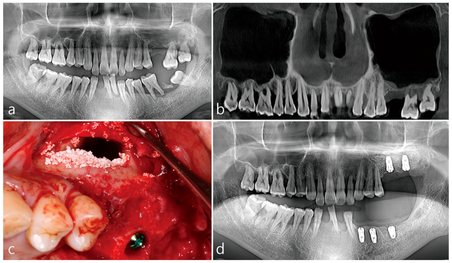
2.2.1. Case 1
2.2.2. Case 2
2.2.3. Case 3
3. Discussion
4. Conclusions
Author Contributions
Funding
Informed Consent Statement
Data Availability Statement
Conflicts of Interest
References
- Starch-Jensen, T.; Aludden, H.; Hallman, M.; Dahlin, C.; Christensen, A.E.; Mordenfeld, A. A systematic review and meta-analysis of long-term studies (five or more years) assessing maxillary sinus floor augmentation. Int. J. Oral Maxillofac. Surg. 2018, 47, 103–116. [Google Scholar] [CrossRef] [PubMed]
- Tatum, O.H. Maxillary sinus grafting for endosseous implants. In Proceedings of the Annual Meeting of Alabama Implant Study Groupe, Birmingham, AL, USA, 13–14 April 1977. [Google Scholar]
- Boyne, P.J.; James, R.A. Grafting of the maxillary sinus floor with autogenous marrow and bone. J. Oral Surg. 1980, 38, 613–616. [Google Scholar] [PubMed]
- Park, W.B.; Kang, K.L.; Han, J.Y. Factors influencing long-term survival rates of implants placed simultaneously with lateral maxillary sinus floor augmentation: A 6- to 20-year retrospective study. Clin. Oral Implants Res. 2019, 30, 977–988. [Google Scholar] [CrossRef] [PubMed]
- Nooh, N. Effect of schneiderian membrane perforation on posterior maxillary implant survival. J. Int. Oral Health. 2013, 5, 28–34. [Google Scholar] [PubMed]
- Anavi, Y.; Allon, D.M.; Avishai, G.; Calderon, S. Complications of maxillary sinus augmentations in a selective series of patients. Oral Surg. Oral Med. Oral Pathol. Oral Radiol. Endod. 2008, 106, 34–38. [Google Scholar] [CrossRef]
- Hernández-Alfaro, F.; Torradeflot, M.M.; Marti, C. Prevalence and management of Schneiderian membrane perforations during sinus-lift procedures. Clin. Oral Implants Res. 2008, 19, 91–98. [Google Scholar] [CrossRef]
- Pikos, M.A. Maxillary sinus membrane repair: Update on technique for large and complete perforations. Implant Dent. 2008, 17, 24–31. [Google Scholar] [CrossRef]
- Park, W.B.; Lim, H.C. Late Developed Unusual Nasal Involvement of Postoperative Maxillary Cyst Following Maxillary Sinus Augmentation: A Case Report. Appl. Sci. 2021, 11, 10730. [Google Scholar] [CrossRef]
- Sakkas, A.; Konstantinidis, I.; Winter, K.; Schramm, A.; Wilde, F. Effect of Schneiderian membrane perforation on sinus lift graft outcome using two different donor sites: A retrospective study of 105 maxillary sinus elevation procedures. GMS Interdiscip. Plast. Reconstr. Surg. DGPW. 2016, 5, Doc11. [Google Scholar] [CrossRef]
- Timmenga, N.M.; Raghoebar, G.M.; Liem, R.S.; Van Weissenbruch, R.; Manson, W.L.; Vissink, A. Effects of maxillary sinus floor elevation surgery on maxillary sinus physiology. Eur. J. Oral Sci. 2003, 111, 189–197. [Google Scholar] [CrossRef]
- Zijderveld, S.A.; Van den Bergh, J.P.; Schulten, E.A.; Ten Bruggenkate, C.M. Anatomical and surgical findings and complications in 100 consecutive maxillary sinus floor elevation procedures. J. Oral Maxillofac. Surg. 2008, 66, 1426–1438. [Google Scholar] [CrossRef]
- Urban, I.A.; Nagursky, H.; Church, C.; Lozada, J.L. Incidence, diagnosis, and treatment of sinus graft infection after sinus floor elevation: A clinical study. Int. J. Oral Maxillofac. Implants. 2012, 27, 449–457. [Google Scholar]
- Chiapasco, M.; Felisati, G.; Zaniboni, M.; Pipolo, C.; Borloni, R.; Lozza, P. The treatment of sinusitis following maxillary sinus grafting with the association of functional endoscopic sinus surgery (FESS) and an intra-oral approach. Clin. Oral Implants Res. 2013, 24, 623–629. [Google Scholar] [CrossRef]
- Schwarz, L.; Schiebel, V.; Hof, M.; Ulm, C.; Watzek, G.; Pommer, B. Risk Factors of Membrane Perforation and Postoperative Complications in Sinus Floor Elevation Surgery: Review of 407 Augmentation Procedures. J. Oral Maxillofac. Surg. 2015, 73, 1275–1282. [Google Scholar] [CrossRef]
- Katranji, A.; Fotek, P.; Wang, H.L. Sinus augmentation complications: Etiology and treatment. Implant Dent. 2008, 17, 339–349. [Google Scholar] [CrossRef]
- Hunter, W.L., IV; Bradrick, J.P.; Houser, S.M.; Patel, J.B.; Sawady, J. Maxillary sinusitis resulting from ostium plugging by dislodged bone graft: Case report. J. Oral Maxillofac. Surg. 2009, 67, 1495–1498. [Google Scholar] [CrossRef]
- Aimetti, M.; Romagnoli, R.; Ricci, G.; Massei, G. Maxillary sinus elevation: The effect of macrolacerations and microlacerations of the sinus membrane as determined by endoscopy. Int. J. Periodontics Restor. Dent. 2001, 21, 581–589. [Google Scholar]
- Toscano, N.J.; Holtzclaw, D.; Rosen, P.S. The effect of piezoelectric use on open sinus lift perforation: A retrospective evaluation of 56 consecutively treated cases from private practices. J. Periodontol. 2010, 81, 167–171. [Google Scholar] [CrossRef]
- Ardekian, L.; Oved-Peleg, E.; Mactei, E.E.; Peled, M. The clinical significance of sinus membrane perforation during augmentation of the maxillary sinus. J. Oral Maxillofac. Surg. 2006, 64, 277–282. [Google Scholar] [CrossRef]
- Testori, T.; Wallace, S.S.; Del Fabbro, M.; Taschieri, S.; Trisi, P.; Capelli, M.; Weinstein, R.L. Repair of large sinus membrane perforations using stabilized collagen barrier membranes: Surgical techniques with histologic and radiographic evidence of success. Int. J. Periodontics Restor. Dent. 2008, 28, 9–17. [Google Scholar]
- Proussaefs, P.; Lozada, J.; Kim, J.; Rohrer, M.D. Repair of the perforated sinus membrane with a resorbable collagen membrane: A human study. Int. J. Oral Maxillofac. Implants. 2004, 19, 413–420. [Google Scholar] [PubMed]
- Van den Bergh, J.P.; Ten Bruggenkate, C.M.; Disch, F.J.; Tuinzing, D.B. Anatomical aspects of sinus floor elevations. Clin. Oral Implants Res. 2000, 11, 256–265. [Google Scholar] [CrossRef] [PubMed]
- Becker, S.T.; Terheyden, H.; Steinriede, A.; Behrens, E.; Springer, I.; Wiltfang, J. Prospective observation of 41 perforations of the Schneiderian membrane during sinus floor elevation. Clin. Oral Implants Res. 2008, 19, 1285–1289. [Google Scholar] [CrossRef]
- Aricioglu, C.; Dolanmaz, D.; Esen, A.; Isik, K.; Avunduk, M.C. Histological evaluation of effectiveness of platelet-rich fibrin on healing of sinus membrane perforations: A preclinical animal study. J. Craniomaxillofac. Surg. 2017, 45, 1150–1157. [Google Scholar] [CrossRef] [PubMed]
- Shlomi, B.; Horowitz, I.; Kahn, A.; Dobriyan, A.; Chaushu, G. The effect of sinus membrane perforation and repair with Lambone on the outcome of maxillary sinus floor augmentation: A radiographic assessment. The Int. J. Oral Maxillofac. Implants. 2004, 19, 559–562. [Google Scholar]
- Barbu, H.M.; Iancu, S.A.; Jarjour Mirea, I.; Mignogna, M.D.; Samet, N.; Calvo-Guirado, J.L. Management of Schneiderian Membrane Perforations during Sinus Augmentation Procedures: A Preliminary Comparison of Two Different Approaches. J. Clin. Med. 2019, 8, 1491. [Google Scholar] [CrossRef]
- Díaz-Olivares, L.A.; Cortés-Bretón Brinkmann, J.; Martínez-Rodríguez, N.; Martínez-González, J.M.; López-Quiles, J.; Leco-Berrocal, I. Meniz-García Management of Schneiderian membrane perforations during maxillary sinus floor augmentation with lateral approach in relation to subsequent implant survival rates: A systematic review and meta-analysis. Int. J. Implant Dent. 2021, 12, 91. [Google Scholar] [CrossRef]
- Nolan, P.J.; Freeman, K.; Kraut, R.A. Correlation between Schneiderian membrane perforation and sinus lift graft outcome: A retrospective evaluation of 359 augmented sinus. J. Oral Maxillofac. Surg. 2014, 72, 47–52. [Google Scholar] [CrossRef]
- Berbéri, A.; Sabbagh, J.; Bou Assaf, R.; Ghassibe-Sabbagh, M.; Al-Nemer, F.; El Majzoub, R.; Fayyad-kazan, M.; Badran, B. Comparing the osteogenic potential of schneiderian membrane and dental pulp mesenchymal stem cells: An in vitro study. Cell Tissue Bank. 2021, 22, 409–417. [Google Scholar] [CrossRef]
- Srouji, S.; Kizhner, T.; Ben David, D.; Riminucci, M.; Bianco, P.; Livne, E. The Schneiderian membrane contains osteoprogenitor cells: In vivo and in vitro study. Calcif. Tissue Int. 2009, 84, 138–145. [Google Scholar] [CrossRef]
- Derjac-Aramă, A.I.; Sarafoleanu, C.; Manea, C.M.; Nicolescu, M.I.; Vrapciu, A.D.; Rusu, M.C. Regenerative potential of human schneiderian membrane: Progenitor cells and epithelial-mesenchymal transition. Anat. Rec. 2015, 298, 2132–2140. [Google Scholar] [CrossRef]
- Jung, J.H.; Choi, B.H.; Jeong, S.M.; Li, J.; Lee, S.H.; Lee, H.J. A retrospective study of the effects on sinus complications of exposing dental implants to the maxillary sinus cavity. Oral Surg. Oral Med. Oral Pathol. Oral Radiol. Endod. 2007, 103, 623–625. [Google Scholar] [CrossRef]
- Zhong, W.; Chen, B.; Liang, X.; Ma, G. Experimental study on penetration of dental implants into the maxillary sinus in different depths. J. Appl. Oral Sci. 2013, 21, 560–566. [Google Scholar] [CrossRef]
- Troedhan, A.; Kurrek, A.; Wainwright, M. Biological Principles and Physiology of Bone Regeneration under the Schneiderian Membrane after Sinus Lift Surgery: A Radiological Study in 14 Patients Treated with the Transcrestal Hydrodynamic Ultrasonic Cavitational Sinus Lift (Intralift). Int. J. Dent. 2012, 2012, 576238. [Google Scholar] [CrossRef]
- Scarano, A.; Piattelli, A.; Iezzi, G.; Varvara, G. Spontaneous bone formation on the maxillary sinus floor in association with surgery to remove a migrated dental implant: A case report. Minerva Stomatol. 2014, 63, 351–359. [Google Scholar]


Publisher’s Note: MDPI stays neutral with regard to jurisdictional claims in published maps and institutional affiliations. |
© 2022 by the authors. Licensee MDPI, Basel, Switzerland. This article is an open access article distributed under the terms and conditions of the Creative Commons Attribution (CC BY) license (https://creativecommons.org/licenses/by/4.0/).
Share and Cite
Park, W.-B.; Crasto, G.J.; Kang, P. Preliminary Approach for Open Lateral Window Technique for Successful Maxillary Sinus Augmentation in the Unrepairable Wide Perforation Area of Schneiderian Membrane. Appl. Sci. 2022, 12, 9725. https://doi.org/10.3390/app12199725
Park W-B, Crasto GJ, Kang P. Preliminary Approach for Open Lateral Window Technique for Successful Maxillary Sinus Augmentation in the Unrepairable Wide Perforation Area of Schneiderian Membrane. Applied Sciences. 2022; 12(19):9725. https://doi.org/10.3390/app12199725
Chicago/Turabian StylePark, Won-Bae, Gazelle Jean Crasto, and Philip Kang. 2022. "Preliminary Approach for Open Lateral Window Technique for Successful Maxillary Sinus Augmentation in the Unrepairable Wide Perforation Area of Schneiderian Membrane" Applied Sciences 12, no. 19: 9725. https://doi.org/10.3390/app12199725