Article

Polysaccharides from Moroccan Green and Brown Seaweed and Their Derivatives Stimulate Natural Defenses in Olive Tree Leaves

1
Center for Agrobiotechnology and Bioengineering, CNRST Labeled Research Unit (Centre AgroBiotech, URL-CNRST 05), Faculty of Science and Technology, Cadi Ayyad University, Marrakech 40000, Morocco
2
Université Clermont Auvergne, Clermont Auvergne INP, CNRS, Institut Pascal, F-63000 Clermont-Ferrand, France
3
Laboratory of Biotechnology and Valorization of Plant Resources, Faculty of Sciences, Chouaib Doukkali University, El Jadida 24000, Morocco
4
Laboratory of Plant Biotechnology Applied to Crop Improvement, Faculty of Sciences of Sfax, University of Sfax, Sfax 3038, Tunisia
5
Laboratory of Enzymatic Engineering and Microbiology, Algae Biotechnology Team, National School of Engineers of Sfax, University of Sfax, Sfax 3038, Tunisia
6
Laboratory of Protection of Ecosystems in Arid and Semi-Arid Zones, Faculty of Natural and Life Sciences BP 511, Ouargla Université, Kasdi Merbah University, Ouargla 30000, Algeria
7
Institute of Chemistry of Clermont-Ferrand, Clermont Auvergne University, CNRS, SIGMA Clermont, 63000 Clermont-Ferrand, France
8
Institut Universitaire de France (IUF), 1 Rue Descartes, 75005 Paris, France
*
Author to whom correspondence should be addressed.
Appl. Sci. 2022, 12(17), 8842; https://doi.org/10.3390/app12178842
Submission received: 29 July 2022 / Revised: 25 August 2022 / Accepted: 31 August 2022 / Published: 2 September 2022

Abstract

:
This study aims to assess for the first time the ability of marine polysaccharides and their derivatives to stimulate natural defenses in olive tree leaves. Alginates, ALSM, and ALCM were isolated from the brown algae Sargassum muticum and Cystoseira myriophylloides, respectively. The OASM and OACM fractions were obtained after radical depolymerization of ALSM and ALCM, respectively. Three sulfated polysaccharides, arabinogalactans (AGB and AGP) and fucoidans (FUCCM) were extracted from the green alga Codium decorticatum and the brown seaweed C. myriophylloides, respectively. The extraction yield of different extracts was in the range of 0.82–32% and the average molecular weight (Mw) varied from 3000 Da to 2173 kDa. The M/G ratios of ALSM and ALCM were 0.87 and 1.12, respectively. FUCCM contained 53% of fucose and 12.83% of sulfates. The AGB and AGP fractions were characterized by the presence of a high degree of sulfation and protein (12–23% (w/w)) and were composed mainly of galactose, glucose, and arabinose. The aqueous saccharide solutions were applied to the leaf discs of the olive tree at 0.5 g/L, 1 g/L, and 2 g/L, for 24 h. These molecules triggered defense responses, by showing a differential capacity to induce the activity of the phenylalanine and tyrosine ammonia-lyase (TAL and PAL), polyphenols, and lignin contents in the leaves of the olive tree. Alginates and their derivatives as well as arabinogalactans exhibited an important induction of TAL activity compared to the PAL. The sulfated polysaccharides were more effective compared to the unsulfated polysaccharides (alginates) which were active at a low concentration. The optimum concentration for most of the studied elicitors was 2 g/L. These results suggest the valorization of these molecules derived from marine biomass as inducers of natural defenses of the olive tree to protect against phytopathogens in the context of sustainable development.

1. Introduction

The olive tree (Olea europaea L.) plays a key socio-economic and ecological role in Mediterranean countries. It is one of the most cultivated crop species and the only one of the Oleaceae family that produces comestible fruits processed for oil and table olive production [1]. In the European Union (EU), the olive-growing area was about 4.59 million hectares (ha) in 2017. Three-quarters of the utilized area were concentrated in Spain, with 2.5 million ha (55%), followed by Italy with 1.07 million ha (23%), and Greece with 15% of the total EU area under olive trees [2]. Spain is the biggest producer of olive oil, providing 59% of total EU production, followed by Italy, Portugal, and Greece with 24.8%, 8.4%, and 7.3%, respectively [3]. Morocco, after Spain, Italy, and Greece, has the fourth-largest olive tree cultivation, covering 78,400 ha and producing 1.48 million tons of olives per year [4]. It is the 2nd largest producer of table olives and the 6th of olive oil [5].
Unfortunately, the olive tree is subjected to several biotic stresses, influencing the health, yield, and quality of the oil and fruits [6,7,8]. Verticillium dahlia (Verticillium wilt) and Spilocaea oleagina (olive leaf spot) are the major pathogenic fungi of the olive tree in the world [7,9]. Anthracnose, caused by Colletotrichum spp., is also one of the most severe diseases of this woody crop [10]. Furthermore, the olive fruit fly poses a serious economic problem. It reduces about 15% of production, equivalent to an average of 800 million dollars per year [11]. Today, pesticides are an important input in the protection of the olive tree [7]. Despite their effectiveness, the residues of these agrochemicals in fruits and olive oil present a major risk to public health [7,12]. Recently, researchers have focused on using biological control agents (BCAs) such as arbuscular mycorrhizal fungi (AMF), plant growth-promoting rhizobacteria (PGPR), plant growth-promoting fungi (PGPF), and endophytes as an eco-friendly alternative to control olive diseases [12]. However, the use of BCAs is qualified by some restrictions, namely negative impact on non-target microorganisms, issues of adaptation to the target ecological niche, and especially direct effects leading to resistance increases in pathogen communities [9]. Therefore, sustainable approaches such as the stimulation of natural defenses of plants using natural elicitors could be a promising alternative to preventing disease development in the olive tree and reducing dependence on pesticides. This strategy aims to induce plant resistance using molecules, such as lipids, glycoproteins, peptides, proteins, oligosaccharides, or polysaccharides [13]. Systemic acquired resistance (SAR) and induced systemic resistance (ISR), are two types of plant resistance involved depending on the nature of the elicitors and the regulatory pathway, leading to protection against a wide range of phytopathogens [14].
Seaweed or marine macroalgae constitute an important source of bioactive products [15]. Due to their abundance, research has been focused on exploiting seaweed extracts and seaweed-derived polymers in agriculture as inducers of plant defense against a wide range of phytopathogens [16,17]. Currently, a great deal of attention has been directed at using algal polysaccharides [16]. Their applications have shown to activate natural defenses in a variety of plants, including tomato, palm date, apple fruits, and tobacco [18,19,20,21,22,23,24]. However, only a few studies have been conducted on the olive tree.
This study aims to characterize the cell wall polysaccharides of the Moroccan brown seaweed S. muticum and C. myriophylloides, and to evaluate for the first time, the ability of alginates, and their derivatives, fucoidans, and glycoproteins from the green seaweed C. decorticatum to induce natural defenses in the leaves of the olive tree (Picholine Marocaine). To our knowledge, this is the first report investigating the elicitor capacity of fucoidan, low Mw alginates, and glycoprotein from algae in the olive tree.

2. Materials and Methods

2.1. Algal Samples

In July 2019, the green seaweed C. decorticatum (CD) and the brown macroalgae S. muticum (SM) and C. myriophylloides (CM) were harvested from the Atlantic coast of El Jadida City, Morocco. The raw material was thoroughly rinsed with distilled water, then oven-dried at 50 °C and finely ground into a fine powder.

2.2. Extraction and Purification of Polysaccharides and Preparation of Oligomers Extracts

The alginates and fucoidans were extracted from (SM) and (CM) according to the Mazumber et al., method with slight modifications [25]. Briefly, 200 g of algal powder were soaked in 2% of formaldehyde for 24 h to 48 h at room temperature under stirring to discard all phenolic compounds and pigment. The dried residues were treated with HCl (pH: 2–3.5) for 3 h at 60 °C to convert alginate salts into alginic acid. The precipitates were suspended in 2–3% of sodium carbonate solution for 3 h at 80 °C to transform the alginic acid into sodium alginates, and the filtrates were used to extract fucoidans (FUCCM) from (CM). Alginates (ALCM and ALSM) and FUCCM fractions were purified by rinsing with ethanol several times at 96° followed by ultrafiltration using the Vivaflow 50R-200 system (Sartorius Lab Instruments GmbH & Co., KG, Goettingen, Germany). The final samples were then lyophilized for 72 h (Figure 1).
The LMW alginates OACM and OASM were obtained from ALCM and ALSM, respectively. The radical hydrolysis was performed using hydrogen peroxide (H2O2) [26]. Briefly, alginates were dissolved in distilled water at a concentration of 10 g/L and shaken for 1–2 h at 70 °C. H2O2 was added to the solution in a 1:1 (w/w) ratio, and the mixture was incubated for 6 h (70 °C, 300 rpm). The reaction was stopped by adding 96° (1/10, v/v) ethanol and stirring for 1 h at room temperature to remove the residual H2O2. The samples were lyophilized, dissolved in distilled water, and washed several times with ethanol (96°). At the end of the experiment, the H2O2 was quantified to ensure its elimination. The oxidative fractions were finally re-dissolved in distilled water and freeze-dried for 72 h.
The sulfated arabinogalactan-rich-protein, crude, and purified fractions (AGB and AGP) were extracted from the green seaweed (CD). As described in the previous study [27], the milled seaweed was pretreated with ethanol and the crude fraction (AGB) was extracted with 20 volumes of distilled water and precipitated with 96° ethanol. The AGP was obtained under the same conditions described above, followed by a step of purification by ultrafiltration using a membrane of 100 kDa MWCO. The resulting AGB and AGP fractions were dissolved in distilled water and lyophilized.

2.3. Biochemical Characterization of Extracted Polysaccharides

2.3.1. Total Sugar Content

The total sugar content of the polysaccharide fractions was measured using the phenol-sulfuric acid method using d-glucose as a standard [28]. Briefly, 200 µL of saccharide solutions (10 g/L) were added to 200 µL of phenol (5%) and 1 mL of sulfuric acid (80%) and then incubated for 30 min at 90 °C. The absorbance was read at 485 nm.

2.3.2. Neutral Sugar and Uronic Acid Content

The sulfuric resorcinol and the m-hydroxydiphenyl (mHDP) methods were used to estimate the neutral and acid carbohydrates, respectively [29,30]. The calibration curve was prepared using d-galactose, d-glucose, l-fucose, and d-glucuronic acid. For the neutral sugar, 200 µL of samples were added to 200 µL of resorcinol (6 g/L) and 1 mL of H2SO4 (80%). The solutions were incubated for 30 min at 90 °C and measured at 450 nm. The quantification of uronic acid was performed by adding 200 µL of polysaccharide solutions to 1 mL of sodium tetraborate (0.12 M) and kept at 90 °C for 1 h, then 200 µL of mHDP were added. After incubation, the absorbance was read at 520 nm. The corrective equations described by Montreuil and Spick were used to calculate sugar contents [31] (Equations (1) and (2)).
[ ON ] = Abs mHDP   β [ AU ]   α
[ AU ] = Abs resorcinol ( α   α ) Abs mHDP   α   β α   β   α
[ON]: neutral sugar concentration; [AU]: uronic acid concentration; α: slope coefficient of d-galactose, d-glucose, or l-fucose obtained after resorcinol assay; β: slope coefficient of d-glucuronic acid obtained after resorcinol assay; α’: slope coefficient of d-galactose, d-glucose, or l-fucose obtained after mHDP assay, β’: slope coefficient of d-glucuronic acid obtained after mHDP assay, AbsmHDP: absorbance of the sample after mHDP assay, Absresorcinol: absorbance of the sample after resorcinol assay.

2.3.3. Sulfates Content

Barium chloride-gelatin assay was performed to determine the degree of sulfation [32]. Briefly, 120 mg of polysaccharides were treated with HCl (3 mL, 2 M) and heated for 2 h at 100 °C, then the hydrolysate was centrifuged at 13000 g for 30 min. One milliliter of the recovered supernatant was added to 1 mL of HCl (0.5 M) and 500 µL of the BaCl2/gelatin reagent already prepared. The mixture was incubated for 30 min at room temperature, and the absorbance was measured at 550 nm. The calibration curve was prepared using K2SO4.

2.3.4. Protein and Phenolic Content

The proteins were quantified according to the Lowry assay using bovine serum albumin (BSA) as a standard [33]. In this case, 500 microliters of samples were added to 700 µL of the Lowry-forming solution (sodium hydroxide + sodium carbonate + copper sulfate + sodium tartrate), then the mixture was incubated in darkness for 20 min at room temperature. Afterward, 100 µL of Folin-Ciocalteu reagent were added, and the samples were kept again for 30 min at room temperature in the dark before measuring absorbance at 750 nm.
The phenolic content was determined based on the Blainski et al., method with slight modifications [34]. In this case, 100 microliters of polysaccharide solutions were added to 250 µL of Folin-Ciocalteu reagent and 500 µL of sodium carbonate (20%). After incubation at 40 °C for 30 min, the absorbance was read at 760 nm. The calibration curve was made using gallic acid.

2.4. Structural Analysis of Saccharides Fractions

The average molecular weight was determined by High-Performance Size-Exclusion Chromatography coupled with on-line Multi-angle Laser Light Scattering (HPSEC–MALLS, Paris, France), using He-Ne laser (HELEOS II, Wyatt Technology Corp., Santa Barbara, CA, USA), differential refractive index (RID 10A, Shimadzu, Japan), viscosimeter (Viscostar II, Wyatt Technology Corp., USA), and OHPAK SB 804-806 HQ columns (Shodex). The average degrees of polymerization (DPs) of alginate and their derivatives were estimated considering the uronic acid (Guluronic acid or mannuronic acid) repeat units of sodium alginates and calculated using the Equation (1):
DP   = M w   216
As described previously, the monosaccharide composition was analyzed by Gas Chromatography-Mass Spectrometry (GC–MS) with cold electron ionization (EI) (GC/MS–EI) (Shimadzu, GCMS-QP2020 NX) [27]. Briefly, polysaccharide extracts were hydrolyzed with Trifluoroacetic acid (2M), then treated with pyridine and N, O-Bis (trimethylsilyl) trifluoroacetamide (BSTFA) (v/v). The final products were dissolved in dichloromethane to be injected into the GCMS–GP2020 NX instrument using the OPTIMA–1MS (30 m, 0.25 mm, 0.25 μm) column (Macherey-Nagel). The standard monosaccharides were determined under the same conditions and the results were expressed in a molar ratio (%).
The 1H NMR analysis was carried out using a Bruker AVANCE III HD 500 MHz spectrometer equipped with a Bruker 5 mm inverse probe TXI (1H/13C/15N) with z-gradient coil probe. Samples were dissolved in deuterium oxide (D2O) (99.99 % D) at 100 g/L and then lyophilized. This step was repeated 3 times to exchange the hydrogen atom for deuterium. 1H NMR spectra were recorded with a number of 128 scans, an impulsion relaxation and acquisition time of 9.7 µs, 4 s, and 3.3 s, respectively, a power of 14 W and a spectral window of 20 ppm, and TD = 64 K data points zero-filled to SI = 131 K before Fourier transformation with 0.3 Hz line broadening.
For alginates, the ratios (M/G) and the frequencies of structural blocks (FG, FM, FGG, FMM, FGM, and FMG) of mannuronic (M) and guluronic acid (G) were estimated based on the signals I, II, and III of the relative area of anomeric regions (G-1, GG-5G, GM-5, and M-1). The following equations were used according to Grasdalen [35]:
F G = A I A II + A III  
F GG = A III A II + A III  
F M = 1 F G
F GM = F MG   = F G F GG
F MM   = F M F MG    
M G = 1 F G F G = F M F G
  η = F GM F M × F G

2.5. Treatment of Leaf Discs of the Olive Tree

In vitro treatments of the olive tree were conducted on the leaf tissue. Healthy leaves from the same olive tree (Picholine Marocaine) were collected, washed with distilled water, dried, and then cut with a sterile cork borer to small discs of 5 mm in diameter. In this case, 20 discs were placed on their abaxial leaf surfaces in Petri dishes containing Whatman filter paper soaked in 3 mL of saccharide solutions at various concentrations (0.5 g/L, 1 g/L, and 2 g/L) and 0.01% of Tween 20 (Figure 2). Afterward, the samples were incubated for 24 h at 30 °C under light. The control discs were immersed in distilled water and Tween 20 (0.01%). Three repetitions were performed for each treatment and concentration.

2.6. Measurement of Enzyme Activities and Protein Assays

After 24 h of incubation, the leaf discs were ground in liquid nitrogen and then weighted. The extraction of phenylalanine and tyrosine ammonia-lyase enzymes (TAL and PAL) was carried out in a 100 mM boric acid buffer at pH 6, containing 1 mM of ethylene diamine tetraacetic acid (EDTA), and 1% of insoluble polyvinylpolypyrrolidone (PVPP) [36]. The enzymatic reactions were performed using 20 mM of l-phenylalanine and l-Tyrosine as substrates. The PAL and TAL activities were estimated by measuring the formation of trans-cinnamic and p-coumaric acid at 290 nm and 310 nm, respectively (Jenway 6305 Spectrophotometer, Jenway, Dunmow, Essex, UK). Specific activities were determined using extinction coefficients of 9116 L·mol−1·cm−1 (trans-cinnamic acid) and 11558 L·mol−1·cm−1 (p-coumaric acid) [37].
Proteins were quantified according to the Bradford assay, using bovine serum albumin (BSA) as a standard [38].

2.7. Quantification of Secondary Metabolites: Phenolic Compounds and Lignin

The leaf disc samples were macerated in acetone (80°) with liquid nitrogen. After centrifugation (10,000× g, 15 min, 4 °C) (UNIVERSAL 320 R, Andreas Hettich GmbH & Co., KG, Tuttlingen, Germany), the recovered supernatants were used to purify the polyphenols, and the pellets were served to extract lignin.
The purification and quantification of total phenolic compounds and lignin content were performed based on the methods described previously [27]. The results were expressed as µg of gallic acid/mg of dry weight (DW) and A280/g of DW for polyphenols and lignin, respectively.

2.8. Statistical Analysis

The comparison of PAL, TAL, polyphenols, and lignin parameters between each treatment at different concentrations was performed using the ANOVA test in SPSS version 25.0 software (IBM SPSS Statistics for Windows, Armonk, NY, USA) at a significance level of 5%. The findings are presented as a mean value ± Standard error (SE) of three replicates.

3. Results and Discussion

3.1. Chemical Composition of Polysaccharide Fractions

As illustrated in Table 1, the extraction yield of the five extracted polysaccharides ranged from 0.87% to 31%. The highest percentage was recorded in alginophytes C. myriophylloides and S. muticum, representing values of 31% and 23%, respectively. The sulfated arabinogalactans AGP and AGB represented 5% and 11% of the dry weight of the green alga C. decorticatum, while the fucoidans yield showed the lowest averages (0.87%).
As far as we know, no previous research has investigated the structural features of C. myriophylloides. The ALCM extraction yield was higher than that reported in some of the Cystoseira genus [39,40,41,42,43,44]. By comparing the alginate content of S. muticum from previous works [25,45,46,47], the ALSM yield was in accordance with that reported by El Atouani et al., [45]. The AGB fraction exhibited an important sulfation degree of 22.68%, while lower averages of 13.21% and 12.83% were observed for AGP and FUCCM, respectively. The presence of 7.29% of sulfates in ALCM could be explained by eventual contamination by fucoidans during the co-extraction step. The FUCCM yield was similar to that reported by Benslima et al., [48]. However, it was lower than that obtained from other Cystoseira species. Higher averages were recorded in C. trinodis (6.34%), C. barbata (5.45%) and C. compressa (5.2%) [43,44,49]. The content of sulfates of FUCCM was within the range of that reported for other Cystoseira species [43,44,48,50]. High averages were found in Fucus spiralis (49.53%) and Bifurcaria bifurcata (45.49%) [19] and lower amounts were obtained from C. schiffneri (4.5–10.7%) [48]. These variations in yields and chemical compositions, even within the same species, could be attributed to a variety of factors, including the temporal and spatial conditions of harvesting and extraction procedures [25,46,48,51].

3.2. SEC–MALLS Analysis of Saccharide Fractions

The five fractions were analyzed using SEC–MALLS and the results are summarized in Table 2. The average molecular weight (Mw) ranged from 3 kDa for oligoalginates OASM obtained from ALSM to 2173 kDa for sulfated arabinogalactans extracted from the green seaweed C. decorticatum (Table 2). The polydispersity index (Ð) was less than 2 for the ALCM (1.3), ALSM (1.2), OACM (1.7), OASM (1.04), and AGP (1.6) fractions, and greater than 2 for the AGB (3.1) and FUCCM (4) fractions. These indicate that the purified arabinogalactans (AGP) and alginates polymers and oligomers are monodisperse with minor degradation or contamination with fucoidans in the case of ALSM and ALCM extracts. For the crude extract (AGB) and FUCCM, the Ð values were higher, meaning that both are polydisperse polymers presenting impurities of other poly/oligosaccharides such as laminarin and especially alginate for FUCCM. The intrinsic viscosity of ALSM and AGP presented the highest averages, whereas the oligoalginates (OASM) exhibited the lowest values compared to other fractions. Usually, the intrinsic viscosity of polysaccharides is dependent on their molecular weight (Mw) and chain dimension, which explains the fluctuation of values of alginates and their derivatives [52]. It was found that the presence of salts in the solution could also decrease the [η], which exhibited a typical polyelectrolyte behavior [53]. This might explain the low intrinsic viscosity of AGB and AGP despite their important Mw. In addition, FUCCM presented an important Mw of 148 kDa, compared to ALCM (76 kDa). However, the [η] was lower (84.4 mL/g) than that found in ALCM (224.2 mL/g). These indicated that the presence of sulfates in polysaccharides could decrease the [η] values due to the charge screening effects, leading to a more compact structure, hence, reducing the hydrodynamic volume of the molecule [54].
The fractions of OACM and OASM were obtained after 6 h of radical depolymerization of ALCM and ALSM, respectively. The Mw exhibited an important fluctuation in which OASM is characterized by a low Mw (3 kDa) and DP of 14 compared to the OACM fraction (Mw = 19 kDa and DP = 88). These indicate that the OASM was hydrolyzed into oligomers in which the DP of ALSM was sharply decreased from 935 to 14, contrary to OACM. As a result, increasing the hydrolysis reaction time is required to generate oligomers from C. myriophylloides alginate. The presence of sulfates contamination in ALCM could be the factor influencing the degradation process. Moseley et al., have suggested that the presence of sulfate could protect molecules against ROS hydrolysis [55]. Under the same conditions, oligomers produced by the degradation of alginate extracted from B. bifurcata, exhibited an Mw of 5.5 kDa and 2 ≤ DP ≤ 24 after 6 h of treatment and 3.5 kDa after 10 h [26], which are different from values found in this study.

3.3. Monosaccharide Composition of Saccharide Fractions

The monosaccharide composition was analyzed by GC–MS–EI and the results are summarized in Table 3. For uronic acids, only the molar ratio of mannuronic acid was defined for alginates and fucoidan. ALCM and ALSM were mainly composed of 74.61% and 87.59% of ManA, respectively, with minor impurities of fucose and glucose. FUCCM presented 53.39% of fucose and 45.70% of ManA, which indicates the contamination by alginate, confirming the colorimetric analysis presented in Section 3.1. In contrast to prior research, fucoidans isolated from Cystoseira spp. and other species contained monosaccharides other than fucose, including galactose, glucose, mannose, xylose, rhamnose, and uronic acids [43,48,56]. On the other hand, sulfated homofucans were isolated from C. crinita, C. compressa, and C. sedoides, of which fucose constituted 43.4%, 61.5%, and 54.5%, respectively [50]. The monosaccharide composition of fucoidans may differ depending on the source of brown macroalgae. The FUCCM isolated from CM is a sulfated homogenous fucoidan made up mainly of fucose.

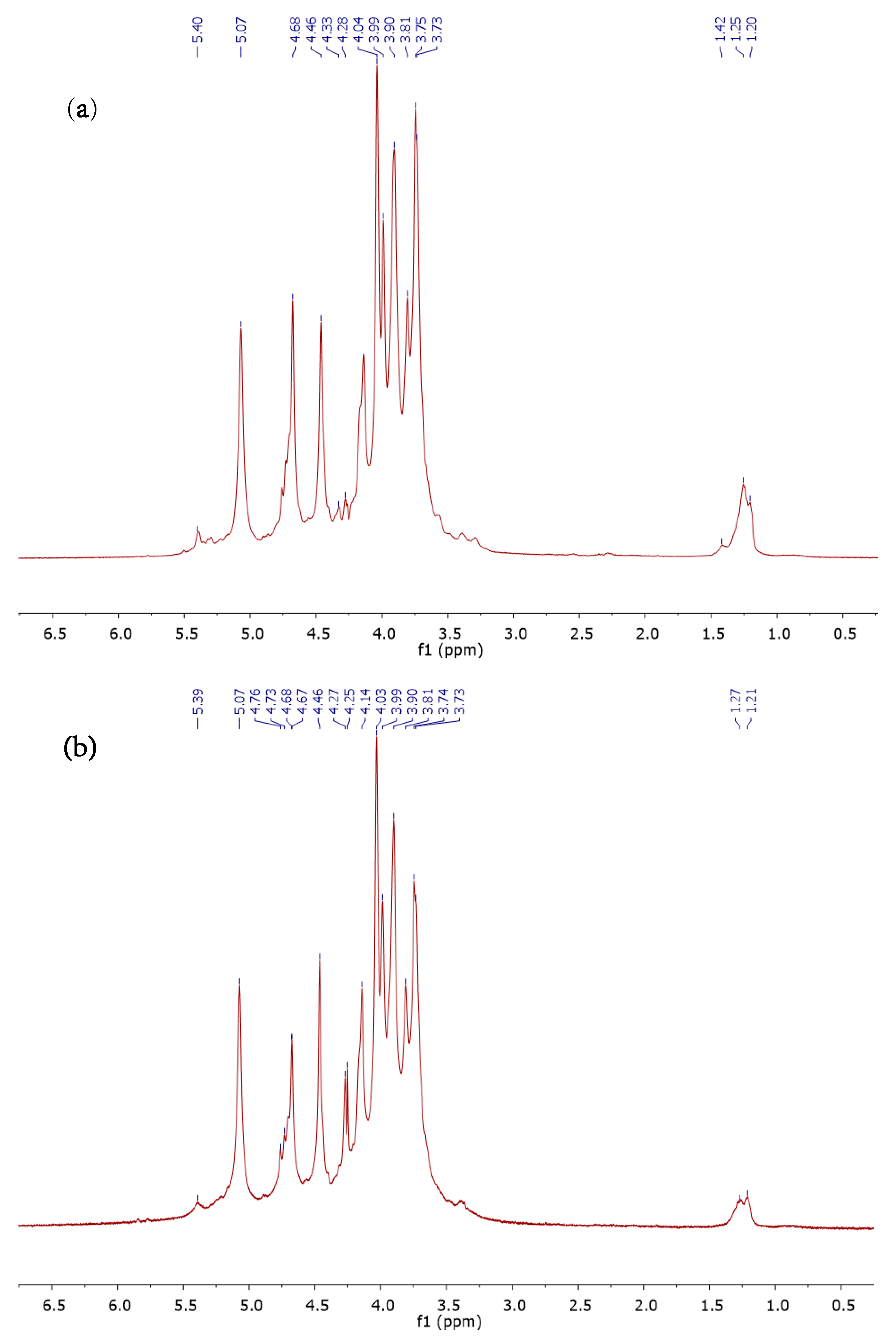
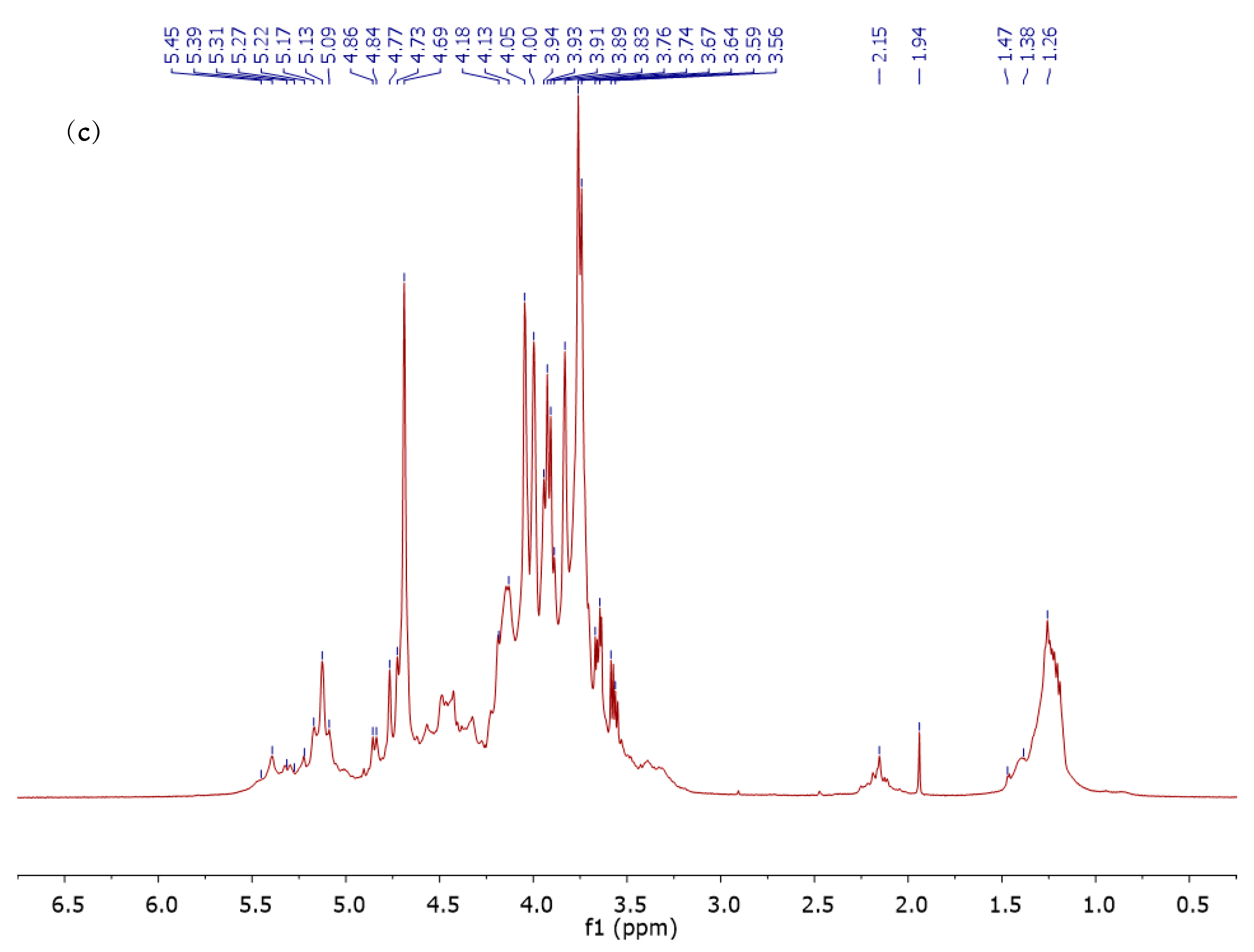
3.4. 1H NMR Analysis of ALCM, ALSM, and FUCCM Fractions

The alginates and fucoidan were analyzed using 1H NMR, and the findings are presented in Figure 3, Table 4 and Table 5.
FUCCM is mainly composed of α-l-fucose presenting intense signals at 1.26–1.47 ppm, which are the characteristic regions of fucopyranose assigned to the CH3 methyl group of H-6 [56] and at 5.13 ppm confirming the α-linked fucose monomers [57]. Peaks at 1.94–2.15 ppm correspond to the CH3 proton of O-acetyl groups [58]. H-1 and C-H protons of O-substituted carbons of α-l-isomers extend from 5.09 to 5.45 ppm, whereas ring proton H-2-H-5 spread out from 3.56 to 4.86 ppm [48]. By comparing with proton chemical shifts of each O-sulfated l-fucose isomer [59], the resonance of H-6 in the region of 1.38 ppm indicates that sulfates could be bonded to C-4 of FUCCM [58] (Table 5). In order to determine more precisely the sulfate group positions, it is required to investigate as well the desulfated fucoidan residues. Other peaks appearing slightly at 4.73 and 4.77 ppm could be attributed to the mannuronic acid anomeric protons due to the presence of alginate traces in the FUCCM extracts.
The 1H NMR spectra of alginates ALCM and ALSM (Figure 3) were used to determine the structural block distribution and the rate of mannuronic and guluronic acid of each fraction through the M/G ratio (Table 4). According to Grasdalen et al. [35], signals at 5.07 and 4.46 ppm (signal I and signal III) were attributed to H-1 and H-5 of guluronic acid (G) residues, respectively. The anomeric proton of mannuronic acid (H-1) and the C-5 of alternating blocks (GM-5) (signal II) imbricated at 4.76–4.68 ppm. Minor signals at 1.21–1.27 ppm for ALSM and 1.20–1.42 ppm for ALCM correspond to fucopyranose residues, which confirm the results obtained by GC–MS–EI analysis, indicating the presence of slight impurities of fucoidan. Each alginate fraction showed a different M/G ratio, which was 0.87 and 1.12 for ALSM and ALCM, respectively. This indicates that alginate extracted from Moroccan S. muticum is rich in guluronic acid (54%), while ALCM extracted from C. myriophylloides exhibits higher levels of mannuronic acid (53%) compared to guluronic acid (47%). The M/G ratio of alginates extracted from S. muticum ranged from 0.31 to 1.08 [25,45,47], these variations within the species are related to harvesting conditions (place and period). In contrast to the current finding, the M/G ratios were < to 1 for C. barbata, C. schiffneri, C. compressa, and C. trinodis [40,41,42,43,44]. The estimated η parameter was < to 1 for both alginate samples, indicating the abundance of homopolymeric sequences (MM and GG) as shown in Table 4.
Table 5. 1H NMR chemical shifts of fucoidan extracted from C.myriophylloides (FUCCM) and sulfated fucopyranoses standard.
FucoseH-1H-2H-3H-4H-5H-6
α-l-Fucp [59]5.203.773.863.814.201.21
α-l-Fucp (FUCCM 1)5.13
(−0.07)
3.76
(−0.01)
3.89
(0.03)
3.83
(0.02)
4.17
(−0.03)
1.26
(0.05)
1 FUCCM: fucoidan extracted from CM.

3.5. Sulfated and Unsulfated Polysaccharides and Oligosaccharides induce Natural Defenses in Olive Tree Leaves

To evaluate the potential effect of the seven saccharide fractions on the natural defenses of the olive tree, leaf discs of 5 mm were used as a model system and were treated for 24 h with different concentrations. The PAL and TAL activities, the phenolic compounds, and the lignin content were then measured. To our knowledge, no study has reported the capacity of alginate (unsulfated polysaccharides), LMW alginates, fucoidan, and sulfated arabinogalactan-rich protein to induce the natural defense of olive trees.
PAL and TAL are monofunctional enzymes belonging to the plant aromatic amino-acid-lyase family [60]. The PAL catalyzes the deamination of l-phenylalanine into trans-cinnamic acid, while the TAL converts the l-tyrosine yielding trans-p-coumarate. PAL is the most widely studied enzyme in plant phenolic synthesis. However, TAL is another enzyme involved in this pathway, that has rarely been investigated. This is the first report exploring the activity of TAL in the leaves of the olive tree.

3.5.1. Evaluation of Enzyme Activities and Secondary Metabolism Synthesis in the Leaf Discs of the Olive Tree in Response to Alginates and Their Oligomers

The application of alginates with different molecular weights, ALCM (202 kDa), ALSM (76 kDa), OACM (19 kDa), and the oligomers OASM (3 kDa), significantly induced the PAL and TAL activities, the phenolic compounds, and the lignin content in the leaves of the olive tree depending on the concentration of saccharide solutions (p < 0.05). After treatment with OASM, the PAL and TAL activities revealed a considerable increase at different concentrations, reaching an upward trend at 2 g/L. The polyphenols followed a similar pattern and rose at 0.5 g/L and 1 g/L, while no significant increment was observed in lignin content. Compared to their native polymer (ALSM) at 2 g/L, the oligoalginates (OASM) showed the induction of PAL and TAL activities, 1.4–1.6 times higher (Figure 4), as well as efficiency at a low concentration (0.5 g/L). On the other hand, the accumulation of polyphenols and lignin in the leaves was significantly increased after the treatment with ALSM polymers. The elicitor activity of the LMW alginates (OACM) was less important compared to the other fractions. In response to the native alginate polymers, ALCM showed significant stimulation of PAL and TAL activities at a low concentration (0.5 g/L) compared to ALSM, while the secondary metabolites (lignin and polyphenols) were strongly promoted after the application of ALSM (Figure 5). Lignin was only promoted in response to high molecular weight alginates polymers (ALCM and ALSM). In this study, the PAL and TAL activities showed a similar trend. In contrast, a considerable fluctuation was observed. All fractions reported a PAL/TAL ratio < to 1, ranging from 0.34 to 0.77, indicating the high expression of TAL in the leaves of the olive tree compared to PAL. As mentioned before, PAL and TAL are the key regulatory enzymes associated with the biosynthesis of various secondary metabolites via the shikimic pathway [61]. Nevertheless, PAL is the most occurs in plants [60], while TAL enzyme is commonly found in Gramineae such as wheat and maize [62,63,64,65]. Recently, TAL has been detected in other plants such as orchids, strawberry fruits, and leaves, ginger and soybean leave in response to biotic and abiotic stresses [61,66,67,68]. As reported by Khan et al., the treatment of soybean by chitin and chitosan oligomers significantly induced the PAL, TAL, and phenolic compounds in leaf tissues, which the specific activity of TAL was lower compared to PAL [67].
In this study, the elicitor capacities of alginate polymers and their derivatives depended on many factors such as concentration, nature of polymers, and chain length. The OASM (3 kDa), ALCM (76 kDa), and OACM (19 kDa) were more effective due to their abilities to activate the PAL and TAL at a low concentration (0.5 g/L) compared to ALSM (202 kDa). However, the OASM (3 kDa) exhibited the highest levels of enzyme activities at 2 g/L. Alginate is an anionic polysaccharide isolated from brown algae. Recently, it has been widely utilized in agriculture as an inducer of the natural defenses of plants. The application of alginates extracted from B. bifurcata and F. spiralis triggered PAL activity and polyphenol synthesis in leaves of tomato seedlings and roots of date palm [18,20]. Few research has focused on the use of LMW alginates as inducers of natural defenses. Partial hydrolysis with HCl of alginates extracted from Lessonia vadose enhanced the PAL and peroxidase (POD) activities in wheat plants, higher levels were recorded after the treatment with polymannuronic enriched fraction [69]. A recent study has reported the effectiveness of oligoalginates (5.5 kDa), obtained after oxidative degradation, compared to their native polymer, showing their capacity to stimulate the PAL activity as well as the phenolic compounds in the leaves of tomato seedlings at a concentration of 1 g/L [18]. It has been conclusively shown that oligoglucuronans and oligoulvans with low DP (2–3) enhanced the activation of PAL, antioxidant-related enzymes as well as the accumulation of lignin and phenolic compounds, protecting apple fruit against Penicillium expansum and Botrytis cinerea. These oligomers were more effective than their native polymers [22,23]. No previous study to date has examined the elicitor effects of alginates and their derivatives in leaf tissues of the olive tree.

3.5.2. Evaluation of Enzyme Activities and Secondary Metabolism Synthesis in the Leaf Discs of the Olive Tree in Response to Sulfated Polysaccharides AGB, AGP and FUCCM

The sulfated polysaccharides, arabinogalactan, and fucoidan extracted from the brown and green seaweed were applied to the olive leaves at different concentrations. The results showed significant induction of the PAL and TAL activities accompanied by the synthesis of lignin and polyphenols (p < 0.05). These responses depend on the nature of the polymers and their concentrations. In response to the tree fractions, the phenolic metabolism was significantly stimulated, at the low concentration (0.5 g/L). FUCCM revealed an important elicitor capacity at 0.5 g/L, showing high levels of PAL, polyphenols, and lignin compared to arabinogalactan (crude and purified extracts). The TAL was less expressed than PAL after the treatment by the fucoidan, indicating a PAL/TAL ratio higher than 1. On the other hand, for the crude (AGB) and the purified (AGP) extracts, the PAL/TAL ratio was in the range of 0.45–0.75, showing high activity of the TAL enzyme. No considerable difference was observed in the elicitor effects between AGB and AGP (Figure 6 and Figure 7). A similar conclusion was reached in the previous work, after the application of sulfated arabinogalactan AGB and AGP for the first time as inducers of PAL activity, phenolic compounds, and lignin content in leaf and root tissues of tomato seedlings [27]. The application of sulfated polysaccharides from marine algae as stimulators of natural defenses of plants has been largely reported. Carrageenan, a sulfated galactan presented in the red algae, was found to be capable of activating the defense mechanisms against a large wide of pathogens in Arabidopsis thaliana, tobacco, and tomato plants [70,71,72,73,74,75]. The sulfated heteropolysaccharides, ulvan, from the green seaweed have been shown to trigger the pathogenesis-related proteins gene expression, reactive oxygen species metabolism, and the octadecanoid pathway, leading to protection against Zymoseptoria tritici in wheat [76]. The treatment of A. thaliana plants by ulvans with sulfate content of 18.9 to 36.6% reduced the severity of Alternaria brassicicola and Colletotrichum higginsianum independently of sulfation degree [77]. These findings are in accordance with those reported in this work and the previous work [27], in which the AGB and AGP showed similarities in responses despite their sulfation variation. Furthermore, several studies have investigated the elicitor effects of ulvans in several plants [21,23,78,79,80]. The ability of fucoidan to provide protection and enhance natural defenses in plants has been previously assessed only to a very limited extent. Fucoidans from the brown macroalgae B. bifurcata and F. spiralis stimulated PAL activity, polyphenols, and lignin content in the roots of the date palm [19]. Detached leaves of Datura stramonium L. and Nicotiana tabacum L. infected by potato virus X (PVX) and tobacco mosaic virus (TMV), respectively, exhibited an inhibition of the development of the virus during the first stage of infection after treatment with fucoidan extracted from the brown alga F. evanescens [81,82]. In addition, fucoidan from Lessonia vadosa activated the PAL, lipooxygenase and glutathione-S-transferase activities in tobacco plants [24].
Compared to alginates (ALCM and ALSM), the sulfated polysaccharides (AGP, AGB, and FUCCM) exhibited an elicitor capacity more effective at low concentrations (0.5 g/L and 1 g/L). In response to fucoidan at 0.5 g/L, the levels of the PAL activity, phenolic compounds, and lignin content were significantly higher compared to AGB, AGP, ALCM, and ALSM. The sulfate group and the low Mw seem to be the principal factors in this fluctuation of plant defense responses. It has been shown before that the elicitation by glucuronan (unsulfated polysaccharide) had no considerable effects on PAL activity of tomato seedlings, while ulvan (sulfated polysaccharides) exhibited significant levels [21]. In addition, the desulfation of ulvan has shown suppression of the elicitor effect in the leaves of tomato plants, indicating the main role of sulfate substituent in the elicitation efficiency [21]. Several studies have found that desulfation reduces biological activities [83,84,85,86].
The use of polysaccharides extracted from seaweeds as olive resistance-inducers has been previously assessed only to a very limited extent. Tziros et al., have reported the evaluation of laminarin (Vacciplant 4.5SL) efficiency, against olive leaf spot (OLS) disease, caused by Fusicladium oleagineum on six-month-old olive plants [87]. The application of laminarin suppressed the disease severity and no symptoms were detected, compared to copper pesticides used as reference treatments. Moreover, the expression of PAL, copper amine oxidase (Cuao), and major pollen allergen (Mpol) genes was induced in laminarin-treated plants [87]. Another study has involved the ability of ulvan to control verticillium wilt of olive (VWO) caused by V. dahlia [88]. In response to this elicitor, the phenolic metabolism (PAL, polyphenols, and lignin) was significantly enhanced, reducing the verticillium wilt in the twigs (10 cm) of the olive tree (Picholine Marocaine) [88]. To the best of our knowledge, no study to date has examined the fucoidan, alginates from the brown macroalgae S. muticum and C. myriophylloides and their derivatives, as well as the sulfated glycoprotein from the green seaweed C. decorticatum, as inducers of natural defenses in the leaves of the olive tree. Nevertheless, further studies are required to evaluate the efficiency of these polysaccharides in stimulating and protecting against vascular and foliar pathogens of the olive tree under field conditions.

4. Conclusions

In the current work, alginates, LMW alginate derivatives, fucoidans from the brown seaweed S. muticum and C. myriophylloides, and the glycoproteins isolated from C. decorticatum were characterized chemically and structurally. These saccharide extracts were investigated for their ability to induce the PAL and TAL activities, polyphenol, and lignin contents in the leaves of the olive tree (Picholine Marocaine). Sulfated polysaccharides (FUCCM, AGB, and AGP) were found to be the most active at a low concentration (0.5 g/L) compared to alginates (unsulfated polysaccharides), indicating the main role of the sulfate group in the efficiency of the elicitor activity. Fucoidan, with an Mw of 148 kDa and 12.83% of sulfates was more effective than the other polymer fractions. The activity of alginate polymers and their derivatives was related to their concentrations and chain length. When compared to ALSM (202 kDa), the low molecular weight alginates OASM (3 kDa), ALCM (76 kDa), and OACM (19 kDa) were more efficient in inducing PAL and TAL activities at low concentrations. In addition, oligoalginates (OASM) manifested the highest levels at 2 g/L. PAL/TAL ratios were < to 1, indicating that the TAL enzyme was highly expressed in the leaves of the olive tree in response to all saccharide fractions except for fucoidan. The expression of the TAL enzyme in olive tree leaf tissue was examined for the first time. These results highlight the use of alginates, LMW alginates, fucoidan, and glycoprotein from marine macroalgae, as inducers of natural defenses of the olive tree, to protect against phytopathogens in the context of sustainable development. However, future research should consider the protective effects of these molecules against biotic and abiotic stresses under field conditions.

Author Contributions

Conceptualization, M.A., Z.E.A.-T., C.D. and C.E.M.; methodology, M.A., Z.E.A.-T., C.D., M.T. and C.E.M.; software, M.A.; validation, M.A., Z.E.A.-T., C.D. and C.E.M.; formal analysis, M.A., Z.E.A.-T., C.D. and C.E.M.; investigation, M.A., Z.E.A.-T., C.D. and C.E.M.; resources, M.A., Z.B., M.D.O.E.-H. and H.R.; data curation, M.A.; writing—original draft preparation, M.A.; writing—review and editing, M.A., Z.E.A.-T., C.D., C.E.M., G.P., P.D. and P.M.; visualization, M.A., Z.E.A.-T., C.D., I.F., S.A. and C.E.M.; supervision, Z.E.A.-T., C.D. and C.E.M.; project administration, G.P. All authors have read and agreed to the published version of the manuscript.

Funding

This work was funded by the French Ministry of Europe and Foreign Affairs and the Moroccan, Algerian, and Tunisian Ministries of Higher Education and Scientific Research, within the framework of the Hubert Curien program (PHC Maghreb), grant number 19MAG36, and by the National Centre for Scientific and Technical Research (CNRST Morocco), grant number 4UCA2018.

Institutional Review Board Statement

Not applicable.

Informed Consent Statement

Not applicable.

Data Availability Statement

Not applicable.

Acknowledgments

We want to thank Didier Le Cerf and Christophe Rihouey from Université de Rouen Normandie (INSA Rouen) for SEC-MALLS analysis.

Conflicts of Interest

The authors declare no conflict of interest.

References

  1. López-Escudero, F.J.; Mercado-Blanco, J. Verticillium wilt of olive: A case study to implement an integrated strategy to control a soil-borne pathogen. Plant Soil 2010, 344, 1–50. [Google Scholar] [CrossRef]
  2. Eurostat. Eurostat Statistics Explained. Available online: https://ec.europa.eu/eurostat/statistics-explained/index.php?title=Agricultural_production_-_orchards#Olive_trees (accessed on 1 April 2022).
  3. Eurostat. Agriculture, Forestry and Fishery Statistics, 2018th ed.; Eurostat: Bietlot Frères, Belgium, 2018; p. 200. [Google Scholar]
  4. Chliyeh, M.; Ouazzani Touhami, A.; Filali-Maltouf, A.; El Modafar, C.; Moukhli, A.; Oukabli, A.; Benkirane, R.; Douira, A.; Bioscience, A. Effect of a composite endomycorrhizal inoculum on the growth of olive trees under nurseries conditions in morocco. Int. J. Pure Appl. Biosci. 2014, 2, 1–14. [Google Scholar]
  5. El Mouhtadi, I.; Agouzzal, M.; Guy, F.J.O. L’olivier au maroc. OCL 2014, 21, D203. [Google Scholar] [CrossRef]
  6. Achbani, E.H.; Benbouazza, A.; Douira, A. First report of olive anthracnose, caused by colletotrichum gloeosporioides, in morocco. Atlas J. Biol. 2013, 2, 172–175. [Google Scholar] [CrossRef]
  7. Rugini, E.; Baldoni, L.; Muleo, R.; Sebastiani, L. The Olive Tree Genome; Springer: Berlin/Heidelberg, Germany, 2016; Volume 978. [Google Scholar]
  8. Kour, D.; Bakshi, P.; Wali, V.K.; Sharma, N.; Sharma, A.; Iqbal, M. Alternate bearing in olive—A review. Int. J. Curr. Microbiol. Appl. Sci. 2018, 7, 2281–2297. [Google Scholar] [CrossRef]
  9. Gómez-Lama Cabanás, C.; Ruano-Rosa, D.; Legarda, G.; Pizarro-Tobías, P.; Valverde-Corredor, A.; Triviño, J.; Roca, A.; Mercado-Blanco, J. Bacillales members from the olive rhizosphere are effective biological control agents against the defoliating pathotype of verticillium dahliae. Agriculture 2018, 8, 90. [Google Scholar] [CrossRef]
  10. Nigro, F.; Antelmi, I.; Labarile, R.; Sion, V.; Pentimone, I. Biological control of olive anthracnose. Acta Hortic. 2018, 1199, 439–444. [Google Scholar] [CrossRef]
  11. Bueno, A.M.; Jones, O. Alternative methods for controlling the olive fly, bactrocera oleae, involving semiochemicals. IOBC Wprs Bull. 2002, 25, 147–156. [Google Scholar]
  12. Bizos, G.; Papatheodorou, E.M.; Chatzistathis, T.; Ntalli, N.; Aschonitis, V.G.; Monokrousos, N. The role of microbial inoculants on plant protection, growth stimulation, and crop productivity of the olive tree (Olea europea L.). Plants 2020, 9, 743. [Google Scholar] [CrossRef]
  13. Dzhavakhiya, V.G.; Shcherbakova, L.A. Control of plant pathogens by biogenic elicitors and possible mechanisms of action. Front. Plant Sci. 2016, 7, 340. [Google Scholar] [CrossRef]
  14. Choudhary, D.K.; Prakash, A.; Johri, B.N. Induced systemic resistance (isr) in plants: Mechanism of action. Indian J. Microbiol. 2007, 47, 289–297. [Google Scholar] [CrossRef] [PubMed]
  15. Bedoux, G.; Hardouin, K.; Burlot, A.S.; Bourgougnon, N. Bioactive components from seaweeds. In Sea Plants; Academic Press: Cambridge, MA, USA, 2014; pp. 345–378. [Google Scholar]
  16. Shukla, P.S.; Borza, T.; Critchley, A.T.; Prithiviraj, B. Seaweed-based compounds and products for sustainable protection against plant pathogens. Mar. Drugs 2021, 19, 59. [Google Scholar] [CrossRef] [PubMed]
  17. Agarwal, P.K.; Dangariya, M.; Agarwal, P. Seaweed extracts: Potential biodegradable, environmentally friendly resources for regulating plant defence. Algal Res. 2021, 58, 102363. [Google Scholar] [CrossRef]
  18. Aitouguinane, M.; Bouissil, S.; Mouhoub, A.; Rchid, H.; Fendri, I.; Abdelkafi, S.; Ould El-Hadj, M.D.; Boual, Z.; Dubessay, P.; Gardarin, C.; et al. Induction of natural defenses in tomato seedlings by using alginate and oligoalginates derivatives extracted from moroccan brown algae. Mar. Drugs 2020, 18, 521. [Google Scholar] [CrossRef]
  19. Bouissil, S.; Alaoui-Talibi, Z.E.; Pierre, G.; Rchid, H.; Michaud, P.; Delattre, C.; El Modafar, C. Fucoidans of moroccan brown seaweed as elicitors of natural defenses in date palm roots. Mar. Drugs 2020, 18, 596. [Google Scholar] [CrossRef]
  20. Bouissil, S.; El Alaoui-Talibi, Z.; Pierre, G.; Michaud, P.; El Modafar, C.; Delattre, C. Use of alginate extracted from moroccan brown algae to stimulate natural defense in date palm roots. Molecules 2020, 25, 720. [Google Scholar] [CrossRef]
  21. El Modafar, C.; Elgadda, M.; El Boutachfaiti, R.; Abouraicha, E.; Zehhar, N.; Petit, E.; El Alaoui-Talibi, Z.; Courtois, B.; Courtois, J. Induction of natural defence accompanied by salicylic acid-dependant systemic acquired resistance in tomato seedlings in response to bioelicitors isolated from green algae. Sci. Hortic. 2012, 138, 55–63. [Google Scholar] [CrossRef]
  22. Abouraïcha, E.F.; El Alaoui-Talibi, Z.; Tadlaoui-Ouafi, A.; El Boutachfaiti, R.; Petit, E.; Douira, A.; Courtois, B.; Courtois, J.; El Modafar, C. Glucuronan and oligoglucuronans isolated from green algae activate natural defense responses in apple fruit and reduce postharvest blue and gray mold decay. J. Appl. Phycol. 2016, 29, 471–480. [Google Scholar] [CrossRef]
  23. Abouraïcha, E.; El Alaoui-Talibi, Z.; El Boutachfaiti, R.; Petit, E.; Courtois, B.; Courtois, J.; El Modafar, C. Induction of natural defense and protection against penicillium expansum and botrytis cinerea in apple fruit in response to bioelicitors isolated from green algae. Sci. Hortic. 2015, 181, 121–128. [Google Scholar] [CrossRef]
  24. Chandia, N.P.; Matsuhiro, B. Characterization of a fucoidan from lessonia vadosa (phaeophyta) and its anticoagulant and elicitor properties. Int. J. Biol. Macromol. 2008, 42, 235–240. [Google Scholar] [CrossRef]
  25. Mazumder, A.; Holdt, S.L.; De Francisci, D.; Alvarado-Morales, M.; Mishra, H.N.; Angelidaki, I. Extraction of alginate from sargassum muticum: Process optimization and study of its functional activities. J. Appl. Phycol. 2016, 28, 3625–3634. [Google Scholar] [CrossRef]
  26. Soukaina, B.; Zainab, E.L.A.-T.; Guillaume, P.; Halima, R.; Philippe, M.; Cherkaoui, E.M.; Cédric, D. Radical depolymerization of alginate extracted from moroccan brown seaweed bifurcaria bifurcata. Appl. Sci. 2020, 10, 4166. [Google Scholar] [CrossRef]
  27. Aitouguinane, M.; El Alaoui-Talibi, Z.; Rchid, H.; Fendri, I.; Abdelkafi, S.; Ould El-Hadj, M.D.; Boual, Z.; Dubessay, P.; Michaud, P.; Le Cerf, D.; et al. A novel sulfated glycoprotein elicitor extracted from the moroccan green seaweed codium decorticatum induces natural defenses in tomato. Appl. Sci. 2022, 12, 3643. [Google Scholar] [CrossRef]
  28. Dubois, M.; Gilles, K.; Hamilton, J.; Rebers, P.; Smith, F.J.N. A colorimetric method for the determination of sugars. Nature 1951, 168, 167. [Google Scholar] [CrossRef]
  29. Monsigny, M.; Petit, C.; Roche, A.-C. Colorimetric determination of neutral sugars by a resorcinol sulfuric acid micromethod. Anal. Biochem. 1988, 175, 525–530. [Google Scholar] [CrossRef]
  30. Blumenkrantz, N.; Asboe-Hansen, G. New method for quantitative determination of uronic acids. Anal. Biochem. 1973, 54, 484–489. [Google Scholar] [CrossRef]
  31. Montreuil, J.; Spik, G.; Chosson, A.; Segard, E.; Scheppler, N. Methods of study of the structure of glycoproteins. J. Pharm. Belg. 1963, 18, 529–546. [Google Scholar] [PubMed]
  32. Dodgson, K.; Price, R.J. A note on the determination of the ester sulphate content of sulphated polysaccharides. Biochem. J. 1962, 84, 106–110. [Google Scholar] [CrossRef] [PubMed]
  33. Lowry, O.H.; Rosebrough, N.J.; Farr, A.L.; Randall, R. Protein measurement with the folin phenol reagent. J. Biol. Chem. 1951, 193, 265–275. [Google Scholar] [CrossRef]
  34. Blainski, A.; Lopes, G.C.; De Mello, J.C.P. Application and analysis of the folin ciocalteu method for the determination of the total phenolic content from Limonium brasiliense L. Molecules 2013, 18, 6852–6865. [Google Scholar] [CrossRef]
  35. Grasdalen, H. High-field, 1H-nmr spectroscopy of alginate: Sequential structure and linkage conformations. Carbohydr. Res. 1983, 118, 255–260. [Google Scholar] [CrossRef]
  36. Liu, H.; Jiang, W.; Bi, Y.; Luo, Y. Postharvest bth treatment induces resistance of peach (Prunus persica L. Cv. Jiubao) fruit to infection by penicillium expansum and enhances activity of fruit defense mechanisms. Postharvest Biol. Technol. 2005, 35, 263–269. [Google Scholar] [CrossRef]
  37. Hendrikse, N.M.; Holmberg Larsson, A.; Svensson Gelius, S.; Kuprin, S.; Nordling, E.; Syren, P.O. Exploring the therapeutic potential of modern and ancestral phenylalanine/tyrosine ammonia-lyases as supplementary treatment of hereditary tyrosinemia. Sci. Rep. 2020, 10, 1315. [Google Scholar] [CrossRef] [PubMed]
  38. Bradford, M.M. A rapid and sensitive method for the quantitation of microgram quantities of protein utilizing the principle of protein-dye binding. Anal. Biochem. 1976, 72, 248–254. [Google Scholar] [CrossRef]
  39. Belattmania, Z.; Kaidi, S.; El Atouani, S.; Katif, C.; Bentiss, F.; Jama, C.; Reani, A.; Sabour, B.; Vasconcelos, V. Isolation and FTIR-ATR and 1H NMR characterization of alginates from the main alginophyte species of the atlantic coast of Morocco. Molecules 2020, 25, 4335. [Google Scholar] [CrossRef]
  40. Trica, B.; Delattre, C.; Gros, F.; Ursu, A.V.; Dobre, T.; Djelveh, G.; Michaud, P.; Oancea, F. Extraction and characterization of alginate from an edible brown seaweed (Cystoseira barbata) harvested in the romanian black sea. Mar. Drugs 2019, 17, 405. [Google Scholar] [CrossRef]
  41. Sellimi, S.; Younes, I.; Ayed, H.B.; Maalej, H.; Montero, V.; Rinaudo, M.; Dahia, M.; Mechichi, T.; Hajji, M.; Nasri, M. Structural, physicochemical and antioxidant properties of sodium alginate isolated from a tunisian brown seaweed. Int. J. Biol. Macromol. 2015, 72, 1358–1367. [Google Scholar] [CrossRef]
  42. Benslima, A.; Sellimi, S.; Hamdi, M.; Nasri, R.; Jridi, M.; Cot, D.; Li, S.; Nasri, M.; Zouari, N. The brown seaweed cystoseira schiffneri as a source of sodium alginate: Chemical and structural characterization, and antioxidant activities. Food Biosci. 2021, 40, 100873. [Google Scholar] [CrossRef]
  43. Hentati, F.; Delattre, C.; Ursu, A.V.; Desbrieres, J.; Le Cerf, D.; Gardarin, C.; Abdelkafi, S.; Michaud, P.; Pierre, G. Structural characterization and antioxidant activity of water-soluble polysaccharides from the tunisian brown seaweed cystoseira compressa. Carbohydr. Polym. 2018, 198, 589–600. [Google Scholar] [CrossRef]
  44. Hifney, A.F.; Fawzy, M.A.; Abdel-Gawad, K.M.; Gomaa, M. Upgrading the antioxidant properties of fucoidan and alginate from cystoseira trinodis by fungal fermentation or enzymatic pretreatment of the seaweed biomass. Food Chem. 2018, 269, 387–395. [Google Scholar] [CrossRef]
  45. El Atouani, S.; Bentiss, F.; Reani, A.; Zrid, R.; Belattmania, Z.; Pereira, L.; Mortadi, A.; Cherkaoui, O.; Sabour, B. The invasive brown seaweedsargassum muticumas new resource for alginate in morocco: Spectroscopic and rheological characterization. Phycol. Res. 2016, 64, 185–193. [Google Scholar] [CrossRef]
  46. Gorham, J.; Lewey, S. Seasonal changes in the chemical composition of sargassum muticum. Mar. Biol. 1984, 80, 103–107. [Google Scholar] [CrossRef]
  47. Davis, T.A.; Llanes, F.; Volesky, B.; Mucci, A. Metal selectivity of Sargassum spp. and their alginates in relation to their alpha-l-guluronic acid content and conformation. Environ. Sci. Technol. 2003, 37, 261–267. [Google Scholar] [CrossRef] [PubMed]
  48. Benslima, A.; Sellimi, S.; Hamdi, M.; Nasri, R.; Jridi, M.; Cot, D.; Li, S.; Nasri, M.; Zouari, N. Brown seaweed cystoseira schiffneri as a promising source of sulfated fucans: Seasonal variability of structural, chemical, and antioxidant properties. Food Sci. Nutr. 2021, 9, 1551–1563. [Google Scholar] [CrossRef]
  49. Sellimi, S.; Kadri, N.; Barragan-Montero, V.; Laouer, H.; Hajji, M.; Nasri, M. Fucans from a tunisian brown seaweed cystoseira barbata: Structural characteristics and antioxidant activity. Int. J. Biol. Macromol. 2014, 66, 281–288. [Google Scholar] [CrossRef] [PubMed]
  50. Hadj Ammar, H.; Lajili, S.; Ben Said, R.; Le Cerf, D.; Bouraoui, A.; Majdoub, H. Physico-chemical characterization and pharmacological evaluation of sulfated polysaccharides from three species of mediterranean brown algae of the genus Cystoseira. DARU J. Pharm. Sci. 2015, 23, 1. [Google Scholar] [CrossRef]
  51. Rahelivao, M.P.; Andriamanantoanina, H.; Heyraud, A.; Rinaudo, M. Structure and properties of three alginates from madagascar seacoast algae. Food Hydrocoll. 2013, 32, 143–146. [Google Scholar] [CrossRef]
  52. Burkus, Z. Determination of the molecular weight of barley β-glucan using intrinsic viscosity measurements. Carbohydr. Polym. 2003, 54, 51–57. [Google Scholar] [CrossRef]
  53. Khouryieh, H.A.; Herald, T.J.; Aramouni, F.; Alavi, S. Intrinsic viscosity and viscoelastic properties of xanthan/guar mixtures in dilute solutions: Effect of salt concentration on the polymer interactions. Food Res. Int. 2007, 40, 883–893. [Google Scholar] [CrossRef]
  54. Rochefort, W.E.; Middleman, S. Rheology of xanthan gum: Salt, temperature, and strain effects in oscillatory and steady shear experiments. J. Rheol. 1987, 31, 337–369. [Google Scholar] [CrossRef]
  55. Moseley, R.; Waddington, R.; Evans, P.; Halliwell, B.; Embery, G. The chemical modification of glycosaminoglycan structure by oxygen-derived species in vitro. Biochim. Biophys. Acta 1995, 1244, 245–252. [Google Scholar] [CrossRef]
  56. Lim, S.J.; Wan Aida, W.M.; Maskat, M.Y.; Latip, J.; Badri, K.H.; Hassan, O.; Yamin, B.M. Characterisation of fucoidan extracted from malaysian sargassum binderi. Food Chem. 2016, 209, 267–273. [Google Scholar] [CrossRef]
  57. Zayed, A.; El-Aasr, M.; Ibrahim, A.S.; Ulber, R. Fucoidan characterization: Determination of purity and physicochemical and chemical properties. Mar. Drugs 2020, 18, 571. [Google Scholar] [CrossRef] [PubMed]
  58. Synytsya, A.; Kim, W.-J.; Kim, S.-M.; Pohl, R.; Synytsya, A.; Kvasnička, F.; Čopíková, J.; Il Park, Y. Structure and antitumour activity of fucoidan isolated from sporophyll of korean brown seaweed undaria pinnatifida. Carbohydr. Polym. 2010, 81, 41–48. [Google Scholar] [CrossRef]
  59. Daniel, R.; Berteau, O.; Chevolot, L.; Varenne, A.; Gareil, P.; Goasdoue, N. Regioselective desulfation of sulfated l-fucopyranoside by a new sulfoesterase from the marine mollusk pecten maximus: Application to the structural study of algal fucoidan (Ascophyllum nodosum). Eur. J. Biochem. 2001, 268, 5617–5626. [Google Scholar] [CrossRef]
  60. Barros, J.; Dixon, R.A. Plant phenylalanine/tyrosine ammonia-lyases. Trends Plant Sci. 2020, 25, 66–79. [Google Scholar] [CrossRef]
  61. Golan, K.; Sempruch, C.; Górska-Drabik, E.; Czerniewicz, P.; Łagowska, B.; Kot, I.; Kmieć, K.; Magierowicz, K.; Leszczyński, B. Accumulation of amino acids and phenolic compounds in biochemical plant responses to feeding of two different herbivorous arthropod pests. Arthropod-Plant Interact. 2017, 11, 675–682. [Google Scholar] [CrossRef]
  62. Jangaard, N.O. The characterization of phenylalanine ammonia-lyase from several plant species. Phytochemistry 1974, 13, 1765–1768. [Google Scholar] [CrossRef]
  63. Xu, Y.; Guo, H.; Geng, G.; Zhang, Q.; Zhang, S. Changes in defense-related enzymes and phenolics in resistant and susceptible common wheat cultivars under aphid stress. Acta Physiol. Plant. 2021, 43, 36. [Google Scholar] [CrossRef]
  64. Rösler, J.; Krekel, F.; Amrhein, N.; Schmid, J. Maize phenylalanine ammonia-lyase has tyrosine ammonia-lyase activity. Plant Physiol. 1997, 113, 175–179. [Google Scholar] [CrossRef]
  65. Siqueira-Soares, R.d.C.; Soares, A.R.; Parizotto, A.V.; Ferrarese, M.d.L.L.; Ferrarese-Filho, O. Root growth and enzymes related to the lignification of maize seedlings exposed to the allelochemical L-DOPA. Sci. World J. 2013, 2013, 134237. [Google Scholar] [CrossRef]
  66. Montero, T.; Mollá, E.; Martín-Cabrejas, M.A.; López-Andréu, F. Effects of gibberellic acid (ga3) on strawberry pal (phenylalanine ammonia-lyase) and tal (tyrosine ammonia-lyase) enzyme activities. J. Sci. Food Agric. 1998, 77, 230–234. [Google Scholar] [CrossRef]
  67. Khan, W.; Prithiviraj, B.; Smith, D.L. Chitosan and chitin oligomers increase phenylalanine ammonia-lyase and tyrosine ammonia-lyase activities in soybean leaves. J. Plant Physiol. 2003, 160, 859–863. [Google Scholar] [CrossRef]
  68. Ganapathy, G.; Keerthi, D.; Nair, R.A.; Pillai, P. Correlation of phenylalanine ammonia lyase (pal) and tyrosine ammonia lyase (tal) activities to phenolics and curcuminoid content in ginger and its wild congener, zingiber zerumbet following pythium myriotylum infection. Eur. J. Plant Pathol. 2016, 145, 777–785. [Google Scholar] [CrossRef]
  69. Chandía, N.P.; Matsuhiro, B.; Mejías, E.; Moenne, A. Alginic acids in lessonia vadosa: Partial hydrolysis and elicitor properties of the polymannuronic acid fraction. J. Appl. Phycol. 2004, 16, 127–133. [Google Scholar] [CrossRef]
  70. Sangha, J.S.; Ravichandran, S.; Prithiviraj, K.; Critchley, A.T.; Prithiviraj, B. Sulfated macroalgal polysaccharides λ-carrageenan and ι-carrageenan differentially alter arabidopsis thaliana resistance to sclerotinia sclerotiorum. Physiol. Mol. Plant Pathol. 2010, 75, 38–45. [Google Scholar] [CrossRef]
  71. Mercier, L.; Lafitte, C.; Borderies, G.; Briand, X.; Esquerré-Tugayé, M.T.; Fournier, J. The algal polysaccharide carrageenans can act as an elicitor of plant defence. New Phytol. 2001, 149, 43–51. [Google Scholar] [CrossRef]
  72. Ghannam, A.; Abbas, A.; Alek, H.; Al-Waari, Z.; Al-Ktaifani, M. Enhancement of local plant immunity against tobacco mosaic virus infection after treatment with sulphated-carrageenan from red alga (Hypnea musciformis). Physiol. Mol. Plant Pathol. 2013, 84, 19–27. [Google Scholar] [CrossRef]
  73. Sangha, J.S.; Khan, W.; Ji, X.; Zhang, J.; Mills, A.A.; Critchley, A.T.; Prithiviraj, B. Carrageenans, sulphated polysaccharides of red seaweeds, differentially affect arabidopsis thaliana resistance to trichoplusia ni (Cabbage looper). PLoS ONE 2011, 6, e26834. [Google Scholar] [CrossRef]
  74. Sangha, J.S.; Kandasamy, S.; Khan, W.; Bahia, N.S.; Singh, R.P.; Critchley, A.T.; Prithiviraj, B. Lambda-carrageenan suppresses tomato chlorotic dwarf viroid (tcdvd) replication and symptom expression in tomatoes. Mar. Drugs 2015, 13, 2875–2889. [Google Scholar] [CrossRef]
  75. Shukla, P.S.; Borza, T.; Critchley, A.T.; Prithiviraj, B. Carrageenans from red seaweeds as promoters of growth and elicitors of defense response in plants. Front. Mar. Sci. 2016, 3, 81. [Google Scholar] [CrossRef]
  76. de Borba, M.C.; Velho, A.C.; Maia-Grondard, A.; Baltenweck, R.; Magnin-Robert, M.; Randoux, B.; Holvoet, M.; Hilbert, J.L.; Flahaut, C.; Reignault, P.; et al. The algal polysaccharide ulvan induces resistance in wheat against zymoseptoria tritici without major alteration of leaf metabolome. Front. Plant Sci. 2021, 12, 703712. [Google Scholar] [CrossRef] [PubMed]
  77. de Freitas, M.B.; Ferreira, L.G.; Hawerroth, C.; Duarte, M.E.; Noseda, M.D.; Stadnik, M.J. Ulvans induce resistance against plant pathogenic fungi independently of their sulfation degree. Carbohydr. Polym. 2015, 133, 384–390. [Google Scholar] [CrossRef] [PubMed]
  78. Jaulneau, V.; Lafitte, C.; Jacquet, C.; Fournier, S.; Salamagne, S.; Briand, X.; Esquerre-Tugaye, M.T.; Dumas, B. Ulvan, a sulfated polysaccharide from green algae, activates plant immunity through the jasmonic acid signaling pathway. J. Biomed. Biotechnol. 2010, 2010, 525291. [Google Scholar] [CrossRef]
  79. Jaulneau, V.; Lafitte, C.; Corio-Costet, M.-F.; Stadnik, M.J.; Salamagne, S.; Briand, X.; Esquerré-Tugayé, M.-T.; Dumas, B. An ulva armoricana extract protects plants against three powdery mildew pathogens. Eur. J. Plant Pathol. 2011, 131, 393–401. [Google Scholar] [CrossRef]
  80. Zheng, F.; Chen, L.; Zhang, P.; Zhou, J.; Lu, X.; Tian, W. Carbohydrate polymers exhibit great potential as effective elicitors in organic agriculture: A review. Carbohydr. Polym. 2020, 230, 115637. [Google Scholar] [CrossRef]
  81. Reunov, A.; Lapshina, L.; Nagorskaya, V.; Zvyagintseva, T.; Shevchenko, N. Effect of fucoidan from the brown alga fucus evanescens on the development of infection induced by potato virus x in Datura stramonium L. Leaves. J. Plant Dis. Prot. 2009, 116, 49–54. [Google Scholar] [CrossRef]
  82. Lapshina, L.A.; Reunov, A.V.; Nagorskaya, V.P.; Zvyagintseva, T.N.; Shevchenko, N.M. Inhibitory effect of fucoidan from brown alga fucus evanescens on the spread of infection induced by tobacco mosaic virus in tobacco leaves of two cultivars. Russ. J. Plant Physiol. 2006, 53, 246–251. [Google Scholar] [CrossRef]
  83. Wei, X.; Cai, L.; Liu, H.; Tu, H.; Xu, X.; Zhou, F.; Zhang, L. Chain conformation and biological activities of hyperbranched fucoidan derived from brown algae and its desulfated derivative. Carbohydr. Polym. 2019, 208, 86–96. [Google Scholar] [CrossRef]
  84. Seto, S.P.; Miller, T.; Temenoff, J.S. Effect of selective heparin desulfation on preservation of bone morphogenetic protein-2 bioactivity after thermal stress. Bioconjugate Chem. 2015, 26, 286–293. [Google Scholar] [CrossRef]
  85. Silchenko, A.S.; Rasin, A.B.; Zueva, A.O.; Kusaykin, M.I.; Zvyagintseva, T.N.; Rubtsov, N.K.; Ermakova, S.P. Discovery of a fucoidan endo-4o-sulfatase: Regioselective 4o-desulfation of fucoidans and its effect on anticancer activity in vitro. Carbohydr. Polym. 2021, 271, 118449. [Google Scholar] [CrossRef] [PubMed]
  86. Takada, T.; Katagiri, T.; Ifuku, M.; Morimura, N.; Kobayashi, M.; Hasegawa, K.; Ogamo, A.; Kamijo, R. Sulfated polysaccharides enhance the biological activities of bone morphogenetic proteins. J. Biol. Chem. 2003, 278, 43229–43235. [Google Scholar] [CrossRef] [PubMed] [Green Version]
  87. Tziros, G.T.; Samaras, A.; Karaoglanidis, G.S. Laminarin induces defense responses and efficiently controls olive leaf spot disease in olive. Molecules 2021, 26, 1043. [Google Scholar] [CrossRef] [PubMed]
  88. Ben Salah, I.; Aghrouss, S.; Douira, A.; Aissam, S.; El Alaoui-Talibi, Z.; Filali-Maltouf, A.; El Modafar, C. Seaweed polysaccharides as bio-elicitors of natural defenses in olive trees against verticillium wilt of olive. J. Plant Interact. 2018, 13, 248–255. [Google Scholar] [CrossRef]
Figure 1. Extraction and purification of sodium alginates (ALCM and ALSM) and fucoidans (FUCCM) from the brown algae S. muticum (SM) and C. myriophylloides (CM).
Applsci 12 08842 g001
Figure 2. Detached leaves of the olive tree “Picholine Marocaine” (a); Leaf discs (5 mm in diameter) placed in the Petri dishes containing saccharide solutions (b).
Applsci 12 08842 g002
Figure 3. 1H NMR analysis of alginate (ALCM) (a) and fucoidan (FUCCM) (c) co-extracted from brow seaweed CM, and ALSM isolated from SM (b).
Applsci 12 08842 g003aApplsci 12 08842 g003b
Figure 4. Effect of alginates extracted from CM (ALCM) and SM (ALSM) and their derivatives (OACM and OASM) on the induction of PAL (a) and TAL (b) activities in the leaves of olive tree after 24 h of treatment. The letters (a, b, c) indicate significant differences between treatments at each concentration using the Tukey-HSD test, at (p < 0.05). Treatments presented with the same letter are not significantly different.
Applsci 12 08842 g004
Figure 5. Effect of alginates extracted from CM (ALCM) and SM (ALSM) and their derivatives (OACM and OASM) on the induction of polyphenol (a) and lignin (b) contents in the leaves of olive tree after 24 h of treatment. The letters (a, b, c) indicate significant differences between treatments at each concentration using the Tukey-HSD test, at (p < 0.05). Treatments presented with the same letter are not significantly different.
Applsci 12 08842 g005
Figure 6. Effect of sulfated polysaccharides AGB and AGP extracted from CE and FUCCM isolated from CM on the induction of PAL (a) and TAL (b) activities in the leaves of olive tree after 24 h of treatment. The letters (a, b, c) indicate significant differences between treatments at each concentration using the Tukey-HSD test, at (p < 0.05). Treatments presented with the same letter are not significantly different.
Applsci 12 08842 g006
Figure 7. Effect of sulfated polysaccharides AGB and AGP extracted from CE and FUCCM isolated from CM on the induction of polyphenol (a) and lignin (b) contents in the leaves of olive tree after 24 h of treatment. The letters (a, b, c) indicate significant differences between treatments at each concentration using the Tukey-HSD test, at (p < 0.05). Treatments presented with the same letter are not significantly different.
Applsci 12 08842 g007
Table 1. Biochemical characterization and extraction yield of polysaccharides extracted from CE, SM, and CM.
FractionsSourceYield (% w/w)Total Sugar (% w/w)Neutral Sugar (% w/w)Uronic Acid (% w/w)Sulfate (% w/w)Protein (% w/w)Polyphenol (% w/w)
ALSM 1S. muticum2344.46 ± 1.672.42 ±1.9663.27 ± 0.17Nd 6TraceNd
ALCM 2C. myriophylloides3157.48 ± 0.3530.27 ± 0.8444.99 ± 3.707.29 ±0.4NdNd
FUCCM 3C. myriophylloides0.8752.11 ± 3.0729.06 ± 0.5223.19 ± 1.5612.83 ± 0.062.93 ±0.17Nd
AGB 4C. decorticatum1137.67 ± 1.1037.56 ± 1.733.05 ± 0.2922.68 ± 2.0820.03 ± 0.29Trace 7
AGP 5C. decorticatum548.38 ± 1.4445.68 ± 0.584.04 ± 0.3513.21 ± 0.2311.89 ± 2.48Trace
1 ALSM: alginate extracted from SM; 2 ALCM: alginate extracted from CM; 3 FUCCM: fucoidan extracted from CM; 4 AGB: sulfated glycoprotein (crude extract) extracted from CD; 5 AGP: sulfated glycoprotein (purified extract) extracted from CD; 6 Nd: Not determined; 7 Trace: % w/w < 1.
Table 2. Average molecular weight determination and DPs distribution of extracted saccharides.
FractionsSourceMn 8 (g/mol)Mw 9 (g/mol)Ð 10Intrinsic Viscosity (mL/g)DP 11
ALCM 1C. myriophylloides59,000
(±1.3%)
76,000
(±0.9%)
1.3
(±1.6%)
224.2
(±1.5%)
352
ALSM 2S. muticum173,000
(±1.7%)
202,000
(±1.7%)
1.2
(±2.3%)
738.6
(±1.0%)
935
OACM 3C. myriophylloides11,000
(±4.9%)
19,000
(±5.6%)
1.7
(±7.4%)
35.8
(±6.0%)
88
OASM 4S. muticum2000
(±9.8%)
3000
(±10.8%)
1.04
(±14.5%)
8.4
(±3.1%)
14
FUCCM 5C. myriophylloides37,000
(±3.0%)
148,000
(±1.0%)
4.0
(±3.1%)
84.4
(±3.7%)
Nd 12
AGP 6C. decorticatum1,295,000
(±0.9%)
2,042,000
(±0.7%)
1.6
(±1.2%)
560.6
(±4.%)
Nd
AGB 7C. decorticatum711,000
(±1.4%)
2,173,000
(±0.4%)
3.1
(±1.4%)
266.7
(±3.0%)
Nd
1 ALCM: alginate extracted from CM; 2 ALSM: alginate extracted from SM; 3 OACM: LMW alginates obtained from ALCM; 4 OASM: LMW alginates obtained from ALSM; 5 FUCCM: fucoidan extracted from CM; 6 AGB: sulfated glycoprotein (crude extract) extracted from CD; 7 AGP: sulfated glycoprotein (purified extract) extracted from CD; 8 Mn: number average molecular weight; 9 Mw: weight average molecular weight; 10 Ð: index of Polydispersity; 11 DP: degree of polymerization; 12 Nd: Not determined.
Table 3. GC/MS–EI analysis of different saccharide extracts.
Monosaccharides
(%Molar Ratio)
AGB 1AGP 2FUCCM 3ALCM 4ALSM 5
Galactose (Gal)46.07 ± 1.4651.55 ± 2.67NdNdNd
Glucose (Glc)38.78 ± 0.2424.56 ± 1.050.91 ± 0.100.55 ± 0.091.13 ± 0.57
Arabinose (Ara)12.08 ± 117.32 ± 1.33NdNdNd
Xylose (Xyl)1.70 ± 0.233.93 ± 0.61NdNdNd
Rhamnose (Rha)1.37 ± 0.132.63 ± 0.15NdNdNd
Fucose (Fuc)Nd 6Nd53.39 ± 4.5724.84 ± 4.7411.28 ± 1.23
Mannuronic acid (ManA)NdNd45.70 ± 4.5274.61 ± 4.8187.59 ± 0.85
1 AGB: sulfated glycoprotein (crude extract) extracted from CD; 2 AGP: sulfated glycoprotein (purified extract) extracted from CD; 3 FUCCM: fucoidan extracted from CM; 4 ALCM: alginate extracted from CM; 5 ALSM: alginate extracted from SM; 6 Nd: Not determined.
Table 4. Structural characterization of ALCM and ALSM extracted from C. myriophylloides and S. muticum.
AlginatesSourceFrequencies of Structural Blocks% M% Gη
FGFMFGGFMMFMGFGMM/G
ALSM 1S. muticum0.540.460.470.400.0630.0630.8746540.25
ALCM 2C. myriophylloides0.470.530.430.490.0380.0381.1253470.15
1 ALSM: alginate extracted from SM; 2 ALCM: alginate extracted from CM.
Publisher’s Note: MDPI stays neutral with regard to jurisdictional claims in published maps and institutional affiliations.

