Article

Assessment of Diagnostic Radiology Facilities Technical Radiation Protection Requirements in KSA

1
Diagnostic Radiology Department, Faculty of Applied Medical Sciences, King Abdulaziz University, P.O. Box 80204, Jeddah 21589, Saudi Arabia
2
Smart Medical Imaging Research Group, King Abdul-Aziz University, P.O. Box 80204, Jeddah 21589, Saudi Arabia
3
Animal House Unit, King Fahd Medical Research Center, King Abdul-Aziz University, P.O. Box 80204, Jeddah 21589, Saudi Arabia
4
Faculty of Engineering, Nuclear Engineering Department, King Abdul-Aziz University, P.O. Box 80204, Jeddah 21589, Saudi Arabia
5
Egyptian Atomic Energy Authority (EAEA), P.O. Box 7551, Cairo 11762, Egypt
*
Authors to whom correspondence should be addressed.
On leave from EAEA.
Appl. Sci. 2022, 12(14), 7284; https://doi.org/10.3390/app12147284
Submission received: 17 June 2022 / Revised: 8 July 2022 / Accepted: 13 July 2022 / Published: 20 July 2022
(This article belongs to the Section Applied Biosciences and Bioengineering)

Abstract

:
The national regulatory body in the state regulates the source of ionizing radiation such as diagnostic radiology to minimize exposure to the operators and the patients. The Saudi Food and Drug Authority (KSA-SFDA) cover all medical regulatory aspects of ionizing radiation such as diagnostic X-ray facilities for all practices and intervention requirements. The study presents an assessment and analysis of the level of technical radiation protection requirements and the status of applying the national regulatory standards for different diagnostic facilities in KSA. Based on the online scientific recent database published in the field of radiation protection regulations and dose assessment for diagnostic radiology in KSA, and from the data published by the KSA-SFDA report in 2015. About 109 diagnostic X-ray facilities were selected from 35 governorates distributed in the kingdom. More than 95% of the examined facilities were in good condition concerning the national radiological protection technical regulation. About 11.9% of the facilities had a radiation leakage or cracks in the wall of the X-ray room/entrance door of the room. 16.5% of the facilities did not have a radiation warning sign written in Arabic/English languages. About 21.9% of the operators did not use any personal radiation dosimeter such as TLD or OSL. More than 40.7% of those facilities do not keep a record of the personal dosimeter reading at their facilities. Only 11.1% of the examined facilities do not have any personal protective tools such as a lead apron or thyroid shield. About 38.2% of the examined facilities do not carry out the annual periodic maintenance for the used X-ray machines at those facilities. From the obtained results, it was concluded that the majority of the radiation protection, radiation safety requirements and physical security measures undertaken in these facilities were in good implementation of the national and international technical regulations. The study suggests a need to apply for a quality control test procedures program regularly at those facilities.

1. Introduction

The radiation exposure from different types of diagnostic radiology machines represents the highest radiation exposure in the population; about 90% of the doses received by personnel come from the artificial sources of ionizing radiation. The main function and the responsibility of the regulatory body are to carry out the inspections for the nuclear and radiation activities including the radiological facility in the state [1,2,3]. The United Nations Scientific Committee on the Effects of Atomic Radiation (UNSCEAR) estimates that the number of X-ray machines examined worldwide every year accounts for 3.6 billion diagnostic radiographs; accordingly, the international radiological survey is important for diagnostic radiology machine inspection [4,5]. The responsibility of the medical physicist in the medical imaging field is to prepare the records and report documents inside the facility. The most important reports include the inventory report, the quality control test document, the calibration of the area radiation survey, and the record of the personal dosimetry for the radiation workers at such facility [6].

1.1. Regulatory Body Inspection and Responsibilities

One of the important inspection tools for the regulatory body to do the inspection process correctly is the inspection checklist. The principal activity of the regulatory body is the so-called planned inspections. The inspection may be either announced or unannounced. An unannounced inspection makes the regulatory body check the actual status of the facility. For this reason, this type of inspection is preferred over the other inspections. The disadvantages of this type of inspection are missing the personnel at the time of inspection or may be part of the inspected facility may not be functioning at that time. The other type of inspection is the announced inspection, which helps the inspectors be able to discuss many points with the users or RSO (Radiation Safety Officer) through an active interview with them. The suggested period for the minimum range of inspection frequencies for computed tomography (CT), interventional radiology (IR), fluoroscopy, and mammography X-ray facilities is 3 years while the corresponding value for conventional X-ray is 5 years [3,7]. The following points must be covered during the inspection process.
The location of the X-ray machine and the layout of the X-ray room concerning the occupation and the adjacent rooms in that place; the availability to apply for the quality control program regularly as specified in the international recommendation. The inspector should randomly select instruments such as portable survey instruments or fixed monitoring equipment and examine them to verify their function and efficiency [3].
The inspector will verify the following topics: (1) The managing organization users have a system to ensure that medical exposures are discussed in an authorized procedure and should be carried out by staff or workers based on the ALARA principle (As Low As Reasonably Achievable). (2) The responsibility for patient protection was assigned in a manner that is suitable for a qualified medical specialist e.g., radiologist. (3) The calibration of the user equipment such as clinical dosimetry, imaging tools, and quality control was designed for a qualified expert e.g., medical physicist. (4) All radiation workers have acceptable qualifications and sufficient training in the field of radiation protection and national regulation. (5) The absorbed dose delivered to the patient must be optimized; a program for quality control test procedures for X-ray machines including the dose measurements must be carried out regularly at the facility [3,8].

1.2. Recommendations in the Design of a Diagnostic Radiology Room

To lower the exposure to workers, the design of the X-ray room should be optimized from the viewpoint of radiation safety, and protection. One of the important parameters in the design of an X-ray room is the shielding tools inside the room. Many factors affect the design of the X-ray room and its radiation shielding which depends on: the directions of the primary beam generated from the X-ray tube; the level of secondary radiation and leakage radiation; the type of the diagnostic examination and the workload of the facility; and the occupancy factor. Based on UK National Health Service recommendations, the British Institute of Radiology (BIR), and KSA-FSDA regulation for the suggested dimension of the X-ray room to be 33, 50, and 15 m2 for conventional, interventional, and mammography facilities respectively [9,10,11,12]. For conventional X-ray rooms, the ceiling-mounted tubes should have a minimum height of 3.1 m between the floor level and the underside of the ceiling support grid. A conventional ceiling height of 2.4 m was sufficient for a dental room. For general X-ray rooms, the approximate lead for the door is mainly 2 mm at 150 kV. However, doors of approximately 3–4 mm lead at 150 kV are recommended for multiple slice CT and angiography X-ray machines [3,10,13,14]. The following points should be considered during the structural shielding parameters of the X-ray room; for a new diagnostic radiology facility, the shielding design requirements for the X-ray room should be approved by the KSA-SFDA for the safety of the environment from radiation risk. The shielding requirements are less for the mammography room due to its lower energy. Most interventional and cardiology rooms use ceiling-suspended X-ray equipment such as C-arm configuration. The recommended size for that type of room ranges from 38 to 50 m2. The shielding requirements in interventional rooms may exceed 2.24 mm lead due to the high scattering radiation arising from the long fluoroscopy times and also high doses to the patient. The expected scattered radiation is high in the CT scan room, therefore, increasing the degree of shielding is recommended in this case. About 3–4 mm lead shield is sufficient for that reason [13,15]. A specific area of 12 m2 is sufficient in the design of the room for the dental panoramic/Orthopantomography unit (OPG) [4,13,16]. The operator’s console area should be near the entrance door of the staff; the variation in the room size will be largely dependent on the space, cost, and modality. The operator should have the ability to observe the patient during the exposure procedure. Personal protective equipment such as lead aprons, thyroid shields, gonad shields, and lead aprons hangers should be in the controlled area. The signs concerning the expectation of women in pregnancy before carrying out the diagnostic examinations should be notified and hung in a clear area. Radiation warning lights should illuminate as an alarm sign in the first preparation step and the second step during the actual exposure of the patient. The existence of worded radiation warning signs must be posted on access doors to the X-ray room; finally, the verification of sufficient interlocks, which fit the safety and security condition and their function work correctly, was recommended [3,13,14,15,17,18].

1.3. Area Classifications

There are two descriptions of radiation areas as a workplace for diagnostic radiology. The first area is the controlled area and the second is the supervised area. The controlled area is an area in which all safety instructions and specific protection measures are essential for controlling the radiation exposures during normal operation and preventing any effect from harmful exposures. In our case, all X-ray rooms are controlled areas and the same for the area of mobile X-ray machines. The supervised area is an area where the exposure to the workers needs to be kept under review; it may be an area surrounding X-ray rooms such as the staff office due to the lower exposure level [18,19,20].

1.4. Posting and Warning Signs, and Lights Requirements

The employer is assigned by law to post signage to warn people of possible radiation hazards against the presence of ionizing radiation such as diagnostic X-rays [16]. A radiation sign should be placed at the entry point to the X-ray room (controlled zone). The doors that lead to the X-ray room should carry a hard material radiation warning post. The inspector should examine the signs at the correct locations and must be in the mother and foreign language [3]. The new radiation-warning symbol was launched in 2007 by the International Organization for Standardization (ISO) and the International Atomic Energy Agency (IAEA) entitled “Ionizing-Radiation Warning-Supplementary Symbol” or ISO Standard number 21482. Based on that standard, the aim of using the new symbol is to help reduce the serious injuries from any incident or accident radiation exposure to large radioactive sources and to reduce needless deaths from that accident [21,22]. The RSO should establish a program that covers the following points: The occupational dose to the facility workers; the patient absorbed dose by using a specific dosimetry system, and the radiation surveys report for the occupied areas in the facility [23,24,25]. The inspector needs to verify that the training courses and workshops carried out at the facility are up to date and regularly done periodically. The inspector needs to verify that the radiation protection concept and safety culture be covered correctly for all radiation workers at the facility. The international recommendation requires radiation workers to receive less than 20 mSv in a year and that members of the public or pregnant women receive less than 1 mSv in one year [10,26,27].

2. Methods

KSA-SFDA regulates the use of diagnostic radiology facilities such as X-ray machines at hospitals. The regulation of SFDA are issued in 2007, it is the main authority to control all the radiation-emitting devices such as diagnostic X-ray machines in the kingdom [15,28]. The ministry of health gives the licensee for the design construction and building of the hospitals that include radiology facilities. The King Abdul-Aziz City for Science and Technology (KACST) regulates and provides the license of the RSO in the field of diagnostic radiology. Based on the KSA-SFDA recommendation, the responsibility of the medical RSO is to assess and ensure the continuity of the radiation protection program within the facility. The inspector must make an open discussion with the radiation safety officer (RSO) and the radiation workers to make sure the following subjects:
  • Personal dosimetry devices are worn by the operators at the correct part of the body such as the chest, front, wrist, fingers, etc.
  • The equipment and devices are matched with the type of radiation and its energy;
  • The used dosimeters must be approved by the national regulatory body and matched with its standards;
  • The operators are periodically informed by their radiation absorbed doses;
  • Radiation records of personal monitoring are kept in existence and in a safe condition; verify that all individual annual doses are below the annual recommended dose limits of 20 mSv/y;
  • The inspector must verify that all personal protective equipment exists and is available at the inspected facility [3,6,17].
There are many instruction guides available for radiation protection and control of radiation activities in the Kingdom for different types of radiation activities, one of them is titled “general instruction for ionizing radiation safety in Saudi Arabia’ first edition of 2007” published by King Abdul-Aziz City for Science and Technology. Another guide was published by KSA-SFDA with the title “SFDA Requirements for Radiological Health Safe practice in health facilities” [12,17]. All these guides cover the radiation regulations and instructions in the field of radiation safety and protection for ionizing radiation such as diagnostic X-ray facilities. Based on the national and international standards, the dose limit is 20 mSv per year for radiation workers and 1 mSv per year for the public in the kingdom. The qualification requirements of the radiation workers, the license for radiation safety officer (RSO), the safety requirements, the shielding requirements for the X-ray room, and the acceptance test requirements for the X-ray machine itself are covered in that guide. Based on the data published in the KSA-SFDA report in 2015, there were 109 diagnostic X-ray facilities in different KSA governorates that were evaluated and analyzed from the viewpoint of radiation protection requirements and national regulations. The inspection was carried out by the team inspector from KSA-SFDA who is authorized to work within the powers described in the regulations to monitor compliance with the regulations. The planning inspection strategy framework of the activity carried out by a team of staff from KSA-SFDA covered the following topics to ensure that:
  • The maintenance program for the used equipment is properly maintained;
  • The X-ray room and machine are all licensed by the regulatory body;
  • Evaluation of the design structure layout (X-ray room) from the viewpoint of radiation safety and security;
  • The degree of qualification, knowledge, licensee, the level of training courses given to the workers, and the total number of the radiation workers in every facility;
  • The records for the radiation survey program;
  • The radiation warning signs should be clear to the user and the public and prominently displayed and the interlock system for the door of the X-ray room is functioning.
  • Ensuring that the radiation safety monitoring program existed at the facility;
  • Ensuring that the radiological emergency plan existed at each facility for any incident action updated periodically;
  • The radiation level outside the X-ray room should not be more than 10 µSv/h during the exposure time.
  • Ensuring that any X-ray room is shielded well against any leakage from the ionizing radiation devices;
  • Review the shielding design of the entrance door, the patient window, the operator shielding and the shielding tools for the X-ray room. Table 1 shows that, the design layout for the facility including the radiation shielding, radiation protection requirements, and radiation safety measures at those facilities [12,17].
Figure 1, shows, the number of the examined diagnostic X-ray facilities covered in the inspection activities at different KSA governorates (35 governorates) during the site visit of the SFDA staff. From Figure 1, the majority of the X-ray machine is located in the four KSA big cities, which are Jeddah, Riyadh, Al-Ahsa’a, and El-Medina due to the high percentage of the population in those cities.

3. Results

Based on the following three criteria: warning light, Arabic warning sign, and English warning sign, about 83.5% of all examined X-ray facilities indicated that they have alarm lights with the availability of radiation warning signs written in Arabic/English language as shown in Table 2, while some of the examined machines have that warning signs written in English languish only and vice versa and shows about 16.5% from the total examined facilities.
The missing warning sign written in Arabic languish represents the highest percentage parameter in the task activities. The inspection site visits show that the apron hangers were not available in some of the inspected facilities; moreover, some facilities do not have appropriate shielding design for preventing any leakage of radiation arising from the exposure. The shielding structure of the wall for some X-ray rooms in the examined facilities was not sufficiently based on the SFDA requirements and shows a percentage of 11.9% of the total number of investigated facilities as shown in Table 2 and Figure 2.

4. Discussion

More than 95% of the list of the examined inspection facilities were accepted based on the national regulations and matched with the standards regulations. The operator should be familiar with the guidance and documents related to the radiation protection program at each facility. The shielding design for the walls and the areas around the entrance to the X-ray room were found to be around 12%. The shielding requirements for the operator console for unnecessary radiation exposure were sufficient. In addition, personal protective tools at each facility such as lead glass and thyroid shield are available. The lead apron holder was available in most of the inspected facilities but not used at all times. The security and storage of all mobile X-ray machines at each facility were found to be in good condition. It is important to mention that protective barriers such as gonad shields and leaded screens were available in most inspected facilities. Overall, the examined facilities show good and sufficient shielding conditions based on SFDA regulation. SFDA staff carried out another site visit inspection program in 2016. The inspection report for that site visit shows a total number of 737 hospitals (governmental, clinic, dental, universities, others) in 83 cities were selected. The SFDA report concluded that about 21.9% of the inspected facilities do not have any personal radiation dosimeter such as a thermoluminescent dosimeter (TLD) or optically stimulated luminescence (OSL) and 40.7% of those facilities do not keep a record for the personal dosimeter reading at their facilities. Only 11.1% of the inspected facilities do not have any personal protective tools such as a lead apron. About 38.2% of the examined facilities do not carry out the annual periodic maintenance for the used X-ray machines at those facilities. Finally, a percentage of 33.9% of the examined facilities do not have any alarm warning light at their entrance to the room [28]. In general, the radiation workers or the operators in all examined X-ray facilities exhibited a very good understanding of the general rules and safety instructions in the medical radiation field. They scored an accepted 95% during the assessment of their radiation protection rules and the radiation safety culture as shown in Table 2. It was found that the highest percentage of the accepted facilities cover the radiation safety and awareness of the radiological practices undertaken in these facilities and matched with the SFDA requirements. The majority of the facilities did not use radiation-warning signs, although these radiation-warning signs were available in those facilities. Overall, the inspected facilities show good and sufficient shielding conditions based on SFDA regulations.
For assessment of the annual dose for diagnostic radiology workers in KSA, we reviewed the data published in the literature for the period from 1998–2022 to predict the value of the annual effective dose for diagnostic radiology facilities in Saudi Arabia. Table 3 provides evidence that the annual effective dose for all monitored workers in diagnostic radiology facilities received an acceptable dose level based on the national and international recommended value (20 mSv/y). These results indicate such facilities have effective radiation protection policies and have good knowledge and are aware of the radiation risk and safety from ionizing radiation.

5. Conclusions

The radiographers in all examined X-ray facilities exhibited a good understanding of the rules and regulations of the radiation safety rules and the security of radiation sources. More than 95% of the examined facilities were accepted based on the national radiological protection regulation and international safety standards. The lower percentage of the leakage radiation in the examined facilities indicates understanding of the concept of justification. The higher percentage for the recommended safe distance between the workers and the X-ray machine indicates a good understanding of the ALARA principle. The total dose to any individual from regulated sources in planned exposure situations other than medical exposure of patients should not exceed the appropriate recommended limits. In this context, radiation protection should be also improved through periodic and continuing training, educational process, and appropriate information in addition to the periodic control and inspection performed by the regulatory bodies. The quality control tests program is essential and required to ensure the performance condition of the X-ray machine and to achieve high image quality with lower radiation doses to the patient.

Author Contributions

Conceptualization, J.A.; data curation, M.H.N.; formal analysis, M.H.N.; investigation, M.H.N.; methodology, J.A. and M.H.N.; resources, J.A.; software, J.A.; supervision, M.H.N.; validation, M.H.N.; visualization, J.A.; writing—original draft, M.H.N.; writing—review and editing, J.A. All authors have read and agreed to the published version of the manuscript.

Funding

This research work was funded by the Institutional Fund Projects under grant no. IFPDP-5-22.

Institutional Review Board Statement

Not applicable.

Informed Consent Statement

Not applicable.

Data Availability Statement

Not applicable.

Acknowledgments

This research work was funded by Institutional Fund Project under grant no. (IFPDP-5-22). Therefore, the authors gratefully acknowledge technical and financial support from Ministry of Education and Deanship of Scientific Research (DSR), King Abdulaziz University (KAU), Jeddah, Saudi Arabia.

Conflicts of Interest

The authors declare no conflict of interest.

References

  1. Hourdakis, C.J.; Kalivas, N.; Kalathaki, M.; Simantirakis, G.; Tritakis, P.; Manousaridis, G.; Vogiatzi, S.; Kipouros, P.; Boziari, A.; Kamenopoulou, V. Performance of medical radiographic X-ray systems in Greece for the time period 1998–2004. Phys. Med. 2007, 23, 107–114. [Google Scholar]
  2. Taha, M.T.; Al-Ghorabie, F.H.; Kutbi, R.A.; Saib, W.K. Assessment of entrance skin doses for patients undergoing diagnostic X-ray examinations in King Abdullah Medical City, Makkah, KSA. J. Radiat. Res. Appl. Sci. 2015, 8, 100–103. [Google Scholar] [CrossRef] [Green Version]
  3. International Atomic Energy Authority (IAEA). Inspection of Radiation Sources and Regulatory Enforcement, IAEA-TECDOC-1526 2007, Supplement to IAEA Safety Standards Series No. GS-G-1.5. Available online: https://www.iaea.org/publications/7627/inspection-of-radiation-sources-and-regulatory-enforcement (accessed on 10 May 2021).
  4. Rehani, M.M.; Ciraj-Bjelac, O.; Al-Naemi, H.M.; Al-Suwaidi, J.S.; El-Nachef, L.; Khosravi, H.R.; Kharita, M.H.; Muthuvelu, P.; Pallewatte, A.S.; San Juan, B.C.; et al. Radiation protection of patients in diagnostic and interventional radiology in Asian countries. Impact of an IAEA project. Eur. J. Radiol. 2012, 81, 982–989. [Google Scholar] [CrossRef] [PubMed]
  5. United Nation Scientific Committee on the Effects of Atomic Radiation (UNSCEAR). Sources Effects of Ionizing Radiation: Report to the General Assembly, with Scientific Annexes; United Nation Publications, 2008; Volume 1, Available online: https://www.unscear.org/unscear/uploads/documents/unscear-reports/UNSCEAR_2008_Report_Vol.I-CORR.pdf (accessed on 15 April 2021).
  6. Meghzifene, A.; Vano, E.; Le Heron, J.; Cheung, K.Y. Roles and responsibilities of medical physicists in radiation protection. Eur. J. Radiol. 2010, 76, 24–27. [Google Scholar] [CrossRef] [PubMed]
  7. International Atomic Energy Authority (IAEA). Radiation Protection and Safety of Radiation Sources; IAEA Safety Standards International. Basic Safety Standards; Interim Edition 2011, General Safety Requirements Part 3. No. GSR Part 3 (Interim); 2011; Available online: https://www-pub.iaea.org/MTCD/publications/PDF/Pub1578_web-57265295.pdf (accessed on 20 June 2021).
  8. Penelope, E. Radiation protection in medical imaging. Radiography 2006, 12, 153–160. [Google Scholar]
  9. International Commission on Radiological Protection (ICRP). Protection against Ionizing Radiation from External Sources Used in Medicine; ICRP Publication 33. Ann. ICRP 9 (1); Pergamum Press: Oxford, UK, 1982. [Google Scholar]
  10. International Commission on Radiological Protection (ICRP). Radiological Protection of the Worker in Medicine and Dentistry; ICRP Publication 57, Ann. ICRP 20 (3), Superseded by ICRP Publication 75; 1990; Available online: https://www.icrp.org/publication.asp?id=ICRP%20Publication%2057 (accessed on 16 June 2022).
  11. Sutton, D.G.; Williams, J.R. Radiation shielding for diagnostic X-rays 2001, Report of a joint BIR/IPEM Working party. Radiology 2001, 219. [Google Scholar]
  12. Saudi Food and Drug Authority (SFDA). Requirements on Radiation Protection and Safety for Healthcare Providers; Version Number: 2.0 Version Date: 23/12/2019; Saudi food and drug authority, 2019; pp. 1–14. Available online: https://beta.sfda.gov.sa/sites/default/files/2021-03/RequirementRadiationProtectionSafetyE.pdf (accessed on 16 June 2022).
  13. Radiological Protection Institute of Ireland. The Design of Diagnostic Medical Facilities Where Ionizing Radiation Is Used; A Code of Practice; 2009; pp. 1–112. Available online: https://inis.iaea.org/collection/NCLCollectionStore/_Public/41/081/41081618.pdf (accessed on 16 June 2022).
  14. Interagency Working Group on Medical Radiation U.S. Environmental Protection Agency Washington. Radiation Protection Guidance for Diagnostic and Interventional X-ray Procedures 2014, Federal Guidance Report No.14; pp. 1–145. Available online: https://www.epa.gov/radiation/federal-guidance-report-no-14-radiation-protection-guidance-diagnostic-and-interventional (accessed on 16 June 2022).
  15. United States Environmental Protection Agency (EPA). Radiation Shielding Design Assessment and Verification Requirements 2015; Radiation Guideline 7.EPA; State of NSW and the Environment Protection Authority, 2015. Available online: www.epa.nsw.gov.au (accessed on 10 June 2021).
  16. United States Environmental Protection Agency (EPA). Protocols: Compliance Testing of Dental X-ray Apparatus Used for Plain Radiography; Environment Protection Authority (EPA), 2011; No. RP 91. Available online: https://www.epa.sa.gov.au/files/4771314_protocols_dental_xray.pdf (accessed on 16 June 2022).
  17. Saudi Food and Drug Authority (SFDA). Requirements for Radiological Health; Safe Practice in Health Facilities, Saudi Food and Drug Authority: Riyadh, Saudi Arabia, 2019; pp. 1–50. [Google Scholar]
  18. Radiation Health Series No.1 Guidance Notes on Radiation Protection for Diagnostic Radiology; Radiation Health Division, Department of Health: Hong Kong, China, 2019; pp. 1–51. Available online: https://www.rhd.gov.hk/en/pdf/Pub1_english.pdf (accessed on 16 June 2022).
  19. International Atomic Energy Agency (IAEA). Applying Radiation Safety Standards in Diagnostic Radiology and Interventional Procedures Using X-rays; Safety Reports Series No.39; International Atomic Energy Agency (IAEA): Vienna, Austria, 2006. [Google Scholar]
  20. Hutchinson, D.E.; Cobb, B.J.; Jacob, C.S. A compliance testing program for diagnostic X-ray equipment. Appl. Radiat. Isot. 1999, 50, 237–245. [Google Scholar] [CrossRef]
  21. ISO Standard Number 21482; Ionizing Radiation Warning Supplementary Symbol. International Organization for Standardization, 2007; pp. 1–10. Available online: https://www.iso.org/standard/40264.html (accessed on 16 June 2022).
  22. Available online: https://www.iaea.org/newscenter/news/new-symbol-launched-warn-public-about-radiation-dangers (accessed on 10 June 2021).
  23. Man, Y.C. Radiation Protection and Regulations for the Nuclear Medicine Physician. Semin. Nucl. Med. 2014, 44, 215–228. [Google Scholar]
  24. Vaz, P. Radiation protection and dosimetry issues in the medical applications of ionizing radiation. Radiat. Phys. Chem. 2014, 104, 23–30. [Google Scholar] [CrossRef]
  25. International Atomic Energy Agency (IAEA). Safety Reports Series No. 47. Radiation Protection in the Design of Radiotherapy Facilities; IAEA: Vienna, Austria, 2006. [Google Scholar]
  26. International Commission on Radiological Protection (ICRP). The 2007 Recommendations of the International Commission on Radiological Protection. ICRP Publication 103. Ann. ICRP 2007, 37, 1–332. [Google Scholar]
  27. International Atomic Energy Agency (IAEA). Radiation Protection and Safety of Radiation Sources: International Basic Safety Standards; IAEA: Vienna, Austria, 2014. [Google Scholar]
  28. Available online: https://www.sfda.gov.sa/ar/news/79176 (accessed on 10 June 2021).
  29. Al-Haj, A.N.; Lagarde, C.S.; Lobriguito, A.M. Variation of Occupational Doses among Subspecialties in Diagnostic Radiology. In Proceedings of the 11th International Congress of the International Radiation Protection Association, Madrid, Spain, 23–28 May 2004; Spanish Radiation Protection Society: Madrid, Spain, 2004; pp. 1–7. [Google Scholar]
  30. Aldhebaib, A.; Singh, O.G.; Haq, F.U.; Alqurbani, S.T.; Albarkheel, A.I.B.; Alshamrani, A.; Alnuwaiser, A.O.; Alsumykhi, M.A.; Alhenaki, I.A.; Vetrayan, J. A Cross-sectional Study Based on the Assessment of the Radiation dose for Medical Radiation Workers. King Khalid Univ. J. Health Sci. 2021, 6, 93–99. [Google Scholar] [CrossRef]
  31. Soliman, K.M.; Alenezi, A.; Alruwaili, T.; Altimyat, S.; Alrushoud, A.; Alkhorayef, M. Five Years Reviof Occupational Dosimetry Program at A Tertiary Care Hospital and Comparison with UNSCEAR 2008 Report. Int. J. Radiol. 2018, 4, 157–160. [Google Scholar] [CrossRef]
  32. Nassef, M.H.; Kinsara, A.A. Occupational Radiation Dose for Medical Workers at a University Hospital. J. Taibah Univ. Sci. 2017, 11, 1259–1266. [Google Scholar] [CrossRef]
  33. Alashban, Y. An assessment of occupational effective dose in several medical departments in Saudi Arabia. J. King Saud Univ. Sci. 2021, 33, 1–5. [Google Scholar] [CrossRef]
  34. Sulieman, A.; Mahmoud, M.Z.; Serhan, O.; Alonazi, B.; Alkhorayef, M.; Alzimami, K.; Bradley, D. CT examination effective doses in Saudi Arabia. Appl. Radiat. Isot. 2018, 141, 261–265. [Google Scholar] [CrossRef] [PubMed]
  35. Shubayr, N.; Alashban, Y.; Almalki, M.; Aldawood, S.; Aldosari, A. Occupational radiation exposure among diagnostic radiology workers. J. King Saud Univ. Sci. 2021, 33, 101249. [Google Scholar] [CrossRef]
Figure 1. The location and the number of examined diagnostic X-ray facilities selected in the site visit at KSA governorates.
Applsci 12 07284 g001
Figure 2. Contribution of each (accepted) category of radiation protection measure to the total inspected facilities with percentage.
Applsci 12 07284 g002
Table 1. The main topics included in the inspection site visit.
Task ActivitiesInspection Procedures
Assessment of Monitoring devices
Personal monitoring devices, survey meters, and equipment
Check all radiation measuring instruments used, Aging of the X-ray machine, calibration, and the type of the personal dosimetry TLD/OSL
Assessment of radiation warning signs and protective tools
Radiation warning signs, personal protective tools, illuminated radiation indicator
Check the existence of the radiation-warning signs in Arabic and English Languish, and evaluation of the personal protective tools (lead apron, thyroid shield, gonad shield, and eye shield.
Assessment of documentation and licensee and maintenance
Diagnostic radiology Records and reports, licensee, the number of radiation workers and their responsibilities, and maintenance reports
Check the availability of records and reports such as safety assessment and calculations report, radiation survey report, Personnel dosimetry report, area survey report;
Check the number of the radiation workers in the facility and their responsibilities, and their degree of qualifications;
Check periodic servicing and maintenance of the machine and equipment
Check the licensee for every individual worker and facility
Check the existence of the radiological emergency plan at the facility (written emergency strategy with final revision and updated periodically)
Assessment of room design and layout
X-ray room design layout and shielding
Check the condition of the design of the X-ray room for safety and security issues and shielding requirements. The new facility should have the approval to assure that the shielding design meets the SFDA requirements. Any barrier (window frame, door, floor, doorframe, wall, ceiling, and protective viewing screen) in various occupational areas must be shielded.
Table 2. Checklist of the observed and registered radiation protection measures during the inspection of the facilities.
Radiation Protection Assessment FrameworkThe Number of Diagnostic Inspected Facilities and Their Percentage
Accepted Not Accepted
Case of the existence of two X-ray machines or more in the same room106 (97.2%)3 (2.8%)
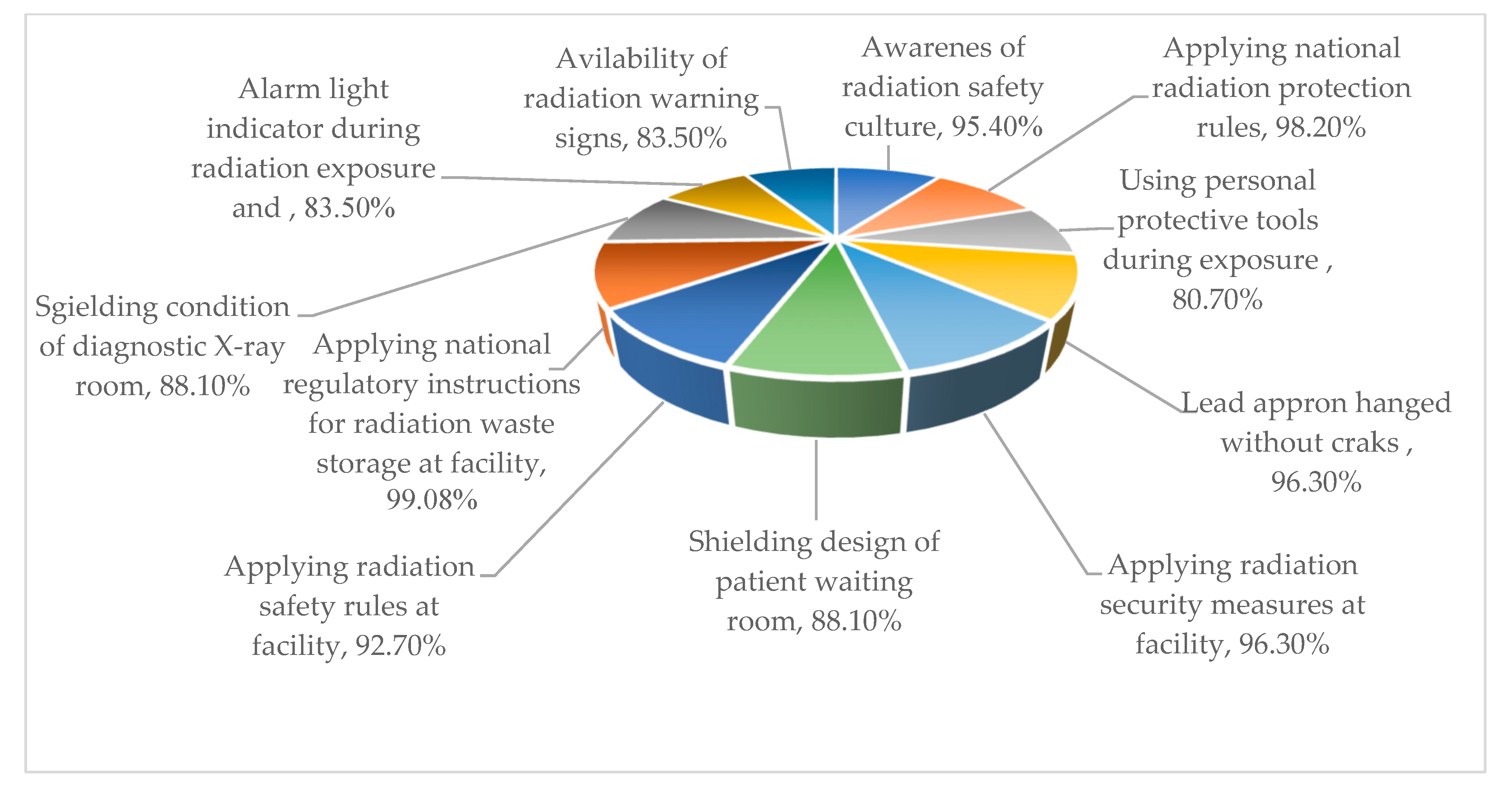
Regular training for the RSO in the field of radiation protection and radiation safety in diagnostic radiology108 (99.08%)1 (0.92%)
Notices of awareness of safety culture such as (eating in the lab., homeland devices, refrigerator, …)104 (95.4%)5 (4.6%)
The facility operator can apply correctly the national radiation protection rules concerning the unnecessary radiation exposure107 (98.2%)2 (1.8%)
Using a lead protective apron as a personal protective barrier during the exposure 88 (80.7%)21 (19.3%)
Keeping lead aprons in hanging position using an apron holder 105 (96.3%)4 (3.7%)
The security of portable X-ray machines and their proper storage condition105 (96.3%)4 (3.7%)
Patient waiting room design layout and the recommended safe distance from the X-ray machine to the patient entrance door (lead-lined door)96 (88.1%)13 (11.9%)
Display of safety work procedures, radiation safety rules, and the staff waiting room design condition 101 (92.7%)8 (7.3%)
Applying national regulatory instructions for radioactive waste storage at the facility and the concept of radiation safety rules108 (99.08%)1 (0.92%)
Check for any leakage of radiation or cracks in the wall of the X-ray room/patient entrance door and the surrounding96 (88.1%)13 (11.9%)
Existence of alarm light (illuminated radiation ON/OFF indicator) during the exposure; radiation warning signs written in Arabic and English language91 (83.5%)18 (16.5%)
Table 3. Annual effective dose in diagnostic radiology facilities in Saudi Arabia during the period from 1998 to 2022.
Time PeriodAnnual Effective Dose for Radiation Workers in KSA Diagnostic Radiology Facilities (mSv)References
1998–20020.48–0.94[29]
2009–20100.66[30]
2012–20160.40[31]
20170.66[32]
20180.96[33]
20181.90[34]
20187.40[34]
20191.24[33]
2018–20190.53[30]
20210.88[35]
202214.35[30]
Publisher’s Note: MDPI stays neutral with regard to jurisdictional claims in published maps and institutional affiliations.

