Article

Roots of Lithospermum erythrorhizon Alleviated Ovalbumin-Induced Allergic Rhinitis and IgE-triggered Degranulation of RBL-2H3 Cells

1
Natural Product Research Center, Korea Institute of Science & Technology, Gangneung 25451, Korea
2
Division of Bio-Medical Science & Technology, KIST School, Korea University of Science and Technology, Gangneung 25451, Korea
3
COSMAX NBT Inc., Seongnam 13486, Korea
*
Authors to whom correspondence should be addressed.
These authors contributed equally to the work.
Appl. Sci. 2022, 12(12), 6116; https://doi.org/10.3390/app12126116
Submission received: 26 May 2022 / Revised: 14 June 2022 / Accepted: 15 June 2022 / Published: 16 June 2022
(This article belongs to the Special Issue Bioactive Compounds from Natural Products - Volume II)

Abstract

:
Lithospermum erythrorhizon (L. erythrorhizon) root is used in traditional medicine for its anti-inflammatory, antibacterial, and antioxidant properties. However, no studies have examined its impact on allergic rhinitis (AR). Here, we explored the protective effects of L. erythrorhizon in immunoglobulin E (IgE)-stimulated RBL-2H3 cells and in an ovalbumin (OVA)-induced AR mouse model. In the latter, we examined nasal mucosal inflammation, allergen-specific cytokine production, and histological changes to the nasal mucosa. In the mouse model, oral administration of an ethanol extract of L. erythrorhizon (LE) led to a marked reduction in rubbing and sneeze frequency, a significant decrease in serum OVA-specific IgE and IgG1 levels, and a significant increase in the IgG2a/IgG1 ratio. LE also reduced expression of interleukin (IL)-4, IL-5, and IL-13 in nasal lavage fluid (NALF), and suppressed inflammatory cell infiltration and epithelial degradation in nasal tissues. In IgE-stimulated RBL-2H3 cells, LE suppressed release of degranulation markers such as β-hexosaminidase and histamine. Based on these findings, we suggest that LE may ameliorate OVA-induced AR by regulating mast cell-mediated inflammatory responses.

Graphical Abstract

1. Introduction

Allergic rhinitis (AR), a frequent allergic condition affecting 30% of the world’s population, is usually a lifelong problem [1]. AR, described as an inflammatory condition of the nasal mucosa caused by allergen exposure, is defined as any combination of rhinorrhea, nasal congestion, sneezing, and nasal itching [2]. Often, AR is associated with acute and chronic inflammatory diseases of the airway, including asthma, which is similarly characterized by antigen hypersensitivity, leading to increased local inflammation, bronchoconstriction, vasomotor changes, and mucus hypersecretion [3]. Although AR may appear insignificant compared with other medical conditions, it can cause emotional instability, insomnia, and a significantly lower quality of life, as well as being a considerable economic burden to the patient due to insufficient management [4]. Treatments such as allergen avoidance, medication, and allergen immunotherapy are commonly used; however, none of these therapies are perfect with respect to symptom alleviation. Although oral antihistamines and intranasal corticosteroids are authorized medications for the treatment of AR, adverse effects continue to represent a danger to patients. Oral antihistamines, including first- and new-generation medications, might have adverse effects on the central nervous system, while intranasal corticosteroids can induce headache, throat irritation, nosebleeds, tingling, burning, and dry nasal passages [5]. Thus, developing an anti-AR medicine that is both safe and effective remains a critical global challenge.
Due to their safety and effectiveness, many traditional medicinal plants have attracted attention over recent years. The dried roots of L. erythrorhizon have been used as a medicinal plant in traditional medicine for centuries in Korea, China, and Japan [6]. Phytochemically, L. erythrorhizon primarily contains naphthoquinone compounds such as shikonin derivatives, which, among others, have a variety of anti-inflammatory, antiviral, antibacterial, and hypoglycemic effects [7,8,9,10]. In addition, shikonin suppresses mast cell activation by blocking calcineurin [11], and data from a mouse model of AR show that acetylshikonin reduces allergic inflammation [12], suggesting that L. erythrorhizon could be an effective treatment for AR. However, no studies on the ability of L. erythrorhizon to ameliorate AR-related symptoms, allergic inflammation, and mast cell activation have been conducted. Here, we focused on the role of L. erythrorhizon in a mouse model of ovalbumin (OVA)-induced AR and in anti-2,4,6-dinitrophenyl-IgE/anti-2,4,6-dinitrophenyl-bovine serum albumin (DNP-IgE/DNP-BSA)-stimulated rat basophil cells (RBL-2H3 cells).

2. Materials and Methods

2.1. Preparation of LE

An ethanol extract of L. erythrorhizon (LE) was obtained from COSMAX NS, INC. (Seongnam, Korea). The dried roots of L. erythrorhizon were exposed to 70% ethanol at 70 °C for four hours, after which they were filtered, and all ethanol was removed under vacuum. The extracts were mixed with maltodextrin, and the sample was dried by spray-drying.

2.2. Animals

The experimental animals were 6-week-old female BALB/c mice (Orient Bio Inc., Seong-nam, Korea). Mice were housed in a room with a temperature of 23 ± 2 °C, relative humidity of 55 ± 10%, and a light–dark cycle of 12 h. KIST’s committee for the care and use of animals reviewed and approved all experimental protocols (Approval NO. 2020-155).

2.3. The OVA-Induced AR Mice Model

BALB/c mice were randomly assigned to one of five groups (n = 10/group): (1) the control (Con) group; (2) the OVA group; (3) the LE 50 group; (4) the LE 100 group; and (5) the ciclesonide (CIC) group. On Days 0, 7, and 14, mice were sensitized by intraperitoneal injection of 100 μg of OVA (Grade V, Sigma-Aldrich, St. Louis, MO, USA) and 2 mg of aluminum hydroxide (Sigma-Aldrich). From Day 21 to Day 27, sensitized mice were challenged by intranasal administration of 500 μg of OVA (20 μL into each nasal cavity). In the OVA group, mice were sensitized with OVA and challenged with OVA and phosphate-buffered saline, respectively. The LE50 and LE100 groups of mice were sensitized and challenged with OVA and orally administered LE (50 or 100 mg/kg, respectively; once daily via oral gavage; 200 μL). The CIC group was sensitized and challenged with OVA and CIC (15 μg) administered intranasally. Mice in the Con group did not undergo sensitization, challenge, or treatment. Mice were anesthetized by ether and sacrificed on Day 28 (Figure 1A).

2.4. Evaluation of Nasal Symptoms following Last Allergen Exposure

Blinded observers recorded the number of nasal rubbing and sneezing occurrences for 10 min after the last OVA challenge.

2.5. Collection and Analysis of NALF

After the final intranasal challenge, NALF was obtained by inserting a tube into the nasal portion of the upper trachea and flushing it with 500 μL of saline. The supernatant of NALF was immediately gathered and frozen at −80 °C. Cytokine levels of IL-4, IL-5, and IL-13 were determined using an ELISA MAXTM Deluxe Set KIT (BioLegend., San Diego, CA, USA).

2.6. Quantification and Collection of Serum OVA-Specific Immunoglobulins

Following the final intranasal challenge, heart blood samples were collected and centrifuged at 13,000× g rpm for 10 min to obtain serum. OVA-specific IgG1, IgG2, and IgE in the serum were determined using an ELISA MAXTM Deluxe Set KIT (BioLegend).

2.7. Histopathological Assessment of Nasal Mucosa

The head tissues were fixed for three days at room temperature in a 4% paraformaldehyde solution (T&I., Seoul, Korea). The samples were then placed in a paraffin block and sliced. The head tissue slides were stained with H&E according to a standard protocol. The slides were treated at room temperature with hematoxylin buffer, washed three times with distilled water, and then dipped in an eosin Y solution containing 1% eosin Y. The slides were then mounted after being washed three times with 95% ethanol. Using a PAS stain kit, periodic acid–Schiff (PAS) staining was performed (ab150680; Abcam, UK). Briefly, slides were treated at room temperature with periodic acid. After 15 min, the slides were washed four times with distilled water and then immersed for 15 min in Schiff’s solution. The slides were then rinsed with hot running water, treated with hematoxylin buffer, washed with distilled water three times, and mounted. Using a standard procedure, the head tissue slides were stained with toluidine blue. Briefly, the slides were treated for three minutes with toluidine blue working solution, washed with distilled water three times, and mounted. Sections were examined using a CKX41 phase contrast microscope (Olympus., Tokyo, Japan).

2.8. Cell Culture

Cell lines for RBL-2H3 were purchased from the Korean cell line research foundation of the Korean cell line bank (Seoul, Korea). The cells were cultured in culture dishes filled with DMEM supplemented with 10% fetal bovine serum plus 100 U/mL penicillin–streptomycin. We kept the cultures at 37 °C in a humidified atmosphere containing 5% CO2.

2.9. Cell Viability Assay

Using the EZ-Cytox kit, the effect of LE on RBL-2H3 cell viability was determined (DOGEN, Seoul, Korea). Briefly, cells (1 × 10⁴ cells/well) were treated with various doses of LE for 24 h. Next, EZ-Cytox was applied at 37 °C for 30 min. Absorbance at 450 nm was measured using a microplate reader.

2.10. β-Exosaminidase and Histamine Release Assay

For IgE-induced activation, 5 × 10⁵ cells/well of RBL-2H3 cells were plated on a 24-well plate, washed, and then incubated with DNP-IgE (50 ng/mL) for 24 h. The cells were washed three times with siraganian buffer (119 mM NaCl, 5.6 mM glucose, 0.4 mM MgCl2, 0.1% BSA, 5 mM KCL, 25 mM PIPES, 1 mM CaCl2, pH 7.2) and then incubated with LE for 1 h before DNP-BSA (100 ng/mL) stimulation for an additional 1 h. β-hexoaminidase activity was measured by a color change of the substrate (1 mM p-nitrophenyl-N-acetyl-β-d-glucosaminide) in citrate buffer. Using an ELISA, the histamine concentration in the supernatant was measured (ab213975; Abcam, UK).

2.11. Statistical Analysis

Using GraphPad Prism 9.0, statistical analyses were conducted (GraphPad Software, San Diego, CA, USA). A paired Student’s t-test or one-way ANOVA was utilized for statistical comparisons. The data for independent experiments are expressed as the mean ± standard error of the mean (SEM).

3. Results

3.1. LE Significantly Ameliorated Nasal Allergy Symptoms and Reduced NALFcytokine Levels in Mice with OVA- Induced AR

Chronic inflammation, itchy nose, sneezing, and rhinorrhea are common symptoms of AR [13]. To evaluate the effectiveness of LE treatment on AR symptoms, we examined the frequency of rubbing and sneezing over a period of 10 min on the final day of intranasal OVA administration. The frequency of rubbing and sneezing was significantly higher in OVA-induced AR mice than in control mice. However, compared with the OVA group, the LE (50 or 100 mg/kg) group showed significantly fewer allergic nasal symptoms. CIC, an intranasal corticoid approved for the treatment of AR [14,15], was used as a positive control; this also reduced allergy symptoms significantly in the OVA model (Figure 1B,C). Next, we examined cytokine levels in NALF samples from OVA-induced AR mice. IL-4 levels in NALF from the OVA group were higher than those in the control group, whereas those in the LE treatment group were lower, albeit not significantly (Figure 1D). Levels of IL-5 and IL-13 in the NALF from the OVA group were significantly higher than those in the control group (Figure 1D–F). However, when compared with OVA, LE (100 mg/kg) and CIC significantly reduced the levels of IL-5 and IL-13 in NALF. These results indicate that LE ameliorates allergic responses in AR mice by suppressing production of cytokines.

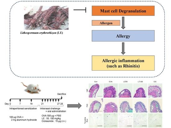
3.2. LE Significantly Reduced Nasal Mucosal Thickness and the Accumulation of Eosinophils, Goblet Cells, and Mast Cells in OVA-Induced AR Mice Nasal Tissues

Using H&E staining, the effects of LE on the nasal mucosa were then evaluated. The nasal mucosa of OVA-induced AR mice exhibited epithelial disruption, inflammatory cell infiltration, and mucosal detachment, whereas the nasal mucosa of LE-treated mice (50 mg/kg and 100 mg/kg) was protected from OVA-induced damage, as observed by measuring the nasal epithelium thickness (Figure 2A,B). Furthermore, LE suppressed OVA-induced swelling of epithelial cells. Eosinophils, crucial effector cells during allergic inflammation, have been linked to development of allergic illnesses such as asthma and rhinitis [16]. Therefore, we asked whether LE prevents eosinophil infiltration in the nasal mucosa. Significantly greater eosinophil infiltration was observed in the OVA group compared to the control group. However, the LE group displayed considerably less eosinophil infiltration than the OVA group (Figure 2C). PAS staining revealed a significant increase in goblet cell hyperplasia in the OVA group, which was significantly reduced by oral administration of LE (Figure 2A,D). To count mast cells, the nasal mucosa was then stained with toluidine blue. In the OVA group, there were significantly more mast cells than in the control group. In contrast, LE decreased nasal mast cell infiltration, indicating that LE treatment inhibits mast cell infiltration into the nasal mucosa of AR mice (Figure 2A,E).

3.3. LE Significantly Suppressed Allergic Responses by Modulating OVA-Specific Immunoglobulins in the Serum of OVA-Induced AR Mice

When exposed to allergens, IgE-primed mast cells degranulate and release a large quantity of histamine, resulting in a rapid and strong allergic response. Repeated exposure to an allergen induces production of additional IgE and IgG1, which enhances immune responses. Th1 cells, which can regulate IgE and IgG1 activity, induce immunoglobulin class switching by B cells, leading to the production of IgG2a [17,18]. Therefore, we used an ELISA to measure the quantity of OVA-specific immunoglobulins in serum. The results show that levels of OVA-specific IgE and IgG1 were considerably greater in the OVA group, but levels of OVA-specific IgG2a in the OVA group tended to be lower than in the control group. However, the amount of OVA-specific IgE in serum fell significantly after administration of 50 mg/kg or 100 mg/kg LE (Figure 3A). At a dose of 100 mg/kg, LE significantly decreased the amount of OVA-specific IgG1 (Figure 3B). Additionally, mice treated with LE (100 mg/kg) had a considerably higher IgG2a/IgG1 ratio than OVA mice (Figure 3D). These data suggest that LE reduces allergy symptoms by stimulating production of Th1-related OVA-specific IgG2a and inhibiting production of OVA-specific IgE and IgG1.

3.4. LE Inhibited IgE-Stimulated Degranulation and Histamine Release by RBL-2H3 Cells

Mast cells contribute to the pathophysiology of inflammatory allergic disorders such as AR. IgE-mediated binding to immunoglobulin Fc epsilon receptor I (FcεRI) receptors induces mast cell degranulation, leading to the production of granule-related substances and cytokines [19,20]. To test whether LE suppresses the activation of mast cells, RBL-2H3 cells were utilized in experiments. First, we used EZ-Cytox to investigate the effect of LE on cellular viability, and confirmed that the reduction in mast cell granulation was not due to LE-mediated cell death. LE concentrations of 50, 100, and 200 μg/mL had no discernible effect on the cell viability (Figure 4A). Next, we tested the antiallergic effects of LE by measuring the amount of β-hexosaminidase and histamine, two markers for mast cell degranulation. Pretreatment of LE at 50, 100, and 200 μg/mL suppressed β-hexosaminidase release by IgE–antigen-complex-stimulated cells in a dose-dependent manner (Figure 4B). Histamine release was significantly lower in the 100 μg/mL LE pretreatment group than in the IgE-triggered group (Figure 4C). These results show that LE suppresses allergic responses in RBL-2H3 cells by inhibiting mast cell degranulation.

4. Discussion

LE is a potent antioxidant with antibacterial, anticancer, antidiabetic, and antioxidative properties. However, it is currently unknown if LE could also help suppress allergic rhinitis. This is the first study to demonstrate that LE treatment can modulate allergic inflammation in a mouse model of AR induced by OVA. The AR mouse model, which induces allergic reactions with OVA, is comparable to human allergic reactions. AR mouse models exhibit allergic behaviors such as increased sneezing and nasal rubbing, as well as production of Th2 cytokines and chemokines and promotion of Th2-type immune responses [21,22]. Since a Japanese research team reported an OVA-induced AR mouse model, the AR mouse model has been utilized in a number of experiments [23]. In this study, the effect of LE on various biomarkers was therefore investigated using an AR mouse model. LE treatment inhibited release of OVA-specific immunoglobulins in serum and the secretion of allergy-associated cytokines IL-4, IL-5, and IL-13 in the NALF. Additionally, LE decreased the thickness of the epithelial mucosa, and reduced infiltration by eosinophils, goblet cells, and mast cells. In AR mice, LE ameliorated allergy symptoms such as nasal rubbing and sneezing. Additionally, treatment with LE decreased RBL-2H3 cell degranulation and histamine release significantly. Due to the fact that our study was performed in animal models, there may be differences in physiological and molecular target homology between mice and humans, which may result in translational limitations. Nevertheless, our data suggest that LE, which is utilized in traditional medicine, could be considered as a potential AR therapeutic material.
In those with allergic disorders, Th2 cells are responsible for inducing production of IgE antibodies by B cells, and for triggering infiltration of eosinophils and mast cells into tissues [24]. During allergic reactions, a number of different inflammatory cells are involved; however, eosinophils and mast cells are the primary players. Eosinophil infiltration is a characteristic feature of mucosal inflammation in AR, which is associated with IL-5 secretion by Th2 cells [25]. Mast cells are found on a variety of tissues, including mucosal epithelial surfaces and connective tissues, and they penetrate inflammatory sites associated with allergic or chronic atopic disorders [26]. The present study shows that LE treatment reduced eosinophil and mast cell infiltration, lowered production of OVA-specific IgE and IgG1, increased the IgG2a/IgG1 ratio, and reduced nasal symptoms such as rubbing and sneezing, indicating that nasal mucosal structures are protected to some extent. Therefore, we suggest that LE may alleviate AR by reducing Th2-related allergic reactions.
As it is widely known that IFN- γ stimulates IgG2a production and IL-4 stimulates IgG1 production, IgG2a is recognized as a marker of Th1 cells while IgG1 is recognized as a marker of Th2 cells [27,28]. Indeed, an increase in the total level of antigen-specific IgE and IgG1 has been observed in an allergy mouse model, and it is understood that the total level of antigen-specific IgE and IgG1 should be reduced in order to diminish the allergic reaction [29]. In addition, an increase in the IgG2a/IgG1 ratio has been reported to correlate with a protective immune response in allergic inflammatory diseases [30]. In our study, we revealed that LE treatment decreased serum IgE and IgG1 levels and increased IgG2a/IgG1 ratios. Consequently, these results suggest that LE may augment the protective immune response in AR.
Polarized Th2 cells play a key role in the development of AR, and Th2 cytokines such as IL-4, IL-5, and IL-13 play a crucial role in regulating allergenic characteristics and immune responses by IgE and inflammatory cells [31]. Among Th2 cytokines, a significant role of IL-4 is to trigger an allergic response by switching Ig classes to IgE, increasing the adhesion molecule expression, and inducing eosinophil migration to sites of inflammation [31]. IL-5 is a major factor in the differentiation and maturation of eosinophils and plays a crucial role in eosinophil activity, development, survival, and cytokine response [32]. In addition, IL-13, a cytokine produced by activated T cells, B cells, eosinophils, and mast cells, induces B-cell maturation and differentiation as well as IgE isotype conversion [33]. Our results show that LE dose-dependently and significantly reduced levels of IL-5 and IL-13 in NALF, whereas IL-4 exhibited a non-significant but decreasing trend. Therefore, we suggest that LE exerts its antiallergic effects on AR through its influence on Th2 cytokines.
Mast cells play an important role in the development of allergic reactions in AR. They accomplish this via expressing FcεRI, a fully functional high-affinity IgE receptor that mediates acute and chronic inflammation after IgE activation by producing allergen mediators [26]. Due to its high expression of FcεRI, the RBL-2H3 cell, a rat basophilic leukemia cell line, is often used to examine IgE-mediated mast cell activation [34]. We examined the in vitro antiallergic activity of LE using these cells. Degranulation is a sign that mast cells are activated and releasing inflammatory mediators. The degranulation markers β-hexosaminidase and histamine are found in cytoplasmic granules. In our study, we observed that LE inhibited the release of β-hexosaminidase and histamine by IgE–antigen-complex-stimulated RBL-2H3 cells, indicating that LE inhibits mast cell degranulation. Thus, our findings suggest that LE has antiallergic properties.
The active ingredients of LE include shikonin, acetylshikonin, and lithospermic acid, which have antiallergic, anti-inflammatory, and antioxidant properties. Understanding how these components of LE reduce allergies can shed light on the mechanism of the protective effects of LE against AR. According to reports, when the mast cells were treated with Shikonin, the activation of the mast cells was suppressed by inhibiting calcineurin and reducing the expression of the mRNA encoding the Nr4a family [11]. Moreover, shikonin can inhibit the expression of transcription factors such as GA-TA-3 and Maf in T cells, consequently lowering the expression of IL-4 and IL-5 and exhibiting antiallergic properties [35]. In an asthma model, shikonin inhibited ERK-NFkB signaling to block the invasion of immune cells and decrease allergic responses [36]. Acetylshikonin has been reported to exhibit antiallergic responses in OVA-induced AR mice [12]. In the OVA-induced AR mouse model, Acetylshikonin attenuated the nasal symptoms, sneezing, and rubbing motion, and decreased IgE and IgG1. Additionally, treatment of it reduced the expression of Th2-related cytokines IL-4, IL-5, and IL-13 in NALF and also suppressed histamine production. It can be said that the phenotype of the acetylshikonin-treated group in AR mice is similar to that of the LE-treated group. However, how acetylshikonin regulates mast cells has not yet been studied, and the mechanism of its antiallergic effect is unknown. Additionally, lithospermic acid possesses anti-inflammatory and antiallergic properties. In an allergic asthma model, lithospermic acid derived from Salvia miltiorrhiza extract reduced eosinophil infiltration and IL-4 and IL-13 release [37]. However, the mechanism by which lithospermic acid inhibits allergic reactions is unknown, as is its effect on mast cells. In our study, we observed that LE suppressed allergic reactions, prevented immune cell infiltration, and reduced mast cell function, but we have not investigated the role of LE’s active components. Therefore, further research utilizing isolated active chemicals from LE is required to determine which components of LE reduce allergic responses and how they function in AR.

5. Conclusions

To summarize, this study shows that LE protected OVA-induced allergic mice by reducing symptoms of rhinitis, reversing nasal mucosa pathological alterations, and reducing IgE production. In addition, LE ameliorated allergy reactions by decreasing degranulation and histamine production by activated mast cells. Taken together, although these findings are limited to immediate clinical translation because they are based on animal research, we propose that LE may be a potential AR therapeutic material.

Author Contributions

Conceptualization, W.-B.L. and S.H.J.; methodology, T.K.K., S.-Y.C. and W.-B.L.; investigation, T.K.K., T.T.L. and H.-W.S.; writing—original draft preparation, W.-B.L. All authors have read and agreed to the published version of the manuscript.

Funding

This work was supported by an intramural grant (2E31890) from the Korea Institute of Science and Technology (KIST), Republic of Korea, and by the Basic Science Research Program through the National Research Foundation of Korea (NRF), funded by the Ministry of Education (2021R1C1C100700711).

Institutional Review Board Statement

Animal experiments were reviewed and approved by the Institutional Animal Care and Use Committee of Korea Institute of Science and Technology (KIST).

Informed Consent Statement

Not applicable.

Data Availability Statement

The data presented in this study are available within the article.

Conflicts of Interest

The authors declare no conflict of interest.

Abbreviations

LEethanol extract of Lithospermum erythrorhizon
OVAovalbumin
ARallergic rhinitis
DNP-IgEanti-2,4,6-dinitrophenyl-IgE
DNP-BSAanti-2,4,6-dinitrophenyl-bovine serum albumin
NALFnasal lavage fluid
H&Ehematoxylin and eosin
PASperiodic acid–Schiff

References

  1. Tohidinik, H.R.; Mallah, N.; Takkouche, B. History of allergic rhinitis and risk of asthma; a systematic review and meta-analysis. World Allergy Organ J. 2019, 12, 100069. [Google Scholar] [CrossRef] [PubMed] [Green Version]
  2. Pawankar, R.; Mori, S.; Ozu, C.; Kimura, S. Overview on the pathomechanisms of allergic rhinitis. Asia Pac. Allergy 2011, 1, 157–167. [Google Scholar] [CrossRef] [PubMed] [Green Version]
  3. Smolensky, M.H.; Lemmer, B.; Reinberg, A.E. Chronobiology and chronotherapy of allergic rhinitis and bronchial asthma. Adv. Drug Deliv. Rev. 2007, 59, 852–882. [Google Scholar] [CrossRef] [PubMed]
  4. Zuberbier, T.; Lotvall, J.; Simoens, S.; Subramanian, S.V.; Church, M.K. Economic burden of inadequate management of allergic diseases in the European Union: A GA(2) LEN review. Allergy 2014, 69, 1275–1279. [Google Scholar] [CrossRef]
  5. Kakli, H.A.; Riley, T.D. Allergic Rhinitis. Prim. Care 2016, 43, 465–475. [Google Scholar] [CrossRef]
  6. Kang, T.K.; Le, T.T.; Kim, K.A.; Kim, Y.J.; Lee, W.B.; Jung, S.H. Roots of Lithospermum erythrorhizon promotes retinal cell survival in optic nerve crush-induced retinal degeneration. Exp. Eye Res. 2021, 203, 108419. [Google Scholar] [CrossRef]
  7. Brigham, L.A.; Michaels, P.J.; Flores, H.E. Cell-specific production and antimicrobial activity of naphthoquinones in roots of lithospermum erythrorhizon. Plant Physiol. 1999, 119, 417–428. [Google Scholar] [CrossRef] [Green Version]
  8. Oberg, A.I.; Yassin, K.; Csikasz, R.I.; Dehvari, N.; Shabalina, I.G.; Hutchinson, D.S.; Wilcke, M.; Ostenson, C.G.; Bengtsson, T. Shikonin increases glucose uptake in skeletal muscle cells and improves plasma glucose levels in diabetic Goto-Kakizaki rats. PLoS ONE 2011, 6, e22510. [Google Scholar] [CrossRef] [Green Version]
  9. Gao, H.; Liu, L.; Qu, Z.Y.; Wei, F.X.; Wang, S.Q.; Chen, G.; Qin, L.; Jiang, F.Y.; Wang, Y.C.; Shang, L.; et al. Anti-adenovirus activities of shikonin, a component of Chinese herbal medicine in vitro. Biol. Pharm. Bull. 2011, 34, 197–202. [Google Scholar] [CrossRef] [Green Version]
  10. Bai, G.Z.; Yu, H.T.; Ni, Y.F.; Li, X.F.; Zhang, Z.P.; Su, K.; Lei, J.; Liu, B.Y.; Ke, C.K.; Zhong, D.X.; et al. Shikonin attenuates lipopolysaccharide-induced acute lung injury in mice. J. Surg Res. 2013, 182, 303–311. [Google Scholar] [CrossRef]
  11. Wang, X.; Hayashi, S.; Umezaki, M.; Yamamoto, T.; Kageyama-Yahara, N.; Kondo, T.; Kadowaki, M. Shikonin, a constituent of Lithospermum erythrorhizon exhibits anti-allergic effects by suppressing orphan nuclear receptor Nr4a family gene expression as a new prototype of calcineurin inhibitors in mast cells. Chem. Biol. Interact. 2014, 224, 117–127. [Google Scholar] [CrossRef] [PubMed]
  12. Fan, X.H.; Cheng, L.; Yan, A.H. Ameliorative effect of acetylshikonin on ovalbumin (OVA)-induced allergic rhinitis in mice through the inhibition of Th2 cytokine production and mast cell histamine release. APMIS 2019, 127, 688–695. [Google Scholar] [CrossRef] [PubMed]
  13. Stokes, J.R.; Romero, F.A., Jr.; Allan, R.J.; Phillips, P.G.; Hackman, F.; Misfeldt, J.; Casale, T.B. The effects of an H3 receptor antagonist (PF-03654746) with fexofenadine on reducing allergic rhinitis symptoms. J. Allergy Clin. Immunol. 2012, 129, 409–412. [Google Scholar] [CrossRef] [PubMed]
  14. Kim, H.S.; Won, S.; Lee, E.K.; Chun, Y.H.; Yoon, J.S.; Kim, J.T.; Kim, H.H. Effect of Proparacaine in a Mouse Model of Allergic Rhinitis. Clin. Exp. Otorhinolaryngol. 2017, 10, 325–331. [Google Scholar] [CrossRef] [PubMed]
  15. Patel, P.; Patel, D.; Kunjibettu, S.; Hall, N.; Wingertzahn, M.A. Onset of action of ciclesonide once daily in the treatment of seasonal allergic rhinitis. Ear Nose Throat J. 2008, 87, 340–353. [Google Scholar] [CrossRef]
  16. Stone, K.D.; Prussin, C.; Metcalfe, D.D. IgE, mast cells, basophils, and eosinophils. J. Allergy Clin. Immunol. 2010, 125, S73–S80. [Google Scholar] [CrossRef]
  17. Grun, J.L.; Maurer, P.H. Different T-Helper Cell Subsets Elicited in Mice Utilizing 2 Different Adjuvant Vehicles—The Role of Endogenous Interleukin-1 in Proliferative Responses. Cell Immunol. 1989, 121, 134–145. [Google Scholar] [CrossRef]
  18. Toda, T.; Yoshino, S. Enhancement of ovalbumin-specific Th1, Th2, and Th17 immune responses by amorphous silica nanoparticles. Int. J. Immunopathol. Pharmacol. 2016, 29, 408–420. [Google Scholar] [CrossRef] [Green Version]
  19. Mendez-Enriquez, E.; Salomonsson, M.; Eriksson, J.; Janson, C.; Malinovschi, A.; Sellin, M.E.; Hallgren, J. IgE cross-linking induces activation of human and mouse mast cell progenitors. J. Allergy Clin. Immunol. 2021, 149, 1458–1463. [Google Scholar] [CrossRef]
  20. Siraganian, R.P. Mast cell signal transduction from the high-affinity IgE receptor. Curr. Opin. Immunol. 2003, 15, 639–646. [Google Scholar] [CrossRef] [Green Version]
  21. Aswar, U.; Shintre, S.; Chepurwar, S.; Aswar, M. Antiallergic effect of piperine on ovalbumin-induced allergic rhinitis in mice. Pharm. Biol. 2015, 53, 1358–1366. [Google Scholar] [CrossRef] [PubMed]
  22. Zhang, N.; Li, H.; Jia, J.; He, M. Anti-inflammatory effect of curcumin on mast cell-mediated allergic responses in ovalbumin-induced allergic rhinitis mouse. Cell Immunol. 2015, 298, 88–95. [Google Scholar] [CrossRef] [PubMed]
  23. Almansouri, H.M.; Yamamoto, S.; Kulkarni, A.D.; Ariizumi, M.; Adjei, A.A.; Yamauchi, K. Effect of dietary nucleosides and nucleotides on murine allergic rhinitis. Am. J. Med. Sci. 1996, 312, 202–205. [Google Scholar] [CrossRef]
  24. Minai-Fleminger, Y.; Levi-Schaffer, F. Mast cells and eosinophils: The two key effector cells in allergic inflammation. Inflamm. Res. 2009, 58, 631–638. [Google Scholar] [CrossRef]
  25. Takatsu, K.; Nakajima, H. IL-5 and eosinophilia. Curr. Opin. Immunol. 2008, 20, 288–294. [Google Scholar] [CrossRef]
  26. Church, M.K.; Levi-Schaffer, F. The human mast cell. J. Allergy Clin. Immunol. 1997, 99, 155–160. [Google Scholar] [CrossRef]
  27. Lewkowich, I.P.; Rempel, J.D.; HayGlass, K.T. Antigen-specific versus total immunoglobulin synthesis: Total IgE and IgG1, but not IgG2a levels predict murine antigen-specific responses. Int. Arch. Allergy Immunol. 2004, 133, 145–153. [Google Scholar] [CrossRef]
  28. Kim, D.K.; Joo, K.H.; Chung, M.S. Changes of cytokine mRNA expression and IgG responses in rats infected with Capillaria hepatica. Korean J. Parasitol. 2007, 45, 95–102. [Google Scholar] [CrossRef] [Green Version]
  29. Mountford, A.P.; Fisher, A.; Wilson, R.A. The profile of IgG1 and IgG2a antibody responses in mice exposed to Schistosoma mansoni. Parasite Immunol. 1994, 16, 521–527. [Google Scholar] [CrossRef]
  30. Rostamian, M.; Sohrabi, S.; Kavosifard, H.; Niknam, H.M. Lower levels of IgG1 in comparison with IgG2a are associated with protective immunity against Leishmania tropica infection in BALB/c mice. J. Microbiol. Immunol. Infect. 2017, 50, 160–166. [Google Scholar] [CrossRef] [Green Version]
  31. Deo, S.S.; Mistry, K.J.; Kakade, A.M.; Niphadkar, P.V. Role played by Th2 type cytokines in IgE mediated allergy and asthma. Lung India 2010, 27, 66–71. [Google Scholar] [CrossRef] [PubMed]
  32. Coffman, R.L.; Seymour, B.W.; Hudak, S.; Jackson, J.; Rennick, D. Antibody to interleukin-5 inhibits helminth-induced eosinophilia in mice. Science 1989, 245, 308–310. [Google Scholar] [CrossRef] [PubMed]
  33. Grunig, G.; Warnock, M.; Wakil, A.E.; Venkayya, R.; Brombacher, F.; Rennick, D.M.; Sheppard, D.; Mohrs, M.; Donaldson, D.D.; Locksley, R.M.; et al. Requirement for IL-13 independently of IL-4 in experimental asthma. Science 1998, 282, 2261–2263. [Google Scholar] [CrossRef] [Green Version]
  34. Passante, E.; Ehrhardt, C.; Sheridan, H.; Frankish, N. RBL-2H3 cells are an imprecise model for mast cell mediator release. Inflamm. Res. 2009, 58, 611–618. [Google Scholar] [CrossRef]
  35. Lee, C.C.; Kang, J.J.; Chiang, B.L.; Wang, C.N.; Cheng, Y.W. Shikonin inhibited mitogen-activated IL-4 and IL-5 production on EL-4 cells through downregulation of GATA-3 and c-Maf induction. Life Sci. 2011, 89, 364–370. [Google Scholar] [CrossRef]
  36. Wang, T.Y.; Zhou, Q.L.; Li, M.; Shang, Y.X. Shikonin alleviates allergic airway remodeling by inhibiting the ERK-NF-kappaB signaling pathway. Int. Immunopharmacol. 2017, 48, 169–179. [Google Scholar] [CrossRef]
  37. Heo, J.Y.; Im, D.S. Anti-allergic effects of salvianolic acid A and tanshinone IIA from Salvia miltiorrhiza determined using in vivo and in vitro experiments. Int. Immunopharmacol. 2019, 67, 69–77. [Google Scholar] [CrossRef]
Figure 1. Effects of LE on nasal symptoms and allergic inflammation in mice with OVA-induced allergic rhinitis (AR). (A) Experimental protocol. (B,C) After the last intranasal administration of OVA, nasal rubbing (B) and sneezing (C) were measured for 10 min. Oral administration of LE (50 or 100 mg/kg) and intranasal challenge with CIC significantly decreased levels of allergy-associated cytokines in NALF. (DF) Using ELISA, the levels of IL-4 (D), IL-5 (E), and IL-13 (F) in NALF were determined. Values represent the mean ± SEM (n = 10 per group). Significant differences with # p < 0.05, ### p < 0.001 versus the control group. * p < 0.05, ** p < 0.01, *** p < 0.001 versus the OVA group.
Applsci 12 06116 g001
Figure 2. Effects of LE on nasal tissue histological changes in OVA-induced AR mice. (A) Hematoxylin and eosin (H&E), periodic acid–Schiff (PAS), and toluidine blue stains were used to evaluate the histological findings in nasal tissues from each group. Scale bars: 400 μm. Histological sections were assessed with respect to (B) epithelial layer thickness (from H&E staining), (C) eosinophil counts in the nasal mucosa, (D) goblet cell counts (from PAS staining), and (E) mast cell counts (from toluidine blue staining). Data represent the mean ± SEM (n = 10 per group). Significant differences at ### p < 0.001 versus the control group. * p < 0.05, *** p < 0.001 versus the OVA group.
Applsci 12 06116 g002
Figure 3. The effects of LE on OVA-specific IgE, IgG1, and IgG2a in the serum of OVA-induced AR mice. Serum measurement of (A) OVA-specific IgE, (B) OVA-specific IgG1, and (C) OVA-specific IgG2a. (D) The IgG2a:IgG1 ratio was calculated for each mouse. The average ratio for each group is shown. All results are represented as the mean ± SEM (n = 10 per group). Significant differences at # p < 0.05, ## p < 0.01, versus the control group. * p < 0.05, ** p < 0.01 versus the OVA group.
Applsci 12 06116 g003
Figure 4. LE suppresses degranulation and production of allergy-related cytokines in RBL-2H3 cells. (A) The viability of cells was assessed 24 h after treatment with 50, 100, and 200 μg/mL LE. (B,C) FcεRI-mediated degranulation assay. The cells were sensitized with anti-DNP-IgE and then treated with LE (50, 100, or 200 μg/mL) for 1 h prior to challenge with DNP-BSA. (B) β-hexosaminidase release was measured after stimulation with DNP-BSA. (C) Histamine was measured in an ELISA. Data are expressed as the mean ± SEM. # p < 0.05, ### p < 0.001, versus the control group. ** p < 0.01, *** p < 0.001 versus the vehicle group.
Applsci 12 06116 g004
Publisher’s Note: MDPI stays neutral with regard to jurisdictional claims in published maps and institutional affiliations.

