Article

Nanomechanical Characterization of Bacterial Polyhydroxyalkanoates Using Atomic Force Microscopy

Dipartimento di Chimica, Materiali e Ingegneria Chimica “Giulio Natta”, Politecnico di Milano, Piazza Leonardo da Vinci 32, 20133 Milan, Italy
*
Author to whom correspondence should be addressed.
Appl. Sci. 2022, 12(10), 4994; https://doi.org/10.3390/app12104994
Submission received: 11 April 2022 / Revised: 6 May 2022 / Accepted: 13 May 2022 / Published: 15 May 2022

Abstract

:
Polyhydroxyalkanoates are a promising class of biopolymers that can allow the production of sustainable plastic materials. The mechanical properties of such materials are very important for possible industrial applications, but the amount of polymer required for common mechanical testing can be orders of magnitude more than what is possible to achieve with a lab-scale process. Nanoindentation with the Atomic Force Microscope allows an estimation of the Elastic Modulus that can be used as a preliminary measurement on PHA when only a limited amount of material is available. Poly(hydroxybutyrate-co-hydroxyvalerate) copolymers were analyzed, with moduli ranging from 528 ± 62 MPa to 1623 ± 172 MPa, according to both the composition and the crystallization kinetics.

1. Introduction

Mechanical characterization of materials is fundamental for gaining information about their microstructure and, hence, their possible applicability. Hardness, elasticity, plasticity, strength, and fracture are commonly measured mechanical characteristics. Well-established techniques such as tensile testing, hardness testing, or dynamic mechanical analysis are employed for this purpose [1].
However, since these measurements require macroscopic samples, they cannot be compatible in cases of poor availability of materials. In addition, they mainly provide macroscopic information about the tested samples, and heterogeneities or features at the nano-scale cannot be detected. A solution to these problems is represented by the use of atomic force microscopy (AFM). The AFM, introduced in 1986 and belonging to the scanning probe microscope (SPM) family [2], consists of a micro-machined cantilever probe, a sharp tip mounted to a piezoelectric actuator and a position-sensitive photodiode detector for receiving the reflected laser beam to provide cantilever deflection feedback [3].
AFM spreading over the years is caused by the possibility to analyze the sample, not only obtaining high-resolution topographic images, but also performing a characterization of the nanomechanical properties [4]. In particular, it can provide valuable information on local properties such as elasticity, viscoelasticity, hardness, and adhesion [5,6,7,8,9,10].
To obtain such information, the so-called force–distance curve must be first obtained through AFM in quasi-static nanoindentation. Force–distance curves can be then fitted with mathematical methods which contain relevant system properties as parameters [3].
This peculiarity caused a strong diffusion of AFM in biology and biomedicine fields, where the understanding of the behavior of complex biological systems is fundamental. In the last decades, AFM was used to evaluate the local elasticity of small, soft, and inhomogeneous samples, such as bacteria, cells but also bone tissues, and a large variety of biomaterials [11,12,13,14]. In addition, recent works include the properties investigation of many classes of materials related to different fields of application. By way of example, it is possible to mention geological materials [15], metallic alloys [16], ceramics [17], polymers [18], nanocomposites [19] and functional nanostructures [20].
Since AFM allows to determine both the microstructure and the local mechanical properties of soft matter, it has been revealed as a particularly interesting tool for probing polymers, whose morphology presents relevant features at the nanoscale [21]. As mentioned above, in the last decade, several pioneering studies using AFM nanomechanical mapping on polymers have been published [22]. Among the large variety of studied polymers, it is possible to cite homopolymers such as polyvinyl acetate, polystyrene or polymethylmethacrylate [23,24], blends like polystyrene/poly(n-butyl methacrylate) or polyolefin elastomer/polyamide [25,26], and even polymer-based composites materials [27,28]. In particular, the Elastic modulus is the most diffusely measured parameter, having been quantitatively deduced for several polymeric bulk samples and thin films with elastic modulus varying in a wide range, from a few kPa, e.g., hydrated hyaluronic acid hydrogels, to several GPa, e.g., polymethylmethacrylate, polyethylene naphthalate, or oxidized polyimides [29,30].
It should be mentioned, the use of AFM for the microstructure mechanical investigation of biopolymers. Indeed, the knowledge of the relationships between structure, properties, and performance is essential for prospective safe applications of biodegradable and/or bio-based polymers [31]. Both natural and synthetic biopolymers have been reported in more recent years. Cellulose fibers, polycaprolactone, polyvinyl acetate, polylactic acid (PLA) and cellulose-reinforced starch–gelatin polymer composite are a few of the investigated materials [32,33,34,35].
In this work, we propose a nanomechanical characterization by force–distance curves obtained via AFM of different biopolyesters belonging to the broad family of hemisynthetic and bacterial polyhydroxyalkanoates (PHA). Parameters, methodologies and accessibility of AFM tests were studied and described in previous experiences in our research group [30]. First, a comparison of the mechanical properties measured via conventional tensile test and tip nanoindentation AFM test are presented, using polylactic acid (PLA) as a standard. Then, elastic moduli obtained by tip indentation AFM test are shown for different poly(hydroxybutyrate-co-hydroxyvalerate (PHBV) copolymers, obtained by bacterial co-fermentation of different volatile fatty acid mixtures. Finally, a correlation of the obtained elastic modulus with composition, microstructure and thermal properties is proposed.

2. Materials and Methods

2.1. Materials

PHA-rich biomasses from a mixed microbial culture (MMC) selected under feast and famine conditions in a Sequencing Batch Reactor (S-SBR) using volatile fatty acids (VFAs) as substrates were kindly supplied by the Department of Biotechnology of Università di Verona (Prof. N. Frison). The PHA-rich biomass was obtained with VFAs investigating different ratios of acetic acid (HAc) to propionic acid (HPr). In particular, the following proportions were studied: 100:0 HAc:HPr; 60:40 HAc:HPr; 50:50 HAc:HPr; 30:70 HAc:HPr; 0:100 HAc:HPr. PHA samples were obtained by extraction in chloroform and recovered after filtration and drying [36].
As reported in a previous work of ours, the extracted PHA samples resulted to be complex blends of poly(hydroxybutyrate-co-hydroxyvalerate) (PHBV), where different phases, having a well-defined ratio between hydroxybutyrate (HB) and hydroxyvalerate (HV), were present. To investigate the properties of a well-defined PHBV phase, a solvent/non-solvent fractionation was also carried out for an extracted PHA batch [37].
The PHAs samples were then characterized, and the nanomechanical properties were tested by AFM.
As a benchmark for AFM testing, polylactic acid (PLA) IngeoTM 4043D was supplied by NatureWorks LLC.

2.2. Methods

In the following, the methods used for the materials characterizations are described.

2.2.1. Spin-Coating

The materials were deposited by spin-coating in the form of thin film on a glass substrate to allow AFM characterization. Parameters (400 rpm, 40 s) were set to guarantee a minimum thickness of ≈1 μm, which is required for reliable AFM analysis. At the end of the deposition process, the wet thin films were dried on a stove at 60 °C in vacuum condition for at least 40 min to guarantee solvent evaporation and to avoid contamination of the wet film.

2.2.2. Atomic Force Microscopy (AFM)

An AFM NscriptorTM DPN-WriterTM was used to study the morphology and nanomechanical properties of thin films obtained by spin-coating. Data were collected by the software SPM COCKPIT (Pacific Nanotechnology Inc., Santa Clara, CA, USA) and analyzed by OriginPro version 2020 (OriginLab Corporation, Northampton, MA, USA). Force–distance curves were performed in air at 0.05 Hz, with ACT tips from AppNano® (Santa Clara, CA, USA) with a nominal radius of curvature less than 10 nm and a half-opening angle of α = 24°. Morphological images were taken using ACT-SS (super sharp) tips from AppNano® with cantilever stiffness kc = 37 N/m, tip radius r = 6 nm, and half-opening angle α = 18°. Scanning was performed over 256 lines, 0.2 lines per second on two different square areas: 50 × 50 (μm) and 2 × 2 (μm).

2.2.3. Nuclear Magnetic Resonance (NMR)

The PHA compositions were determined using 1H-NMR spectroscopy. Spectra were recorded in deuterochloroform (CDCl3) (10 mg mL−1 for 1H-NMR) at 25 °C on a Bruker SampleXpress spectrometer. Relative peak intensities for this purpose were analyzed using the peak fitting program MNova. Peak shifts were referenced to the CDCl3 peak at 7.26 ppm.

2.2.4. Gel Permeation Chromatography (GPC)

Molecular weight measurements on extracted and fractionated PHA samples were performed using a Waters 510 Gel Permeation Chromatography (GPC) apparatus working in tetrahydrofuran (THF) at 40 °C, equipped with a set of Ultrastyragel® columns and a Waters 410 differential diffractometer as a detector. The calibration was performed with monodisperse fractions of polystyrene.

2.2.5. Dilute Solution Viscometry

Dilute solution viscometry was carried out for one extracted PHA batch, insoluble in THF for GPC analysis. The sample was dissolved in CHCl3 and the efflux time of the solution from a capillary in calibrated glass was measured at 30 °C. The intrinsic viscosity was computed with a double extrapolation at zero concentration of two semi-empirical equations (Huggins and Kraemer equations). Then, Mark–Houwink’s empirical equation was exploited to obtain the viscosity average molecular weight (Mη) assumed to be equal to the weight average molecular weight Mw as a first approximation. The empirical constants of PHB in CHCl3 at 30 °C were used, with K = 0.0118 mL/g and α = 0.78 [30].

2.2.6. Differential Scanning Calorimetry (DSC)

Thermal properties analysis was carried out using DSC for extracted and fractionated PHA. Differential scanning calorimetry (DSC) was performed with a DSC 823 e (Mettler-Toledo, Columbus, OH, USA). All runs were performed on 5–10 mg samples in a nitrogen atmosphere. The data obtained were used to measure characteristic temperatures and their enthalpies. The DSC thermal history consisted of: (i) first heating run from 25 °C to 200 °C (20 °C min−1); (ii) cooling step from 200 °C to 170 °C (20 °C min−1), from 170 °C to 0 °C (10 °C min−1) to stimulate the polymer crystallization, from 0 °C to −50 °C (20 °C min−1); (iii) second heating run from −50 °C to 200 °C (20 °C min−1).

2.2.7. Uniaxial Tensile Tests

PLA tensile properties were determined at room temperature using a Zwick/Roell Z010 (Zwick Roell Italia, Genova, Italy) equipped with a 10 kN load cell and a long-stroke extensometer following the standard test method ASTM D638-14.

3. Results

In this section, surface morphology investigation by AFM in tapping mode, raw data acquisition by tip indentation AFM test, data elaboration, and elastic moduli of the extracted PHA blends are presented. Also, the elastic modulus evolution with time is reported for a fractionated sample of PHA. For further information about PHA materials production methods and compositional, morphological, and thermal characterization, please refer to the previous work [37].
In addition, the validation of the elastic moduli extrapolation is carried out by comparing it with the Elastic modulus measured by conventional tensile test for PLA, used as a benchmark.

3.1. Surface Morphology

When deposited in the form of thin film, PHA samples were transparent to visible light at the macroscopic scale. To isolate homogenous portions of material for the tip indentation AFM test, the surface morphology of the PHA samples was observed.
The PHA morphology was investigated by tapping mode AFM, once deposited by spin-coating on a glass substrate having an arithmetic mean height Sa and a root mean squared height Sq equal to 16.7 nm and 22.8 nm, respectively. Note that 50 × 50 (µm) and 2 × 2 (µm) height and demodulation images are reported in Figure 1.
The 50 × 50 (µm) height image is coupled with a line profile (Figure 2) to highlight the typical roughness showed by the PHA samples.
Choosing a reference plane, whose area is A, and defining Z(x,y) as the distance between the points and the reference plane, roughness parameters are defined as follows:
S a = 1 A | Z ( x , y ) | dx dy
S q = 1 A Z 2 ( x , y ) dx dy
where Sa and Sq are the arithmetic mean height and root mean squared height, respectively. For the surface morphology shown in Figure 2, the surface roughness parameters, specifically the arithmetic mean height Sa and root mean squared height Sq, were calculated. Sa and Sq resulted to be equal to 51.4 nm and 66.2 nm, respectively.

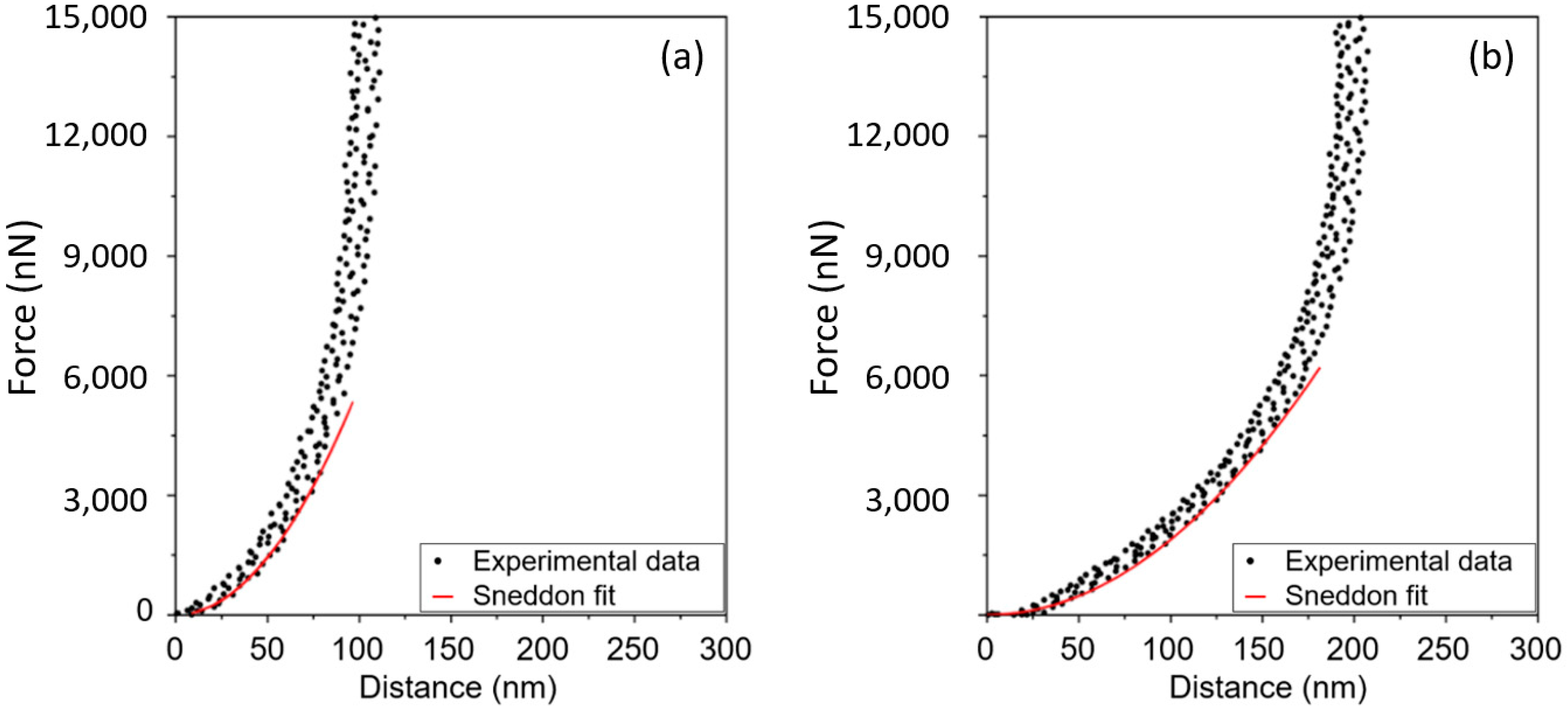
3.2. Raw Data and Data Elaboration

AFM was used to perform force–distance curves, as reported in Figure 3. The cantilevers used in the analyses were calibrated on silicon slabs before the tests on the samples. To evaluate the elastic modulus, the Sneddon model was used [38]. The equation used is the following:
F = 2 π tan α E s ( 1 ν s 2 ) δ 2
where F is the applied force, α is the half-opening of the tip, considered conical in the ending part, Es and νs are the elastic modulus and the Poisson’s ratio of the sample (assumed equal to 0.35), respectively, and δ is the indentation depth, as previously described in other studies [9].
The force was calculated as follows:
F = k C Z C
where kc is the cantilever spring constant and Zc is the cantilever deflection.
The cantilever spring constant was calculated using the Sader method, that for rectangular cantilevers defines kc as follows [39]:
k C = 0.1906   ρ   b 2   L   Q   Γ i ( ω R )   ( ω R ) 2
where ρ is the density of the fluid surrounding the cantilever, b and L are the cantilever width and length, respectively, ωR and Q are the radial resonant frequency and quality factor in a fluid of the fundamental flexural mode, respectively, and ΓiR) is the imaginary part of the dimensionless hydrodynamic function evaluated at resonant frequency [40]. The quality factor describes the relative width of the resonance peak, and it is equal to the resonant frequency divided by the half-width of the square amplitude resonance peak.
The fitting of the curves was performed with OriginPro 8.5 software. The quality of the fitting was evaluated in terms of the coefficient of correlation R2. The adjusted R2 coefficient was considered a valid measure for the reliability of interpolation, and it is defined by the following equation:
adjusted   R 2 = i = 1 n ( y i   ^ y ) 2 ( n k ) i = 1 n ( y i   ¯ y ) 2 ( n 1 )
where yi and ŷ are the original measured data values and modeled values, respectively, ȳ is the mean of the observed data, n is the number of dataset points and k is the number of parameters calculated by the interpolation. Fitting was selected to have adjusted R2 higher than 0.990.
Every sample had a total of eight indentations at different points, leading to eight force–distance curves, from which the elastic modulus was extrapolated. Figure 3 shows an example of such fittings.

3.3. Comparison between Tensile Test and Tip Indentation AFM Test

To verify the reliability of elastic modulus measurements extrapolated by the nanomechanical properties tested by AFM, as shown in Figure S1, tensile tests with a dynamometer according to the ASTM D638-14 standard were carried out with a commercial PLA sample.
Young’s modulus of 2450 ± 360 MPa for dumbbell-shaped specimens of PLA was obtained from the tensile test. On the other hand, the elastic modulus from the AFM indentation test was estimated to be 2400 ± 324 MPa. This comparison highlighted an excellent agreement: the tensile property measured by the conventional tensile test was shown to be correspondent to the nanomechanical property extrapolated by the AFM test.

3.4. Elastic Modulus Measurements in PHA

Nanomechanical characterization was carried out on extracted and fractionated PHA. The elastic modulus of the extracted PHA was calculated with force–distance curves data fitting. Table 1 lists elastic moduli extrapolated for the extracted PHA along with the average composition, molecular weights distribution and thermal properties.
The elastic properties of the tested materials are very similar, except the PHA produced with a substrate of 100:0 HAc:HPr. P(3 HB-co-2% 3 HV) showed an elastic modulus of 1623 ± 172 MPa, while all the other PHA samples showed lower values.
The PHA produced with a substrate composition ranging from 60:40 to 0:100 HAc:HPr exhibited similar elastic responses to the indentation tests: elastic moduli ranged between 528 ± 62 MPa and 885 ± 176 MPa, without a specific trend with the HV content, and with an average value of 702 ± 145 MPa.

3.5. Elastic Modulus Evolution with Time of PHA

A nanomechanical characterization was also carried out on a PHA fraction obtained by a solvent/non-solvent fractionation of an extracted PHA sample, to guarantee the strict compositional homogeneity of the material. In particular, P(3 HB-co-88% 3 HV) with HV content of 88 mol% and a single well-defined peak at 106 °C was tested. This analysis allowed the evaluation of the elastic modulus of the pure copolymer P(3 HB-co-88% 3 HV), and not of the native blend of copolymers.
The indentation tests by AFM were performed one, three, and seven days after the deposition of the thin film. Figure 4 shows the elastic moduli extrapolated for P(3 HB-co-88% 3 HV) after one, three and seven days after the deposition of the thin film.
As shown in Figure 4, P(3 HB-co-88% 3 HV) showed an increasing elastic modulus with time. P(3 HB-co-88% 3 HV) after one day exhibited an elastic modulus of 217 ± 31 MPa, which gradually increased to 285 ± 30 MPa after three days and 708 ± 78 MPa after seven days.

4. Discussion

In this section, results are discussed in terms of PHA surface morphology, correlation among extrapolated elastic moduli, thermal properties and microstructure of the extracted PHA blends. Also, indirect considerations about crystallization are provided for the fractionated sample of PHA.

4.1. Surface Morphology

The morphology of the PHA surface (Figure 1) appears as small grains, whose size is about 1 μm. The height of those grains is usually between 50 and 100 nm, as it is possible to observe in the line profile of the surface. The demodulation images show no significant phase separation in the samples. This morphology allowed us to carry out indentation tests by AFM.

4.2. Elastic Modulus Measurements in PHA

The PHA produced with a substrate of 100:0 HAc:HPr shows the highest elastic modulus. The latter, indeed, differently from all the others, has a minimal amount of HV units (2 mol%). Therefore, it is reasonable to think that its mechanical properties are not far from those exhibited by poly-3-hydroxybutyrate homopolymer (PHB), which is reported in the literature to have Elastic modulus always above 1 GPa, usually between 1 and 4 GPa [41,42]. In our case, P(3 HB-co-2% 3 HV) shows an elastic modulus of 1623 ± 172 MPa.
All the other PHA samples, having a higher amount of HV units than the case of the PHA produced with a substrate of 100:0 HAc:HPr, show lower and similar values of elastic modulus, without a specific trend. This result is in line with the study of Arcos-Hernández et al. [43], who found that PHBV blends produced by mixed cultures, despite the compositional differences, had similar Elastic modulus, measured by tensile test.
This apparently unexpected behavior can be understood by referring to the crystal phases of the extracted PHA. Despite the compositional variation in terms of HV fraction among the different batches, all PHA highlight three main faster-crystallizing phases, having similar melting temperatures tested by DSC, and therefore similar compositions. Since mechanical properties tend to be dominated by the properties of the crystalline phases, no large variation of elastic moduli from one batch to another is expected. The only exception is represented by the case of P(3 HB-co-2% 3 HV). This sample, indeed, has a content of HV low enough to exclude the formation of blends, and a single phase rich in HB units.
The main variation of elastic moduli for the extracted PHA, excluding P(3 HB-co-2% 3 HB), can be noticed for the batches produced with 30:70 HAc:HPr and 0:100 HAc:HPr. They show average values of 528 and 885 MPa, respectively, with an average difference of 357 MPa. Such modulus is rather small. However, it must be considered that the crystallization kinetics of each sample is not identical and usually slow. Therefore, it is possible that tests were not performed on materials with a crystallinity fully developed.

4.3. Elastic Modulus Evolution with Time of PHA

The PHA fraction of P(3 HB-co-88% 3 HV) shows an elastic modulus of 708 ± 78 MPa after seven days. This value, as expected, is lower than the elastic modulus usually exhibited by pure PHB: indeed, the increase of HV content allows a reduction of the elastic modulus. For example, in this work, P(3 HB-co-2% 3 HV) shows an elastic modulus of 1623 ± 172 MPa. The introduction of 88 mol% leads to an average reduction of the elastic modulus of 915 MPa.
In addition, the measure of the elastic modulus with time for the PHA fraction, P(3 HB-co-88% 3 HV), allows having indirect information about the crystallization kinetics of this sample. The gradual increase of the elastic modulus with time, from 217 ± 31 MPa after one day, to 708 ± 78 MPa after seven days, suggests that the crystallization kinetics are sluggish. This phenomenon is not uncommon for the PHA [42] and agrees with the thermal analyses. DSC studies, indeed, highlight the absence of crystallization in the second heating scan, indicating a long time required for the copolymer to form crystals [37].

5. Conclusions

A series of PHA with different carbonaceous feeds were analyzed from the nanomechanical point of view and the results were interpreted together with calorimetric results. The AFM proved to be an interesting instrument to evaluate the mechanical properties of PHA when only a small amount of material is available. The results suggest that polymers obtained with a feed of acetic and propionic acid are crystalline and stiff and could be considered for sustainable packaging or other technical applications. By changing the feedstock of the bacteria that produce PHA, it is possible to modulate the chemical composition of such polymers and obtain a certain degree of variability in the mechanical properties of the extracted PHA.

Supplementary Materials

The following supporting information can be downloaded at: https://www.mdpi.com/article/10.3390/app12104994/s1, Figure S1: Example of stress-strain curves for PLA.

Author Contributions

Conceptualization, S.T.; validation, S.B. and R.C.; writing—original draft preparation, S.B.; writing—review and editing, R.C.; supervision, S.T. All authors have read and agreed to the published version of the manuscript.

Funding

This research was funded by the Cariplo Foundation, grant number 2018-0992.

Institutional Review Board Statement

Not applicable.

Informed Consent Statement

Not applicable.

Data Availability Statement

The data presented in this study are available on request from the corresponding author. The data are not publicly available due to privacy.

Acknowledgments

We thank Nicola Frison (Università degli Studi di Verona, Dipartimento di Biotecnologie) for kindly providing us with PHA-rich biomasses. We also thank Elena Ficara (Politecnico di Milano, Dipartimento di Ingegneria Civile e Ambientale) for kindly allowing the PHA extraction from biomasses. This work was funded by the Cariplo Foundation.

Conflicts of Interest

The authors declare no conflict of interest.

References

  1. Wiederhorn, S.; Fields, R.; Low, S.; Bahng, G.-W.; Wehrstedt, A.; Hahn, J.; Tomota, Y.; Miyata, T.; Lin, H.; Freeman, B.; et al. Mechanical Properties. In Springer Handbook of Materials Measurement Methods; Czichos, H., Saito, T., Smith, L., Eds.; Springer: Berlin/Heidelberg, Germany, 2006; pp. 283–397. ISBN 978-3-540-30300-8. [Google Scholar]
  2. Binnig, G.; Quate, C.F.; Gerber, C. Atomic Force Microscope. Phys. Rev. Lett. 1986, 56, 930–933. [Google Scholar] [CrossRef] [PubMed] [Green Version]
  3. Jalili, N.; Laxminarayana, K. A Review of Atomic Force Microscopy Imaging Systems: Application to Molecular Metrology and Biological Sciences. Mechatronics 2004, 14, 907–945. [Google Scholar] [CrossRef]
  4. Garcia, R. Nanomechanical Mapping of Soft Materials with the Atomic Force Microscope: Methods, Theory and Applications. Chem. Soc. Rev. 2020, 49, 5850–5884. [Google Scholar] [CrossRef] [PubMed]
  5. Zemła, J.; Danilkiewicz, J.; Orzechowska, B.; Pabijan, J.; Seweryn, S.; Lekka, M. Atomic Force Microscopy as a Tool for Assessing the Cellular Elasticity and Adhesiveness to Identify Cancer Cells and Tissues. Semin. Cell Dev. Biol. 2018, 73, 115–124. [Google Scholar] [CrossRef]
  6. Garcia, P.D.; Garcia, R. Determination of the Elastic Moduli of a Single Cell Cultured on a Rigid Support by Force Microscopy. Biophys. J. 2018, 114, 2923–2932. [Google Scholar] [CrossRef] [Green Version]
  7. Chim, Y.H.; Mason, L.M.; Rath, N.; Olson, M.F.; Tassieri, M.; Yin, H. A One-Step Procedure to Probe the Viscoelastic Properties of Cells by Atomic Force Microscopy. Sci. Rep. 2018, 8, 14462. [Google Scholar] [CrossRef] [Green Version]
  8. Walker, J.; Umer, J.; Mohammadpour, M.; Theodossiades, S.; Bewsher, S.R.; Offner, G.; Bansal, H.; Leighton, M.; Braunstingl, M.; Flesch, H.G. Asperity Level Characterization of Abrasive Wear Using Atomic Force Microscopy. Proc. R. Soc. A Math. Phys. Eng. Sci. 2021, 477, 20210103. [Google Scholar] [CrossRef]
  9. Suriano, R.; Ciapponi, R.; Griffini, G.; Levi, M.; Turri, S. Fluorinated Zirconia-Based Sol-Gel Hybrid Coatings on Polycarbonate with High Durability and Improved Scratch Resistance. Surf. Coat. Technol. 2017, 311, 80–89. [Google Scholar] [CrossRef]
  10. Ciapponi, R. Sol-Gel Hybrid Fluorinated Coatings: Preparation, Characterisation and Correlation with Human Perceived Sensorial Properties. Master Thesis, Politecnico di Milano, Milano, Italy, 29 April 2015. [Google Scholar]
  11. Chang, K.; Chiang, Y.; Yang, C.; Liou, J. Atomic Force Microscopy in Biology and Biomedicine. Tzu Chi Med. J. 2012, 24, 162–169. [Google Scholar] [CrossRef] [Green Version]
  12. Smith, J.R.; Olusanya, T.O.B.; Lamprou, D.A. Characterization of Drug Delivery Vehicles Using Atomic Force Microscopy: Current Status. Expert Opin. Drug Deliv. 2018, 15, 1211–1221. [Google Scholar] [CrossRef] [Green Version]
  13. Deng, X.; Xiong, F.; Li, X.; Xiang, B.; Li, Z.; Wu, X.; Guo, C.; Li, X.; Li, Y.; Li, G.; et al. Application of Atomic Force Microscopy in Cancer Research. J. Nanobiotechnology 2018, 16, 102. [Google Scholar] [CrossRef] [PubMed] [Green Version]
  14. Weber, A.; Iturri, J.; Benitez, R.; Toca-Herrera, J.L. Measuring Biomaterials Mechanics with Atomic Force Microscopy. 1. Influence of the Loading Rate and Applied Force (Pyramidal Tips). Microsc. Res. Tech. 2019, 82, 1392–1400. [Google Scholar] [CrossRef] [PubMed] [Green Version]
  15. Wang, K.; Taylor, K.G.; Ma, L. Advancing the Application of Atomic Force Microscopy (AFM) to the Characterization and Quantification of Geological Material Properties. Int. J. Coal Geol. 2021, 247, 103852. [Google Scholar] [CrossRef]
  16. Muhammad, H.; Massab, J.; Muhammad, S.; Alkuhayli, A.; Noman, A.M.; Al-Shamma’a, A.A. Indentation Creep Behavior of Pulsed Tungsten Inert Gas Welded Ti-5Al-2.5Sn Alloy Joints by Nanoindentation and Atomic Force Microscopy. Proc. Inst. Mech. Eng. Part E J. Process Mech. Eng. 2022. [Google Scholar] [CrossRef]
  17. Deng, B.; Luo, J.; Harris, J.T.; Smith, C.M.; Wilkinson, T.M. Toward Revealing Full Atomic Picture of Nanoindentation Deformation Mechanisms in Li2O-2SiO2 Glass-Ceramics. Acta Mater. 2021, 208, 116715. [Google Scholar] [CrossRef]
  18. Morozov, I.A. Atomic Force Microscopy Nanoindentation Kinetics and Subsurface Visualization of Soft Inhomogeneous Polymer. Microsc. Res. Tech. 2021, 84, 1959–1966. [Google Scholar] [CrossRef]
  19. Arora, G.; Pathak, H. Nanoindentation Characterization of Polymer Nanocomposites for Elastic and Viscoelastic Properties: Experimental and Mathematical Approach. Compos. Part C Open Access 2021, 4, 100103. [Google Scholar] [CrossRef]
  20. Sobola, D.; Ramazanov, S.; Konečný, M.; Orudzhev, F.; Kaspar, P.; Papež, N.; Knápek, A.; Potoček, M. Complementary SEM-AFM of Swelling Bi-Fe-O Film on HOPG Substrate. Materials 2020, 13, 2402. [Google Scholar] [CrossRef]
  21. Collinson, D.W.; Sheridan, R.J.; Palmeri, M.J.; Brinson, L.C. Best Practices and Recommendations for Accurate Nanomechanical Characterization of Heterogeneous Polymer Systems with Atomic Force Microscopy. Prog. Polym. Sci. 2021, 119, 101420. [Google Scholar] [CrossRef]
  22. Wang, D.; Russell, T.P. Advances in Atomic Force Microscopy for Probing Polymer Structure and Properties. Macromolecules 2018, 51, 3–24. [Google Scholar] [CrossRef]
  23. Stafford, C.M.; Vogt, B.D.; Harrison, C.; Julthongpiput, D.; Huang, R. Elastic Moduli of Ultrathin Amorphous Polymer Films. Macromolecules 2006, 39, 5095–5099. [Google Scholar] [CrossRef] [Green Version]
  24. Miyake, K.; Satomi, N.; Sasaki, S. Elastic Modulus of Polystyrene Film from near Surface to Bulk Measured by Nanoindentation Using Atomic Force Microscopy. Appl. Phys. Lett. 2006, 89, 031925. [Google Scholar] [CrossRef]
  25. Cappella, B.; Kaliappan, S.K. Determination of Thermomechanical Properties of a Model Polymer Blend. Macromolecules 2006, 39, 9243–9252. [Google Scholar] [CrossRef]
  26. Wang, D.; Fujinami, S.; Liu, H.; Nakajima, K.; Nishi, T. Investigation of Reactive Polymer-Polymer Interface Using Nanomechanical Mapping. Macromolecules 2010, 43, 5521–5523. [Google Scholar] [CrossRef]
  27. Niu, Y.-F.; Yang, Y.; Gao, S.; Yao, J.-W. Mechanical Mapping of the Interphase in Carbon Fiber Reinforced Poly(Ether-Ether-Ketone) Composites Using Peak Force Atomic Force Microscopy: Interphase Shrinkage under Coupled Ultraviolet and Hydro-Thermal Exposure. Polym. Test. 2016, 55, 257–260. [Google Scholar] [CrossRef]
  28. Liang, X.; Ito, M.; Nakajima, K. Reinforcement Mechanism of Carbon Black-filled Rubber Nanocomposite as Revealed by Atomic Force Microscopy Nanomechanics. Polymers 2021, 13, 3922. [Google Scholar] [CrossRef]
  29. Passeri, D.; Rossi, M.; Tamburri, E.; Terranova, M.L. Mechanical Characterization of Polymeric Thin Films by Atomic Force Microscopy Based Techniques. Anal. Bioanal. Chem. 2013, 405, 1463–1478. [Google Scholar] [CrossRef]
  30. Suriano, R.; Credi, C.; Levi, M.; Turri, S. AFM Nanoscale Indentation in Air of Polymeric and Hybrid Materials with Highly Different Stiffness. Appl. Surf. Sci. 2014, 311, 558–566. [Google Scholar] [CrossRef]
  31. Rydz, J.; Šišková, A.; Andicsová Eckstein, A. Scanning Electron Microscopy and Atomic Force Microscopy: Topographic and Dynamical Surface Studies of Blends, Composites, and Hybrid Functional Materials for Sustainable Future. Adv. Mater. Sci. Eng. 2019, 2019, 6871785. [Google Scholar] [CrossRef] [Green Version]
  32. Nguyen, H.K.; Fujinami, S.; Nakajima, K. Elastic Modulus of Ultrathin Polymer Films Characterized by Atomic Force Microscopy: The Role of Probe Radius. Polymer 2016, 87, 114–122. [Google Scholar] [CrossRef]
  33. Iqbal, Q.; Bernstein, P.; Zhu, Y.; Rahamim, J.; Cebe, P.; Staii, C. Quantitative Analysis of Mechanical and Electrostatic Properties of Poly(Lactic) Acid Fibers and Poly(Lactic) Acid-Carbon Nanotube Composites Using Atomic Force Microscopy. Nanotechnology 2015, 26, 105702. [Google Scholar] [CrossRef] [PubMed]
  34. Chlanda, A.; Kijeńska-Gawrońska, E.; Zdunek, J.; Swieszkowski, W. Internal Nanocrystalline Structure and Stiffness Alterations of Electrospun Polycaprolactone-Based Mats after Six Months of in Vitro Degradation. An Atomic Force Microscopy Assay. J. Mech. Behav. Biomed. Mater. 2020, 101, 103437. [Google Scholar] [CrossRef] [PubMed]
  35. Rodríguez-Castellanos, W.; Flores-Ruiz, F.J.; Martínez-Bustos, F.; Chiñas-Castillo, F.; Espinoza-Beltrán, F.J. Nanomechanical Properties and Thermal Stability of Recycled Cellulose Reinforced Starch-Gelatin Polymer Composite. J. Appl. Polym. Sci. 2015, 132, 41787. [Google Scholar] [CrossRef]
  36. Kourmentza, C.; Plácido, J.; Venetsaneas, N.; Burniol-Figols, A.; Varrone, C.; Gavala, H.N.; Reis, M.A.M. Recent Advances and Challenges towards Sustainable Polyhydroxyalkanoate (PHA) Production. Bioengineering 2017, 4, 55. [Google Scholar] [CrossRef] [PubMed] [Green Version]
  37. Bagatella, S.; Ciapponi, R.; Ficara, E.; Frison, N.; Turri, S. Production and Characterization of Polyhydroxyalkanoates from Wastewater via Mixed Microbial Cultures and Microalgae. Sustainability 2022, 14, 3704. [Google Scholar] [CrossRef]
  38. Sneddon, I.N. The Relation between Load and Penetration in the Axisymmetric Boussinesq Problem for a Punch of Arbitrary Profile. Int. J. Eng. Sci. 1965, 3, 47–57. [Google Scholar] [CrossRef]
  39. Sader, J.E.; Sanelli, J.A.; Adamson, B.D.; Monty, J.P.; Wei, X.; Crawford, S.A.; Friend, J.R.; Marusic, I.; Mulvaney, P.; Bieske, E.J. Spring Constant Calibration of Atomic Force Microscope Cantilevers of Arbitrary Shape. Rev. Sci. Instrum. 2012, 83, 103705. [Google Scholar] [CrossRef] [Green Version]
  40. Van Eysden, C.A.; Sader, J.E. Frequency Response of Cantilever Beams Immersed in Viscous Fluids with Applications to the Atomic Force Microscope: Arbitrary Mode Order. J. Appl. Phys. 2007, 101, 044908. [Google Scholar] [CrossRef]
  41. Khatami, K.; Perez-Zabaleta, M.; Owusu-Agyeman, I.; Cetecioglu, Z. Waste to Bioplastics: How Close Are We to Sustainable Polyhydroxyalkanoates Production? Waste Manag. 2021, 119, 374–388. [Google Scholar] [CrossRef]
  42. Laycock, B.; Halley, P.; Pratt, S.; Werker, A.; Lant, P. The Chemomechanical Properties of Microbial Polyhydroxyalkanoates. Prog. Polym. Sci. 2014, 39, 397–442. [Google Scholar] [CrossRef]
  43. Arcos-Hernández, M.V.; Laycock, B.; Donose, B.C.; Pratt, S.; Halley, P.; Al-Luaibi, S.; Werker, A.; Lant, P.A. Physicochemical and Mechanical Properties of Mixed Culture Polyhydroxyalkanoate (PHBV). Eur. Polym. J. 2013, 49, 904–913. [Google Scholar] [CrossRef]
Figure 1. Images of surface morphology acquired by AFM in tapping mode for PHA thin films. Top: height (left) and demodulation (right) 50 × 50 (µm) images; bottom: height (left) and demodulation (right) 2 × 2 (µm) images.
Applsci 12 04994 g001
Figure 2. Height 50 × 50 (µm) image of surface morphology acquired by AFM in tapping mode for PHA thin films sample (left) and its line profile (right).
Applsci 12 04994 g002
Figure 3. Force–distance curves of the tested bio-polyesters: (a) PLA; (b) P(3 HB-co-88% 3 HV).
Applsci 12 04994 g003
Figure 4. Elastic modulus from AFM force–distance curves of P(3 HB-co-88% 3 HV) one, three, and seven days after the deposition of the thin film.
Applsci 12 04994 g004
Table 1. Extracted PHA molar composition, elastic modulus from AFM force–distance curves on thin film, weight-average molecular weight Mw, polydispersity index and thermal properties.
HAc:HPrElastic ModulusMolar CompositionMwPolydispersity IndexMelting TemperaturesCrystallinity Degree
[g-COD/L:g-COD/L][MPa][HV mol%][105 Da] [°C][-]
100:01623 ± 1722%7.47 *-17348%
30:70528 ± 6212%1.952.7817438%
60:40693 ± 7415%3.061.5395/138/17055%
0:100885 ± 17640%2.181.6697/132/17255%
50:50700 ± 13047%2.451.3398/140/17555%
* Mw estimated by dilute solution viscometry.
Publisher’s Note: MDPI stays neutral with regard to jurisdictional claims in published maps and institutional affiliations.

