Article

Cytotoxic Influence of Khat (Catha edulis (Vahl) Forssk. ex Endl) on Oral Fibroblasts, Squamous Carcinoma Cells, and Expression of α Smooth Muscle Actin

1
Academic Unit of Restorative Dentistry, The School of Clinical Dentistry, University of Sheffield, Claremont Crescent, Sheffield S10 2TA, UK
2
Department of Restorative Dentistry and Endodontics, College of Dentistry, King Faisal University, Al-Ahsa 31982, Saudi Arabia
3
Preventive Dental Sciences Department, Community Dentistry Division, College of Dentistry, Princess Nourah Bint Abdulrahman Universty, Riyadh 11671, Saudi Arabia
4
Department of Periodontics and Community Dentistry, College of Dentistry, King Saud University, Riyadh 11545, Saudi Arabia
5
Department of Prosthodontics, College of Dentistry, King Khalid University, Abha 61421, Saudi Arabia
6
Department of Restorative Dental Science, College of Dentistry, King Saud University, Riyadh 11545, Saudi Arabia
7
Department of Prosthetic Dental Science, College of Dentistry, King Saud University, Riyadh 11545, Saudi Arabia
*
Author to whom correspondence should be addressed.
Appl. Sci. 2021, 11(8), 3524; https://doi.org/10.3390/app11083524
Submission received: 19 March 2021 / Revised: 8 April 2021 / Accepted: 9 April 2021 / Published: 14 April 2021
(This article belongs to the Special Issue New Insights in Oral Health and Diets)

Abstract

:
The aim was to determine the cytotoxicity of Khat (Catha edulis (Vahl) Forssk. ex Endl) on normal oral fibroblasts (NOFs) and SCC4 (squamous carcinoma cells) along with expression of α-smooth muscle actin (α-SMA) in fibroblasts. Khat filtrate was prepared to obtain a concentrated viscous solution. NOFs and SCC4 cells were cultured in biological cabinets and were grown in Dulbeccos’ modified Eagles medium. Frozen cells were thawed at 37 °C and cell seeding was performed. NOFs and SCC4 cells were seeded on 96 well plates and allowed to attach. The medium was removed and a fresh medium containing different concentrations of Khat was added. The group without Khat served as a negative control and 4% paraformaldehyde as the positive control. Cell viability was assessed using the MTT assay and effect of Khat on fibroblast and SCC4 phenotypes was evaluated by immunostaining. Analysis of variance was used to assess data (p < 0.05). NOF 316 showed cell death in response to 4% paraformaldehyde, 12.5, 6.25, and 3.12 mg/mL of Khat. The highest concentration of Khat (25 mg/mL) failed to cause cytotoxicity of NOF 316. NOF 319 and NOF 26 displayed cell death at all concentrations of Khat, however, cytotoxicity was not dose dependent. NOF 18 and SCC4 cells showed dose-dependent cell death. NOF 316 showed α-SMA expression after 1 mg/mL of Khat exposure. Not all fibroblasts were α-SMA-positive, suggesting specific activation of a subset of fibroblasts. Khat is cytotoxic to NOF and SCC4 cells. Furthermore, it can also cause activation and phenotypic changes in oral fibroblasts, indicating a potential role in progression of oral squamous cell carcinoma.

1. Introduction

Oral cancer is considered to be a significant health problem, as around ninety percent of all oral cancer cases are oral squamous cell carcinomas (OSCC) [1]. It has a worldwide distribution and is the sixth most commonly occurring cancer globally [2]. The major risk factors associated with oral cancer are tobacco smoking and alcohol consumption. Other established risk factors i.e., betel quid chewing, use of smokeless tobacco, human papilloma viruses (types 16 and 18), nutrient deficiency, exposure to solar radiation, and genetic predisposition also causes OSCC [3]. However, there exists a great difference in inter- and intra-country distribution of these causative factors, which possibly explains the different geographical pattern of the disease [1]. One such product was reported to cause malignant and pre malignant oral lesions and is routinely used in the Arabian Peninsula as Khat [4].
The increasing use of Khat (Catha edulis (Vahl) Forssk. ex Endl) is associated with grave health hazards [5]. Chronic consumption of Khat affects almost every organ of the human body. It also leads to oral histopathological changes i.e., hyperkeratosis, epithelial hyperplasia, and mild dysplasia [6]. Lukandu et al. revealed that Khat induces abnormal epithelial differentiation and decreases basal cell proliferation [7]. Similarly, a study conducted by Al-Ahdal et al. reported that the cytotoxicity and mutagenicity of Khat leaves extract on human cells [8]. Khat performs its genotoxic activity by inhibiting de novo synthesis of proteins, RNA, and DNA, to reduce free radical metabolizing enzymes, induces oxidative stress, and provokes caspase-dependent apoptosis in various leukemic cells [9,10]. However, studies on its toxicological potential remain scarce.
Previously, it was believed that epithelial genetic changes were the sole cause of oral pre-cancer and cancer, but recent reports showed that progression of carcinoma is derived from the epithelial component as well as mesenchymal tissue [11]. The connective tissue stroma in the vicinity of the tumor plays an important part in oral cancer progression [12]. Normal oral fibroblasts (NOFs) are cells of mesenchymal origin and are omnipresent in almost all tissues within the stroma or connective tissue [13]. In precancerous and cancerous lesions, NOF are modified or ‘activated’ into myofibroblasts (MFs) and express α-smooth muscle actin (α-SMA) due to mechanical cell stress and tumor stimulating factors [14]. These MFs tend to differentiate in the presence of the transforming growth factor (TGF)-β, another factor that increases in fibrotic lesions. Studies showed that the stroma surrounding the tumor assists in metastasis and malignant progression [15]. However, the role of mesenchymal tissue and NOFs in disease progression due to Khat is not yet determined.
Considering the available indexed literature, it was found that numerous studies showed adverse effects of Khat on oral mucosa. Whereas, its role in OSCC, particularly in relation to connective tissue/stromal changes is largely uncharacterized. Therefore, it is hypothesized that Khat is cytotoxic to the NOFs and SCC4 cells (squamous carcinoma cells). It is also hypothesized that it will exhibit positive expression for α-SMA in fibroblasts. Hence, the aim of the current study was to determine the cytotoxicity of Khat on NOFs and SCC4 cells, along with the expression of α-SMA in fibroblasts.

2. Materials and Methods

2.1. Khat Preparation

Khat (mature) used in the present study was commercially obtained from Sana, Yemen. The Khat extraction method was similar, as previously explained by Aziz et al. [16]. Small pieces of frozen Khat leaves (100 gm) were dissolved in 100 mL of 95% ethanol. The prepared solution was centrifuged at 5000 rpm for 5 min and filtered using Whatman filter paper (no.1). After filtration, 100 mL of ethanol were added to the remaining leaves and the procedure was repeated. Rotary evaporator (EYELA, N-1001S-W, Tokyo, Japan) at 70 rpm was used to concentrate ethanolic Khat at 30 °C, until 30% of ethanol was left. The concentrated viscous solution obtained was then diluted using 100 mL of distilled water and stirred at ambient temperature for 1 h. The filtrate was freezed for 24 h by keeping it at −80 °C and then dried by lyophilization (Labconco, Kansas City, MO, USA). Every 100 g of dried Khat leaves gives 8 g of Khat in powder form. Liquid chromatography was performed to confirm the presence of alkaloids, i.e., 80% cathine and 20% norephedrine. Cathinone was not discovered in the analysis. All materials and equipment used in the study are presented in Appendix A. Dimethyl sulfoxide (DMSO) was used a vehicle for suspension of Khat extract.

2.2. Culture of NOFs and SCC4 Cells

2.2.1. Cell Lines and Culturing

NOFs in the present study were obtained after ethical approval from patients undergoing third molar extractions at Charles Clifford Dental Hospital. NOFs and SCC4 cells (American Type Culture Collection/ATCC CRL-1624) were cultured in Biological Class II sterile laminar flow cabinets. In order to maintain cell viability, the confluence medium was routinely changed and cells were maintained at 37 °C in a 5% CO2 incubator. Details of cells used in this study are provided in Table 1.

2.2.2. Cell Growth Medium

All cells were grown in Dulbeccos’ Modified Eagles Medium (DMEM) (Invitrogen) with supplements (Table 2).

2.2.3. Thawing and Seeding Cell Lines

Frozen cell vials were removed from liquid nitrogen and thawed in a water bath at 37 °C. Thawed cells were moved to a sterile universal tube containing 9 mL of pre-warmed medium and centrifuged at 1000 rpm for 5 min. The supernatant was discarded, the cell pellet was re-suspended in 10 mL fresh culture medium, and moved to a 75 cm2 tissue culture flask.

2.2.4. Adherent Cell Sub-Culturing and Passaging

Inverted microscope (Axioinvert Carl Zeiss) was used to visualize the cell growth. Sub-culturing was performed when 80% of confluence was reached. To passage the cells, the medium was removed; cells were washed twice in a 10 mL solution of Ca2+ and Mg2+ free PBS. Trypsin/EDTA solution was added and flasks were placed in the incubator for 10 min. Trypsin was neutralized by fresh medium and the cells were centrifuged, resuspended, and counted using a hemocytometer.

2.3. MTT Assay

2.3.1. Seeding Cells in 96 Well Plates

1.5 × 10⁴ NOFs and 1 × 10⁴ SCC4 cells (in 100 µL per well) were seeded on 96 well plates and allowed to get attached for 24 h.

2.3.2. Khat Exposure

Khat at a concentration of 25 mg/mL was used throughout this study, which was diluted accordingly. The extract was filtered using 0.2 µm Merck Millipore express filters in a liquid form. After 24 h, 96 well plates were checked under the inverted microscope to ensure cell attachment. The medium was removed and a fresh medium containing 25, 12.5, 6.25, 3.125, 1.56, 0.78, 0.39, and 0.19 mg/mL of Khat was added. The group without Khat served as a negative control and 4% paraformaldehyde as the positive control. Paraformaldehyde was dissolved in DMSO and the control wells were also treated with it. The 96 well plate was incubated for a further 24 h at 37 °C in a 5% CO2 incubator. In addition, a triplicate of each concentration was run and the average values were taken.

2.3.3. Determination of Cytotoxicity/Metabolic Activity

Cell viability was assessed using the MTT (3-(4, 5-dimethylthiazolyl-2)-2, 5-diphenyltetrazolium bromide) assay, a yellow tetrazole. MTT solution (Sigma Aldrich) (0.5 mg/mL in PBS–100 µL/well) was added after removing the medium and washing the cell, using PBS solution. The unreacted MTT solution was removed after one hour and newly formed purple intracellular formazan salt was solubilized using acidified isopropanol (50 µL/well). Absorbance was measured at 540 nm with a reference at 630 nm, using a spectrophotometer (Tecan) and Magellan software. Background subtraction was done and optical densities were normalized to a control sample of the untreated cells. Data were entered and analyzed through Microsoft Excel. Analysis of variance (ANOVA) was used to determine the significance of data obtained from MTT assays.

2.4. Effect of Khat on Fibroblast Phenotype

2.4.1. Immunostaining Preparation

2 × 10⁴ cells per well were seeded on sterile coverslips in 24 well plates. The plate was incubated for 24 h in 5% CO2 at 37 °C. the old medium was removed from all wells and the cells were washed with 1 mL of medium. Cells were then exposed to Khat concentrations of 1 mg/mL and 0.5 mg/mL; and the 40 µg/mL of Transforming Growth Factor- β (TGF-β) acted as a positive control. A total of 500 µL of medium without Khat or TGF-β acted as negative control.

2.4.2. Immunofluorescence for SMA

The medium from the 24 well plate was removed and the cells were washed twice in PBS solution, followed by fixation in 100% 1 mL methanol for 10 min. The coverslips were washed once in 0.5 mL of 4 mM sodium deoxycylate in PBS and permeabilized in the same solution for 10 min. Sodium deoxycylate was removed and the coverslips were blocked using 500 µL of 2.5% Bovine Serum Albumin (BSA) in PBS for 30 min, followed by an incubation with 0.5 mL of FITC-conjugated anti-alpha smooth muscle actin antibody (Sigma, Clone1A.4, dilution 1:1000) for one hour at 37 °C, in dark. An IgG isotype antibody was used as a negative control. After 1 h, the coverslips were washed in PBS and placed on glass slides containing 50 µL DAPI (vectorized). Glass slides were kept at 3–4 °C for a day and were viewed using a fluorescence microscope (Zeisss Axioplan 2, imaging with software Proplus 7.0.1. Presence of a green staining was considered to be positive.

3. Results

3.1. MTT Assay

3.1.1. NOF 316

MTT Assay was performed to determine the viability of NOF 316 primary cells, after being exposed to different concentrations of Khat. Cell death was seen in response to 4% paraformaldehyde (positive control), 12.5, 6.25, and 3.12 mg/mL concentrations of Khat, as compared to the negative control (p < 0.05). However, the lower doses of Khat (1.56, 0.781, 0.39, and 0.195 mg/mL) did not display significant cytotoxicity. Interestingly, at the highest dose, i.e., 25 mg/mL, Khat did not display a significant increase in cell death (Figure 1).

3.1.2. NOF 26

Results for NOF 26 were somewhat different from NOF316, as nearly all concentrations of Khat caused a significant increase in cell death, as compared to the negative control (p < 0.01). A similar response was observed in the positive control and cytotoxicity appeared to be dose dependent (Figure 2).

3.1.3. NOF 319

NOF 319 cells showed a similar pattern to NOF 26 cells, as all concentrations of Khat caused significant cell death (p < 0.01). However, cytotoxicity did not appear to be dose-dependent. However, the positive control caused significantly more cell death, as compared to Khat (p < 0.01) (Figure 3).

3.1.4. NOF 18

Exposure of NOF 18 cells to different Khat concentrations resulted in a significant increase in cell death, as compared to the negative control (p < 0.01). In addition, the extent of cell cytotoxicity was dependent on Khat concentration, showing a dose-dependent response (Figure 4).

3.1.5. Cytotoxicity of Different NOF’s
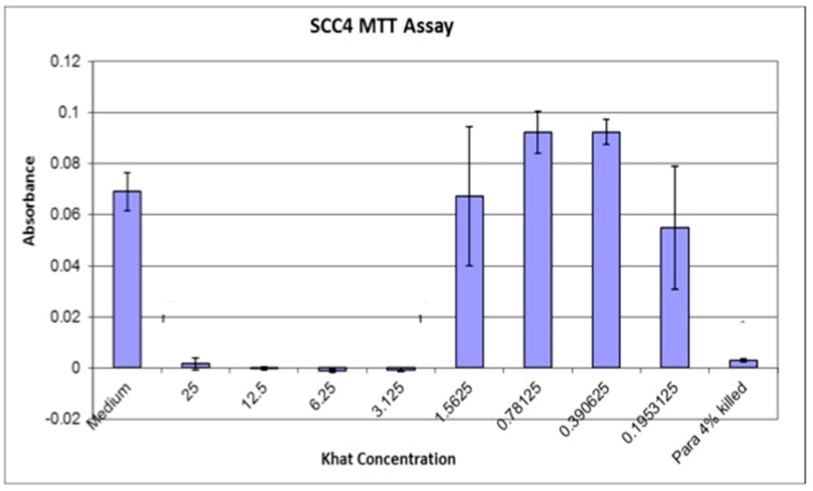
A comparison of the different batches of NOFs used showed a somewhat similar trend with the highest Khat toxicity seen between 1.56 to 12.5 mg/mL concentrations. Surprisingly, in some cells, the highest concentration (25 mg/mL) failed to cause cell death. However, a significant variation between the different cells was observed (Figure 5).

3.2. SCC4 Cells

The SCC4 cells also displayed concentration-dependent cytotoxicity. The highest cytotoxicity was seen after cells were incubated with 25–3.125 mg/mL of Khat, which was significantly different from the negative control (p < 0.005). However, lower doses of Khat (1.56–0.19 mg/mL) failed to elicit a significant response, with an increase in number of cells seen with two concentrations (1.5625 and 0.1953125) (Figure 6).

SCC4 (Late Passage)

Somewhat different results were obtained when late passage of SCC4 cells were performed. A significant reduction in absorbance was seen with all doses of Khat (except 1.56 mg/mL) indicating toxicity (p < 0.05). No obvious difference was seen between the different doses used, suggesting variability in cell behavior in late passages (Figure 7).

3.3. Comparison of NOF’s and SCC4 Assays

Comparison between the average cell number for all NOFs and repeat SCC4 assays showed no significant difference between the two cell types, with a similar dose response curve (Figure 8).

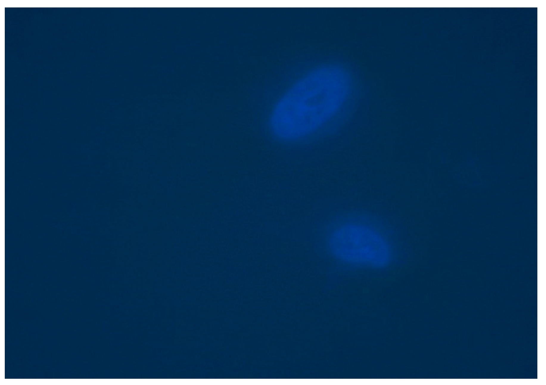
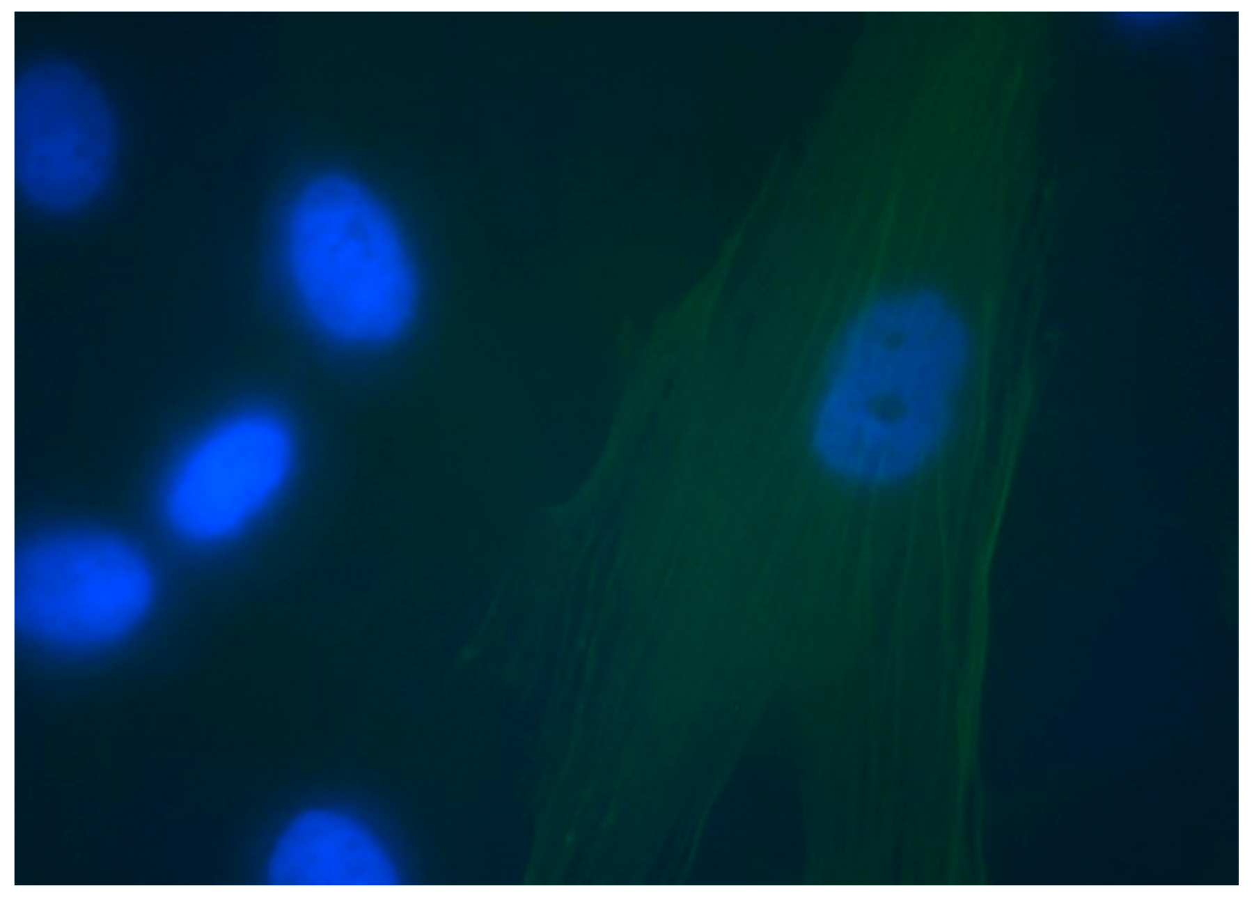
3.4. Detection of α-SMA

No α-SMA staining was seen with the negative control IgG isotype antibodies for NOF 316 (Figure 9). This suggested that 1 mg/mL of Khat concentration was sufficient to alter the fibroblast phenotype. Figure 10 shows a clear positive staining of α-SMA fibers running as vertical strands throughout the cell body of a fibroblast. Not all fibroblasts were α-SMA-positive, suggesting specific activation of a subset of fibroblasts. Similar results were seen when cells were stimulated with TGF-β, as this is known to increase the expression of α-SMA. SMA staining was seen in NOF 316, after incubation with 40 µg/mL of TGF-β (Figure 11). TGF-β is a protein that regulates proliferation and cellular differentiation, and causes apoptosis in tumor cells.

4. Discussion

The present study was based on the hypothesis that Khat is cytotoxic to NOFs and SSC4 cells. It was also hypothesized that Khat would stimulate positive expression of α-SMA in fibroblasts. Interestingly, the study revealed the cytotoxicity of Khat for both NOFs and SSC4 cells, and increased cell death occurred as compared to the negative control. Moreover, fibroblasts also stained positive for α-SMA after Khat exposure. Therefore, both postulated hypotheses were accepted. For cytotoxicity assessment, MTT assay was employed, as the technique was convenient, rapid, and reproducible [17]. Similarly, immunofluorescence was used to determine α-SMA positive staining in fibroblastic cells, as it showed high sensitivity and specificity over other techniques [18].
Khat was studied in relation to its damaging effects on oral mucosal keratinocytes, but its potential influence on the oral fibroblasts and cancer cell line was not studied. Multiple studies stated that Khat chewing is associated with oral hyperplasia, hyperkeratinization, and oral cancers [19]. The results of the present study showed that Khat caused cell death of both NOFs and SCC4 cells. The exact mechanism for Khat-induced cell death is not known. However, the available literature focused on the presence of alkaloids (cathine and other phytochemicals) as a cause of its cytotoxicity and suggests that Khat could induce cell death even in short tissue exposure [6]. In addition, Khat also triggers intracellular generation of free radical, i.e., reactive oxygen species (ROS) and Glutathione (GSH) depletion, which initiates programmed cell death (apoptosis) by causing injury to the DNA [20]. Furthermore, in the present study, it was also observed that higher concentrations (25 mg/mL) of Khat displayed variable outcomes of cytotoxicity, not showing significant cell death of NOF 316 cells, as compared to the other tested cells.
Interestingly, Khat showed dose-dependent cell death of NOF 18, NOF 26, and SCC4 cells, indicating higher toxicity at higher doses. In contrast, NOF 316 and NOF 319 did not exhibit dose-dependent cytotoxicity due to Khat. A possible explanation for this might be derived from the fact that Khat (25 mg/mL) used in the present study was in the frozen from and might have lost some of its activity [21]. Moreover, the variations in backgrounds and oral habits of the patients from which NOFs were obtained, could have resulted in such outcomes [22]. Similar findings were also reported in studies by Abderrahman and Modallal [23] and Lakundu et al. [24]. Furthermore, it was reported that Khat caused genetic material damage and its genotoxic effect on normal keratinocytes and fibroblasts increased with higher dose [25]. Their findings supported the outcomes of the present study. Therefore, clinically speaking, oral lesions including mucosal keratosis, pigmentation, and leukoplakia were not only induced by Khat, but could also progress and differentiate due to increased frequency and the extent of Khat chewing habit. Hence, patients should be educated that the Khat chewing increases the risk and incidence of oral lesions and a cessation of the habit should be encouraged. The present study reports a positive expression for α-SMA staining in fibroblast, indicating a phenotypic change. SMA is a well appreciated marker for MFs differentiation in tumor stroma, particularly in relation to OSCC [26]. MFs display intermediate physical characteristics between fibroblasts and smooth muscle cells [27]. Various laboratory-based studies demonstrate the role of cancer cell line in acquisition of fibroblast transformation in MFs, by secreting various extracellular matrix proteins, matrix-degrading enzymes, and growth factors [28]. These MFs modulate the growth, adhesion, migration, invasion, and differentiation of cancer cells. MFs tend to differentiate in the presence of TGF-β, another factor that increases in fibrotic lesions [29]. Recently, studies showed that suppression of α-SMA stops the connective tissue growth factor (CTGF) activity linked with reduced nuclear factor kappa B (NF-κB) translocation in different areas of progenecity, indicating the importance of MFs in tissue fibrosis and cell death [30]. In addition, a study conducted by Etemad-Moghadam et al., reported that MFs appear more frequently in OSCCs and are detected more towards the invasive front [31]. These findings indicate that Khat influences the progression and development of pre-malignant and malignant lesions, as it express α-SMA in fibroblasts. Weak and anecdotal evidence exists in relation to Khat chewing and oral cancer, in light of findings from retrospective and descriptive studies [32,33]. Our findings further the clinical notion, that Khat usage (expression of α-SMA) facilitates differentiation and progression of pre-malignant lesions. Therefore, Khat users (with tobacco use) should be warned of the risk of possible development of pre-malignant lesions and their progress to malignancy. Hence, further randomized controlled trials investigating the effect Khat usage with and without tobacco smoking, on the incidence of malignant and pre-malignant lesions are recommended.
Khat showed cytotoxicity at almost all concentrations, as compared to the negative control but there are certain limitations, which should be considered in the interpretation of the outcomes of the present study. This investigation was in-vitro, and cell cytotoxicity was performed under ideal conditions. In the present study, decreased cell viability was observed, which indicated induction of differentiation or cell cycle arrest not necessarily indicating cell death. Therefore, for strictly assessing cell death, other assays including trypan blue exclusion assay is recommended in future studies. For the cytotoxicity of cells, MTT assay, and for α-SMA, immunofluorescence was performed. However, VELscope® (Visually Enhanced Lesion scope) is a relatively new method to screen oral lesions and is recognized as an efficient tool by World Health Organization (WHO) in oral cancer prevention [34]. Therefore, further clinical trials investigating the influence of Khat on inducing oral lesions using VELscope® as a tool are recommended. In addition, confounding factors, i.e., smoking, tobacco chewing, oral hygiene, and chronic diseases are critical in the development of OSSC, and result in accelerated cell death in the presence of Khat. However, these factors were not assessed in the present study. Moreover, this study used control, Khat, and TGF-β (as a positive control), therefore, an interaction of Khat and TGF-β by incubating cells pretreated with TGF-β with Khat should be performed in future studies. Additionally, the effect of dose-, duration-, and friction-associated trauma in Khat chewers is of great importance in assessing its clinical influence on mucosal differentiation. Therefore, further clinical randomized controlled trials assessing the oral cellular changes among Khat users in oral epithelial and mesenchymal cells with the associated factors (smoking, tobacco chewing, oral hygiene, chronic diseases, and trauma) are recommended to clinically translate the findings of the existing study.

5. Conclusions

Results from the present study showed that Khat is cytotoxic to oral fibroblasts. It also causes cell death of oral cancer cells (i.e., SCC4 cells). Furthermore, it can also cause activation and phenotypic changes in oral fibroblasts, indicating a potential role in progression of OSCC.

Author Contributions

A.U.Y.S., M.A.A. and E.I.A., data collection, study design, data assessment, and manuscript writing, manuscript revision. M.A.-A., A.M.A. and R.J., data inference, experiment performance (SEM and RS), study design, manuscript drafting, data analysis, and manuscript revision. A.R.A. and S.A.M., experiment (MTBS and DC), data collection, funding, resources, data interpretation, writing, revise, editing, and final manuscript approval. N.A., F.V. and T.A., experiment (MTBS and DC), data collection, data interpretation, funding, resources, software, writing, revise, manuscript revisions, and final manuscript approval. All authors have read and agreed to the published version of the manuscript.

Funding

The authors extend their appreciation to the Deputyship for Research & Innovation, “Ministry of Education” in Saudi Arabia for funding this research work through the project number IFKSURG-1438-075.

Data Availability Statement

The study data is available from the corresponding author on request.

Acknowledgments

The authors extend their appreciation to the Deputyship for Research & Innovation, “Ministry of Education” in Saudi Arabia for funding this research work through the project number IFKSURG-1438-075.

Conflicts of Interest

The authors declare that they have no conflict of interest and all authors have read and approved the final draft.

Appendix A

Table A1. Materials and equipment used in the study.
Materials/EquipmentCompany
Dulbecco’s Modified Eagles Medium, Cell culture mediumGibco by Life technologies Invitrogen
PBS solution (without Ca2+ and Mg2+)Sigma Aldrich
Trypsin/EDTAInvitrogen
Acidified IsopropanolFisher Scientific, Leicestershire, UK
MTT, Thiazolyl Blue Tetrazolium BromideSigma Aldrich
Galaxy R CO2 IncubatorBrunswick lab, (UK)
MSE Harrier 18/80 Refrigerated CentrifugeMSE, UK
25 cm2 and 75 cm2 tissue culture flasks (Cellstar®)Greiner Bio-One Ltd. (Gloucestershire, UK)
96 well tissue culture platesFisher Scientific UK Ltd. (Loughborough, UK)
Integra Biosciences PIPETBOYpro Thermo Scientific, UK Ltd.
Disposable sterile pipettes (Costar®)Corning Incorporated (USA)
MicropipettesThermo Scientific and Gilson lab (UK)
Labpette pipettesManufactured by Labnet (NJ, USA)
Pipette tipsSTARLAB, Ltd. and Gilson. (UK)
Universal containersSARSTEDT Ltd. (Leicester, UK) & Sterilin UK
Manual Desktop CounterRyman, UK
Cell counting chamber slides/HaemocytometerInvitrogen
Zeiss Axiovert 200 M inverted microscopeCarl Zeiss Ltd. (Hertfordshire, UK)
Syringe and Its filter unitsMillex®GP, Ireland
Eppendorf centrifuge tubesEppendorf, UK
24 well tissue culture platesFisher Scientific UK Ltd. (Loughborough, UK)
Glass slidesThermo, UK
Axioplan 2 Imagin softwareOperated by University of Sheffield
Spectrophotometer plate reader, Infinite M200Tecan, UK
Magellan™ data analysis softwareTecan, UK
100% MethanolProvided by tissue culture lab, Dental School, University of Sheffield
Sodium deoxycholateProvided by tissue culture lab, Dental School, University of Sheffield
Bovine Serum Albumin (BSA)Sigma Aldrich, UK
Monoclonal Anti-Actin, α-Smooth Muscle - FITC antibody produced in mouse
clone 1A4, purified immunoglobulin, buffered aqueous solution
C2 Sigma Aldrich, UK
DAPI nuclear stainLife Technologies, Invitrogen
Monoclonal Anti-Human IgG1−FITC antibody produced in mouseSigma Aldrich, UK
TGF-βSigma Aldrich, UK

References

  1. Gharat, S.A.; Momin, M.; Bhavsar, C. Oral Squamous Cell Carcinoma: Current Treatment Strategies and Nanotechnology-Based Approaches for Prevention and Therapy. Crit. Rev. Ther. Drug Carr. Syst. 2016, 33, 363–400. [Google Scholar] [CrossRef]
  2. Pires, F.R.; Ramos, A.B.; De Oliveira, J.B.C.; Tavares, A.S.; Da Luz, P.S.R.; Dos Santos, T.C.R.B. Oral squamous cell carcinoma: Clinicopathological features from 346 cases from a single Oral Pathology service during an 8-year period. J. Appl. Oral Sci. 2013, 21, 460–467. [Google Scholar] [CrossRef] [PubMed]
  3. Wang, Z.-Q.; Liu, K.; Huo, Z.-J.; Li, X.-C.; Wang, M.; Liu, P.; Pang, B.; Wang, S.-J. A cell-targeted chemotherapeutic nanomedicine strategy for oral squamous cell carcinoma therapy. J. Nanobiotechnol. 2015, 13, 1–10. [Google Scholar] [CrossRef] [Green Version]
  4. Al-Maweri, S.A.; Warnakulasuriya, S.; Samran, A. Khat (Catha edulis) and its oral health effects: An updated review. J. Investig. Clin. Dent. 2018, 9, e12288. [Google Scholar] [CrossRef] [PubMed]
  5. Yarom, N.; Epstein, J.; Levi, H.; Porat, D.; Kaufman, E.; Gorsky, M. Oral manifestations of habitual khat chewing: A case-control study. Oral Surg. Oral Med. Oral Pathol. Oral Radiol. Endodontol. 2010, 109, e60–e66. [Google Scholar] [CrossRef]
  6. Abdelwahab, S.I.; Alsanosy, R.; Taha, M.M.E.; Mohan, S. Khat Induced Toxicity: Role on Its Modulating Effects on Inflammation and Oxidative Stability. BioMed Res. Int. 2018, 2018, 1–7. [Google Scholar] [CrossRef]
  7. Lukandu, O.M.; Bredholt, T.; Neppelberg, E.; Gjertsen, B.T.; Johannessen, A.C.; Vintermyr, O.K.; Costea, D.E. Early loss of mitochondrial inner transmembrane potential in khat-induced cell death of primary normal human oral cells. Toxicology 2009, 263, 108–116. [Google Scholar] [CrossRef]
  8. Al-Ahdal, M.N.; McGarry, T.J.; Hannan, M.A. Cytotoxicity of Khat (Catha edulis) extract on cultured mammalian cells: Effects on macromolecule biosynthesis. Mutat. Res. Toxicol. 1988, 204, 317–322. [Google Scholar] [CrossRef]
  9. Al-Akwa, A.A.; Shaher, M.; Al-Akwa, S.; Aleryani, S.L. Free radicals are present in human serum of Catha edulis Forsk (Khat) abusers. J. Ethnopharmacol. 2009, 125, 471–473. [Google Scholar] [CrossRef]
  10. Dimba, E.A.; Gjertsen, B.T.; Bredholt, T.; Fossan, K.O.; Costea, D.E.; Francis, G.W.; Johannessen, A.C.; Vintermyr, O.K. Khat (Catha edulis)-induced apoptosis is inhibited by antagonists of caspase-1 and -8 in human leukaemia cells. Br. J. Cancer 2004, 91, 1726–1734. [Google Scholar] [CrossRef] [PubMed] [Green Version]
  11. Williams, E.D.; Thompson, E.W. Epithelial mesenchymal plasticity in carcinoma metastasis. Clin. Exper. Metastasis 2015, 32, 188. [Google Scholar] [CrossRef]
  12. Daly, A.J.; McIlreavey, L.; Irwin, C.R. Regulation of HGF and SDF-1 expression by oral fibroblasts—Implications for invasion of oral cancer. Oral Oncol. 2008, 44, 646–651. [Google Scholar] [CrossRef]
  13. Lee, C.H.; Shah, B.; Moioli, E.K.; Mao, J.J. CTGF directs fibroblast differentiation from human mesenchymal stem/stromal cells and defines connective tissue healing in a rodent injury model. J. Clin. Investig. 2010, 120, 3340–3349. [Google Scholar] [CrossRef] [Green Version]
  14. Grotendorst, G.R.; Rahmanie, H.; Duncan, M.R. Combinatorial signaling pathways determine fibroblast proliferation and myofibroblast differentiation. FASEB J. 2004, 18, 469–479. [Google Scholar] [CrossRef] [Green Version]
  15. Karagiannis, G.S.; Poutahidis, T.; Erdman, S.E.; Kirsch, R.; Riddell, R.H.; Diamandis, E.P. Cancer-Associated Fibroblasts Drive the Progression of Metastasis through both Paracrine and Mechanical Pressure on Cancer Tissue. Mol. Cancer Res. MCR 2012, 10, 1403–1418. [Google Scholar] [CrossRef] [PubMed] [Green Version]
  16. Aziz, K.H.; Peh, Y.T.-O. Research & Clinical Practice, Undefined 2011, Herbal Delivery System for Treatment of Obesity Administration of Encapsulated Khat-Extracts on Body Weight of Rats, Elsevier. (n.d.). Available online: https://www.sciencedirect.com/science/article/pii/S1871403X11000159?casa_token=uYkaq5-2G3kAAAAA:UuoMUGEn2084syGHl66wrIy191u1_XAyt76GNQCYWbFuwfmic63JFuCumGXiU75wgLyxLBUJk2g (accessed on 1 February 2021).
  17. Macedo, A.L.; da Silva, D.P.; Moreira, D.L.; de Queiroz, L.N.; Vasconcelos, T.R.; Araujo, G.F.; Kaplan, M.A.C.; Pereira, S.S.; de Almeida, E.C.; Valverde, A.L.; et al. Cytotoxicity and selectiveness of Brazilian Piper species towards oral carcinoma cells. Biomed. Pharmacother. 2019, 110, 342–352. [Google Scholar] [CrossRef] [PubMed]
  18. Paul, B.D.; Cole, K.A. Cathinone (Khat) and Methcathinone (CAT)in Urine Specimens: A Gas Chromatographic-Mass Spec-trometric Detection Procedure*. 2001. Available online: https://academic.oup.com/jat/article/25/7/525/729486 (accessed on 27 January 2021).
  19. Alsanosy, R.; Alhazmi, H.A.; Sultana, S.; Abdalla, A.N.; Ibrahim, Y.; Al Bratty, M.; Banji, D.; Khardali, I.; Khalid, A. Phytochemical Screening and Cytotoxic Properties of Ethanolic Extract of Young and Mature Khat Leaves. J. Chem. 2020, 2020, 1–9. [Google Scholar] [CrossRef]
  20. Lukandu, O.M.; Costea, D.E.; Neppelberg, E.; Johannessen, A.C.; Vintermyr, O.K. Khat (Catha edulis) Induces Reactive Oxygen Species and Apoptosis in Normal Human Oral Keratinocytes and Fibroblasts. Toxicol. Sci. 2008, 103, 311–324. [Google Scholar] [CrossRef] [PubMed] [Green Version]
  21. Atlabachew, M.; Chandravanshi, B.; Redi-Abshiro, M.; Torto, N.; Chigome, S.; Pule, B. Evaluation of the effect of various drying techniques on the composition of the psychoactive phenylpropylamino alkaloids of khat (Catha edulis Forsk) chewing leaves. Bull. Chem. Soc. Ethiop. 2013, 27, 347–358. [Google Scholar] [CrossRef]
  22. Al-Habori, M. The potential adverse effects of habitual use of Catha edulis (khat). Expert Opin. Drug Saf. 2005, 4, 1145–1154. [Google Scholar] [CrossRef]
  23. Abderrahman, S.M.; Modallal, N. Genotoxic Effects of Catha edulis (Khat) Extract on Mice Bone Marrow Cells. Jordan J. Biol. Sci. 2008, 1, 165–172. [Google Scholar]
  24. Lukandu, O.; Neppelberg, E.; Vintermyr, O.; Johannessen, A.; Costea, D. Khat Alters the Phenotype of in vitro-reconstructed Human Oral Mucosa. J. Dent. Res. 2009, 89, 270–275. [Google Scholar] [CrossRef] [PubMed]
  25. Nyongesa, A.W.; Oduma, J.A.; Nakajima, M.; Odongo, H.O.; Adoyo, P.A.; Al’Absi, M. Dose-response inhibitory effects of purified cathinone from khat (Catha edulis) on cortisol and prolactin release in vervet monkeys (Chlorocebus aethiops). Metab. Brain Dis. 2013, 29, 451–458. [Google Scholar] [CrossRef] [PubMed]
  26. Hinz, B.; Dugina, V.; Ballestrem, C.; Wehrle-Haller, B.; Chaponnier, C. α-smooth muscle actin is crucial for focal adhesion mat-uration in myofibroblasts. Mol. Biol. Cell 2003, 14, 2508–2519. [Google Scholar] [CrossRef] [Green Version]
  27. Biela, S.A.; Su, Y.; Spatz, J.P.; Kemkemer, R. Different sensitivity of human endothelial cells, smooth muscle cells and fibroblasts to topography in the nano–micro range. Acta Biomater. 2009, 5, 2460–2466. [Google Scholar] [CrossRef] [PubMed]
  28. Giannoni, E.; Bianchini, F.; Masieri, L.; Serni, S.; Torre, E.; Calorini, L.; Chiarugi, P. Reciprocal activation of prostate cancer cells and cancer-associated fibroblasts stimulates epithelial-mesenchymal transition and cancer stemness. Cancer Res. 2010, 70, 6945–6956. [Google Scholar] [CrossRef] [Green Version]
  29. Gold, L.I. The role for transforming growth factor-β (TGF-β) in human cancer. Crit. Rev. Oncog. 1999, 10, 303–360. Available online: https://europepmc.org/article/med/10654929 (accessed on 27 January 2021).
  30. Cunningham, J.L.; Tsolakis, A.V.; Jacobson, A.; Janson, E.T. Connective tissue growth factor expression in endocrine tumors is associated with high stromal expression of α-smooth muscle actin. Eur. J. Endocrinol. 2010, 163, 691–697. [Google Scholar] [CrossRef] [PubMed] [Green Version]
  31. Etemad-Moghadam, S.; Khalili, M.; Tirgary, F.; Alaeddini, M. Evaluation of myofibroblasts in oral epithelial dysplasia and squamous cell carcinoma. J. Oral Pathol. Med. 2009, 38, 639–643. [Google Scholar] [CrossRef]
  32. Alan, H.; Ağaçayak, S.; Kavak, G.; Özcan, A. Verrucous carcinoma and squamous cell papilloma of the oral cavity: Report of two cases and review of literature. Eur. J. Dent. 2015, 9, 453–456. [Google Scholar] [CrossRef] [Green Version]
  33. Nasr, A.H.; Khatri, M.L. Head and neck squamous cell carcinoma in Hajjah, Yemen. Saudi Med. J. 2000, 21, 565–568. [Google Scholar] [PubMed]
  34. Cicciù, M.; Cervino, G.; Fiorillo, L.; D’Amico, C.; Oteri, G.; Troiano, G.; Zhurakivska, K.; Muzio, L.L.; Herford, A.S.; Crimi, S.; et al. Early Diagnosis on Oral and Potentially Oral Malignant Lesions: A Systematic Review on the VELscope® Fluorescence Method. Dent. J. 2019, 7, 93. [Google Scholar] [CrossRef] [PubMed] [Green Version]
Figure 1. Absorbance from MTT Assay for NOF 316 cells treated with different concentrations of Khat solution (25 mg/mL to 0.195 mg/mL). Medium with only NOF 316 was used as a negative control while 4% paraformaldehyde was used as a positive control showing complete cell death.
Applsci 11 03524 g001
Figure 2. Absorbance from MTT Assay for NOF 26 cells treated with different concentrations of Khat solution (25 mg/mL to 0.195 mg/mL). Medium with only NOF 26 was used as a negative control, while 4% paraformaldehyde was used as a positive control showing complete cell death.
Applsci 11 03524 g002
Figure 3. Absorbance from MTT Assay for NOF 319 cells treated with different concentrations of Khat solution (25 mg/mL to 0.195 mg/mL). Medium with only NOF 319 was used as a negative control, while 4% paraformaldehyde was used as a positive control showing complete cell death.
Applsci 11 03524 g003
Figure 4. Absorbance from MTT Assay for NOF 18 cells treated with different concentrations of Khat solution (25 mg/mL to 0.195 mg/mL). Medium with only NOF 18 was used as a negative control while 4% paraformaldehyde was used as a positive control showing complete cell death.
Applsci 11 03524 g004
Figure 5. Comparison of absorbance values from MTT Assay for NOF 18, NOF 26, NOF 319, and NOF 316 cells treated with different concentrations of the Khat solution (25 mg/mL to 0.195 mg/mL). Medium only with cells specific for each experiment was used as a negative control, while 4% paraformaldehyde was used as a positive control showing complete cell death. Aqua dotted curve = average of the 4 assays.
Applsci 11 03524 g005
Figure 6. Absorbance from MTT Assay for SCC4 cells treated with different concentrations of Khat solution (25 mg/mL to 0.195 mg/mL). Medium with only SCC4 cells was used as a negative control while 4% paraformaldehyde was used as a positive control, showing complete cell death.
Applsci 11 03524 g006
Figure 7. Absorbance from MTT Assay for SCC4 cells (late passage) treated with different concentrations of Khat solution (25 mg/mL to 0.195 mg/mL). Medium with only SCC4 cells was used as a negative control, while 4% paraformaldehyde was used as a positive control showing complete cell death.
Applsci 11 03524 g007
Figure 8. Comparison of dose response curves (MTT Assay) for NOFs and SCC4 cells treated with different concentrations of Khat solution (25 mg/mL to 0.195 mg/mL). Medium was used as a negative control while 4% paraformaldehyde acted as a positive control.
Applsci 11 03524 g008
Figure 9. NOF 316 showing nuclear DAPI staining with the IgG antibody. (magnification ×100, Fluorescence at 1.3 s, DAPI at 0.07 s).
Applsci 11 03524 g009
Figure 10. NOF 316 showing SMA expression after stimulation with 1 mg/mL Khat (magnification ×100, Fluorescence at 1.3 s, DAPI at 0.07 s).
Applsci 11 03524 g010
Figure 11. SMA expression in NOF 316 after incubation with 40 µg/mL of TGFβ (magnification ×100, Fluorescence at 1.6 s, DAPI at 0.100 s).
Applsci 11 03524 g011
Table 1. Cells used in the study.
Cell LineTypeOrigin
NOF 18Primary cellsNormal Oral Fibroblasts
NOF 26Primary cellsNormal Oral Fibroblasts
NOF 316Primary cellsNormal Oral Fibroblasts
NOF 319Primary cellsNormal Oral Fibroblasts
SCC4Cell lineSquamous Cell Carcinoma
Table 2. Medium used for growing primary cells and SCC4 cell line.
CompositionVolumeRemarks
Dulbecco’s Modified Eagles Medium (DMEM) supplemented by 4500 mg/L glucose, GlutaMAX™ I and Sodium Pyruvate450 mLInvitrogen, UK
lot- 1250148
lot- 1122288
lot- 1369047
10% Fetal Bovine Serum (FBS)50 mLBiosera, East Sussex, UK
Pen/Strep (Antibiotics)5 mL
100 IU/mL Penicillin, 100 µg/mL Streptomycin
Sigma Aldrich, Dorset, UK
Publisher’s Note: MDPI stays neutral with regard to jurisdictional claims in published maps and institutional affiliations.

