Article

Role of Ovalbumin/β-Cyclodextrin in Improving Structural and Gelling Properties of Culter alburnus Myofibrillar Proteins during Frozen Storage

1
Angel Yeast CO., LTD., Yichang 443003, China
2
College of Life Sciences, Yan’an University, Yan’an 716000, China
3
College of Food Science and Technology, Zhejiang University of Technology, Hangzhou 310014, China
4
College of Plant Science and Technology, Huazhong Agricultural University, Wuhan 430070, China
5
Shenzhen Key Laboratory of Marine Microbiome Engineering, Institute for Advanced Study, Shenzhen University, Shenzhen 518060, China
6
National Institute of Food Science and Technology, University of Agriculture, Faisalabad 38000, Pakistan
7
Instituto de Farmacia, Facultad de Ciencias, Campus Isla Teja, Universidad Austral de Chile, Valdivia 5090000, Chile
8
Center for Interdisciplinary Studies on the Nervous System (CISNe), Universidad Austral de Chile, Valdivia 5090000, Chile
9
Centro Tecnológico de la Carne de Galicia, Avd. Galicia nº 4, Parque Tecnológico de Galicia, San Cibrao das Viñas, 32900 Ourense, Spain
10
Área de Tecnología de los Alimentos, Facultad de Ciencias de Ourense, Universidad de Vigo, 32004 Ourense, Spain
11
Department of Clinical Pharmacy, College of Pharmacy, King Saud University, Riyadh 11451, Saudi Arabia
*
Authors to whom correspondence should be addressed.
Appl. Sci. 2021, 11(24), 11815; https://doi.org/10.3390/app112411815
Submission received: 20 October 2021 / Revised: 29 November 2021 / Accepted: 3 December 2021 / Published: 13 December 2021

Abstract

:
This study aimed to analyze the cryoprotective effect of a ovalbumin (OVA) and β-cyclodextrin (βCD) mixture (3:1, OVA/βCD) on the structure, rheology and gelling properties of myofibrillar proteins (MPs) during 90 days of frozen storage. A mixture of OVA/βCD at different concentrations (0, 2, 4, and 6%) was added to MPs and stored at −18 °C for 90 days. The addition of OVA/βCD significantly decreased the sulfhydryl contents while it increased the surface hydrophobicity, which was closely connected with tertiary structural changes. Circular dichroism analysis showed that the addition of OVA/βCD enhanced the stability of the secondary structure by inhibiting the decline in the α-helix. Rheological properties analysis indicated that 6% OVA/βCD treatment showed better storage modulus (G’) and loss modulus (G”). In addition, treatment of OVA/βCD showed better gel forming properties than the control group (0%), helping to form a homogeneous and denser gel network. The results proved that 6% OVA/βCD could be act as a promising cryoprotectant, which can improve the structure and gel behavior of Culter alburnus MPs during frozen storage. Moreover, OVA/βCD could be a potential alternative to conventional cryoprotectants at the industrial level to increase the economic and commercial values of seafood products.

1. Introduction

Culter alburnus, also called as topmouth culter, is an important Chinese freshwater fish due to its abundant availability and consumption [1]. It has higher nutritional values, low cholesterol and low polyunsaturated fatty acids that make it important for preservation and commercialization. Freezing and frozen storage is one of the widely used method for long term preservation of fish with minimum deteriorative changes in the quality. Frozen storage inhibits enzymatic activity, chemical reactions and crystallization in order to prevent deteriorative changes [2]. However, during frozen storage, fish proteins undergo various changes that can affect the quality of fish, such as protein and lipid oxidation, resulting in protein denaturation and aggregation. Moreover, the uncontrolled formation of ice crystals causes protein denaturation and results in more water-soluble proteins [3,4]. Fish are comprised of different proteins such as sarcoplasmic, myofibrillar and stromal proteins. Myofibrillar proteins (MPs) is the most important proteins, and account for 65–75% of total proteins. Some properties of fish MPs that deteriorate during frozen storage, such as water-holding capacity and gel-forming abilities, are connected with the formation of disulfide bonds and the exposure to hydrophobic residues [5]. These changes can be prevented with the addition of different cryoprotectants. Various cryoprotectants have been reported in many studies to prevent freeze-induced changes in proteins [6].
The more widely used cryoprotectants include saccharides and polyphosphates. The hydroxyl groups in saccharide molecules can react with certain groups of protein molecules to avoid aggregation and denaturation between protein molecules, and can change the state of bound water embedded in protein molecules [7]. Sucrose and sorbitol are the most commercially used cryoprotectants, however, they impart a sweet taste, which is undesirable to consumers. Therefore, low-sweetness alternative cryoprotectants have received increasing attention in the surimi industries.
Cyclodextrins (CDs) are cyclic oligosaccharides composed of glucopyranose groups. Adding CDs as cryoprotectants to MPs can improve the stability of MPs during freezing. CDs can form complexes with macromolecules through hydrophobic interactions or hydrogen bonding [8]. The CDs containing 7 glucopyranose units include β-cyclodextrin (βCD).
Walayat et al. [9] have reported the protective effect of adding egg white proteins (EWP) and βCD to MPs during frozen storage. But as the main protein component of EWP, ovalbumin (OVA), OVA can better reflect the functional characteristics of EWP. OVA is a high-quality protein due to its excellent functional properties, which can improve the characteristics of MPs by filling the gaps and making the gel structure denser [10,11]. No study has reported on the use of OVA/βCD as a potential cryoprotectant and its structural, gelling and water mobility characteristics in association with antioxidative properties. Therefore, this study will further analyze the cryoprotective effect of OVA/βCD on Culter alburnus MPs to broaden the range of research utilization of OVA.
The main objective of this study was to analyze the effect of OVA/βCD on the structure, rheology and gel properties of MPs during 90 days of frozen storage. The results demonstrate the broad potential of the OVA/βCD mixture as a promising new cryoprotectant to extend the economic value of Culter alburnus, which has not been reported before in the surimi or seafood industries.

2. Materials and Methods

2.1. Materials

Fifteen Culter alburnus samples (2.3–2.7 kg) were obtained from the Fisheries Research Institute of Wuhan Academy of Agricultural Sciences. Deceased Culter alburnus were transported to the laboratory within 30 min. The MP samples were extracted according to Park et al. [12] with several modifications. Other chemicals were obtained from Sinopharm Chemical Reagent Co., Ltd., Shanghai, China. Ovalbumin (OVA) was purchased from Aladdin Chemical Reagent Co., Ltd., Shanghai, China (purification >80%).

2.2. Preparation of Ovalbumin and β-Cyclodextrin Mixture

The OVA powder and βCD were dissolved in distilled water at a mass ratio of 3:1 and magnetically stirred for 1 h at 4 °C. The concentration of the resulting mixtures was 5% (w/w) and it was freeze-dried for further MP sample preparation.

2.3. Extraction of MPs

The fish muscles were processed into surimi and dissolved in 4 times (w/v) low-salt buffer containing 0.05 M NaCl and 20 mM tris-maleate, homogenized (pH 7.0) for 1 min at 4000 r/min, and then centrifuged at 8000× g at 4 °C for 10 min (Model AG-22331, Eppendorf, Hamburg, Germany). The supernatant was discarded, the pellet was washed again, centrifuged twice, and then homogenized with 4 times high salt buffer containing 0.6 M NaCl and 20 mM tris-maleate (pH 7.0) and centrifuged at 4 °C at 8000× g for 10 min. The supernatant was mixed with 10 times distilled water (4 °C) to obtain a precipitate and further centrifuged at 8000× g for 10 min at 4 °C, then the sediment obtained was named MP. The resulting protein concentration of 76 mg/mL was measured by the Biuret method [13]. Finally, different concentrations of OVA/βCD (2, 4 and 6%) were added to the obtained MPs respectively, then stored in a refrigerator at 18 °C for further analysis. Additionally, the 0% OVA/βCD treatment was used as control sample.

2.4. Preparation of MP Gel

The MPs (50 mg/mL) were mixed with buffer (0.6 M NaCl, 50 mM sodium phosphate, pH 7.0) to obtain sample solutions. The sample solutions were transferred into glass beakers (10 mL), then heated at 40 °C for 1 h and 90 °C for 45 min using water bath. Finally, the formed MP gel was cooled and incubated in the refrigerator (4 °C) overnight.

2.5. Changes in MPs

2.5.1. Determination of Sulphydryl Content

Determinations of total sulfhydryl and free sulfhydryl contents (SH) of MP samples were conducted based on the method described by Ellman [14]. MP samples (4 mg/mL) were diluted with phosphate buffer solution (pH 7.0). One milliliter of MP solution (4 mg/mL) was mixed with 9 mL of 0.2 M tris-HCl buffer (containing 10 mM EDTA, 0.6 M NaCl, 2% SDS, 8 M urea, pH 7.0), then 4 mL of the above mixed solution was reacted with 0.4 mL of 0.2 M tris-HCl (containing 0.1% DTNB, PH 7.0) in a water bath (40 °C) for 25 min. Measurement of the free SH group contents were in line with the above, apart from the presence of urea. Total SH and free SH contents were determined using a spectrophotometer (Shimadzu UV1800, Tokyo, Japan) at a wavelength of 412 nm.
Free   SH   content   ( nmol / mg ) = A × D B × C
where A represents the absorbance at 412 nm, B represents the concentration of sample protein, C represents the absorbance coefficient (13,600 M−1 cm−1), and D represents the dilution times before the water bath.

2.5.2. Determination of Surface Hydrophobicity

The surface hydrophobicity (S0) of MPs were determined by the ANS fluorescence probe method as described by Poowakanjana and Park [15] using a Fluorescence Spectrophotometer (F-4600, Hitiachi High Technologies Co., Tokyo, Japan). MPs samples were diluted to 0.2, 0.3, 0.5 and 1 mg/mL with 0.6 M NaCl, respectively, and 10 mL samples were mixed with 40 μL of 2 mM ANS (0.2 mol/L PBS, pH 7.4) and incubated without light for 30 min at 25 °C. The S0 of MPs was measured with an emission at 470 nm at a 390 nm excitation and calculated from the initial slope of the net relative fluorescence intensity versus the MP sample concentration.

2.5.3. UV Absorption Spectra

The UV absorption spectra was determined to monitor the tertiary structural changes of the MPs, following the method of Qiu et al. [16]. MPs samples (1 mg/mL) were diluted with 0.6 M NaCl. The scan wavelength range was 225–400 nm with a scan rate of 10 nm/s using a UV-1800PC spectrophotometer.

2.5.4. Intrinsic Fluorescence Intensity

According to the method of Li et al. [17] the intrinsic fluorescence intensity (IF) of OVA/βCD treated MPs was determined using a Hitachi F-4600 fluorescence spectrophotometer (Tokyo, Japan). MP samples (0.25 mg/mL) were diluted with 0.6 M NaCl while NaCl solution was used as blank. The excitation wavelength was set at 300 nm with slit width 5 nm; the emission wavelength range was 300–450 nm.

2.5.5. Circular Dichroism

The far-UV circular dichroism (CD) spectrum was used to detect the secondary structures of MPs. MP samples were diluted in 0.6 M NaCl solution to 1 mg/mL. The samples were scanned from 200 to 250 nm with speed 100 nm/min using CD spectrophotometer (model J-1500, JASCO, Tokyo, Japan) based on the method described by Yahaghi et al. [18]. The percentage of α-helical in secondary structural was calculated using the estimation program (Yang’s method) in a CD spectropolarimeter, and CD properties of the MPs were exhibited by molecular ellipticity θ in deg cm2/d mol.

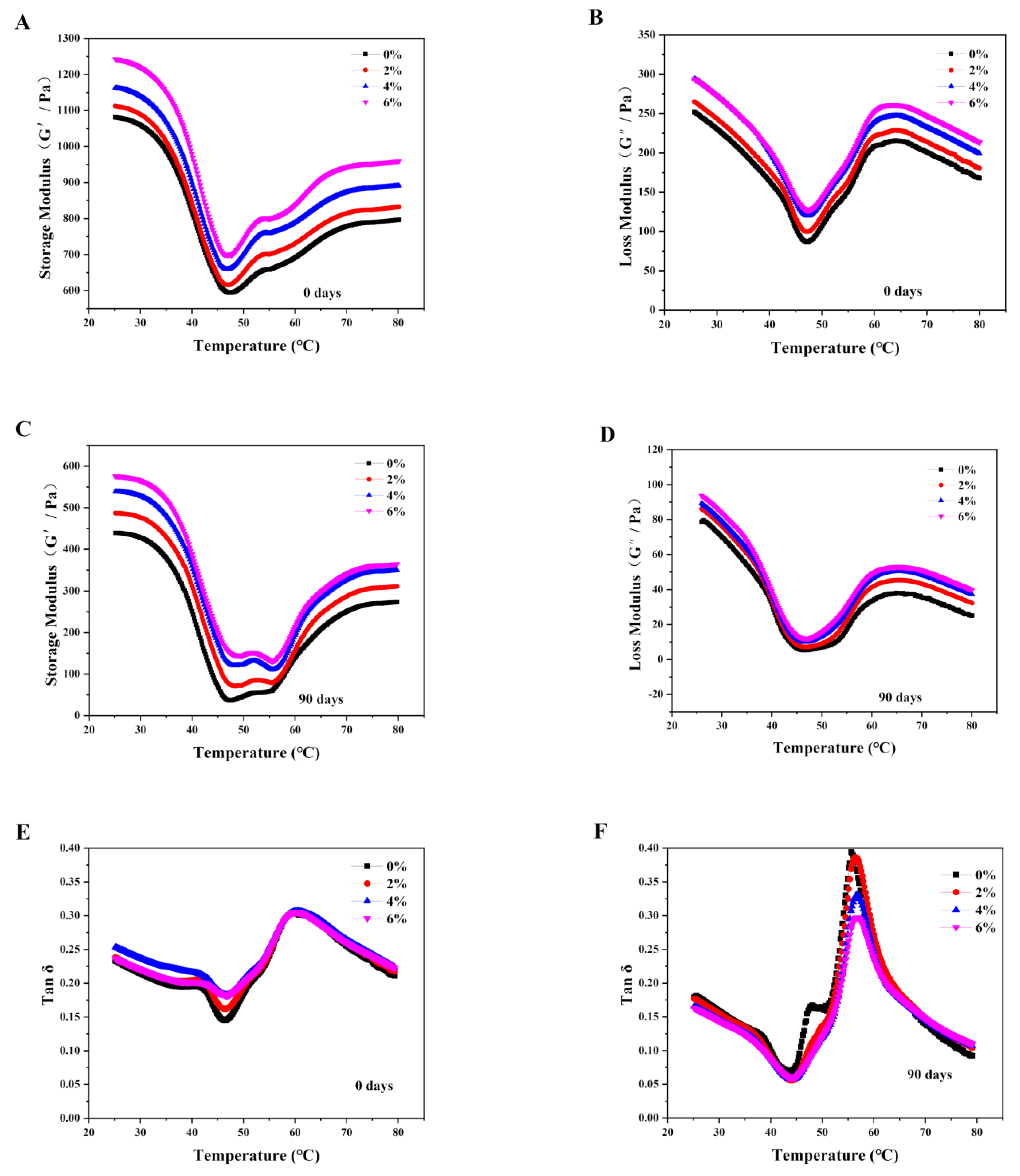
2.5.6. Rheological Properties

The rheological properties of MPs were measured according to Chen et al. [19] with a DHR2 rotary rheometer (TA Instruments, West Sussex, UK). The MP samples (about 3 mL, 76 mg/mL) were evenly dispersed on the lower plate of the rheometer using a spoon. The gap of the upper plate (40 mm Aluminum plate) was slowly reduced to 1.0 mm, and the rim was covered with silicon oil to prevent dehydration. The rheological behavior of the MPs was evaluated by the storage modulus (G′) and loss modulus (G′′) values obtained during heating at a rate of 2 °C/min from 25 to 80 °C with a 1.0% strain and 0.1 Hz frequency.

2.6. Changes in Myofibrillar Protein Gels

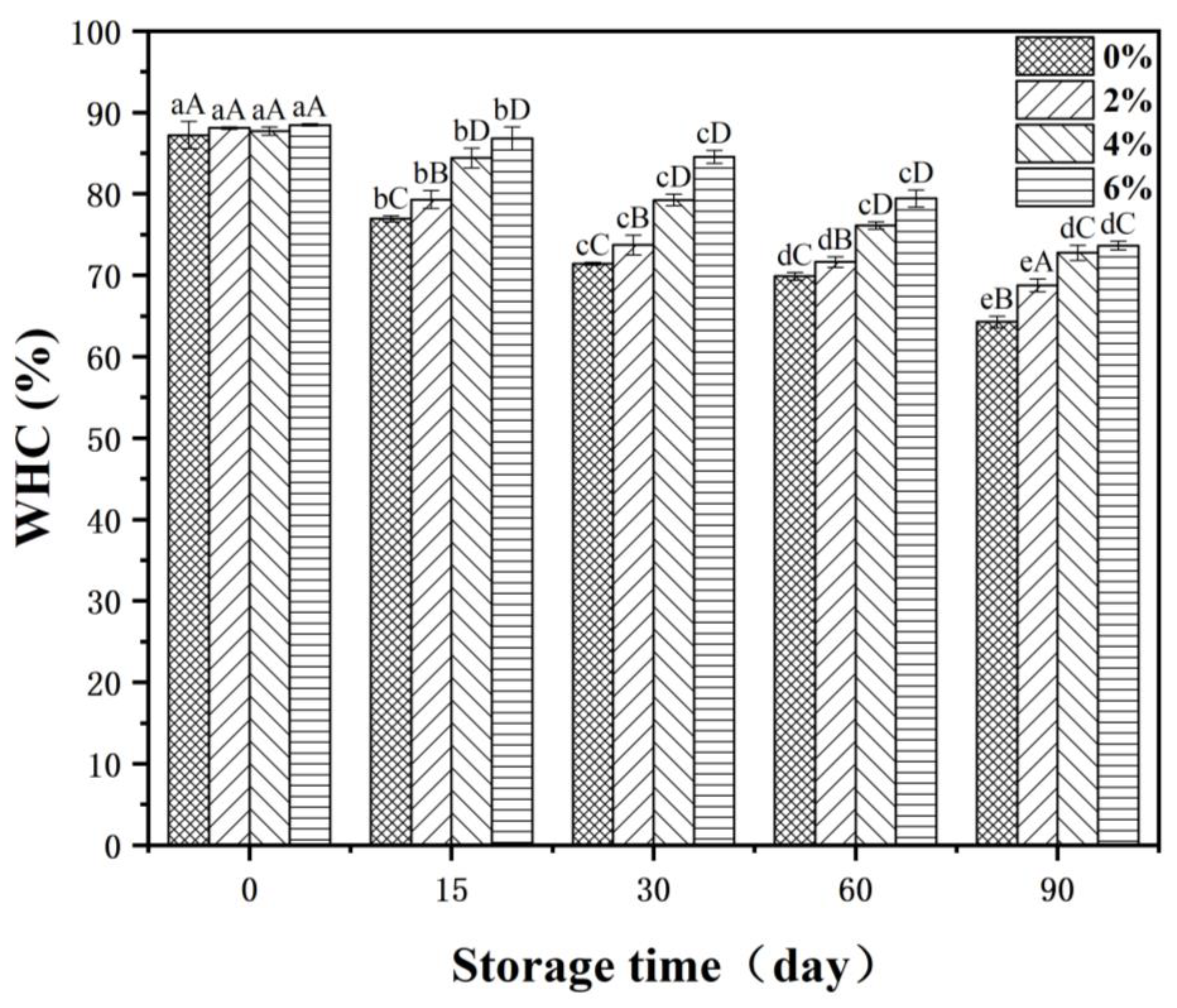
2.6.1. Determination of Water Holding Capacity

The water holding capacity (WHC) (%) of the prepared MP gels were determined according to Jia et al. [20]. Aliquots of 3.0 g MP gels were put into polypropylene tubes and then centrifuged for 8 min at 3000× g and 4 °C. WHC (%) was calculated as
WHC   ( % ) = 100 × M 1 M M 0 M
where M represents the weight of the tube, M0 represents the weight of the tube and the gel before centrifugation, and M1 represents the weight of the tube and the gel after centrifugation.

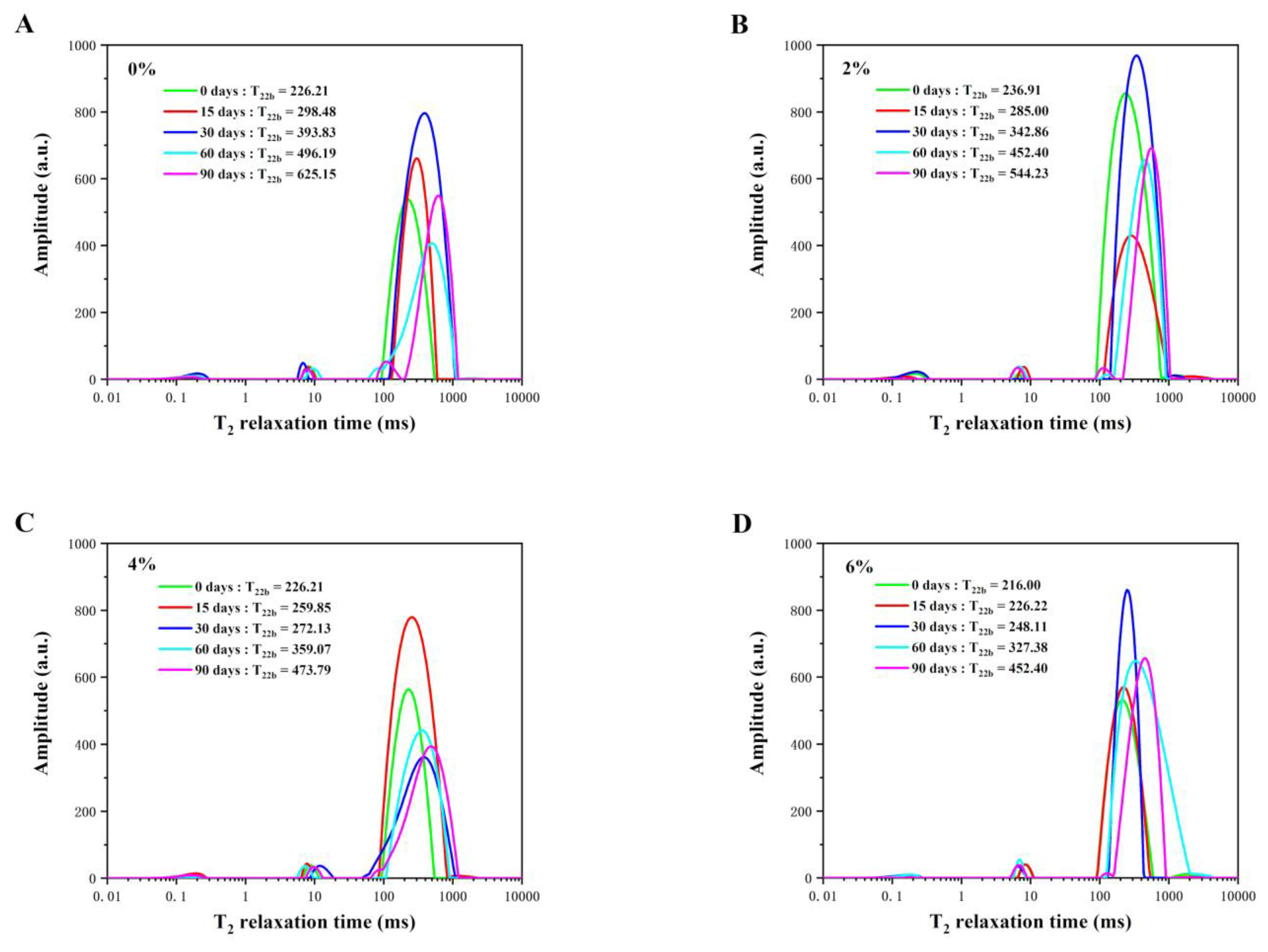
2.6.2. Low-Field Nuclear Magnetic Resonance (LF-NMR) Proton Relaxation

According to the method of Zhang et al. [21] the sample was formed in cylindrical glass tubes (10 mm in diameter) after heating in a water bath to determine relaxation time (T2) using a LF-NMR analyzer (Bruker, Ettlingen, Germany). The main parameters were adjusted appropriately: SF = 20 MHz, TW = 5000 ms, TE = 0.2 ms, NS = 8 and NECH = 10,000.

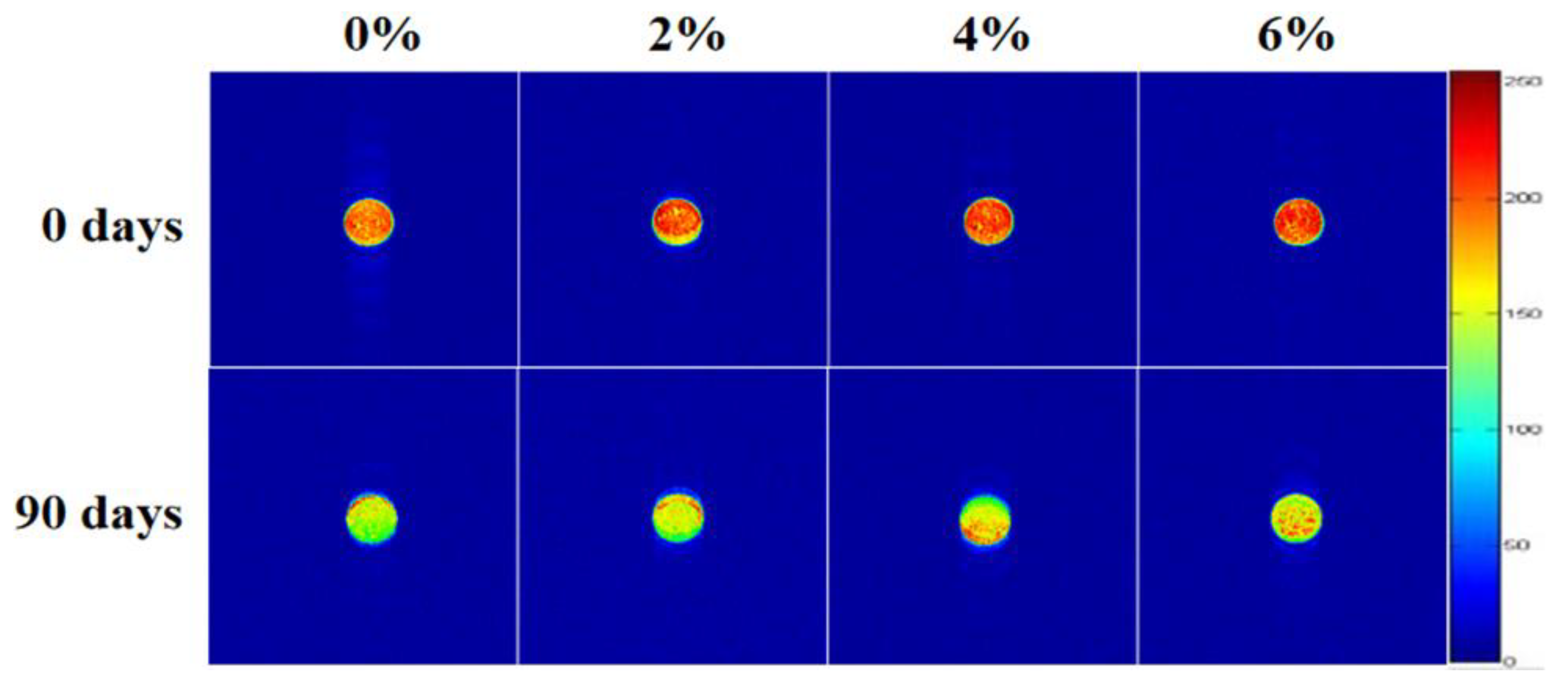
2.6.3. Proton Density Weighted Pseudocolor Images

Proton density images of the MP gels were acquired using a MINI MR-60 instrument (Bruker, Ettlingen, Germany) and analyzed by image evaluation computer software (Shanghai Niumag Corporation, Shanghai, China). Briefly, the width of the layer was 8.0 mm, and the section gap was 3.2 mm.

2.7. Statistical Analysis

Statistical analysis ANOVA (one-way ANOVA) and mean comparison analyses carried through a Duncan post hoc test using SPSS 22.0. All measurements were carried out in triplicate, and data were expressed as mean ± SD. The significance level was p < 0.05.

3. Results and Discussion

3.1. Myofibrillar Proteins

3.1.1. Sulphydryl Content

Total sulphydryl (SH) of MPs are the pivotal reactive groups. The total SH group includes a free SH group and other hidden SH groups which are present in the myosin head and light chains. Myosin is the major component of MPs. Cao and Xiong [22] mentioned that myosin is rich in SH groups and is easily converted into disulfide bonds (SS) under oxidative stress, resulting in decreased total SH and free SH content of the system. The degree of protein oxidative degeneration can be reflected on measuring the SH content of MPs. Table 1 showed the effect of different concentrations of OVA/βCD on the total SH and free SH content of MPs after 90 days of frozen storage. With the extension of frozen time, the total SH and free SH contents of the MPs decreased significantly (p < 0.05). All samples treated with OVA/βCD showed a significant decrease (p < 0.05), but these were still higher than control samples, indicating that OVA/βCD could contribute to the cryoprotection of Culter alburnus MPs. This may have been caused by the cryoprotectant, which improves the stability of SH during frozen storage. Lin et al. [23] also mentioned that cryoprotectants could inhibit the decrease in total SH content because of their ability to prevent the structural changes caused by crystallization.
In the early stage of frozen storage, the free SH content of the MPs decreased sharply. This could be due to the fact that the oxidization on the surface free SH groups prompted a reduction in the SH content. Meanwhile, it was found that the 6% OVA/βCD treatment had higher stability during the storage. Cryoprotectants reduce the protein oxidation owing to the active hydroxyl groups binding the functional groups on the protein molecules by hydrogen bonding, thus inhibiting protein molecule aggregation and denaturation [24].
The results indicated that the 6% OVA/βCD treatment could effectively inhibit the reduction of the free SH content and OVA/βCD could be added to prevent SH from oxidation.

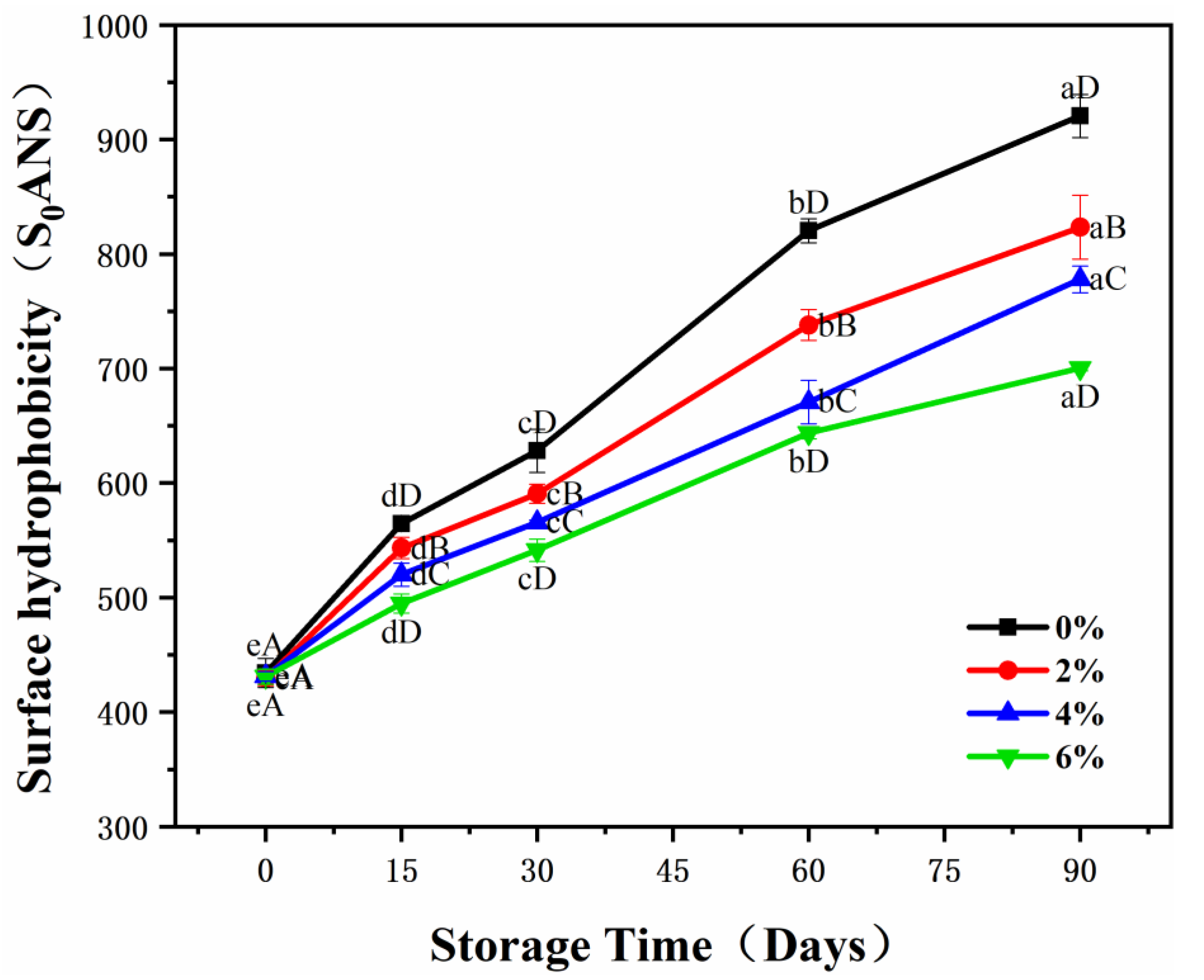
3.1.2. Surface Hydrophobicity

Surface hydrophobicity can be used as indicator that reflects conformational alteration in MP structures. The structural properties of the MPs that would be changed adversely include the alteration in the surface hydrophobicity and SH groups under frozen conditions. In the initial stage of storage, there was no significant (p > 0.05) difference in the surface hydrophobicity of the samples in each group, as shown in Figure 1. However, from day 15 onwards, the surface hydrophobicity increased significantly (p < 0.05), particularly in the samples with lower OVA/βCD concentrations. The samples with 0% and 2% OVA/βCD treatments showed an obvious rise in surface hydrophobicity during the frozen storage. At the same time, the 6% treatment exhibited stronger stability in structure. The changes to surface hydrophobicity were due to the stretching and unfolding of unstable protein molecules during frozen storage, facilitating the exposure of the hydrophobic amino acid residues that are previously buried in the internal region, and thereby triggering the alteration in hydrophobic regions on the protein surface, leading to increases to the surface hydrophobicity [25]. Meanwhile, from the current results, the outcome of the surface hydrophobicity and SH contents indicated that the cryoprotective effect of the 6% OVA/βCD treatment was more obvious. These results may be due to cryoprotectants binding to protein through hydrogen bonds and enhancing the hydrophobic interaction of protein to stabilize the protein structure.

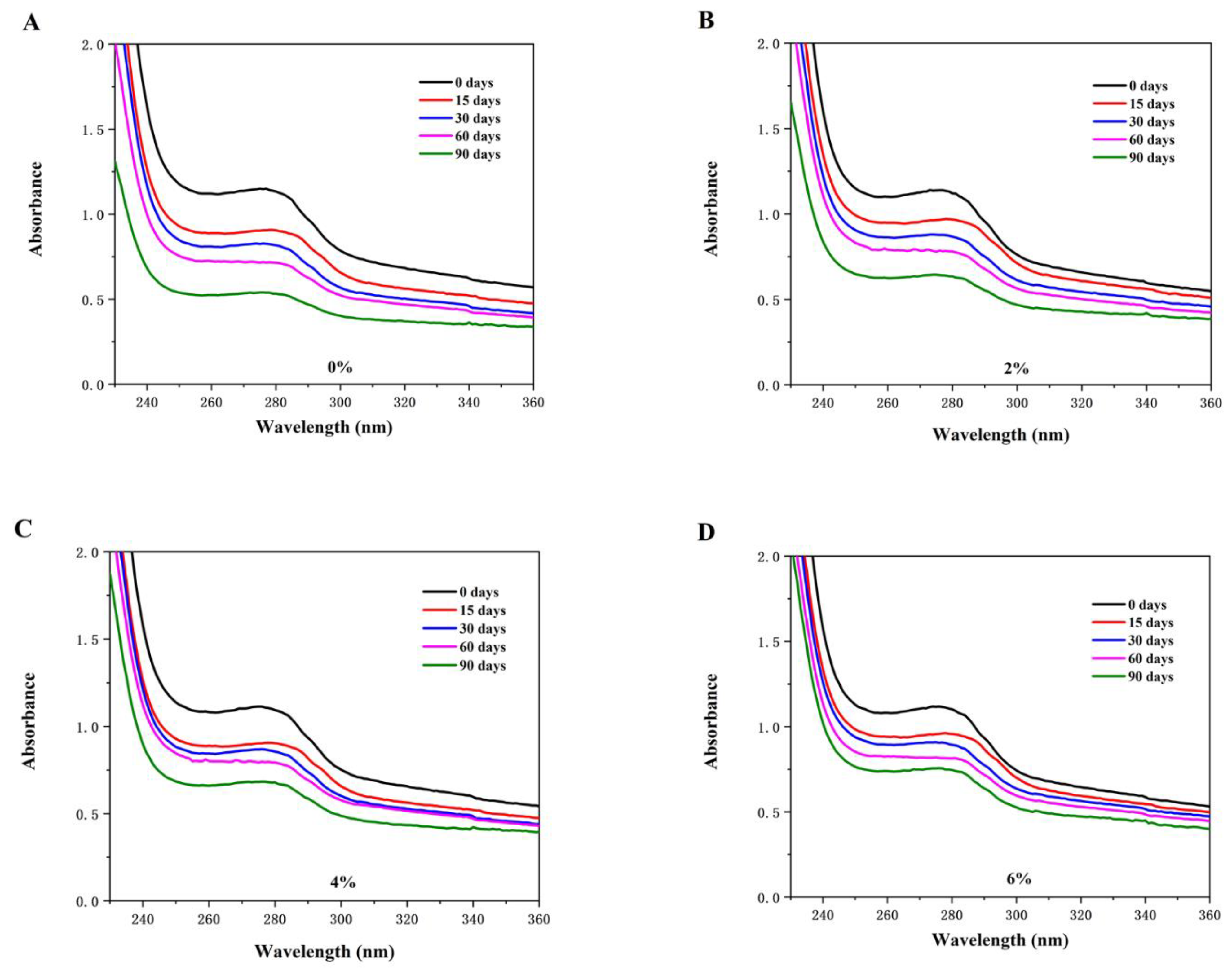
3.1.3. UV Absorption Spectra

Protein molecules contain some chromophores, tyrosine (Tyr) and tryptophan (Trp) residues, among others. The side chain groups of Trp and Tyr residues have an absorption effect on ultraviolet light, generating corresponding UV absorption spectra [26]. UV spectra measurement is an effective instrument used for identifying protein changes in conformation, hydration, dissociation or denaturation during frozen storage [27].
Figure 2 shows the evolution of the UV absorption spectra of MPs during frozen storage. The maximum absorption wavelength of MPs was near 275 nm, and the absorption spectra of the samples in each group were red-shifted after 90 days of frozen storage. The fluctuation of the maximum absorption wavelength may have been caused by the alteration in the Tyr microenvironment [28].
As the frozen storage progressed, the UV absorption intensity of the MPs gradually decreased. The UV absorption intensity of the control (0%) significantly declined. This was due to the formation of MP aggregates during the freezing process, which caused the chromogenic amino acid groups exposed to the outside to be hidden inside again, resulting in a decrease in UV absorption intensity [29]. Meanwhile, the aggregation and denaturation of MPs were among the reason leading to a lower MP gel-forming ability. Moreover, the OVA/βCD treatment showed a smoother decline in UV absorption intensity. Therefore, it could be speculated that the OVA/βCD treatment prevented the MPs from aggregation as well as denaturation, and maintained stability of the MPs tertiary structures during frozen storage. The above-mentioned results are in accordance with the conclusion on the changes of the SH group mentioned in Table 1. The SH group belongs to a weak secondary bond which helps to stabilize the tertiary structure of the protein. The results showed that the lower SH oxidation process observed was in the MPs treated with OVA/βCD, which maintained the integrity of the tertiary structure of MPs.

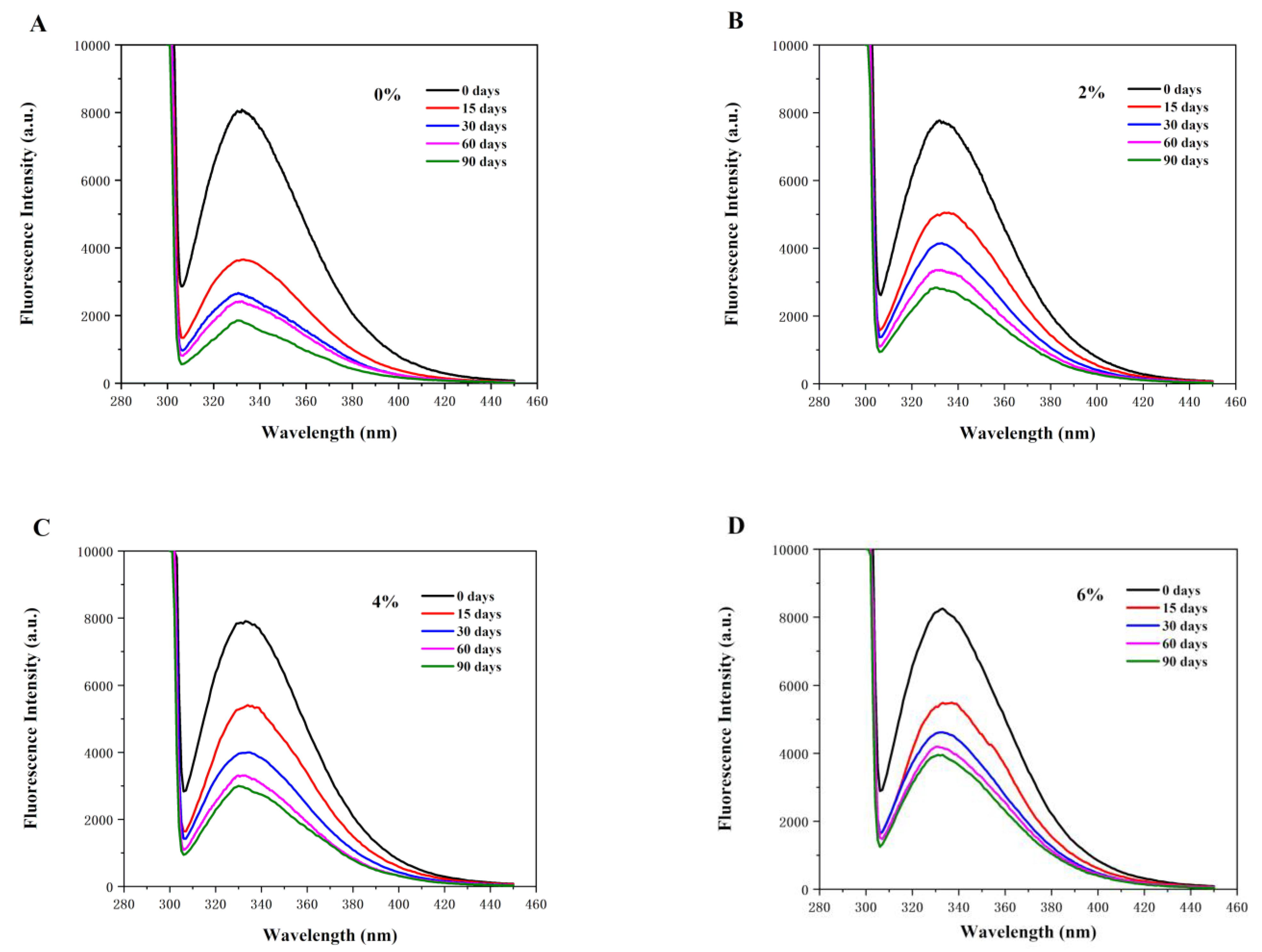
3.1.4. Intrinsic Fluorescence Intensity

Both the tail and the head of MPs contain Trp. While the structure of MP changes, Trp residues will be exposed to the solvent in the microenvironment, resulting in changed IF [16]. During frozen storage, the structure of the MPs was gradually unfolded and Trp residues were exposed to the solvent, resulting in decreased FI. Figure 3 shows the change to MPs IF during frozen storage. The broad band at 336 nm corresponds to Trp. After 90 days of frozen storage, the fluorescence intensity of all MP samples treated with OVA/βCD was higher than that of the the MP samples in control (0%). The 6% treatment decreased slightly, while the control (0%) was significantly reduced, which indicated that the control (0%) had a higher degree of degeneration. Wang et al. [30] reported that the denaturation of Trp promoted the interaction between protein molecules, resulting in the production of protein polymers, which then masked some nonpolar aromatic amino acid residues, leading to a decrease in FI. This analysis agreed with the data regarding surface hydrophobicity (Figure 1), because the increase in the surface hydrophobicity could be affected by denaturation of Trp that strengthened the hydrophobic interactions. Thus, the present outcomes indicate that OVA/βCD acts as a shield against denaturation in Try, preventing it from being exposed to the freezing environment.

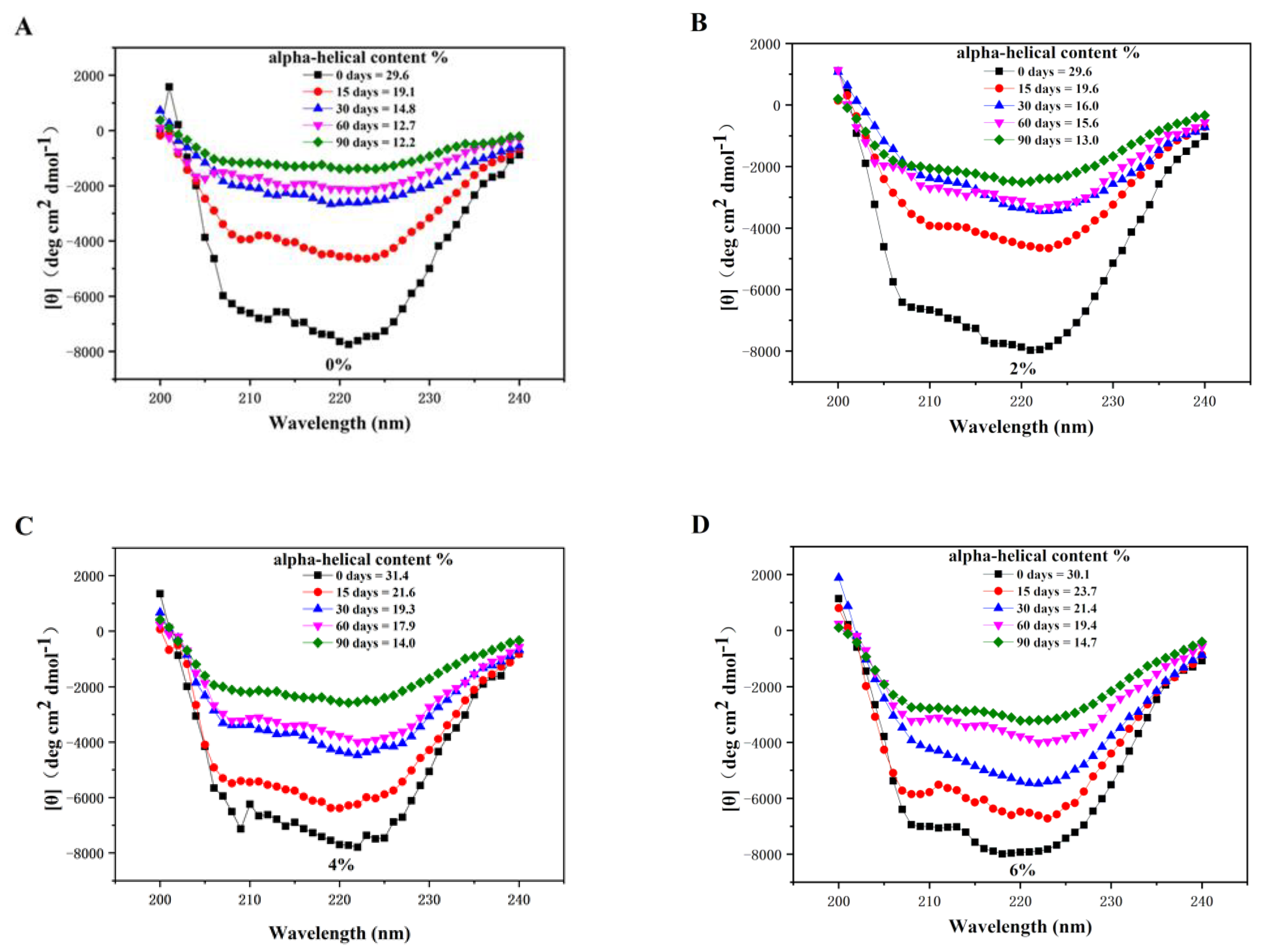
3.1.5. Circular Dichroism

Generally, the secondary structure of MPs is stabilized by intermolecular hydrogen bonds. However, the freezing process destroys the intramolecular hydrogen bindings, resulting in conversion among different types of secondary structures [31]. Therefore, the determination of secondary structural changes in MPs is crucial. Different secondary structures in natural proteins are exhibited under CD analysis. The changes to the secondary structures in the MP samples re shown in Figure 4. The α-helical structure is an important conformation which is mainly comprised of total myosin protein, which maintains the secondary structure stability of proteins mainly through hydrogen bonds [32]. The α-helical structure has one positive peak at 190 nm as well as two negative peaks at 204 nm and 221 nm. During frozen storage, the content of the α-helix of the MP secondary structures gradually decreased. This reduction in the α-helix is attributed to the changes in myosin during frozen storage [33].
After being frozen for 90 days, the α-helix content of all MPs samples decreased, ranging from 29.6 to 12.2% for the control sample, and from 30.1 to 14.7% for 6% the OVA/βCD treated samples. The α-helix content of the control (0%) decreased, while the α-helix content of the 6% OVA/βCD treatment was more stable. As the time frozen increases, the protein unfolds and the hydrogen bond weakens, resulting in α-helix decrease [34]. Therefore, addition of OVA/βCD protected MPs from deterioration by binding with protein molecules through hydrogen bonding.
Zhang et al. [35] reported that the disruption of α-helix structures would be accompanied by the molecular structure loosening, resulted in increased surface hydrophobicity, which might promote protein aggregation, which would further influence the water retention of the MP gel.

3.1.6. Rheological Properties

Rheological behavior alterations were used to analyze the correlations from the shifting to the MP gel network of MP [9]. The storage modulus (G’) represents elastic solid behavior and can reflect mechanical strength, while the loss modulus (G”) represents the fluid viscosity ratio. Figure 5 showed the effect of OVA/βCD treatments on MPs rheological properties (G’ and G”) after 90 days of frozen storage. All samples proved that the G’ values dropped sharply from 35 to 45 °C, then gradually increased, decreased slightly at 53 °C, and subsequently increased. These two peaks are attributed to the higher activity of protease enzymes that degrade myosin, resulting in the breakage of gel strength, or the conformational change of myosin disrupting the integrity of the protein network. The initial decline might be explained by partial MP degradation in the gel, which was attributed to the denaturation of myosin. A slight drop occurred at 55 °C; this sudden drop might be due to the deterioration of the myosin rod sub-fragmentation and the destruction of the temporary protein network during the heating process of the MPs. The subsequent rise may be explained by the establishment of a more stable gel network through protein–protein cross-linking [36]. The MPs treated with 6% OVA/βCD had higher G’ values than the MPs treated with 2% and 4%, indicating that 6% treatment showed better MP gel stability. These results can be related to the disulfide bonds formed in myosin, which resulted in lower gel properties.
Owing to the viscoelastic properties of MP gel, the thermal gel performance of MPs can be monitored by the G” values. The change in the trend of G” is roughly similar to that of G’. It started to decrease until temperature reached 47 °C, and then gradually increased until temperature reached 65 °C, after which it finally decreased. The final decline might have been due to the increase in the heat denaturation of the MPs, caused by the formation of protein–protein bonds that were surpassed by the destruction of the protein–protein, resulted in a decrease in G”. The values of G’ and G” tended to increase gradually as the OVA/βCD concentrations increased, indicating that the addition of OVA/βCD improved the strength of the gel network and made that gel network structure more stable and elastic [37]. Thence, from the above-mentioned results it can be confirmed that OVA/βCD contributed to a stronger gelation process. These conclusions are similar to the previously reported work indicating that cryoprotectant interaction with MPs produced gel networks with viscoelastic properties, which improved the gelation performance of the MP gels [19].

3.2. Myofibrillar Proteins Gels

3.2.1. Water Holding Capacity of MP Gel

WHC is a useful indicator which reflects the important functional properties in MP gel systems. In the protein gel network, WHC indicates the capacity of a gel to hold water, which can reflect the roughness of the protein gel’s network structure [38]. Figure 6 shows that the WHC of MP gels treated with OVA/βCD was significantly (p < 0.05) higher than that of the control samples (0%). After frozen storage for 90 days, the experiment showed that the control (0%) reduced from 87.23% to 66.58% with a significant decline (p < 0.05), while the 6% OVA/βCD treatment only decreased from 88.47% to 76.42%, indicating the structure of MP gel network without the OVA/βCD treatment was coarse and poor. During the freezing process, the hydrophobic interaction of the system increased, weakening the forces of the water molecule and the gel. Under centrifugal conditions, the water molecules were easily released, resulting in the decreased WHC of the MP gel.

3.2.2. Low-Field Nuclear Magnetic Resonance

The T2 relaxation time of the MP sample was monitored by LF-NMR, which identified the distribution of each water component in the sample matrix system [39,40]. The alteration in water migration and state was influenced by the structural changes of MPs gel. The T2 relaxation time of MP gel was distributed between 0.1 and 1000 ms with 3 peaks, namely combined water (T21: 0.02–0.30 ms), and non-flowable water (T22a: 5.88–12.31 ms, T22b: 94.03–1088.42 ms). The non-flowable water T22b was the main existing state of water in the MP gel, so the migration and state of this part directly reflects the change to the gel network structure during frozen storage. The shorter the relaxation time of T2, the tighter the water molecules. A longer T2 relaxation time indicates that the interaction between protein gel macromolecules and water molecules is weak [41]. It can be seen from Figure 7 that the relaxation time of MPs gels gradually prolonged with the decrease in OVA/βCD concentration. In the early stage of frozen storage, the T22b relaxation time of the MP gel samples was kept at about 220, and after 90 days, the relaxation time of the control (0%) increased to 625, but the relaxation time of the 6% OVA/βCD treatment sample only increased to 452. These results proved that the addition of 6% OVA/βCD could effectively enhance the binding of the protein macromolecules and water molecules, which led to lower T22b relaxation time and formed a well-structured MP gel. The results of the LF-NMR were in accordance with the above-mentioned changes in structure and viscoelasticity.

3.2.3. Proton Density Weighted Pseudo-Color Images

MRI technology is a non-destructive analytical tool, which can reflect the distribution of H protons in the samples by the formation of an image. The intensity of the color in the pseudo-color image reflects the relaxation signal intensity difference caused by alterations in proton density distribution [42]. Magnetic resonance imaging (MRI) will be brighter and redder if there are more hydrogen protons within the image [43]. In the present study, the protons mainly came from water molecules the in MP gel samples. Therefore, the proton density image can be used to intuitively observe the spatial distribution of water in the MP gel samples. MRI images of the changes to the MP gels are displayed in Figure 8. In the initial stage of frozen storage, all MPs samples had dense, red bright spots. With the extension of freezing time, the bright red spots in the H proton density image gradually disappeared. At 90 days of frozen storage, the control (0%) and the 2% treatment had only sporadic bright spots, while the 4% treatment and 6% treatment samples had more red bright spots, which indicated that addition of an appropriate concentration of OVA/βCD enhanced the water retention capability of the MP gel. The water retention of the MP gels was mainly related to their microstructure, hence we deduced that the structure of the control (0%) group gel was rough, porous and had poor water retention, thus affecting the water distribution of its MP gel. The deteriorated changes of microstructure may have been caused by oxidation in the myosin, accelerating the formation of a poor gel network structure [44]. Meanwhile, the addition of 6% OVA/βCD preserved the MP gels structure and helped set up a homogeneous, dense gel network by preventing protein aggregation. These outcomes were in agreement with the findings in the WHC examination (Figure 6). Chen, Kong, Guo, Xia, Diao and Li [19] mentioned that cryoprotectants, as protective materials, may bind with the one of functional groups in protein molecules, either by hydrogen or ionic bonds, to prevent the oxidation of MPs during frozen storage and prompt the formation of more ordered and finelt structured gel networks.

4. Conclusions

The results of the study demonstrated the cryoprotective effect of OVA/βCD on Culter alburnus MPs during frozen storage. The structural changes in the MPs were confirmed by changes in surface hydrophobicity (434.20 to 920.47 S0ANS) and sulfhydryl content (59.75 to 31.72 nmol/mg) in the control MP samples throughout the freezing process. The secondary and tertiary structural properties decreases were due to protein aggregation, denaturation and exposure of aromatic amino acids to the freezing environment. Meanwhile, OVA/βCD treatment increased the storage modulus (G’) and loss modulus (G”) and enhanced the MP gel properties, as confirmed with improved WHC (88.47 to 73.64%) and LF-NMR analyses, by interacting at the functional sites of the protein molecules and reducing the availability of free water molecules. These outcomes were related to the gelling and structural properties of MPs protected by OVA/βCD as a cryoprotectant. Overall, 6% OVA/βCD can be considered as an effective commercial cryoprotectant concentration, offering greatly improved structural properties and gel behavior of MPs during frozen storage; it is expected to be used as an effective new cryoprotectant in frozen fishing applications. Moreover, suitable cryoprotectants with reduced sweetness and caloric values are needed to improve the quality, safety and commercial value of seafood products. The limitation of this study is that the use of OVA/βCD is still under consideration for use in product development and sensory analyses.

Author Contributions

Conceptualization, M.L. and Z.Z.; data curation, M.L. and M.H.S.; formal analysis, N.W., Z.Z., A.N., R.M.A., M.J.S. and J.M.L.; funding acquisition, A.N., M.H.S. and R.M.A.; investigation, N.W., Z.Z., S.A., M.J.S. and J.M.L.; project administration, R.M.A.; resources, M.L., N.W. and S.A.; software, X.W., M.H.S. and R.M.A.; supervision, X.W. and S.A.; validation, Z.Z.; visualization, X.W., M.H.S., S.A. and B.L.J.; writing—original draft, M.L., X.W., N.W. and B.L.J.; writing—review and editing, A.N., M.J.S., J.M.L. and B.L.J. All authors have read and agreed to the published version of the manuscript.

Funding

The publication of the present work is supported by the Natural Science Basic Research Program of Shaanxi Province (grant no. 2018JQ5218) and the National Natural Science Foundation of China (51809224), Top Young Talents of Shaanxi Special Support Program.

Institutional Review Board Statement

Not applicable.

Informed Consent Statement

Not applicable.

Data Availability Statement

Data is contained within the article.

Acknowledgments

This study was financially supported by the Nature Science Foundation of Hubei Province (2018CFB269), National Key R & D Program of China (2018YFD0400702) and the Major technical innovation special project of Hubei (2018ABA100). The authors would like to extend their sincere appreciation to the Researchers Supporting Project Number (RSP-2021/168), King Saud University, Riyadh, Saudi Arabia.

Conflicts of Interest

The authors have no conflict of interest to declare. All co-authors have seen and agree with the contents of the manuscript. We certify that the submission is original work and is not under review at any other publication.

Abbreviation

β-CDβ-Cyclodextrin
OVAOvalbumin
MRIMagnetic Resonance Imaging
PBSPhosphate Buffer Saline
LF-NMR Low Field Nuclear Magnetic Resonance

References

  1. Zhang, Z.; Xiong, Z.; Lu, S.; Walayat, N.; Hu, C.; Xiong, H. Effects of oxidative modification on the functional, conformational and gelling properties of myofibrillar proteins from Culter alburnus. Int. J. Biol. Macromol. 2020, 162, 1442–1452. [Google Scholar] [CrossRef]
  2. Walayat, N.; Wang, X.; Nawaz, A.; Zhang, Z.; Abdullah, A.; Khalifa, I.; Saleem, M.H.; Mushtaq, B.S.; Pateiro, M.; Lorenzo, J.M.; et al. Ovalbumin and Kappa-Carrageenan Mixture Suppresses the Oxidative and Structural Changes in the Myofibrillar Proteins of Grass Carp (Ctenopharyngodon idella) during Frozen Storage. Antioxidants 2021, 10, 1186. [Google Scholar] [CrossRef] [PubMed]
  3. Boonsupthip, W.; Lee, T.C. Application of antifreeze protein for food preservation: Effect of type III antifreeze protein for preservation of gel-forming of frozen and chilled actomyosin. J. Food Sci. 2003, 68, 1804–1809. [Google Scholar] [CrossRef]
  4. Ashraf, H.N.; Walayat, N.; Saleem, M.H.; Niaz, N.; Hafeez, A.; Atiq, M.N.; Chattha, M.S.; EL-Sheikh, M.A.; Ali, S. Determination of pesticide residues from grapes procured from different markets using through high performance liquid chromatography (hplc). Pak. J. Bot. 2021, 54, 2. [Google Scholar] [CrossRef]
  5. Wang, H.; Luo, Y.; Shen, H. Effect of frozen storage on thermal stability of sarcoplasmic protein and myofibrillar protein from common carp (Cyprinus carpio) muscle. Int. J. Food Sci. Technol. 2013, 48, 1962–1969. [Google Scholar] [CrossRef]
  6. Walayat, N.; Xiong, H.; Xiong, Z.; Moreno, H.M.; Nawaz, A.; Niaz, N.; Randhawa, M.A. Role of Cryoprotectants in Surimi and Factors Affecting Surimi Gel Properties: A Review. Food Rev. Int. 2020. [Google Scholar] [CrossRef]
  7. Campo-Deano, L.; Tovar, C.A.; Jesus Pombo, M.; Teresa Solas, M.; Javier Borderias, A. Rheological study of giant squid surimi (Dosidicus gigas) made by two methods with different cryoprotectants added. J. Food Eng. 2009, 94, 26–33. [Google Scholar] [CrossRef]
  8. Quan, W.; He, W.; Qie, X.; Chen, Y.; Zeng, M.; Qin, F.; Chen, J.; He, Z. Effects of beta-cyclodextrin, whey protein, and soy protein on the thermal and storage stability of anthocyanins obtained from purple-fleshed sweet potatoes. Food Chem. 2020, 320, 126655. [Google Scholar] [CrossRef] [PubMed]
  9. Walayat, N.; Xiong, Z.; Xiong, H.; Moreno, H.M.; Li, Q.; Nawaz, A.; Zhang, Z.; Wang, P.; Niaz, N. The effectiveness of egg white protein and beta-cyclodextrin during frozen storage: Functional, rheological and structural changes in the myofibrillar proteins of Culter alburnus. Food Hydrocoll. 2020, 105, 105842. [Google Scholar] [CrossRef]
  10. Fradique de Lyra, A.C.; dos Santos Silva, A.L.; dos Santos, E.C.L.; Queijeiro Lopez, A.M.; da Silva, J.C.S.; Figueiredo, I.M.; Caldas Santos, J.C. Molecular interaction of sulfonamides and ovalbumin, an allergenic egg protein, exploring biophysical, theoretical and biological studies. Spectrochim. Acta Part A Mol. Biomol. Spectrosc. 2020, 228, 117747. [Google Scholar] [CrossRef]
  11. Walayat, N.; Xiong, Z.; Xiong, H.; Moreno, H.M.; Nawaz, A.; Niaz, N.; Hu, C.; Taj, M.I.; Mushtaq, B.S.; Khalifa, I. The effect of egg white protein and β-cyclodextrin mixture on structural and functional properties of silver carp myofibrillar proteins during frozen storage. LWT 2021, 135, 109975. [Google Scholar] [CrossRef]
  12. Park, D.; Xiong, Y.L.; Alderton, A.L. Concentration effects of hydroxyl radical oxidizing systems on biochemical properties of porcine muscle myofibrillar protein. Food Chem. 2007, 101, 1239–1246. [Google Scholar] [CrossRef]
  13. Torten, J.; Whitaker, J.R. Evaluation of biuret + dye-binding methods for protein determination in meats. J. Food Sci. 1964, 29, 168. [Google Scholar] [CrossRef]
  14. Ellman, G.L. Tissue sulfhydryl groups. Arch. Biochem. Biophys. 1959, 82, 70–77. [Google Scholar] [CrossRef]
  15. Poowakanjana, S.; Park, J.W. Biochemical characterisation of Alaska pollock, Pacific whiting, and threadfin bream surimi as affected by comminution conditions. Food Chem. 2013, 138, 200–207. [Google Scholar] [CrossRef] [PubMed]
  16. Qiu, C.; Xia, W.; Jiang, Q. Pressure-induced changes of silver carp (Hypophthalmichthys molitrix) myofibrillar protein structure. Eur. Food Res. Technol. 2014, 238, 753–761. [Google Scholar] [CrossRef]
  17. Li, Y.; Kong, B.; Xia, X.; Liu, Q.; Diao, X. Structural changes of the myofibrillar proteins in common carp (Cyprinus carpio) muscle exposed to a hydroxyl radical-generating system. Process. Biochem. 2013, 48, 863–870. [Google Scholar] [CrossRef]
  18. Yahaghi, Z.; Shirvani, M.; Nourbakhsh, F.; De La Pena, T.C.; Pueyo, J.J.; Talebi, M. Isolation and Characterization of Pb-Solubilizing Bacteria and Their Effects on Pb Uptake by Brassica juncea: Implications for Microbe-Assisted Phytoremediation. J. Microbiol. Biotechnol. 2018, 28, 1156–1167. [Google Scholar] [CrossRef] [Green Version]
  19. Chen, H.; Kong, B.; Guo, Y.; Xia, X.; Diao, X.; Li, P. The Effectiveness of Cryoprotectants in Inhibiting Multiple Freeze-Thaw-Induced Functional and Rheological Changes in the Myofibrillar Proteins of Common Carp (Cyprinus carpio) Surimi. Food Biophys. 2013, 8, 302–310. [Google Scholar] [CrossRef]
  20. Jia, N.; Zhang, F.; Liu, Q.; Wang, L.; Lin, S.; Liu, D. The beneficial effects of rutin on myofibrillar protein gel properties and related changes in protein conformation. Food Chem. 2019, 301, 125206. [Google Scholar] [CrossRef]
  21. Zhang, H.; Xiong, Y.; Bakry, A.M.; Xiong, S.; Yin, T.; Zhang, B.; Huang, J.; Liu, Z.; Huang, Q. Effect of yeast beta-glucan on gel properties, spatial structure and sensory characteristics of silver carp surimi. Food Hydrocoll. 2019, 88, 256–264. [Google Scholar] [CrossRef]
  22. Cao, Y.; Xiong, Y.L. Chlorogenic acid-mediated gel formation of oxidatively stressed myofibrillar protein. Food Chem. 2015, 180, 235–243. [Google Scholar] [CrossRef] [PubMed]
  23. Lin, J.; Hong, H.; Zhang, L.; Zhang, C.; Luo, Y. Antioxidant and cryoprotective effects of hydrolysate from gill protein of bighead carp (Hypophthalmichthys nobilis) in preventing denaturation of frozen surimi. Food Chem. 2019, 298, 124868. [Google Scholar] [CrossRef] [PubMed]
  24. Guangquan, X.; Wei, C.; Lixiu, Y.; Xin, D.; Ming, Z.; Ruotai, L.; Shengrong, G.; Mingli, C.; Corke, H.; Yi-Zhong, C. Effects of konjac glucomannan on physicochemical properties of myofibrillar protein and surimi gels from grass carp (Ctenopharyngodon idella). Food Chem. 2009, 116, 413–418. [Google Scholar]
  25. Wang, Z.; He, Z.; Gan, X.; Li, H. The Effects of Lipid Oxidation Product Acrolein on the Structure and Gel Properties of Rabbit Meat Myofibrillar Proteins. Food Biophys. 2018, 13, 374–386. [Google Scholar] [CrossRef]
  26. Lange, R.; Balny, C. UV-visible derivative spectroscopy under high pressure. Biochim. Biophys. Acta Protein Struct. Mol. Enzymol. 2002, 1595, 80–93. [Google Scholar] [CrossRef]
  27. Chang, C.; Li, X.; Li, J.; Niu, F.; Zhang, M.; Zhou, B.; Su, Y.; Yang, Y. Effect of enzymatic hydrolysis on characteristics and synergistic efficiency of pectin on emulsifying properties of egg white protein. Food Hydrocoll. 2017, 65, 87–95. [Google Scholar] [CrossRef]
  28. Zhang, M.; Li, F.; Diao, X.; Kong, B.; Xia, X. Moisture migration, microstructure damage and protein structure changes in porcine longissimus muscle as influenced by multiple freeze-thaw cycles. Meat Sci. 2017, 133, 10–18. [Google Scholar] [CrossRef] [PubMed]
  29. Ruso, J.M.; Gonzalez-Perez, A.; Prieto, G.; Sarmiento, F. Study of the interactions between lysozyme and a fully-fluorinated surfactant in aqueous solution at different surfactant-protein ratios. Int. J. Biol. Macromol. 2003, 33, 67–73. [Google Scholar] [CrossRef]
  30. Wang, K.-Q.; Luo, S.-Z.; Zhong, X.-Y.; Cai, J.; Jiang, S.-T.; Zheng, Z. Changes in chemical interactions and protein conformation during heat-induced wheat gluten gel formation. Food Chem. 2017, 214, 393–399. [Google Scholar] [CrossRef]
  31. Chen, X.; Zhou, R.; Xu, X.; Zhou, G.; Liu, D. Structural modification by high-pressure homogenization for improved functional properties of freeze-dried myofibrillar proteins powder. Food Res. Int. 2017, 100, 193–200. [Google Scholar] [CrossRef] [PubMed]
  32. Xu, Y.; Xia, W.; Jiang, Q. Aggregation and structural changes of silver carp actomyosin as affected by mild acidification with D-gluconic acid delta-lactone. Food Chem. 2012, 134, 1005–1010. [Google Scholar] [CrossRef] [PubMed]
  33. Walayat, N.; Xiong, Z.; Xiong, H.; Moreno, H.M.; Niaz, N.; Ahmad, M.N.; Hassan, A.; Nawaz, A.; Ahmad, I.; Wang, P.-K. Cryoprotective effect of egg white proteins and xylooligosaccharides mixture on oxidative and structural changes in myofibrillar proteins of Culter alburnus during frozen storage. Int. J. Biol. Macromol. 2020, 158, 865–874. [Google Scholar] [CrossRef]
  34. Puppo, C.; Chapleau, N.; Speroni, F.; de Lamballerie-Anton, M.; Michel, F.; Anon, C.; Anton, M. Physicochemical modifications of high-pressure-treated soybean protein isolates. J. Agric. Food Chem. 2004, 52, 1564–1571. [Google Scholar] [CrossRef] [PubMed]
  35. Zhang, R.; Xiong, S.; You, J.; Hu, Y.; Liu, R.; Yin, T. Effects of Ozone Treatments on the Physicochemical Changes of Myofibrillar Proteins from Silver Carp (Hypophthalmichthys molitrix) during Frozen Storage. J. Food Qual. 2017, 2017, 9506596. [Google Scholar] [CrossRef] [Green Version]
  36. Zhou, T.; Zhao, Y.; Fu, S.; Wang, W.; Liu, A. Effects of Pig Skin and Coconut Powder Mixture on Gelling and Rheological Properties of Composite Gel Prepared with Squid Myofibrillar Protein and Lard. Int. J. Food Eng. 2018, 14. [Google Scholar] [CrossRef]
  37. Feng, J.; Cao, A.; Cai, L.; Gong, L.; Wang, J.; Liu, Y.; Zhang, Y.; Li, J. Effects of partial substitution of NaCl on gel properties of fish myofibrillar protein during heating treatment mediated by microbial transglutaminase. Lwt Food Sci. Technol. 2018, 93, 1–8. [Google Scholar] [CrossRef]
  38. Wang, L.; Zhang, M.; Bhandari, B.; Gao, Z. Effects of malondialdehyde-induced protein modification on water functionality and physicochemical state of fish myofibrillar protein gel. Food Res. Int. 2016, 86, 131–139. [Google Scholar] [CrossRef] [Green Version]
  39. Pereira, J.; Malairaj, S.; Brohi, S.A.; Boateng, E.F.; Zhang, W. Impact of unripe banana flour on water states, rheological behaviour and structural properties of myofibrillar protein composite gel. Lwt Food Sci. Technol. 2020, 125, 109276. [Google Scholar] [CrossRef]
  40. Perveen, R.; Wang, X.; Jamil, Y.; Ali, Q.; Ali, S.; Zakaria, M.Q.; Afzaal, M.; Kasana, R.A.; Saleem, M.H.; Fiaz, S. Quantitative Determination of the Effects of He–Ne Laser Irradiation on Seed Thermodynamics, Germination Attributes and Metabolites of Safflower (Carthamus tinctorius L.) in Relation with the Activities of Germination Enzymes. Agronomy 2021, 11, 1411. [Google Scholar] [CrossRef]
  41. Zhuang, X.; Jiang, X.; Zhou, H.; Chen, Y.; Zhao, Y.; Yang, H.; Zhou, G. Insight into the mechanism of physicochemical influence by three polysaccharides on myofibrillar protein gelation. Carbohydr. Polym. 2020, 229, 115449. [Google Scholar] [CrossRef]
  42. Marcone, M.F.; Wang, S.; Albabish, W.; Nie, S.; Somnarain, D.; Hill, A. Diverse food-based applications of nuclear magnetic resonance (NMR) technology. Food Res. Int. 2013, 51, 729–747. [Google Scholar] [CrossRef]
  43. Yang, H.; Ding, L.; An, L.; Xiang, Z.; Chen, M.; Zhou, J.; Li, F.; Wu, D.; Yang, S. A d-f heteronuclear complex for dual-mode phosphorescence and magnetic resonance imaging. Biomaterials 2012, 33, 8591–8599. [Google Scholar] [CrossRef]
  44. Li, Y.; Li, X.; Wang, J.-Z.; Zhang, C.-H.; Sun, H.-M.; Wang, C.-Q.; Xie, X.-L. Effects of Oxidation on Water Distribution and Physicochemical Properties of Porcine Myofibrillar Protein Gel. Food Biophys. 2014, 9, 169–178. [Google Scholar] [CrossRef]
Figure 1. Surface hydrophobicity of myofibrillar proteins treated with different concentrations of OVA/βCD during 90 days of frozen storage at −18 °C. a–d mean significant differences (p < 0.05) within the similar treatments with different storage times. A–D mean significant differences (p < 0.05) within the same storage period of different concentrations.
Applsci 11 11815 g001
Figure 2. UV spectra of myofibrillar proteins treated with different concentrations of OVA/βCD during 90 days of frozen storage at −18 °C. (A) 0% OVA/βCD treatment. (B) 2% OVA/βCD treatment. (C) 4% OVA/βCD treatment. (D) 6% OVA/βCD treatment.
Applsci 11 11815 g002
Figure 3. Fluorescence intensity of myofibrillar proteins treated with different concentrations of OVA/βCD during 90 days of frozen storage at −18 °C. (A) 0% OVA/βCD treatment. (B) 2% OVA/βCD treatment. (C) 4% OVA/βCD treatment. (D) 6% OVA/βCD treatment.
Applsci 11 11815 g003
Figure 4. Circular dichroism of myofibrillar protein treated with different concentrations of OVA/βCD during 90 days of frozen storage at −18 °C. (A) 0% OVA/βCD treatment. (B) 2% OVA/βCD treatment. (C) 4% OVA/βCD treatment. (D) 6% OVA/βCD treatment.
Applsci 11 11815 g004
Figure 5. Dynamic rheological properties of myofibrillar proteins treated with different concentrations of OVA/βCD during 90 days of frozen storage at −18 °C. (A) Storage modulus at 0 days. (B) Loss modulus at 0 days. (C) Storage modulus at 90 days. (D) Loss modulus at 90 days. (E) The tanδ at 0 days. (F) The tanδ at 90 days. G’ indicates the storage modulus and G” indicates the loss modulus.
Applsci 11 11815 g005
Figure 6. Water-holding capacity (%) of myofibrillar proteins gel treated with different concentrations of OVA/βCD during 90 days of frozen storage at −18 °C. a–d mean significant differences (p < 0.05) within the similar treatments with different storage times. A–D mean significant differences (p < 0.05) within the same storage period of different concentrations.
Applsci 11 11815 g006
Figure 7. Relaxation time T2 of myofibrillar protein gels treated with different concentrations of OVA/βCD during 90 days of frozen storage at −18 °C. (A) 0% OVA/βCD treatment. (B) 2% OVA/βCD treatment. (C) 4% OVA/βCD treatment. (D) 6% OVA/βCD treatment.
Applsci 11 11815 g007
Figure 8. Proton density images of myofibrillar protein gels treated with different concentrations of OVA/βCD during 90 days of frozen storage at −18 °C.
Applsci 11 11815 g008
Table 1. The total sulfhydryl content and active sulfhydryl content of myofibrillar proteins treated with different concentrations of OVA/βCD during 90 days of frozen storage at −18 °C.
DaysTotal Sulphydryl Content (nmol/mg)Free Sulphydryl Content (nmol/mg)
0%2%4%6%0%2%4%6%
OVA/βCDOVA/βCDOVA/βCDOVA/βCDOVA/βCDOVA/βCDOVA/βCDOVA/βCD
059.75 ± 0.67 aA59.85 ± 1.17 aA59.96 ± 1.53 aA60.28 ± 0.93 aA44.54 ± 0.81 aA44.75 ± 0.75 aA44.97 ± 0.97 aA44.86 ± 0.49 aA
1552.20 ± 0.99 bC55.00 ± 0.86 bB57.26 ± 0.65 bA58.34 ± 0.93 aA38.39 ± 1.04 bB40.66 ± 0.93 bA41.09 ± 0.86 bA41.84 ± 0.67 bA
3040.44 ± 1.35 cC43.59 ± 1.12 cB46.01 ± 0.68 cA47.27 ± 0.78 bA34.15 ± 0.82 cC35.14 ± 1.09 cBC37.12 ± 0.87 cAB38.46 ± 1.22 cA
6033.25 ± 1.84 dB35.86 ± 1.5 dB38.91 ± 0.62 dA40.98 ± 1.5 cA31.18 ± 1.02 cd32.89 ± 1.24 dBC34.60 ± 0.68 dAB35.77 ± 1.36 dA
9031.72 ± 1.8 dC33.79 ± 1.63 dBC36.04 ± 1.8 eAB37.57 ± 0.95 dA29.30 ± 1.02 dB30.38 ± 0.95 eB33.04 ± 1.32 dA33.88 ± 1.27 dA
a–d means significant differences (p < 0.05) within the similar treatments with different storage times. A–D means significant differences (p < 0.05) within the same storage period of different concentrations.
Publisher’s Note: MDPI stays neutral with regard to jurisdictional claims in published maps and institutional affiliations.

