Review

Stimuli-Responsive Polymeric Nanosystems for Controlled Drug Delivery

1
College of Food Engineering and Biotechnology, Hanshan Normal University, Chaozhou 521041, China
2
Center for Biomedical Optics and Photonics (CBOP) & College of Physics and Optoelectronic Engineering, Key Laboratory of Optoelectronic Devices and Systems, Shenzhen University, Shenzhen 518060, China
3
CAS Key Laboratory for Biomedical Effects of Nanomaterials & Nanosafety, CAS Center for Excellence in Nanoscience, National Center for Nanoscience and Technology, Beijing 100190, China
*
Authors to whom correspondence should be addressed.
Appl. Sci. 2021, 11(20), 9541; https://doi.org/10.3390/app11209541
Submission received: 15 September 2021 / Revised: 6 October 2021 / Accepted: 12 October 2021 / Published: 14 October 2021
(This article belongs to the Special Issue Polymeric Nanoparticles in Drug Delivery)

Abstract

:
Biocompatible nanosystems based on polymeric materials are promising drug delivery nanocarrier candidates for antitumor therapy. However, the efficacy is unsatisfying due to nonspecific accumulation and drug release of the nanoparticles in normal tissue. Recently, the nanosystems that can be triggered by tumor-specific stimuli have drawn great interest for drug delivery applications due to their controllable drug release properties. In this review, various polymers and external stimuli that can be employed to develop stimuli-responsive polymeric nanosystems are discussed, and finally, we delineate the challenges in designing this kind of Nanomedicine to improve the therapeutic efficacy.

1. Introduction

The progress of nanotechnology in the 21st century has tremendously promoted the Nanomedicine development, leading to pharmaceutical improvements and healthcare enhancements. The nanoparticle systems that are predominantly developed for drug delivery are liposomes, nanomicelles, dendrimers, nanocrystals, and polymeric nanoparticles [1,2,3,4,5]. In particular, owing to their good biocompatibility and high designability, polymer-based nanoparticles have been extensively employed as drug delivery nanocarriers. Moreover, the biostability of polymeric nanosystems seems to be better than other nanocarriers, such as liposomes, and the delivery features can be manipulated by engineering the polymer composition and structure. A lot of polymers including natural and synthetic ones such as polycaprolactone and chitosan have been explored as building blocks for nanocarriers development. In particular, due to their good biocompatibility, availability, and FDA approved status, poly(lactic-co-glycolic acid) (PLGA) and poly(lactic acid) (PLA) are the most extensively studied polymers. The PLGA- and PLA-based nanocarriers are easy to synthesize, and have been investigated for the delivery of many therapeutic agents [6,7,8]. Unfortunately, the drug encapsulated in these nanoparticles can be released in a relatively rapid speed (more than 50% release in 10 to 48 h) in both lesions and normal tissue [9]. The lack of spatiotemporal control over the nanosystems in determining where and when the loaded drug is released can significantly compromise the merits of these nanomedicines [10,11]. To this end, the capability to modulate the drug release activity of nanocarriers by using specific triggers including both exogenous and endogenous stimuli can largely overcome the weakness of these conventional carriers and confer spatiotemporal control over their drug release.
The last few years have witnessed tremendous progress in designing stimuli-responsive drug delivery nanocarriers to improve their tumor specificity when administered systemically [12]. A variety of stimuli-responsive polymeric nanocarriers that assemble or disassemble in response to endogenous stimuli such as pH [13], GSH [14] and protease [15,16] or exogenous stimuli such as light [17], temperature [18] and ultrasound [19] have been developed for controlled drug release at the right site and time. For example, capitalizing on the slightly acidic microenvironment in tumor tissue in comparison to physiological condition (pH 7.4) in normal organs, pH-responsive nanocarriers have been designed for pH-triggered drug release at tumor site selectively [13,20,21,22]. The highly hypoxic tumor microenvironment that contains over 4-fold higher levels of reducing glutathione (GSH) than normal tissues is the advantage to develop GSH-responsive polymeric drug delivery systems [14,23,24,25,26]. In addition, owing to the overexpression of many proteases in cancer and their key roles in tumor progression, a number of protease-responsive polymeric nanosystems have been developed by integrating polymer materials with peptide modules to impart the nanoparticles with protease-selectivity, allowing for protease-unlocked drug release at tumor sites [16,27,28,29]. On the other hand, the nanoparticles that can be triggered by exogenous stimuli enable the spatiotemporal control over the drug release through a remote equipment [30]. For example, light, especially near infrared (NIR) light, has frequently been exploited as stimulus to trigger the drug release of some well-designed polymeric nanosystems both in vitro and in vivo [31,32,33,34]. A variety of nanoparticles that are sensitive to temperature have been designed for thermo-controlled drug release based on thermo-sensitive polymeric materials such as poly(N-iso-propylacrylamide) (PNIPAAm) [35,36,37]. Ultrasound is also a widely explored stimulus for activatable polymeric drug delivery nanocarriers development due to the non-invasiveness and deep tissue penetration [38,39,40]. These stimuli-responsive polymeric nanosystems have shown great potential in improving the tumor-specificity of drug delivery and enhancing the anti-tumor efficacy.
In this article, we will review the recent advances and current state of employing stimuli-responsive polymeric materials for developing various kinds of activatable nanocarriers for controlled drug delivery applications. Firstly, typical polymers that can be exploited to design activatable drug delivery nanosystems are summarized (Table 1). Next, various polymeric drug delivery nanoplatforms that can be triggered by external stimuli including endogenous and exogenous targets are surveyed.

2. Typical Polymers for Controlled Drug Delivery

2.1. Poly(caprolactones)

Most synthetic polymers are programmable and their properties can be easily modulated by chemical modification. However, most of their backbones are composed of amide or carbon bonds that are difficult to be degraded in physiological conditions, and their biocompability is not good. Poly(caprolactone) (PCL) is the widely used synthetic aliphatic polyester for developing nanoparticles for drug delivery applications. It is synthesized from ring-opening polymerization based on ε-caprolactone by employing tin octoate as catalyst. Up to now, PCL has not only been applied in developing nanosystems for drug delivery applications but also commonly utilized as scaffold matrix material in tissue engineering [41]. As the degradation rates and solubility of PCL is low, the strategies such as synthesis of block copolymers and modification with other polymers have been employed to improve its solubility and reactivity properties [42,43]. The PCL-based amphiphilic copolymers synthesized by modifying the hydrophobic PCL with hydrophilic polymers such as PEG, poly(2-ethyl-2-oxazoline) (PEtOz), poly (acrylic acid) (PAA) or poly(N,N-dimethylamino-2-ethyl methacrylate) (PDMAEMA) can be used to establish micelles [44]. For example, the diblock copolymers PCL-PEG that are functionalized with integrin-targeting peptide RGD have been used to design nanomicelles for tumor-targeted doxorubicin delivery [45]. Furthermore, a number of PCL-based block copolymers have been used to formulate stimuli-responsive nanomicelles for tumor-specific drug release applications [46].

2.2. PLGA Polymers

The poly (D,L-lactic-co-glycolic acid) (PLGA) polymer is another extensively studied aliphatic polyester with ester bond in the backbone, which can be hydrolyzed in water [8,47]. The PLGA with a diverse range of molecular weights and lactide to glycolide ratios can be synthesized by ring-opening polymerization. This polymer has been widely used as building blocks to develop nanoparticles for drug delivery applications, and the biocompatibility and blood circulation time can be largely improved when modified with PEG, the clinically approved polymer, to obtain PLGA-PEG copolymers [48]. The versatility and solubility of PEG motif allows for modification with various functional ligands at the terminal ends, leading to the established nanosystems with specific functions [49,50,51]. Additionally, the viscous gels that are responsive to temperature can be prepared based on PLGA triblock copolymers with ester linkages in the backbone, which is a promising strategy to develop thermo-responsive drug delivery nanocarriers [52].

2.3. Poly(amides)

The poly(amino acids) polymers are the most commonly used poly(amides) for designing an enzyme, especially protease-responsive drug delivery nanosystems, due to their good biocompatibility and cleavability by enzymes [53]. Moreover, the synthetic route is facile and relatively mature, making poly (amino acids) an ideal polymeric material to build various kinds of nanosystems for drug delivery applications [54]. It is convenient to integrate enzyme-responsive substrates into the polymers due to the high programmability of poly(amino acids), making them attractive candidates for design enzyme-responsive drug delivery nanosystems [55]. The most extensively utilized poly (amino acids) polymers are synthesized with a single type of amino acid and poly(γ-glutamic acid) [56,57,58,59]. Capitalizing on the positive charges of lysine that can interact with nucleic acids through electrostatic interactions, the poly(L-lysine) has been widely used to design gene delivery nanosystems [60,61,62,63,64]. It can facilitate endosomal escape of DNA or RNA through the proton sponge effect. Although some cytotoxicity may be produced by the polymers due to their highly cationic properties, their biocompatibility can be improved by modifying them with other polymers to mask the positive charges [65,66]. The other highly programmable poly(amino acids) that can be functionalized to establish versatile nanoparticles is Poly(γ-glutamic acid). Notably, the carboxylate groups on their side chains can be modified with functional moieties such as tumor targeting ligands, and drugs [66]. Previous studies demonstrated that the poly(γ-glutamic acid)-based nanoparticles can be used for the delivery a variety of drugs including chemotherapeutics, therapeutic protein, agents, and immunotherapeutics [67,68,69].

2.4. Poly(ortho esters)

Poly(ortho esters) (POEs), including POE I−IV, are synthetic polymer materials that were used as surgical sutures in the last century [70]. Previous studies demonstrated that their hydrolysis rates could be manipulated by acidic excipients, which confer the POE-based nanosystems with acidity control over their drug release activities [71]. For POE I−III, they have been less investigated as polymeric materials for drug delivery applications due to their limited water penetrability and highly hydrophobicity. The POE IV with their backbone instilled with glycolic and lactic monomers has demonstrated great potential in developing drug delivery nanosystems for delivering nucleic acid therapeutics and small molecules. By producing glycolic or lactic acid monomers during degradation, the hydrolysis of polymeric ester linkages would be promoted. Moreover, the degradation rate of POE IV could be manipulated by adjusting the property of latent acid diols and diol R group [72].

2.5. Poly(ester amides)

Poly(ester amides) (PEAs) are the combination of polyesters and polyamides, which confers them with enzyme-responsive properties. In particular, the amino acid-based PEAs are promising candidates for biomedical applications including drug delivery due to their favorable biological and mechanical properties [73,74,75]. Of note, their properties such as hydrophility/hydrophobicity and substrate recognition are adjustable by programming the constitution and sequence of amino acids [76]. Owing to the ester and amide bonds in the backbone of PEAs, they can be efficiently hydrolyzed by enzymes such as proteases [77]. In addition, due to the high programmability of PEAs, large libraries of the polymers with a variety of backbones could be built, enabling high throughput screening of PEAs with specific properties for DNA or siRNA delivery [78,79].

2.6. Poly(phosphoesters)

The other polymers that are promising to be utilized as building blocks for drug nanocarrier construction is poly(phosphoesters) due to their good biocompatibility [80,81]. The phosphorus atom in poly(phosphoesters) allows for modification with a number of drug reagents, such as small molecular drugs and protein therapeutics. Meanwhile, modification with different molecules enables the physicochemical modulation of poly(phosphoesters), which may improve their properties for drug delivery applications [82]. Because of the structure similarities between poly(phosphoesters) and nucleic acids, they are favorable polymers for DNA/RNA delivery and the release activity could be controlled by enzymes [83]. Furthermore, their potential in drug delivery applications could be enhanced due to their capability of copolymerization with other polymers such as polyesters or polyethers, and thus, this has gained much attention [84,85].

2.7. Natural Polymers

The other class of polymers that have been applied in drug delivery are natural occurring ones such as albumin, collagen, hyaluronic acid (HA), chitosan, dextran, and cyclodextrins due to their good biocompatibility and abundance in nature [86]. Compared with synthetic polymers, the natural ones are more easily degraded in the physiological conditions and more biocompatible, while their programmability are limited. Among these natural polymers, the HA and chitosan are the two most widely used materials in drug delivery applications. Specifically, chitosan is derived from chitin that is naturally found in crustacean exoskeleton [87]. Chitosan has been widely employed to develop nanosystems for oral drug delivery [88]. It is broken down by lysozyme and chitosan polymers with low degrees of acetylation and can remain in vivo for several months. It has been used for DNA delivery and pulmonary drug delivery due to its positive charge and mucoadhesive properties [89,90]. Additionally, the ease of the side group modification and integration with many other polymeric materials making chitosan a versatile building block for development of a variety of polymeric nanocarriers for controlled drug release [91,92,93]. HA is the other extensively studied natural polysaccharide polymer that is composed of N-acetyl-D-glucosamine disaccharide and dglucuronic acid [94]. HA has been widely utilized in biomedical applications including tissue engineering and drug delivery due to its favorable properties such as good biocompatibility and low immunogenicity [95,96,97]. The conjugation of HA with other hydrophobic therapeutics can improve the drug solubility and blood circulation time. Owing to its highly hydrophilicity and capability to expand the volume (up to 1000-fold) after absorbing water, HA has been widely used to develop hydrogel-based drug delivery systems [98,99].
To make it clearer, the above-mentioned polymers, their applications, advantages, and drawbacks are summarized in the Table 1 bellow.
Table 1. Polymers for drug delivery applications.
Name of PolymerApplicationsStimuliBenefitsDrawbacksReferences
PCLDrug delivery, tissue engineeringHeat, pHMultifunctional adjustabilityLow degradation rates and solubility[41,42,43,44,45,46]
PLGADrug deliveryHeatEase of synthesis, hydrolyzableLow biocompatibility[8,47,48,49,50,51,52]
Poly(amides)Drug deliveryEnzyme, ROSCleavability by enzymesCytotoxicity[53,54,55,56,57,58,59,60,61,62,63,64,65,66,67,68,69]
POEsSurgical sutures, drug deliverypH, lightAcidity controlHighly hydrophobicity[70,71,72]
PEAsDrug deliveryEnzyme, USHigh programmabilityBiocompatibility[73,74,75,76,77,78,79]
Poly(phosphoesters)Drug deliveryEnzymeGood biocompatibility, multifunctional adjustabilitySynthesis complexity[80,81,82,83,84,85]
Natural polymersDrug delivery, tissue engineeringEnzyme, pHGood biocompatibilityProgrammability[86,87,88,89,90,91,92,93,94,95,96,97,98,99]

3. Stimuli-Responsive Polymeric Nanosystems

Stimuli-responsive nanoparticles are degradable nanosystems engineered from specific polymeric materials that undergo functional or structural changes after being triggered by external stimuli. After being trigged by specific stimuli, the loaded (encapsulated or conjugated) drugs will be released by deshedding or breaking the linking bonds, thus realizing controlled drug release [100,101]. The tumor hallmarks that can be exploited as stimuli such as tumor overexpressed enzymes, mild acidic pH, and redox potential are called endogenous stimuli and the triggers such as light, thermo, and ultrasound that are from outside of the body are termed exogenous stimuli. Both endogenous and exogenous stimuli can be introduced to polymeric nanosystems to develop stimuli-sensitive nanomicelles to minimize the side effects by controlling their drug release activities with spatiotemporal precision, hence enhancing the anti-tumor efficacy. In the following part, we will present the widely used stimuli-responsive polymeric nanosystems including endogenous platforms: enzyme-responsive, pH-responsive, redox-responsive, and exogenous ones: light-responsive, thermo-responsive, and ultrasound-responsive choices.

3.1. Endogenous Stimuli-Responsive Polymeric Nanosystems

3.1.1. Enzyme-Responsive Polymeric Nanosystems

Enzymes, proteins with catalytical activities, play critical roles in the regulation of almost all physiological processes [102,103,104]. The aberrations of enzymes activity have been implicated in a variety of diseases [103]. In particular, a variety of enzymes such as proteases are frequently overexpressed in cancers and have been recognized as biomarkers of tumors [27,105,106]. Besides their biomarker role in malignancies, most enzymes can catalyze the chemical reactions in a mild condition in comparison to the harsh conditions of many traditional chemical reactions [107]. Additionally, the enzymes show high specificity to their substrates, allowing for selective chemical reactions catalyzed by enzymes [108]. The last decade has witnessed great progress in developing stimuli-responsive drug delivery nanocarriers controlled by tumor-overexpressed enzymes such as proteases [109].
Proteases, the enzymes that degrade proteins by catalyzing the hydrolysis of peptide bonds, are frequently dysregulated in pathologies. A lot of proteases such as matrix metalloproteases (MMPs) and cathepsin B (CatB) are overexpressed in tumors, and they are widely recognized as biomarkers for tumor diagnosis and therapy [27,105]. The peptide substrates that can be specifically recognized by tumor-overexpressed proteases such as MMP2/9 and CatB have been widely employed to design protease-responsive polymeric drug delivery systems, [28,110,111,112] and several of them are in clinical trials [112,113,114]. Considering the key role proteases play in tumorigenesis, progression, and metastasis, the advantages of protease-triggered drug delivery systems are high specificity and efficacy. In one case, Gu et al. developed a smart drug delivery nanosystems by masking the positive charges on low molecular weight protamine with a polyanionic peptide (E10) via a MMP-2/9-hydrolyzable peptide linker sequence (Pro-Leu-Gly-Leu-Ala-Gly, PLGLAG) to obtain an activatable low molecular weight protamine (ALMWP, E10-PLGLAG-VSRRRRRRGGRRRR) [115]. The ALMWP was further conjugated to PEG-PCL drug delivery nanosystems to enhance the tumor accumulation and treatment efficacy of the nanoformulation. Apart from MMPs, cathepsins are the other useful class of proteases that have drawn great interest in developing a variety of stimuli-responsive nanomicelles for drug delivery applications. The past decades have witnessed great progress in designing and constructing a range of cathepsin-responsive polymeric drug delivery systems, which are predominantly engineered by introducing a tetra-peptide module (Gly-Phe-Leu-Gly, GFLG) that can be specifically cleaved by CatB to polymeric materials [116,117]. The pioneering work of Kopeck et al. conjugated the drug to poly(N-(2-hydroxypropyl) methacrylamide (PHPMA) with Gly-Phe-Leu-Gly peptide substrate in the polymer backbone and side-chains to enable tumor-selective drug release and improve the therapeutic efficacy [118,119]. This strategy was extended to a two-drug (gemcitabine and paclitaxel) combination system linked to PHPMA copolymers to achieve a synergistic antitumor effect (Figure 1) [118]. Most recently, Pu et al. reported a semiconducting polymer nano-PROTAC (SPNpro) system that is composed of a semiconducting polymer core conjugated with proteolysis targeting chimera (PROTAC) segments via peptide fragment to synergize phototherapy with CatB-triggered protein degradation for photo-immunometabolic cancer therapy (Figure 2) [120].
The other enzymes that have been widely used as promising stimuli for developing activatable drug delivery systems include oxidoreductase and phospholipases [121,122,123]. For example, Gu et al. employed glucose oxidase as gate keeper to design nanosystems that are sensitive to glucose levels in blood for controlled release of insulin encapsulated in the core [124,125]. This glucose-activatable nanoformulation allowed for diabetes management in the self-regulated manner. Owing to their frequently upregulation in tumors, the phospholipase A2 (PLA2) has drawn great interest as a target for developing activatable drug delivery nanosystems. Capitalizing on this, Andresen et al. developed the secretory phospholipase A2 (sPLA2)-responsive liposome drug-delivery nanosystems (Figure 3) [126]. In this case, the antitumor ether lipids (AELs) were masked as prodrugs. The prodrugs of AELs (proAELs) were synthesized for liposome preparation and the loaded prodrugs could be triggered by sPLA2 at tumor microenvironment selectively, which not only minimized the undesired side effects but also maximized the antitumor efficacy.

3.1.2. pH-Responsive Polymeric Nanosystems

The high rate of glycolysis in tumor cells and the overproduction of lactic acid lead to the mild acidity (pH 6.2–6.8) in tumor microenvironment compared to the physiological condition in normal tissues (pH 7.2–7.4) [127,128]. The pH difference between the tumor microenvironment and normal tissue has led to the development of pH-activatable polymeric nanoplatforms for controlled drug release at tumor sites. Additionally, the high acidity of endosomes and lysosomes (pH 4.0−6.0) has been utilized as endogenous stimulus to trigger the intracellular cargo release [21,129]. The amine groups-rich polymers that can switch between hydrophobic and hydrophilic states in response to pH changes have been extensively explored for the establishment of pH-responsive nanoplatforms. This kind of polymeric nanomicelle would be disassembled in response to pH changes, leading to the cargo release at a specific microenvironment. Recently, Gao et al. reported a dual pH-responsive nanomicelle drug delivery nanoplatform self-assembled from poly(2-(dimethylamino) ethyl methacrylate)-block-poly(2-(diisopropylamino)ethyl methacrylate) (PDMA-b-PDPA) diblock copolymers for both amphotericin B and siRNA delivery (Figure 4) [130]. In addition, the polymers beard with acid-responsive functionalities such as boronic acid esters, oxime, and hydrazone bonds are widely explored to construct pH-sensitive drug nanocarriers.

3.1.3. Redox-Responsive Polymeric Nanosystems

The uncontrolled proliferation of tumor cells is often associated with the redox difference between tumor cells and normal cells. For example, the high levels of reactive oxygen species (ROS) and glutathione (GSH) in tumor cells are considered as promising targets for construction of tumor-selective drug release nanosystems [131,132]. H2O2 is the most abundant ROS species in tumor cells due to its longer half-life than others, making it a potential target for preparation of ROS-responsive nanoplatforms [133]. To establish ROS-responsive systems, the polymers are often incorporated with arylboronic or thioketal esters. Xia et al. reported an safe and efficient gene delivery polymeric material poly(amino thioketal) (PATK) with ROS responsibility synthesized through polymerization of oligoamines with acrylamide thioketal [134]. The polymer could be degraded by the high level of ROS in tumor cells, leading to the disassembly of DNA/PATK complexes and DNA release.
GSH, a tripeptide composed of glycine, cysteine, and glutamate, is the other most explored redox stimuli in tumor cells for stimuli-sensitive nanocarriers development [135]. The integration of disulfide bond in polymers is the widely explored strategy for GSH-responsive nanosystem construction [136]. Of note, poly(disulfide)s, the intriguing disulfide-containing polymers synthesized through ring-opening disulfide exchange polymerization, are biodegradable GSH-responsive polymeric materials for intracellular delivery of a variety of drug agents including nucleic acids, proteins, and small molecular drugs [137,138,139,140]. Moreover, the disulfide linkers within poly(disulfide)s could improve the cytosol delivery through thiol-mediated cellular uptake, which bypassed endosomal and lysosomal entrapment and degradation. Most recently, Ping et al. reported the poly(disulfide)s-based platform synthesized through copolymerization of diethylenetriamine moieties containing monomer 1 and guanidyl ligands containing monomer 2 for efficient cytosol delivery of both nucleic acids and proteins (Figure 5) [141]. The thiol-mediated uptake of the poly(disulfide)s-based nanoplatforms and the cleavage of disulfide linkers within poly(disulfide)s by the upregulated intracellular GSH contributes to its excellent delivery performance. Additionally, the degradation of the polymers by GSH in cytosol not only promoted the loaded cargo release but also avoided the cytotoxicity of the polymer accumulation in the cytosol.

3.2. Exogenous Stimuli-Responsive Polymeric Nanosystems

3.2.1. Light-Responsive Polymeric Nanosystems

Due to its high spatiotemporal controllability regarding the dosage, wavelength, time and space, light has emerged as a fascinating tool for controlling the drug release activity of polymeric drug delivery nanocarriers [33,34]. To develop light-controlled drug release nanosystems, the most widely explored strategy is incorporation of photolabile or photoswitchable moieties in the polymers to impart light-responsive property to the building blocks.
The past few decades have witnessed tremendous progress in developing UV light (340–380 nm) responsive polymers. Several functional moieties have been widely incorporated into polymers to endow them with physical or chemical changes upon being irradiated with UV light. Azobenzene can undergo photoisomerization upon UV light irradiation by transiting from trans to cis forms, and the transition can be reversed upon visible light (420–490 nm) irradiation. The UV light triggered conformational transition and the change in hydrophilicity of polymers led to the disassembly of the nanoplatforms established from the azobenzene incorporated polymers, and thus triggered the drug release [142]. Oriol et al. reported the UV light-responsive nanovesicles self-assembled from the 4-isobutyloxyazobenzene units (AZO) containing amphiphilic linear-dendritic block copolymers (LDBCs) [143]. Upon UV irradiation, the nanovesicles will be deformed and the membrane permeability increased, leading to the release of encapsulated fluorescent molecules. In addition to azobenzene, spiropyran can also switch between hydrophobic closed form and hydrophilic open form after being irradiated with UV and visible light, respectively [144,145]. Besides the photoisomerization of azobenzene and spiropyran in response to UV light irradiation, the coumarin, cinnamic ester, or cinnamic acid incorporated polymers can also respond to UV light through cross-linking of the polymers [146,147]. Despite the progress made in developing UV light-responsive nanosystems, their biomedical applications are limited by the superficial tissue penetration depth of UV light and the cytotoxicity. Near-infrared (NIR) light (750–1000 nm) offers the advantages of deeper tissue penetration and lower photocytotoxicity, which makes it an appealing external stimulus to trigger the drug release of nanocarriers [148,149]. Zhao et al. reported a NIR light-responsive nanoplatform for controlled cargo release by disrupting the block copolymer micelles (Figure 6) [150]. The nanoplatform was engineered by loading NaYF4:TmYb upconversion nanoparticles (UCNPs) and hydrophobic cargos into the poly(ethylene oxide)-block-poly(4,5-dimethoxy-2-nitrobenzyl methacrylate) copolymer micelles. Upon 980 nm light irradiation, the UCNPs would convert the NIR light to short wavelength UV light which could be absorbed by o-nitrobenzyl groups in the copolymer, leading to the photocleavage of the copolymer. Then, the encapsulated cargos would be released owing to the NIR light triggered disassembly of the micelles. The NIR light can also be utilized to trigger the payload release through photothermal effect based on gold nanorods that can act as NIR-to-heat nanotransducer. The drug release that is triggered by heat will be described in the following section.

3.2.2. Thermo-Responsive Polymeric Nanosystems

Heat is the other extensively explored stimulus for developing activatable drug delivery nanoplatforms. This kind of nanocarrier is engineered from the polymers incorporated with the functional moieties that undergo a structure change in response to thermo stimulus. These polymers exhibit a phase transition in response to different solution temperatures, leading to a dramatic change in their solubility [151,152]. In the case of lower critical solution temperature (LCST), the polymer is miscible with the solvent when the temperature is below LCST, while the polymer will be hydrophobic when the temperature is above LCST. The other case is upper critical solution temperature (UCST). In this condition, the polymers will undergo a phase transition when the temperature is cooled below UCST. Poly(N-isopropyl acrylamide) (pNIPAAm) is one of the most extensively explored thermo-sensitive polymers with an LCST of 32 °C below body temperature, indicating that it is not a good candidate for construction of drug delivery nanoplatforms [153,154]. To regulate the LCST of pNIPAAm, the copolymers with hydrophobic comonomers were synthesized and a variety of polyNIPAAm-based thermo-sensitive polymeric materials with the desired LCST range have been obtained [151]. Okano et al. constructed the thermo-sensitive polymeric micelles based on poly(N-isopropylacrylamide-co-N,N-dimethylacrylamide)-b-poly(D,L-lactide) (P(IPAAm-DMAAm)-b-PLA) and studied their cellular uptake mechanism induced by temperature [155]. The Hydrodynamic size of prepared micelles is about 20 nm and the LCST is about 39 °C. When the temperature is below the LCST, their cellular uptake can be significantly inhibited. Interestingly, the micelles can be internalized into cells and localize at Golgi apparatus when the temperature is above the LCST (42 °C). Additionally, the other thermo-responsive polymers that can be used for drug delivery applications include poly(oligo(ethylene glycol)(methyl ether) (meth)acrylate)s (POEGMA) [156] poly(ether)s [157] poly(2-alkyl-2-oxazoline)s [158] poly(N,N-dimethylaminoethyl methacrylate) [159] and poly(N-vinyl amide)s (mainly poly(N-vinylcaprolactam), PVCL) [160].

3.2.3. Ultrasound-Responsive Polymeric Nanosystems

Owing to its deep tissue penetration, noninvasiveness and good biocompatibility at low frequency, ultrasound (US) has emerging as a promising tool to trigger the drug release of nanocarriers with high spatiotemporal selectivity [161]. Great efforts have been made to develop biocompatible polymers that undergo degradation upon US treatment [162]. Recently, the concept of mechanophores-functionalities offers novel paths to prepare stress-responsive materials by incorporating the functional moieties into polymer materials [163,164,165], which leads to the study of US-responsive mechanolabile moieties [165]. For example, the copolymer PEO−THPMA derived from 2-tetrahydropyranyl methacrylate (THPMA) can be hydrolyzed upon treated with the high-intensity focused US (HIFU) [166]. Based on the property of this copolymer, Zhao et al. reported the HIFU-responsive nanosystems constructed from THPMA based amphiphilic block copolymer [167]. In addition, Xia et al. reported ultrasound and redox-responsive copolymer micelles by functionalizing pluronic type copolymer with ester and disulfide bonds to impart the copolymer with UV response [168]. The copolymer micelles demonstrate a slow redox-triggered release and fast HIFU-triggered release property, and the cleavage sites of the two cases are deferent.

4. Conclusions

In this review, we summarized a variety of polymers that have been extensively explored for polymeric drug delivery nanosystems development, and discussed both endogenous and exogenous stimuli-responsive drug nanocarriers that are reported for controlled drug release in antitumor therapy. Polymeric nanosystems have received much attention for drug delivery applications due to their biocompatibility, nano-size structure, biodegradability, non-toxicity, drug loading capability. Moreover, the pharmaceutical properties of the loaded drugs can be improved and the therapeutic efficacy would be enhanced by controlling the drug release at the site of interest while minimizing the undesired side effects. As we presented above, the proteases, pH, and redox species are particularly promising targets that can be leveraged to improve the tumor-selectivity of the delivered drugs, and several of them are in clinical trials. Despite the progress made, the stimuli toolbox is limited and there is a compelling need to explore more biological stimuli to further improve the drug delivery specificity. Additionally, the multicomponent designs of stimuli-responsive polymeric nanoplatforms make the polymer synthesis complicated and difficult. Besides these challenges, there are some other issues such as tumor heterogeneity that should be considered when designing stimuli-trigged polymeric nanosystems. Therefore, more research works on stimuli-responsive polymeric nanosystems need to be done at both academic and pharmaceutical level to further improve the anti-tumor efficacy of these nanotherapeutics.

Author Contributions

Conceptualization and Supervision, J.S.; Writing—original draft preparation, Z.X.; Writing—review and editing, Z.X. and M.L. All authors have read and agreed to the published version of the manuscript.

Funding

This research received no external funding.

Institutional Review Board Statement

Not applicable.

Informed Consent Statement

Not applicable.

Data Availability Statement

Not applicable.

Conflicts of Interest

The authors declare no conflict of interest.

References

  1. Panyam, J.; Labhasetwar, V. Biodegradable nanoparticles for drug and gene delivery to cells and tissue. Adv. Drug Deliv. Rev. 2003, 55, 329–347. [Google Scholar] [CrossRef]
  2. Torchilin, V.P. Recent advances with liposomes as pharmaceutical carriers. Nat. Rev. Drug Discov. 2005, 4, 145–160. [Google Scholar] [CrossRef] [PubMed]
  3. Davis, M.E.; Chen, Z.G.; Shin, D.M. Nanoparticle therapeutics: An emerging treatment modality for cancer. Nat. Rev. Drug Discov. 2008, 7, 771–782. [Google Scholar] [CrossRef]
  4. Lammers, T.; Aime, S.; Hennink, W.E.; Storm, G.; Kiessling, F. Theranostic nanomedicine. Acc. Chem. Res. 2011, 44, 1029–1038. [Google Scholar] [CrossRef] [PubMed]
  5. Shi, J.; Kantoff, P.W.; Wooster, R.; Farokhzad, O.C. Cancer nanomedicine: Progress, challenges and opportunities. Nat. Rev. Cancer 2017, 17, 20–37. [Google Scholar] [CrossRef] [PubMed]
  6. Makadia, H.K.; Siegel, S.J. Poly lactic-co-glycolic acid (PLGA) as biodegradable controlled drug delivery carrier. Poymers 2011, 3, 1377–1397. [Google Scholar] [CrossRef] [PubMed]
  7. Pandita, D.; Kumar, S.; Lather, V. Hybrid poly(lactic-co-glycolic acid) nanoparticles: Design and delivery prospectives. Drug Discov. Today 2015, 20, 95–104. [Google Scholar] [CrossRef]
  8. Singh, L.; Kumar, V.; Ratner, B.D. Generation of porous microcellular 85/15 poly (DL-lactide-co-glycolide) foams for biomedical applications. Biomaterials 2004, 25, 2611–2617. [Google Scholar] [CrossRef]
  9. Colson, Y.L.; Grinstaff, M.W. Biologically responsive polymeric nanoparticles for drug delivery. Adv. Mater. 2012, 24, 3878–3886. [Google Scholar] [CrossRef]
  10. Sahoo, S.K.; Panyam, J.; Prabha, S.; Labhasetwar, V. Residual polyvinyl alcohol associated with poly (D,L-lactide-co-glycolide) nanoparticles affects their physical properties and cellular uptake. J. Control. Release 2002, 82, 105–114. [Google Scholar] [CrossRef]
  11. Deng, C.; Jiang, Y.; Cheng, R.; Meng, F.; Zhong, Z. Biodegradable polymeric micelles for targeted and controlled anticancer drug delivery: Promises, progress and prospects. Nano Today 2012, 7, 467–480. [Google Scholar] [CrossRef]
  12. Ganta, S.; Devalapally, H.; Shahiwala, A.; Amiji, M. A review of stimuli-responsive nanocarriers for drug and gene delivery. J. Control. Release 2008, 126, 187–204. [Google Scholar] [CrossRef]
  13. Deirram, N.; Zhang, C.; Kermaniyan, S.S.; Johnston, A.P.R.; Such, G.K. pH-responsive polymer nanoparticles for drug delivery. Macromol. Rapid Commun. 2019, 40, e1800917. [Google Scholar] [CrossRef] [Green Version]
  14. Hsu, P.H.; Almutairi, A. Recent progress of redox-responsive polymeric nanomaterials for controlled release. J. Mater. Chem. B 2021, 9, 2179–2188. [Google Scholar] [CrossRef]
  15. Zhang, X.X.; Eden, H.S.; Chen, X. Peptides in cancer nanomedicine: Drug carriers, targeting ligands and protease substrates. J. Control. Release 2012, 159, 2–13. [Google Scholar] [CrossRef] [Green Version]
  16. Chan, Y.C.; Hsiao, M. Protease-activated nanomaterials for targeted cancer theranostics. Nanomedicine 2017, 12, 2153–2159. [Google Scholar] [CrossRef]
  17. Shum, P.; Kim, J.M.; Thompson, D.H. Phototriggering of liposomal drug delivery systems. Adv. Drug Deliv. Rev. 2001, 53, 273–284. [Google Scholar] [CrossRef]
  18. Chilkoti, A.; Dreher, M.R.; Meyer, D.E.; Raucher, D. Targeted drug delivery by thermally responsive polymers. Adv. Drug Deliv. Rev. 2002, 54, 613–630. [Google Scholar] [CrossRef]
  19. Huang, S.L. Liposomes in ultrasonic drug and gene delivery. Adv. Drug Deliv. Rev. 2008, 60, 1167–1176. [Google Scholar] [CrossRef]
  20. Sun, Q.; Bi, H.; Wang, Z.; Li, C.; Wang, X.; Xu, J.; Zhu, H.; Zhao, R.; He, F.; Gai, S.; et al. Hyaluronic acid-targeted and pH-responsive drug delivery system based on metal-organic frameworks for efficient antitumor therapy. Biomaterials 2019, 223, 119473. [Google Scholar] [CrossRef]
  21. Gao, W.; Chan, J.M.; Farokhzad, O.C. pH-Responsive nanoparticles for drug delivery. Mol. Pharm. 2010, 7, 1913–1920. [Google Scholar] [CrossRef] [PubMed]
  22. Du, J.Z.; Li, H.J.; Wang, J. Tumor-acidity-cleavable maleic acid amide (TACMAA): A powerful tool for designing smart nanoparticles to overcome delivery barriers in cancer nanomedicine. Acc. Chem. Res. 2018, 51, 2848–2856. [Google Scholar] [CrossRef]
  23. Shen, W.; Liu, W.; Yang, H.; Zhang, P.; Xiao, C.; Chen, X. A glutathione-responsive sulfur dioxide polymer prodrug as a nanocarrier for combating drug-resistance in cancer chemotherapy. Biomaterials 2018, 178, 706–719. [Google Scholar] [CrossRef] [PubMed]
  24. Chang, Y.; Yang, K.; Wei, P.; Huang, S.; Pei, Y.; Zhao, W.; Pei, Z. Cationic vesicles based on amphiphilic pillar arene capped with ferrocenium: A redox-responsive system for drug/siRNA co-delivery. Angew. Chem. Int. Ed. 2014, 53, 13126–13130. [Google Scholar] [CrossRef] [PubMed]
  25. Wang, Y.; Zhu, L.; Wang, Y.; Li, L.; Lu, Y.; Shen, L.; Zhang, L.W. Ultrasensitive GSH-responsive ditelluride-containing poly(ether-urethane) nanoparticles for controlled drug release. ACS Appl. Mater. Interfaces 2016, 8, 35106–35113. [Google Scholar] [CrossRef]
  26. Liu, X.; Shao, W.; Zheng, Y.; Yao, C.; Peng, L.; Zhang, D.; Hu, X.Y.; Wang, L. GSH-Responsive supramolecular nanoparticles constructed by β-d-galactose-modified pillar arene and camptothecin prodrug for targeted anticancer drug delivery. Chem. Commun. 2017, 53, 8596–8599. [Google Scholar] [CrossRef] [Green Version]
  27. Turk, B. Targeting proteases: Successes, failures and future prospects. Nat. Rev. Drug Discov. 2006, 5, 785–799. [Google Scholar] [CrossRef]
  28. Dheer, D.; Nicolas, J.; Shankar, R. Cathepsin-sensitive nanoscale drug delivery systems for cancer therapy and other diseases. Adv. Drug Deliv. Rev. 2019, 151, 130–151. [Google Scholar] [CrossRef] [Green Version]
  29. Xiong, J.; Gao, H. Matrix metalloproteases-responsive nanomaterials for tumor targeting diagnosis and treatment. J. Mcroencapsul. 2017, 34, 440–453. [Google Scholar] [CrossRef]
  30. Mura, S.; Nicolas, J.; Couvreur, P. Stimuli-responsive nanocarriers for drug delivery. Nat. Mater. 2013, 12, 991–1003. [Google Scholar] [CrossRef]
  31. Zang, C.; Wang, H.; Li, T.; Zhang, Y.; Li, J.; Shang, M.; Du, J.; Xi, Z.; Zhou, C. A light-responsive, self-immolative linker for controlled drug delivery via peptide- and protein-drug conjugates. Chem. Sci. 2019, 10, 8973–8980. [Google Scholar] [CrossRef] [Green Version]
  32. He, X.; Yang, X.; Li, D.; Cao, Z. Red and NIR light-responsive polymeric nanocarriers for on-demand drug delivery. Curr. Med. Chem. 2020, 27, 3877–3887. [Google Scholar] [CrossRef]
  33. Zhao, W.; Zhao, Y.; Wang, Q.; Liu, T.; Sun, J.; Zhang, R. Remote light-responsive nanocarriers for controlled drug delivery: Advances and perspectives. Small 2019, 15, e1903060. [Google Scholar] [CrossRef]
  34. Zhou, Y.; Chen, R.; Yang, H.; Bao, C.; Fan, J.; Wang, C.; Lin, Q.; Zhu, L. Light-responsive polymersomes with a charge-switch for targeted drug delivery. J. Mater. Chem. B 2020, 8, 727–735. [Google Scholar] [CrossRef]
  35. Chung, J.E.; Yokoyama, M.; Yamato, M.; Aoyagi, T.; Sakurai, Y.; Okano, T. Thermo-responsive drug delivery from polymeric micelles constructed using block copolymers of poly(N-isopropylacrylamide) and poly(butylmethacrylate). J. Control. Release 1999, 62, 115–127. [Google Scholar] [CrossRef]
  36. Zhang, K.; Yang, J.; Sun, Y.; He, M.; Liang, J.; Luo, J.; Cui, W.; Deng, L.; Xu, X.; Wang, B.; et al. Thermo-sensitive dual-functional nanospheres with enhanced lubrication and drug delivery for the treatment of osteoarthritis. Chemistry 2020, 26, 10564–10574. [Google Scholar] [CrossRef]
  37. Chatterjee, S.; Chi-Leung Hui, P. Review of stimuli-responsive polymers in drug delivery and textile application. Molecules 2019, 24, 2547. [Google Scholar] [CrossRef] [Green Version]
  38. Chandan, R.; Mehta, S.; Banerjee, R. Ultrasound-responsive carriers for therapeutic applications. ACS Biomater. Sci. Eng. 2020, 6, 4731–4747. [Google Scholar] [CrossRef]
  39. Xia, H.; Zhao, Y.; Tong, R. Ultrasound-mediated polymeric micelle drug delivery. Adv. Exp. Med. Biol. 2016, 880, 365–384. [Google Scholar]
  40. Al-Jawadi, S.; Thakur, S.S. Ultrasound-responsive lipid microbubbles for drug delivery: A review of preparation techniques to optimise formulation size, stability and drug loading. Int. J. Pharm. 2020, 585, 119559. [Google Scholar] [CrossRef]
  41. Dash, T.K.; Konkimalla, V.B. Poly-є-caprolactone based formulations for drug delivery and tissue engineering: A review. J. Control. Release 2012, 158, 15–33. [Google Scholar] [CrossRef]
  42. Dash, T.K.; Konkimalla, V.B. Polymeric modification and its implication in drug delivery: Poly-ε-caprolactone (PCL) as a model polymer. Mol. Pharm. 2012, 9, 2365–2379. [Google Scholar] [CrossRef]
  43. Huang, M.H.; Chou, A.H.; Lien, S.P.; Chen, H.W.; Huang, C.Y.; Chen, W.W.; Chong, P.; Liu, S.J.; Leng, C.H. Formulation and immunological evaluation of novel vaccine delivery systems based on bioresorbable poly(ethylene glycol)-block-poly(lactide-co-epsilon-caprolactone). J. Biomed. Mater. Res. B Appl. Biomater. 2009, 90, 832–841. [Google Scholar] [CrossRef]
  44. Peng, C.L.; Shieh, M.J.; Tsai, M.H.; Chang, C.C.; Lai, P.S. Self-assembled star-shaped chlorin-core poly(epsilon-caprolactone)-poly(ethylene glycol) diblock copolymer micelles for dual chemo-photodynamic therapies. Biomaterials 2008, 29, 3599–3608. [Google Scholar] [CrossRef]
  45. Nasongkla, N.; Shuai, X.; Ai, H.; Weinberg, B.D.; Pink, J.; Boothman, D.A.; Gao, J. cRGD-functionalized polymer micelles for targeted doxorubicin delivery. Angew. Chem. Int. Ed. 2004, 43, 6323–6327. [Google Scholar] [CrossRef]
  46. Wei, X.; Gong, C.; Gou, M.; Fu, S.; Guo, Q.; Shi, S.; Luo, F.; Guo, G.; Qiu, L.; Qian, Z. Biodegradable poly(epsilon-caprolactone)-poly(ethylene glycol) copolymers as drug delivery system. Int. J. Pharm. 2009, 381, 1–18. [Google Scholar] [CrossRef]
  47. Kapoor, D.N.; Bhatia, A.; Kaur, R.; Sharma, R.; Kaur, G.; Dhawan, S. PLGA: A unique polymer for drug delivery. Ther. Deliv. 2015, 6, 41–58. [Google Scholar] [CrossRef]
  48. Wang, H.; Zhao, Y.; Wu, Y.; Hu, Y.L.; Nan, K.; Nie, G.; Chen, H. Enhanced anti-tumor efficacy by co-delivery of doxorubicin and paclitaxel with amphiphilic methoxy PEG-PLGA copolymer nanoparticles. Biomaterials 2011, 32, 8281–8290. [Google Scholar] [CrossRef]
  49. Khalil, N.M.; do Nascimento, T.C.; Casa, D.M.; Dalmolin, L.F.; de Mattos, A.C.; Hoss, I.; Romano, M.A.; Mainardes, R.M. Pharmacokinetics of curcumin-loaded PLGA and PLGA-PEG blend nanoparticles after oral administration in rats. Colloids Surf. B. Biointerfaces 2013, 101, 353–360. [Google Scholar] [CrossRef]
  50. Saffer, E.M.; Tew, G.N.; Bhatia, S.R. Poly(lactic acid)-poly(ethylene oxide) block copolymers: New directions in self-assembly and biomedical applications. Curr. Med. Chem. 2011, 18, 5676–5686. [Google Scholar] [CrossRef]
  51. Kissel, T.; Li, Y.; Unger, F. ABA-triblock copolymers from biodegradable polyester A-blocks and hydrophilic poly(ethylene oxide) B-blocks as a candidate for in situ forming hydrogel delivery systems for proteins. Adv. Drug Deliv. Rev. 2002, 54, 99–134. [Google Scholar] [CrossRef]
  52. Maeda, T.; Kitagawa, M.; Hotta, A.; Koizumi, S. Thermo-responsive nanocomposite hydrogels based on PEG-b-PLGA diblock copolymer and laponite. Polymers 2019, 11, 250. [Google Scholar] [CrossRef] [PubMed] [Green Version]
  53. Khan, W.; Muthupandian, S.; Farah, S.; Kumar, N.; Domb, A.J. Biodegradable polymers derived from amino acids. Macromol. Biosci. 2011, 11, 1625–1636. [Google Scholar] [CrossRef]
  54. Kamaly, N.; Yameen, B.; Wu, J.; Farokhzad, O.C. Degradable controlled-release polymers and polymeric nanoparticles: Mechanisms of controlling drug release. Chem. Rev. 2016, 116, 2602–2663. [Google Scholar] [CrossRef] [PubMed] [Green Version]
  55. Law, B.; Weissleder, R.; Tung, C.H. Peptide-based Biomaterials for protease-enhanced drug delivery. Biomacromolecules 2006, 7, 1261–1265. [Google Scholar] [CrossRef]
  56. Eom, K.D.; Park, S.M.; Tran, H.D.; Kim, M.S.; Yu, R.N.; Yoo, H. Dendritic alpha, epsilon-poly(L-lysine)s as delivery agents for antisense oligonucleotides. Pharm. Res. 2007, 24, 1581–1589. [Google Scholar] [CrossRef]
  57. Shih, I.L.; Van, Y.T.; Shen, M.H. Biomedical applications of chemically and microbiologically synthesized poly(glutamic acid) and poly(lysine). Mini Rev. Med. Chem. 2004, 4, 179–188. [Google Scholar] [CrossRef]
  58. Dey, R.K.; Ray, A.R. Synthesis, characterization, and blood compatibility of polyamidoamines copolymers. Biomaterials 2003, 24, 2985–2993. [Google Scholar] [CrossRef]
  59. Seth, A.; Heo, M.B.; Lim, Y.T. Poly (γ-glutamic acid) based combination of water-insoluble paclitaxel and TLR7 agonist for chemo-immunotherapy. Biomaterials 2014, 35, 7992–8001. [Google Scholar] [CrossRef]
  60. Zhao, J.; Ullah, I.; Gao, B.; Guo, J.; Ren, X.K.; Xia, S.; Zhang, W.; Feng, Y. Agmatine-grafted bioreducible poly(l-lysine) for gene delivery with low cytotoxicity and high efficiency. J. Mater. Chem. B 2020, 8, 2418–2430. [Google Scholar] [CrossRef]
  61. Thomas, T.J.; Tajmir-Riahi, H.A.; Pillai, C.K.S. Biodegradable polymers for gene delivery. Molecules 2019, 24, 3744. [Google Scholar] [CrossRef] [PubMed] [Green Version]
  62. Li, Y.; Zhu, Y.; Xia, K.; Sheng, R.; Jia, L.; Hou, X.; Xu, Y.; Cao, A. Dendritic poly(L-lysine)-b-Poly(L-lactide)-b-dendritic poly(L-lysine) amphiphilic gene delivery vectors: Roles of PLL dendritic generation and enhanced transgene efficacies via termini modification. Biomacromolecules 2009, 10, 2284–2293. [Google Scholar] [CrossRef] [PubMed]
  63. Dai, J.; Zou, S.; Pei, Y.; Cheng, D.; Ai, H.; Shuai, X. Polyethylenimine-grafted copolymer of poly(l-lysine) and poly(ethylene glycol) for gene delivery. Biomaterials 2011, 32, 1694–1705. [Google Scholar] [CrossRef] [PubMed]
  64. Urello, M.A.; Xiang, L.; Colombo, R.; Ma, A.; Joseph, A.; Boyd, J.; Peterson, N.; Gao, C.; Wu, H.; Christie, R.J. Metabolite-based modification of poly(l-lysine) for improved gene delivery. Biomacromolecules 2020, 21, 3596–3607. [Google Scholar] [CrossRef]
  65. Tiera, M.J.; Shi, Q.; Winnik, F.M.; Fernandes, J.C. Polycation-based gene therapy: Current knowledge and new perspectives. Curr. Gene Ther. 2011, 11, 288–306. [Google Scholar] [CrossRef] [PubMed]
  66. Lochmann, D.; Jauk, E.; Zimmer, A. Drug delivery of oligonucleotides by peptides. Eur. J. Pharm. Biopharm. 2004, 58, 237–251. [Google Scholar] [CrossRef]
  67. Lollo, G.; Rivera-Rodriguez, G.R.; Bejaud, J.; Montier, T.; Passirani, C.; Benoit, J.P.; García-Fuentes, M.; Alonso, M.J.; Torres, D. Polyglutamic acid-PEG nanocapsules as long circulating carriers for the delivery of docetaxel. Eur. J. Pharm. Biopharm. 2014, 87, 47–54. [Google Scholar] [CrossRef] [Green Version]
  68. Zhu, Y.; Akagi, T.; Akashi, M. Self-assembling stereocomplex nanoparticles by enantiomeric poly(γ-glutamic acid)-poly(lactide) graft copolymers as a protein delivery carrier. Macromol. Biosci. 2014, 14, 576–587. [Google Scholar] [CrossRef]
  69. Meng, L.; Ji, B.; Huang, W.; Wang, D.; Tong, G.; Su, Y.; Zhu, X.; Yan, D. Preparation of pixantrone/poly(γ-glutamic acid) nanoparticles through complex self-assembly for oral chemotherapy. Macromol. Biosci. 2012, 12, 1524–1533. [Google Scholar] [CrossRef]
  70. Heller, J.; Barr, J. Poly(ortho esters)—From concept to reality. Biomacromolecules 2004, 5, 1625–1632. [Google Scholar] [CrossRef]
  71. Heller, J.; Barr, J.; Ng, S.Y.; Abdellauoi, K.S.; Gurny, R. Poly(ortho esters): Synthesis, characterization, properties and uses. Adv. Drug Deliv. Rev. 2002, 54, 1015–1039. [Google Scholar] [CrossRef]
  72. Qi, M.; Li, X.; Yang, Y.; Zhou, S. Electrospun fibers of acid-labile biodegradable polymers containing ortho ester groups for controlled release of paracetamol. Eur. J. Pharm. Biopharm. 2008, 70, 445–452. [Google Scholar] [CrossRef] [PubMed]
  73. Deng, M.; Wu, J.; Reinhart-King, C.A.; Chu, C.C. Synthesis and characterization of biodegradable poly(ester amide)s with pendant amine functional groups and in vitro cellular response. Biomacromolecules 2009, 10, 3037–3047. [Google Scholar] [CrossRef]
  74. He, P.; Liu, H.; Tang, Z.; Deng, M.; Yang, Y.; Pang, X.; Chen, X. Poly(ester amide) blend microspheres for oral insulin delivery. Int. J. Pharm. 2013, 455, 259–266. [Google Scholar] [CrossRef]
  75. Villamagna, I.J.; Gordon, T.N.; Hurtig, M.B.; Beier, F.; Gillies, E.R. Poly(ester amide) particles for controlled delivery of celecoxib. J. Biomed. Mater. Res. A 2019, 107, 1235–1243. [Google Scholar] [CrossRef]
  76. Yamanouchi, D.; Wu, J.; Lazar, A.N.; Kent, K.C.; Chu, C.C.; Liu, B. Biodegradable arginine-based poly(ester-amide)s as non-viral gene delivery reagents. Biomaterials 2008, 29, 3269–3277. [Google Scholar] [CrossRef]
  77. Ghaffar, A.; Draaisma, G.J.; Mihov, G.; Dias, A.A.; Schoenmakers, P.J.; van der Wal, S. Monitoring the in vitro enzyme-mediated degradation of degradable poly(ester amide) for controlled drug delivery by LC-ToF-MS. Biomacromolecules 2011, 12, 3243–3251. [Google Scholar] [CrossRef]
  78. Akinc, A.; Lynn, D.M.; Anderson, D.G.; Langer, R. Parallel synthesis and biophysical characterization of a degradable polymer library for gene delivery. J. Am. Chem. Soc. 2003, 125, 5316–5323. [Google Scholar] [CrossRef]
  79. Siegwart, D.J.; Whitehead, K.A.; Nuhn, L.; Sahay, G.; Cheng, H.; Jiang, S.; Ma, M.; Lytton-Jean, A.; Vegas, A.; Fenton, P.; et al. Combinatorial synthesis of chemically diverse core-shell nanoparticles for intracellular delivery. Proc. Natl. Acad. Sci. USA 2011, 108, 12996–13001. [Google Scholar] [CrossRef] [Green Version]
  80. Lim, Y.H.; Heo, G.S.; Rezenom, Y.H.; Pollack, S.; Raymond, J.E.; Elsabahy, M.; Wooley, K.L. Development of a vinyl ether-functionalized polyphosphoester as a template for multiple postpolymerization conjugation chemistries and study of core degradable polymeric nanoparticles. Macromolecules 2014, 47, 4634–4644. [Google Scholar] [CrossRef] [PubMed]
  81. Zhang, L.; Shi, D.; Shi, C.; Kaneko, T.; Chen, M. Supramolecular micellar drug delivery system based on multi-arm block copolymer for highly effective encapsulation and sustained-release chemotherapy. J. Mater. Chem. B 2019, 7, 5677–5687. [Google Scholar] [CrossRef]
  82. Zhang, F.; Zhang, S.; Pollack, S.F.; Li, R.; Gonzalez, A.M.; Fan, J.; Zou, J.; Leininger, S.E.; Pavía-Sanders, A.; Johnson, R.; et al. Improving paclitaxel delivery: In vitro and in vivo characterization of PEGylated polyphosphoester-based nanocarriers. J. Am. Chem. Soc. 2015, 137, 2056–2066. [Google Scholar] [CrossRef]
  83. Zhao, Z.; Wang, J.; Mao, H.Q.; Leong, K.W. Polyphosphoesters in drug and gene delivery. Adv. Drug Deliv. Rev. 2003, 55, 483–499. [Google Scholar] [CrossRef]
  84. Liu, J.; Huang, W.; Pang, Y.; Zhu, X.; Zhou, Y.; Yan, D. Hyperbranched polyphosphates for drug delivery application: Design, synthesis, and in vitro evaluation. Biomacromolecules 2010, 11, 1564–1570. [Google Scholar] [CrossRef]
  85. Lim, Y.H.; Tiemann, K.M.; Heo, G.S.; Wagers, P.O.; Rezenom, Y.H.; Zhang, S.; Zhang, F.; Youngs, W.J.; Hunstad, D.A.; Wooley, K.L. Preparation and in vitro antimicrobial activity of silver-bearing degradable polymeric nanoparticles of polyphosphoester-block-poly(L-lactide). ACS Nano 2015, 9, 1995–2008. [Google Scholar] [CrossRef] [Green Version]
  86. Pillai, O.; Panchagnula, R. Polymers in drug delivery. Curr. Opin. Chem. Biol. 2001, 5, 447–451. [Google Scholar] [CrossRef]
  87. Kean, T.; Roth, S.; Thanou, M. Trimethylated chitosans as non-viral gene delivery vectors: Cytotoxicity and transfection efficiency. J. Control. Release 2005, 103, 643–653. [Google Scholar] [CrossRef]
  88. Fonte, P.; Araújo, F.; Silva, C.; Pereira, C.; Reis, S.; Santos, H.A.; Sarmento, B. Polymer-based nanoparticles for oral insulin delivery: Revisited approaches. Biotechnol. Adv. 2015, 33, 1342–1354. [Google Scholar] [CrossRef]
  89. Molinaro, R.; Wolfram, J.; Federico, C.; Cilurzo, F.; Di Marzio, L.; Ventura, C.A.; Carafa, M.; Celia, C.; Fresta, M. Polyethylenimine and chitosan carriers for the delivery of RNA interference effectors. Expert Opin. Drug Deliv. 2013, 10, 1653–1668. [Google Scholar] [CrossRef]
  90. Benediktsdóttir, B.E.; Baldursson, Ó.; Másson, M. Challenges in evaluation of chitosan and trimethylated chitosan (TMC) as mucosal permeation enhancers: From synthesis to in vitro application. J. Control. Release 2014, 173, 18–31. [Google Scholar] [CrossRef]
  91. Hu, L.; Sun, Y.; Wu, Y. Advances in chitosan-based drug delivery vehicles. Nanoscale 2013, 5, 3103–3111. [Google Scholar] [CrossRef]
  92. Ragelle, H.; Vandermeulen, G.; Préat, V. Chitosan-based siRNA delivery systems. J. Control. Release 2013, 172, 207–218. [Google Scholar] [CrossRef]
  93. Chen, M.C.; Mi, F.L.; Liao, Z.X.; Hsiao, C.W.; Sonaje, K.; Chung, M.F.; Hsu, L.W.; Sung, H.W. Recent advances in chitosan-based nanoparticles for oral delivery of macromolecules. Adv. Drug Deliv. Rev. 2013, 65, 865–879. [Google Scholar] [CrossRef]
  94. Kogan, G.; Soltés, L.; Stern, R.; Gemeiner, P. Hyaluronic acid: A natural biopolymer with a broad range of biomedical and industrial applications. Biotechnol. Lett. 2007, 29, 17–25. [Google Scholar] [CrossRef]
  95. Dosio, F.; Arpicco, S.; Stella, B.; Fattal, E. Hyaluronic acid for anticancer drug and nucleic acid delivery. Adv. Drug Deliv. Rev. 2016, 97, 204–236. [Google Scholar] [CrossRef] [PubMed]
  96. Huang, G.; Huang, H. Application of hyaluronic acid as carriers in drug delivery. Drug Deliv. 2018, 25, 766–772. [Google Scholar] [CrossRef] [PubMed]
  97. Huang, G.; Huang, H. Hyaluronic acid-based biopharmaceutical delivery and tumor-targeted drug delivery system. J. Control. Release 2018, 278, 122–126. [Google Scholar] [CrossRef]
  98. Xu, X.; Sabanayagam, C.R.; Harrington, D.A.; Farach-Carson, M.C.; Jia, X. A hydrogel-based tumor model for the evaluation of nanoparticle-based cancer therapeutics. Biomaterials 2014, 35, 3319–3330. [Google Scholar] [CrossRef] [Green Version]
  99. Trombino, S.; Servidio, C.; Curcio, F.; Cassano, R. Strategies for hyaluronic acid-based hydrogel design in drug delivery. Pharmaceutics 2019, 11, 407. [Google Scholar] [CrossRef] [Green Version]
  100. Tang, Q.; Yu, B.; Gao, L.; Cong, H.; Song, N.; Lu, C. Stimuli responsive nanoparticles for controlled anti-cancer drug release. Curr. Med. Chem. 2018, 25, 1837–1866. [Google Scholar] [CrossRef]
  101. Liu, X.; Yang, Y.; Urban, M.W. Stimuli-responsive polymeric nanoparticles. Macromol. Rapid Commun. 2017, 38, 1700030. [Google Scholar] [CrossRef] [Green Version]
  102. Rawlings, N.D.; Morton, F.R.; Kok, C.Y.; Kong, J.; Barrett, A.J. MEROPS: The peptidase database. Nucleic Acids Res. 2008, 36, D320–D325. [Google Scholar] [CrossRef] [Green Version]
  103. Drag, M.; Salvesen, G.S. Emerging principles in protease-based drug discovery. Nat. Rev. Drug Discov. 2010, 9, 690–701. [Google Scholar] [CrossRef] [Green Version]
  104. Neefjes, J.; Dantuma, N.P. Fluorescent probes for proteolysis: Tools for drug discovery. Nat. Rev. Drug Discov. 2004, 3, 58–69. [Google Scholar] [CrossRef]
  105. Kessenbrock, K.; Plaks, V.; Werb, Z. Matrix metalloproteinases: Regulators of the tumor microenvironment. Cell 2010, 141, 52–67. [Google Scholar] [CrossRef] [Green Version]
  106. Lee, M.; Fridman, R.; Mobashery, S. Extracellular proteases as targets for treatment of cancer metastases. Chem. Soc. Rev. 2004, 33, 401–409. [Google Scholar] [CrossRef]
  107. Hu, J.; Zhang, G.; Liu, S. Enzyme-responsive polymeric assemblies, nanoparticles and hydrogels. Chem. Soc. Rev. 2012, 41, 5933–5949. [Google Scholar] [CrossRef]
  108. Yang, J.; Wei, J.; Luo, F.; Dai, J.; Hu, J.J.; Lou, X.; Xia, F. Enzyme-responsive peptide-based AIE bioprobes. Topics Curr. Chem. 2020, 378, 47. [Google Scholar] [CrossRef]
  109. Hu, Q.; Katti, P.S.; Gu, Z. Enzyme-responsive nanomaterials for controlled drug delivery. Nanoscale 2014, 6, 12273–12286. [Google Scholar] [CrossRef]
  110. Vandooren, J.; Opdenakker, G.; Loadman, P.M.; Edwards, D.R. Proteases in cancer drug delivery. Adv. Drug Deliv. Rev. 2016, 97, 144–155. [Google Scholar] [CrossRef] [Green Version]
  111. Zhang, D.; Qi, G.B.; Zhao, Y.X.; Qiao, S.L.; Yang, C.; Wang, H. In situ formation of nanofibers from purpurin18-peptide conjugates and the assembly induced retention effect in tumor sites. Adv. Mater. 2015, 27, 6125–6130. [Google Scholar] [CrossRef]
  112. Vasey, P.A.; Kaye, S.B.; Morrison, R.; Twelves, C.; Wilson, P.; Duncan, R.; Thomson, A.H.; Murray, L.S.; Hilditch, T.E.; Murray, T.; et al. Phase I clinical and pharmacokinetic study of PK1 [N-(2-hydroxypropyl)methacrylamide copolymer doxorubicin]: First member of a new class of chemotherapeutic agents-drug-polymer conjugates. cancer research campaign phase I/II committee. Clin. Cancer Res. 1999, 5, 83–94. [Google Scholar]
  113. Seymour, L.W.; Ferry, D.R.; Anderson, D.; Hesslewood, S.; Julyan, P.J.; Poyner, R.; Doran, J.; Young, A.M.; Burtles, S.; Kerr, D.J. Hepatic drug targeting: Phase I evaluation of polymer-bound doxorubicin. J. Clin. Oncol. 2002, 20, 1668–1676. [Google Scholar] [CrossRef]
  114. Veltkamp, S.A.; Witteveen, E.O.; Capriati, A.; Crea, A.; Animati, F.; Voogel-Fuchs, M.; van den Heuvel, I.J.; Beijnen, J.H.; Voest, E.E.; Schellens, J.H. Clinical and pharmacologic study of the novel prodrug delimotecan (MEN 4901/T-0128) in patients with solid tumors. Clin. Cancer Res. 2008, 14, 7535–7544. [Google Scholar] [CrossRef] [Green Version]
  115. Gu, G.; Xia, H.; Hu, Q.; Liu, Z.; Jiang, M.; Kang, T.; Miao, D.; Tu, Y.; Pang, Z.; Song, Q.; et al. PEG-co-PCL nanoparticles modified with MMP-2/9 activatable low molecular weight protamine for enhanced targeted glioblastoma therapy. Biomaterials 2013, 34, 196–208. [Google Scholar] [CrossRef]
  116. Olson, O.C.; Joyce, J.A. Cysteine cathepsin proteases: Regulators of cancer progression and therapeutic response. Nat. Rev. Cancer 2015, 15, 712–729. [Google Scholar] [CrossRef]
  117. Lee, S.J.; Jeong, Y.I.; Park, H.K.; Kang, D.H.; Oh, J.S.; Lee, S.G.; Lee, H.C. Enzyme-responsive doxorubicin release from dendrimer nanoparticles for anticancer drug delivery. Int. J. Nanomed. 2015, 10, 5489–5503. [Google Scholar]
  118. Wang, D.; Kopecková, J.P.; Minko, T.; Nanayakkara, V.; Kopecek, J. Synthesis of starlike N-(2-hydroxypropyl)methacrylamide copolymers: Potential drug carriers. Biomacromolecules 2000, 1, 313–319. [Google Scholar] [CrossRef]
  119. Wang, D.; Pechar, M.; Li, W.; Kopecková, P.; Brömme, D.; Kopecek, J. Inhibition of cathepsin K with lysosomotropic macromolecular inhibitors. Biochemistry 2002, 41, 8849–8859. [Google Scholar] [CrossRef]
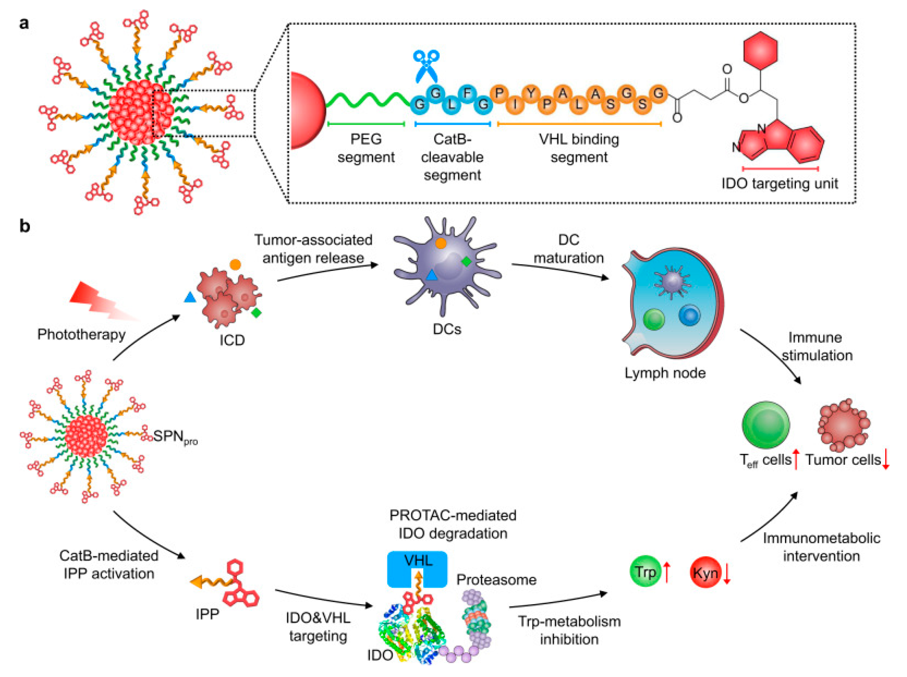
  120. Zhang, C.; Zeng, Z.; Cui, D.; He, S.; Jiang, Y.; Li, J.; Huang, J.; Pu, K. Semiconducting polymer nano-PROTACs for activatable photo-immunometabolic cancer therapy. Nat. Commun. 2021, 12, 2934. [Google Scholar] [CrossRef]
  121. Mo, R.; Jiang, T.; Di, J.; Tai, W.; Gu, Z. Emerging micro- and nanotechnology based synthetic approaches for insulin delivery. Chem. Soc. Rev. 2014, 43, 3595–3629. [Google Scholar] [CrossRef]
  122. Wells, A.; Grandis, J.R. Phospholipase C-gamma1 in tumor progression. Clin. Exp. Metastasis 2003, 20, 285–290. [Google Scholar] [CrossRef]
  123. Graff, J.R.; Konicek, B.W.; Deddens, J.A.; Chedid, M.; Hurst, B.M.; Colligan, B.; Neubauer, B.L.; Carter, H.W.; Carter, J.H. Expression of group IIa secretory phospholipase A2 increases with prostate tumor grade. Clin. Cancer Res. 2001, 7, 3857–3861. [Google Scholar]
  124. Gu, Z.; Aimetti, A.A.; Wang, Q.; Dang, T.T.; Zhang, Y.; Veiseh, O.; Cheng, H.; Langer, R.S.; Anderson, D.G. Injectable nano-network for glucose-mediated insulin delivery. ACS Nano 2013, 7, 4194–4201. [Google Scholar] [CrossRef] [PubMed]
  125. Gu, Z.; Dang, T.T.; Ma, M.; Tang, B.C.; Cheng, H.; Jiang, S.; Dong, Y.; Zhang, Y.; Anderson, D.G. Glucose-responsive microgels integrated with enzyme nanocapsules for closed-loop insulin delivery. ACS Nano 2013, 7, 6758–6766. [Google Scholar] [CrossRef]
  126. Andresen, T.L.; Davidsen, J.; Begtrup, M.; Mouritsen, O.G.; Jørgensen, K. Enzymatic release of antitumor ether lipids by specific phospholipase A2 activation of liposome-forming prodrugs. J. Med. Chem. 2004, 47, 1694–1703. [Google Scholar] [CrossRef]
  127. Webb, B.A.; Chimenti, M.; Jacobson, M.P.; Barber, D.L. Dysregulated pH: A perfect storm for cancer progression. Nat. Rev. Cancer 2011, 11, 671–677. [Google Scholar] [CrossRef]
  128. Cardone, R.A.; Casavola, V.; Reshkin, S.J. The role of disturbed pH dynamics and the Na+/H+ exchanger in metastasis. Nat. Rev. Cancer 2005, 5, 786–795. [Google Scholar] [CrossRef]
  129. Yameen, B.; Choi, W.I.; Vilos, C.; Swami, A.; Shi, J.; Farokhzad, O.C. Insight into nanoparticle cellular uptake and intracellular targeting. J. Control. Release 2014, 190, 485–499. [Google Scholar] [CrossRef] [Green Version]
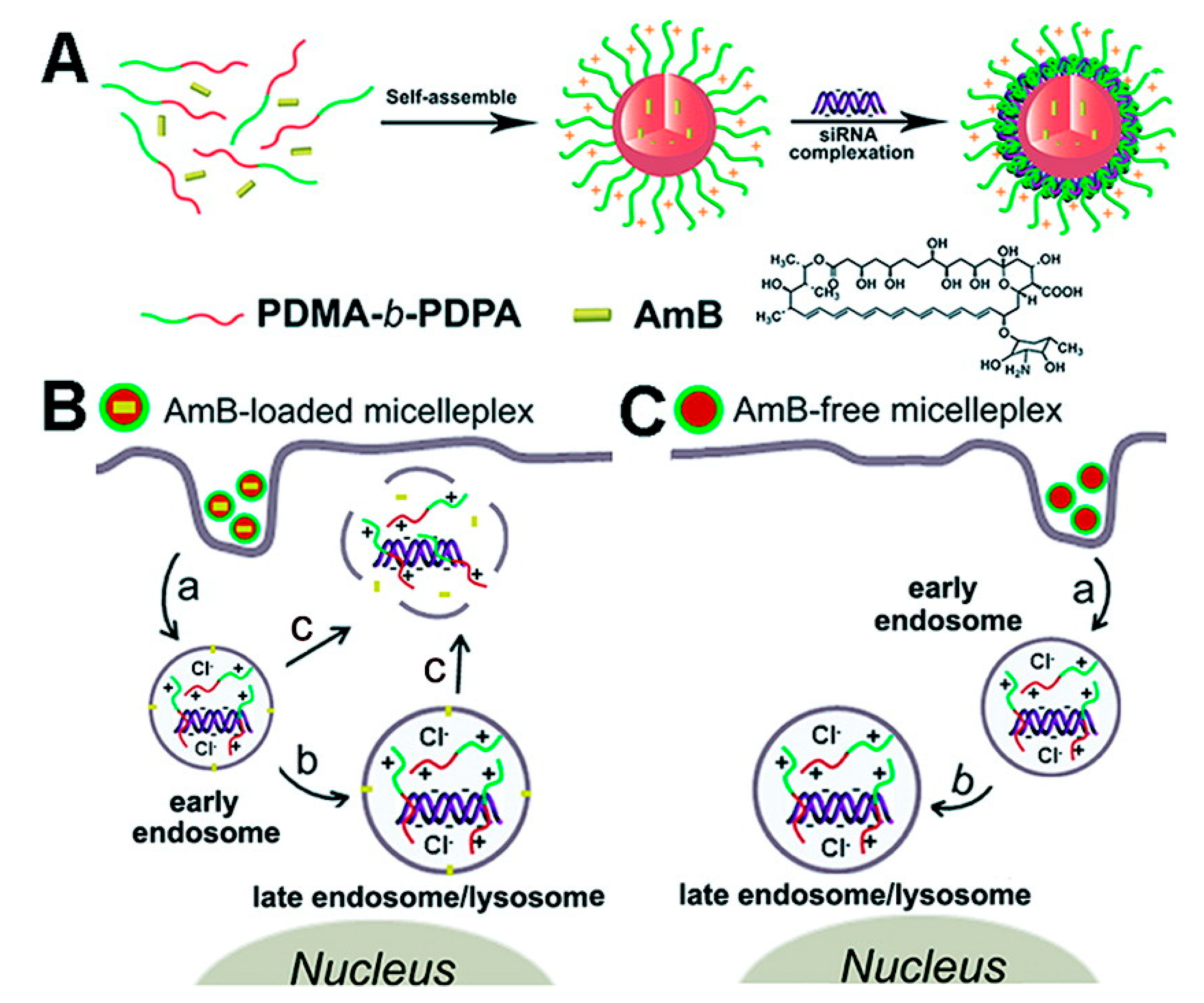
  130. Yu, H.; Zou, Y.; Wang, Y.; Huang, X.; Huang, G.; Sumer, B.D.; Boothman, D.A.; Gao, J. Overcoming endosomal barrier by amphotericin B-loaded dual pH-responsive PDMA-b-PDPA micelleplexes for siRNA delivery. ACS Nano 2011, 5, 9246–9255. [Google Scholar] [CrossRef] [Green Version]
  131. Saito, G.; Swanson, J.A.; Lee, K.D. Drug delivery strategy utilizing conjugation via reversible disulfide linkages: Role and site of cellular reducing activities. Adv. Drug Deliv. Rev. 2003, 55, 199–215. [Google Scholar] [CrossRef]
  132. Kuppusamy, P.; Li, H.; Ilangovan, G.; Cardounel, A.J.; Zweier, J.L.; Yamada, K.; Krishna, M.C.; Mitchell, J.B. Noninvasive imaging of tumor redox status and its modification by tissue glutathione levels. Cancer Res. 2002, 62, 307–312. [Google Scholar]
  133. Parvez, S.; Long, M.J.C.; Poganik, J.R.; Aye, Y. Redox Signaling by Reactive Electrophiles and Oxidants. Chem. Rev. 2018, 118, 8798–8888. [Google Scholar] [CrossRef]
  134. Shim, M.S.; Xia, Y. A reactive oxygen species (ROS)-responsive polymer for safe, efficient, and targeted gene delivery in cancer cells. Angew. Chem. Int. Ed. 2013, 52, 6926–6929. [Google Scholar] [CrossRef]
  135. Gaucher, C.; Boudier, A.; Bonetti, J.; Clarot, I.; Leroy, P.; Parent, M. Glutathione: Antioxidant properties dedicated to nanotechnologies. Antioxidants 2018, 7, 62. [Google Scholar] [CrossRef] [Green Version]
  136. Lee, M.H.; Yang, Z.; Lim, C.W.; Lee, Y.H.; Dongbang, S.; Kang, C.; Kim, J.S. Disulfide-cleavage-triggered chemosensors and their biological applications. Chem. Rev. 2013, 113, 5071–5109. [Google Scholar] [CrossRef]
  137. Gasparini, G.; Matile, S. Protein delivery with cell-penetrating poly(disulfide)s. Chem. Commun. 2015, 51, 17160–17162. [Google Scholar] [CrossRef] [Green Version]
  138. Gasparini, G.; Bang, E.K.; Molinard, G.; Tulumello, D.V.; Ward, S.; Kelley, S.O.; Roux, A.; Sakai, N.; Matile, S. Cellular uptake of substrate-initiated cell-penetrating poly(disulfide)s. J. Am. Chem. Soc. 2014, 136, 6069–6074. [Google Scholar] [CrossRef]
  139. Bang, E.K.; Gasparini, G.; Molinard, G.; Roux, A.; Sakai, N.; Matile, S. Substrate-initiated synthesis of cell-penetrating poly(disulfide)s. J. Am. Chem. Soc. 2013, 135, 2088–2091. [Google Scholar] [CrossRef] [Green Version]
  140. Qian, L.; Fu, J.; Yuan, P.; Du, S.; Huang, W.; Li, L.; Yao, S.Q. Intracellular delivery of native proteins facilitated by cell-penetrating poly(disulfide)s. Angew. Chem. Int. Ed. 2018, 57, 1532–1536. [Google Scholar] [CrossRef]
  141. Guo, J.; Wan, T.; Li, B.; Pan, Q.; Xin, H.; Qiu, Y.; Ping, Y. Rational design of poly(disulfide)s as a universal platform for delivery of CRISPR-Cas9 machineries toward therapeutic genome editing. ACS Cent. Sci. 2021, 7, 990–1000. [Google Scholar] [CrossRef] [PubMed]
  142. del Barrio, J.; Oriol, L.; Alcalá, R.; Sánchez, C. Azobenzene-containing linear−dendritic diblock copolymers by click chemistry: Synthesis, characterization, morphological study, and photoinduction of optical anisotropy. Macromolecules 2009, 42, 5752–5760. [Google Scholar] [CrossRef]
  143. Blasco, E.; Serrano, J.L.; Pinol, M.; Oriol, L. Light responsive vesicles based on linear-dendritic block copolymers using azobenzene-aliphatic codendrons. Macromolecules 2013, 46, 5951–5960. [Google Scholar] [CrossRef]
  144. Buback, J.; Kullmann, M.; Langhojer, F.; Nuernberger, P.; Schmidt, R.; Würthner, F.; Brixner, T. Ultrafast bidirectional photoswitching of a spiropyran. J. Am. Chem. Soc. 2010, 132, 16510–16519. [Google Scholar] [CrossRef]
  145. Klajn, R. Spiropyran-based dynamic materials. Chem. Soc. Rev. 2014, 43, 148–184. [Google Scholar] [CrossRef] [Green Version]
  146. Yang, H.; Jia, L.; Wang, Z.; Di-Cicco, A.; Lévy, D.; Keller, P. Novel photolabile diblock copolymers bearing truxillic acid derivative junctions. Macromolecules 2011, 44, 159–165. [Google Scholar] [CrossRef]
  147. Lendlein, A.; Jiang, H.; Jünger, O.; Langer, R. Light-induced shape-memory polymers. Nature 2005, 434, 879–882. [Google Scholar] [CrossRef]
  148. Weissleder, R. A clearer vision for in vivo imaging. Nat. Biotechnol. 2001, 19, 316–317. [Google Scholar] [CrossRef]
  149. Liu, G.-Y.; Chen, C.-J.; Li, D.-D.; Wang, S.-S.; Ji, J. Near-infrared light-sensitive micelles for enhanced intracellular drug delivery. J. Med. Chem. 2012, 22, 16865–16871. [Google Scholar] [CrossRef]
  150. Yan, B.; Boyer, J.C.; Branda, N.R.; Zhao, Y. Near-infrared light-triggered dissociation of block copolymer micelles using upconverting nanoparticles. J. Am. Chem. Soc. 2011, 133, 19714–19717. [Google Scholar] [CrossRef]
  151. Roy, D.; Brooks, W.L.; Sumerlin, B.S. New directions in thermoresponsive polymers. Chem. Soc. Rev. 2013, 42, 7214–7243. [Google Scholar] [CrossRef] [PubMed]
  152. Biswas, S.; Kumari, P.; Lakhani, P.M.; Ghosh, B. Recent advances in polymeric micelles for anti-cancer drug delivery. Eur. J. Pharm. Sci. 2016, 83, 184–202. [Google Scholar] [CrossRef] [PubMed]
  153. Talelli, M.; Hennink, W.E. Thermosensitive polymeric micelles for targeted drug delivery. Nanomedicine 2011, 6, 1245–1255. [Google Scholar] [CrossRef] [PubMed]
  154. Scarpa, J.S.; Mueller, D.D.; Klotz, I.M. Slow hydrogen-deuterium exchange in a non-.alpha.-helical polyamide. J. Am. Chem. Soc. 1967, 89, 6024–6030. [Google Scholar] [CrossRef]
  155. Akimoto, J.; Nakayama, M.; Sakai, K.; Okano, T. Thermally controlled intracellular uptake system of polymeric micelles possessing poly(N-isopropylacrylamide)-based outer coronas. Mol. Pharm. 2010, 7, 926–935. [Google Scholar] [CrossRef]
  156. Legout, P.; Lefebvre, G.; Bonnin, M.; Gimel, J.C.; Benyahia, L.; Colombani, O.; Calvignac, B. Synthesis of PDMS-b-POEGMA diblock copolymers and their application for the thermoresponsive stabilization of water-supercritical carbon dioxide emulsions. Langmuir 2020, 36, 12922–12932. [Google Scholar] [CrossRef]
  157. Boffito, M.; Torchio, A.; Tonda-Turo, C.; Laurano, R.; Gisbert-Garzarán, M.; Berkmann, J.C.; Cassino, C.; Manzano, M.; Duda, G.N.; Vallet-Regí, M.; et al. Hybrid injectable sol-gel systems based on thermo-sensitive polyurethane hydrogels carrying pH-sensitive mesoporous silica nanoparticles for the controlled and triggered release of therapeutic agents. Front. Bioeng. Biotechnol. 2020, 8, 384. [Google Scholar] [CrossRef]
  158. Haladjova, E.; Halacheva, S.; Momekova, D.; Moskova-Doumanova, V.; Topouzova-Hristova, T.; Mladenova, K.; Doumanov, J.; Petrova, M.; Rangelov, S. Polyplex particles based on comb-like polyethylenimine/poly(2-ethyl-2-oxazoline) copolymers: Relating biological performance with morphology and structure. Macromol. Biosci. 2018, 18, e1700349. [Google Scholar] [CrossRef]
  159. Karanikolopoulos, N.; Zamurovic, M.; Pitsikalis, M.; Hadjichristidis, N. Poly(DL-lactide)-b-poly(N,N-dimethylamino-2-ethyl methacrylate): Synthesis, characterization, micellization behavior in aqueous solutions, and encapsulation of the hydrophobic drug dipyridamole. Biomacromolecules 2010, 11, 430–438. [Google Scholar] [CrossRef]
  160. Zavgorodnya, O.; Carmona-Moran, C.A.; Kozlovskaya, V.; Liu, F.; Wick, T.M.; Kharlampieva, E. Temperature-responsive nanogel multilayers of poly(N-vinylcaprolactam) for topical drug delivery. J. Colloid. Interface Sci. 2017, 506, 589–602. [Google Scholar] [CrossRef]
  161. Leighton, T.G. What is ultrasound? Prog. Biophys. Mol. Biol. 2007, 93, 3–83. [Google Scholar] [CrossRef] [PubMed]
  162. Husseini, G.A.; Pitt, W.G. Ultrasonic-activated micellar drug delivery for cancer treatment. J. Pharm. Sci. 2009, 98, 795–811. [Google Scholar] [CrossRef] [Green Version]
  163. Davis, D.A.; Hamilton, A.; Yang, J.; Cremar, L.D.; van Gough, D.; Potisek, S.L.; Ong, M.T.; Braun, P.V.; Martínez, T.J.; White, S.R.; et al. Force-induced activation of covalent bonds in mechanoresponsive polymeric materials. Nature 2009, 459, 68–72. [Google Scholar] [CrossRef]
  164. Caruso, M.M.; Davis, D.A.; Shen, Q.; Odom, S.A.; Sottos, N.R.; White, S.R.; Moore, J.S. Mechanically-induced chemical changes in polymeric materials. Chem. Rev. 2009, 109, 5755–5798. [Google Scholar] [CrossRef]
  165. Hickenboth, C.R.; Moore, J.S.; White, S.R.; Sottos, N.R.; Baudry, J.; Wilson, S.R. Biasing reaction pathways with mechanical force. Nature 2007, 446, 423–427. [Google Scholar] [CrossRef]
  166. Wang, J.; Pelletier, M.; Zhang, H.; Xia, H.; Zhao, Y. High-frequency ultrasound-responsive block copolymer micelle. Langmuir 2009, 25, 13201–13205. [Google Scholar] [CrossRef]
  167. Xuan, J.; Boissière, O.; Zhao, Y.; Yan, B.; Tremblay, L.; Lacelle, S.; Xia, H.; Zhao, Y. Ultrasound-responsive block copolymer micelles based on a new amplification mechanism. Langmuir 2012, 28, 16463–16468. [Google Scholar] [CrossRef] [PubMed]
  168. Tong, R.; Lu, X.; Xia, H. A facile mechanophore functionalization of an amphiphilic block copolymer towards remote ultrasound and redox dual stimulus responsiveness. Chem. Commun. 2014, 50, 3575–3578. [Google Scholar] [CrossRef] [PubMed]
Figure 1. Structure of cathepsin-responsive HPMA copolymer drug conjugates: left, P(GFLG)-DOX; right, ST-PHPMA-DOX-COOH. Reproduced from ref. [118] with permission from the American Chemical Society.
Applsci 11 09541 g001
Figure 2. Schematic illustration of SPNpro-mediated IDO degradation for cancer photo-immunometabolic therapy. (a) Structure and CatB-specific activation mechanism of SPNpro. (b) SPNpro-mediated activatable photo-immunometabolic therapy with two processes: (i) a series of cancer immune responses including immunogenic cell death (ICD), tumor-associated antigen release, DC maturation, and effector T (Teff) cell activation upon NIR photoirradiation; (ii) SPNpro-mediated immunometabolic intervention processes including CatB-specific activation of IPP, IDO and VHL targeting, proteasome recruitment, IDO degradation, Trp upregulation and Kyn depletion, and Teff cell activation. Reproduced from ref. [120] with permission from the Nature Publishing Group.
Applsci 11 09541 g002
Figure 3. Chemical structures of two different pro-antitumor ether lipids (proAELs) that have been synthesized and investigated with respect to their physical properties and ability to constitute a novel liposome-based drug-delivery system. Secretory phospholipase A2 (sPLA2) hydrolyzes the liposome membrane, thereby releasing both activated antitumor ether lipids (AELs) and the encapsulated drug. The AELs are cytotoxic to cancer cells. Furthermore, synergistically with the generated fatty acids, they function as permeability enhancers that promote drug uptake by the cancer cells. Reproduced from ref. [126] with permission from the American Chemical Society.
Applsci 11 09541 g003
Figure 4. Schematic diagram of AmB-loaded dual pH-responsive micelleplexes for siRNA delivery with enhanced siRNA endosomal escape ability. (A) Production of AmBloaded PDMA-b-PDPA micelleplexes. AmB was loaded in the hydrophobic PDPA core, and siRNA was complexed with the PDMA corona shell. (B) AmB-facilitated endosome disruption and siRNA cytoplasmic release (a: AmB-loaded micelleplexes dissociated in early endosomes after cell uptake, and AmB molecules are inserted into endosomal membranes; b: protonated PDMA-b-PDPA unimers complexed with siRNA and trafficked from early endosomes into late endosome/lysosomes, causing vesicle swelling; c: AmB-enhanced siRNA release from endosomes into cytoplasm via membrane destabilization). (C) In the case of AmB-free micelleplexes, polymer/siRNA complexes were entrapped in late endosomes or lysosomes without efficient cytoplasmic siRNA release. Reproduced from ref. [130] with permission from the American Chemical Society.
Applsci 11 09541 g004
Figure 5. Schematic illustration of the preparation of poly(disulfide)s, the complexation of genome editing biomacromolecules (Cas9 plasmid, Cas9 mRNA, and Cas9 ribonucleoprotein) by poly(disulfide)s, and their intracellular delivery processes for genome editing. Reproduced from ref. [141] with permission from the American Chemical Society.
Applsci 11 09541 g005
Figure 6. (a) Schematic illustration of using NIR light excitation of UCNPs to trigger dissociation of BCP micelles. (b) NIR light-triggered photoreaction with the used BCP of PEO-b-PNBMA and UCNPs of NaYF4:TmYb. Reproduced from ref. [150] with permission from the American Chemical Society.
Applsci 11 09541 g006
Publisher’s Note: MDPI stays neutral with regard to jurisdictional claims in published maps and institutional affiliations.

