Article

Regenerative Healing of Chitosan Scaffold Impregnated with Simvastatin in Repairing Furcal Perforation

by
Ghaliah M. Alsawah
1,*,
Manal Alsheddi
2,*,
Ebtissam M. Al-Madi
1 and
Mohammad I. Al-Obaida
1
1
Department of Restorative Dental Sciences, College of Dentistry, King Saud University, Riyadh 11527, Saudi Arabia
2
Department of Basic Science, College of Dentistry, Princess Nourah Bint Abdulrahman University, Riyadh 11671, Saudi Arabia
*
Authors to whom correspondence should be addressed.
Appl. Sci. 2021, 11(19), 8992; https://doi.org/10.3390/app11198992
Submission received: 17 August 2021 / Revised: 18 September 2021 / Accepted: 22 September 2021 / Published: 27 September 2021

Abstract

:
This study aimed to evaluate the healing of furcation when repaired with Chitosan (CS) scaffold impregnated or not with Simvastatin (SIM) compared with CollaCote (CL) in goat premolar teeth. Root canal treatment was performed in 52 mandibular premolars followed by furcal perforation induction. The perforation was repaired with CL, CS, or CS with SIM after leaving it untreated for 4 weeks. White mineral trioxide aggregate was carried into the furcal site followed by a 2–3 mm resin-modified glass ionomer. The perforation was left untreated, and the access cavity was left open without coronal filling in the control group. The animals were sacrificed after one and three months. Block sections of the premolars were prepared and examined histologically to evaluate the inflammation and type of healing. Hard tissue formation was found in CL, CS, and CS/SIM groups in both periods. At one month, no significant differences were detected among the experimental groups, whereas at three months, CS without SIM showed significantly better performance compared to CL and CS/SIM groups (p = 0.040). Therefore, repairing furcal perforation with CS scaffolds shows desirable biological responses and healing characteristics in favor of bone regeneration at three months.

1. Introduction

Furcal perforations are serious complications of iatrogenic errors throughout endodontic access opening or pathological processes that can adversely affect tooth prognosis. The perforation size, location, and time before repair are all noteworthy factors determining the prognosis of perforated teeth [1]. When perforation is not managed promptly, periodontal destruction will occur around the perforation site, resulting in the surrounding bone resorption [2]. Different materials have been used to seal such perforations, with mineral trioxide aggregate (MTA) showing superiority in terms of sealing ability and biocompatibility; therefore, it has become the gold standard [3,4]—yet MTA has a delayed setting time and poor handling characteristics [5]. Moreover, MTA is difficult to control and can extrude into the periodontium, especially in large furcal perforations which act as a “bottomless pit” [6] causing mechanical and chemical irritation, triggering tissue inflammation, interfering with the periodontal reattachment, and impairing treatment prognosis [7,8]. Accordingly, internal matrices have been introduced to restrict MTA overextension [9], and this approach presented favorable outcomes of perforation repair when compared to matrix-free treatment [10].
Chitosan (CS), a natural biocompatible and biodegradable material has generated great interest in medicine and has been used in tissue engineering (TE) and wound healing due to its hemostatic, anti-inflammatory, and antimicrobial properties. In addition, it can enhance cell adhesion, proliferation, and differentiation [11]. CS’s advantageous physical properties such as the adequate pore size, its ability to be modified, and its ability to be combined with other polymers allowed the development of a wide range of scaffolds that have been used in bone, neural and vascular tissues, cartilage, and skin regeneration [12]. A porous CS scaffold was fabricated and was able to promote the regenerative potential of dental pulp cells and osteoblastic differentiation [13]. Furthermore, a recent study presented CS to be a viable therapeutic option to hinder root resorption while enhancing healing outcomes in cases of severe inflammatory root resorption [14]. Simvastatin (SIM), a widely used statin and a well-established cholesterol-lowering drug [15], has increased the expression of bone morphogenic protein 2 (BMP-2) in osteoblasts and encouraged bone formation [16]. Its ease of purchase and affordable price make its selection more convenient than laboratory bone-driven costly stimulators. In addition, obesity and metabolic syndrome are characterized by lipid metabolism disorders holding the risk of increasing developing periodontal disease [17,18], which is known as the most common chronic infection in the world [19]. In periodontics, SIM has been reported to stimulate the proliferation and osteoblastic differentiation of human periodontal ligament cells (PDL) [20] and to offer aid to mechanical periodontal therapy (MPT) in improving periodontal parameters compared with MPT alone or in association with placebo [21].
To our knowledge, no study has investigated the effect of CS scaffold, alone or with SIM impregnation, in repairing furcal perforations in vivo. This study aimed to histologically evaluate the inflammatory reactions and tissue responses to the experimental CS scaffold, with and without SIM, when used to repair furcal perforation and compare it to the absorbable collagen wound dressing most commonly used in endodontic wound healing, CollaCote (CL). CollaCote is a soft, white, pliable, nonfriable sponge used in dental surgery due to its coherent structure, bleeding control, and protecting properties of the wound bed while accelerating the healing process [22]. The null hypothesis was that there is no difference between CS scaffold and CL in repairing furcal perforations and bone healing with and without SIM impregnation.

2. Materials and Methods

The study was carried out in agreement with the protocols of King Saud University, Riyadh, Saudi Arabia. All animal procedures were conducted according to the National Institute of Health guide for the care and use of laboratory animals (NIH Publications No. 85–23, revised 1985). The proposal was approved by the Ethical Committee and Institutional Review Board, King Saud University (E-19-3930) and the College of Dentistry Research Center of King Saud University (PR 0090) in Riyadh, Saudi Arabia. The experimental procedure was carried out at the animal house of King Khaled University Hospital of King Saud University.

2.1. Sample Size Estimation

The sample size calculation was performed using G*Power 3.1.9.2 software based on an effect size of 0.91, α   error of 0.05, and power of 80%. A minimum sample size of 10 goats was sufficient to detect significant differences between the groups. When 25% dropout was included, the required sample size was 13 goats.

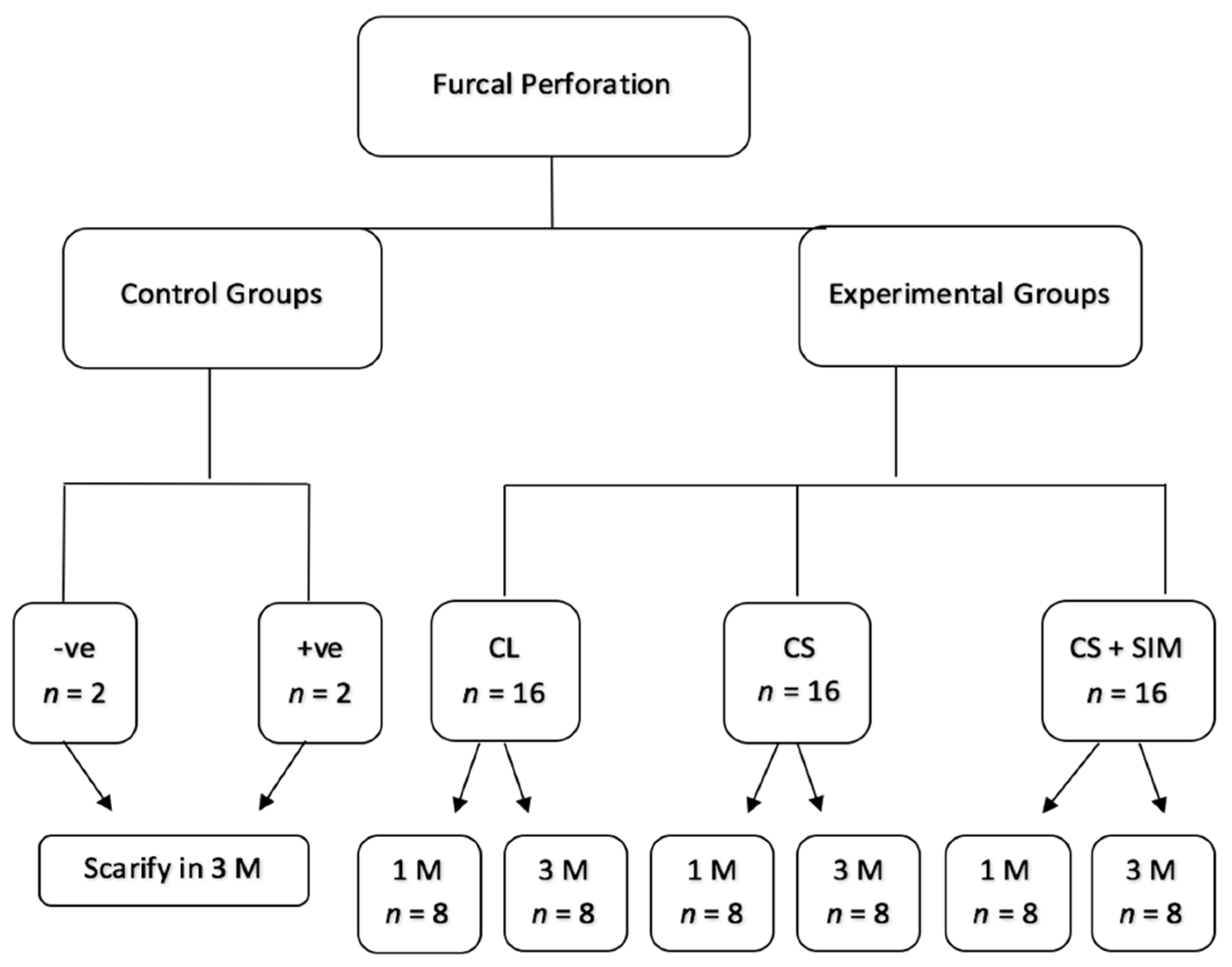
2.2. Sample Selection, Animal Preparation, Randomization, and Grouping

A total of 52 mandibular premolars from 13 healthy adult male goats were used. The teeth were randomly distributed in five groups according to the scaffold used in repairing the perforation: CL, CS, CS with SIM, negative control, and untreated positive control. The mandibular first and second premolars of each side were used. The teeth were intact from caries, free from periodontal diseases, and had complete root development. Inflammation and healing responses were examined at two-time points. The animals were housed under the supervision of veterinary specialists in the Faculty of Agriculture’s yard, King Saud University, Riyadh, Saudi Arabia. They had access to food and water throughout the study period.

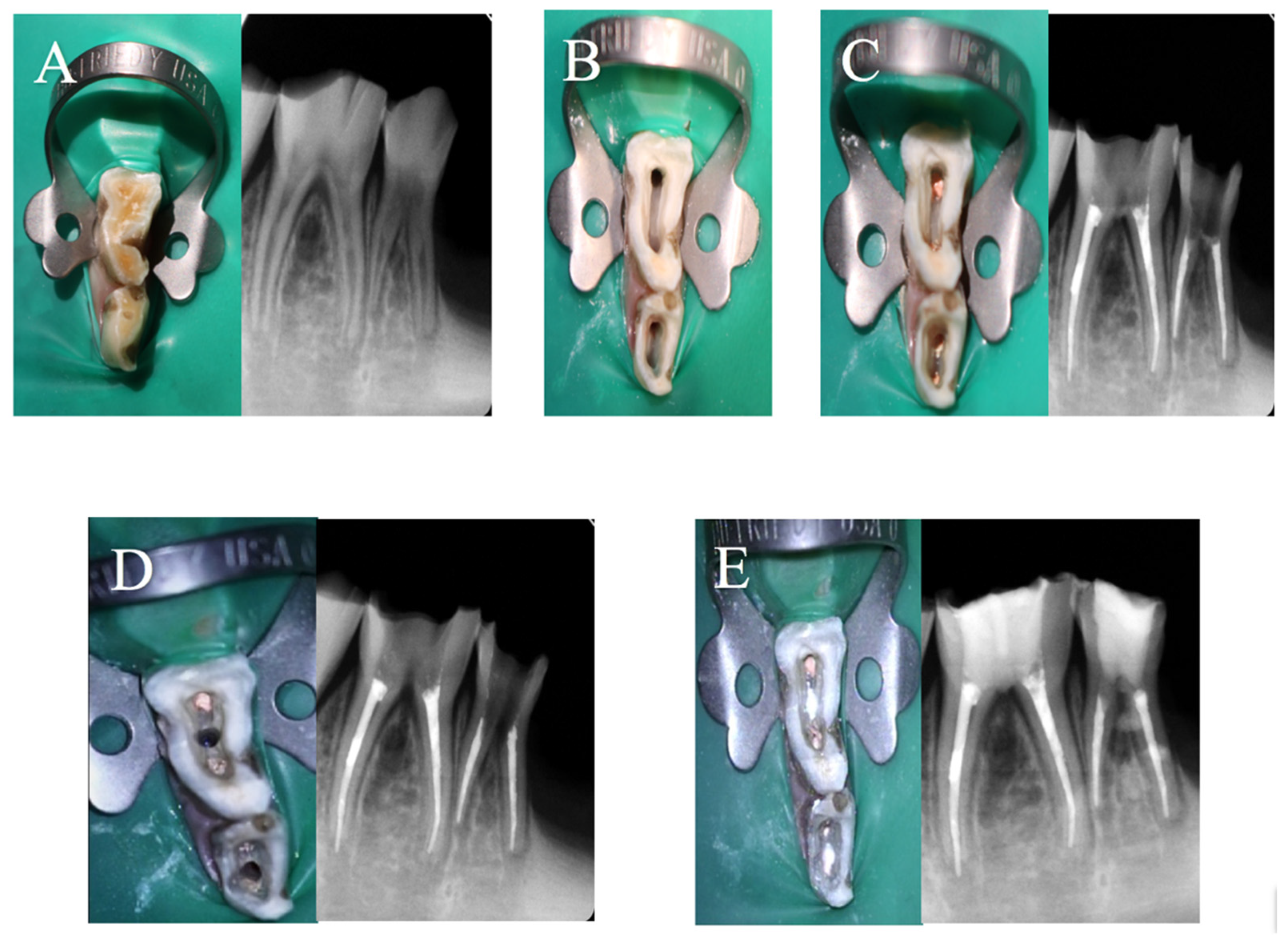
2.3. Operative Procedure

The CS scaffolds were fabricated according to the protocol in our previous study [23]. Intravenous injection of Xylazine (Seton, Barcelona, Spain) at a dose of 2% mg/kg was introduced in the jugular vein using a 25-gauge needle 1 cc/mL to sedate the animals. Subsequent injections of ketamine (TEKAM®, London, UK) were maintained every half an hour and controlled by a veterinarian [24]. Pre-operative radiographs were taken to confirm pathosis absence. Next, teeth were locally anesthetized with infiltration technique using Xylocaine Dental Adrenaline 20 mg/mL + 12.5 mg/mL Standard, 1.8 mL (Dentsply Ltd., Surrey, UK). Teeth were isolated with a rubber dam and disinfected with cotton soaked in 75% ethanol. Under a 4X microscope (System Contraves & Sec, Carl Zeiss, Thornwood, NY, USA), access was opened using a sterile water-cooled tapered diamond bur (Komet Dental, Lemgo, Germany), and root canal treatment was performed with Protaper Gold rotary files (Dentsply Sirona, Baden, Switzerland). The canals were irrigated with 2.5% sodium hypochlorite (NaOCL) and dried with sterile paper points then obturated with single matching gutta-percha cones (Dentsply Sirona, York, NC, USA) using AH26 silver-free sealer (Dentsply De- Trey GmbH, Konstanz, Germany). A 2 mm diameter perforation (equal to the width of the used round diamond bur to standardize the size among all teeth) was created in the pulp chamber floor, except for the negative control group. The depth of the perforation was between 2 and 4 mm according to the depth of the tooth’s pulpal floor to invade completely through the furcal dentin into the periodontium. Irrigation protocol using 2.5% NaOCL was followed. Afterwards, teeth were left open for four weeks to ensure inflammatory lesion formation in the furcation area. No medication was used to inhibit new variables’ effects. After a month, animals were sedated following the same previous protocol and under aseptic technique and rubber dam isolation, the perforation site was curetted using a small spoon excavator to remove the food remnants, debris, and inflamed tissue, cleaned with 2.5% NaOCL, and dried with paper points and cotton pellets. The teeth were randomly treated as follows:
  • Group 1: Negative control. No perforation was created in the pulp chamber after finishing the root canal filling. The access cavity was left open without coronal filling;
  • Group 2: Positive control. The furcal perforation and access cavity were left open without coronal filling;
  • Group 3: CollaCote (CollaCote®; Zimmer, Warsaw, IN, USA). CL was placed in the furcal perforation then white MTA (ProRoot MTA; Dentsply Tulsa Dental Specialties, Johnson City, TN, USA) mixed with sterile saline (3 parts powder to 1 part liquid) was carried into the furcal site with amalgam carrier and compacted with a size 1 Buchanan hand plugger with light pressure, followed by a 2–3 mm resin-modified glass ionomer (GC Fuji II LC Capsule; GC Corporation, Tokyo, Japan);
  • Group 4: Chitosan. CS scaffold with no SIM was placed in the furcal perforation site then the same procedure was repeated as in group 3;
  • Group 5: Chitosan scaffold impregnated with SIM was placed in the furcal site, then the same procedure was repeated as in group 3. Group distribution is illustrated in Figure 1.
The procedures were all performed by one operator. The goats were continuously monitored for any changes in habits and food intake during the post-treatment evaluation periods. The clinical steps and radiographic images were documented in one animal for illustration purposes (Figure 2).

2.4. Sample Preparation

All animals were coded and clustered into two subgroups according to their time of sacrifice; one and three months. The goats were sacrificed in the slaughterhouse, and the mandibular jaw was sectioned using a diamond disc and hand saw (35 cm) to obtain blocks composed of the treated teeth and the surrounding alveolar bone free from soft tissues.

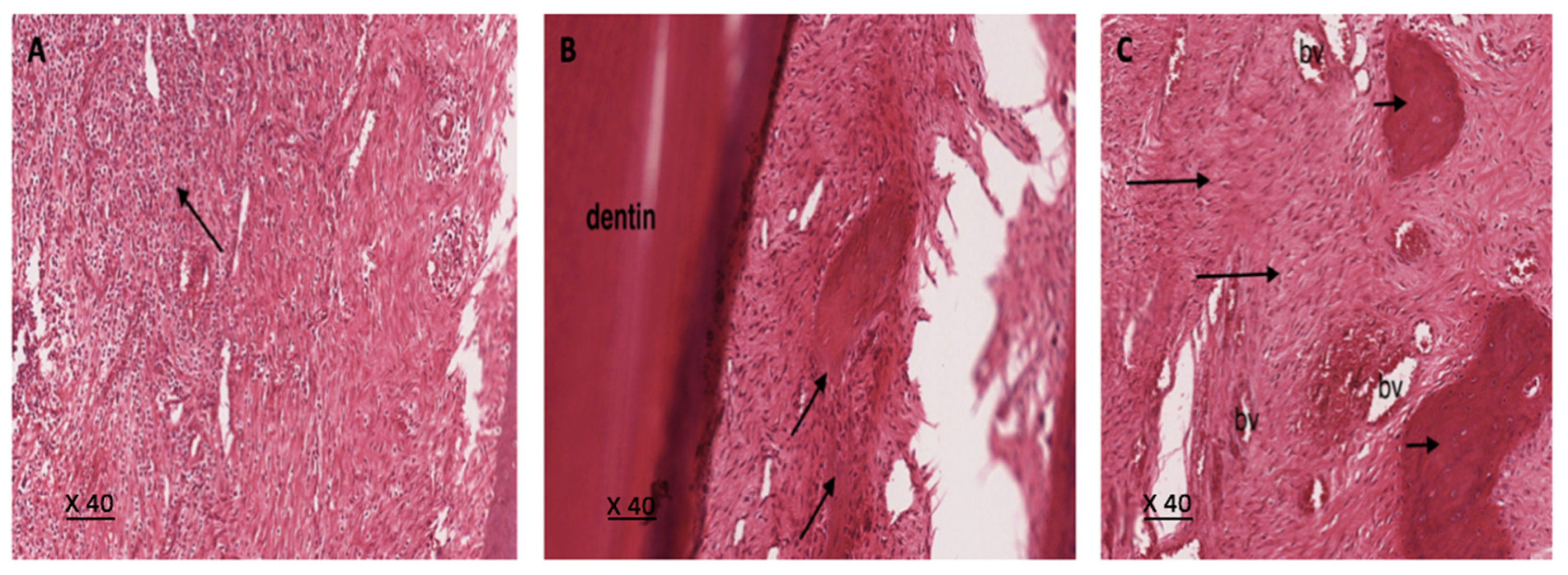
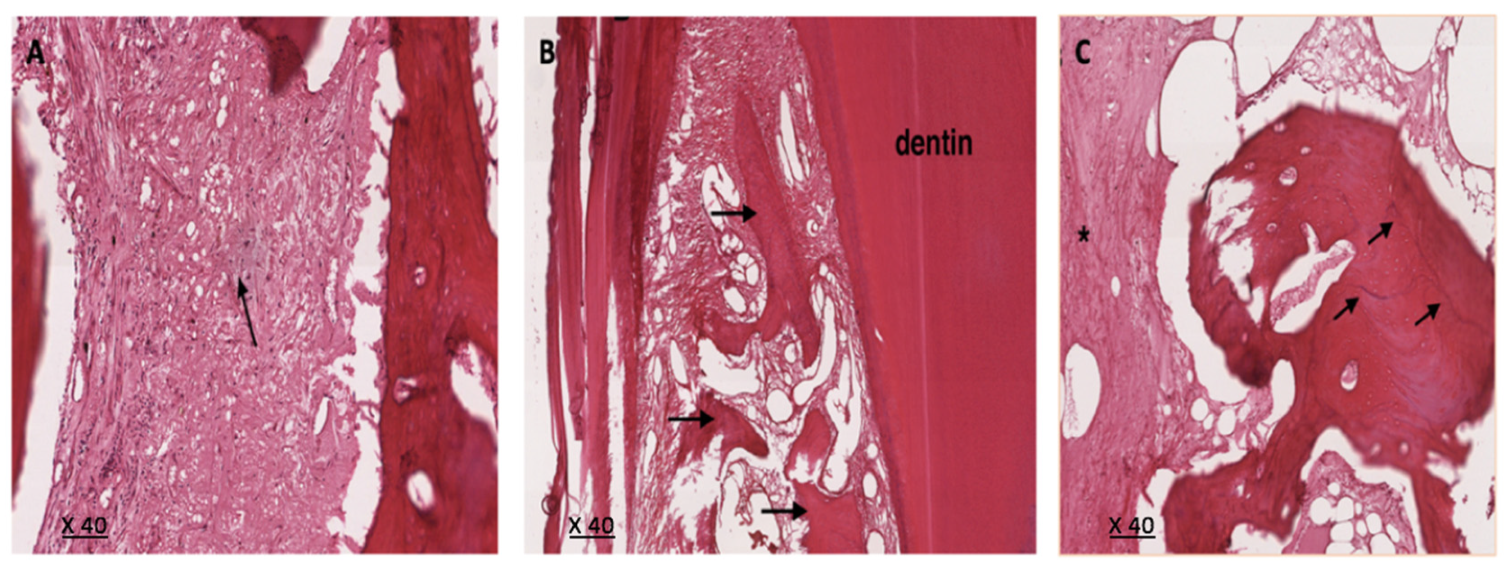
2.5. Histological Examination

The blocks were coded, fixed in 4% paraformaldehyde solution (SecurBiop®, Teramo, Italy) for two days, and then decalcified for four weeks using 17% EDTA/phosphate-buffered saline solution. The tissues were dehydrated in graded ethanol and embedded in paraffin wax (Leica Biosystems, Richmond, IL, USA). Serial sections were cut at a 5-micrometer thick buccolingual direction with a microtome (Leica, RM2255, Nussloch, Germany), and the defect site was reached and stained with hematoxylin–eosin (H&E) according to the conventional pathological protocols (Baso Diagnostic Inc, Zhuhai, China). The area of interest was examined by light microscope (Leitz, Laborlux S, Binocular microscope, Wetzlar, Germany) and scanned (ScanScope® AT, Vista, CA, USA) for the tissue assessment. The following parameters were examined:
  • The inflammation quantification by counting the ratio of inflammatory cells in 100 cells under the high-power magnification (×400) and scored as follows [25]:
    • Score 0: <10%;
    • Score 1: 10–30%;
    • Score 2: 30–50%;
    • Score 3: >50%.
  • Bone formation was marked with the presence or absence of osteoid cells;
  • The osteoclastic activity was marked as absent or present;
  • Foreign body reaction was marked with the presence or absence of macrophages or giant cells;
  • Granulation tissue was marked as absent or present.
The histological features were blindly evaluated by one trained pathologist.

2.6. Statistical Analysis

Most outcomes were categorical, they were summarized using frequencies and proportions. Comparison between groups was performed using Fisher’s Exact test. The overall index was summarized using descriptive statistics such as mean, median, standard deviation (SD), and interquartile range (IQR). Index scores were compared between three experimental groups using the Kruskal–Wallis test (the non-parametric equivalent of one-way ANOVA). The STATA software (ver.14) was used for data analysis. Statistical significance was set at 0.05.

3. Results

The follow-up of the goats showed no changes in their habits. All goats ate and drank well throughout the study except for one goat who got sick for 8 days and died; therefore, four specimens in the CS/SIM-1-month group were excluded. The scoring results of the histological findings are summarized in Table 1.
Overall, an index was created between the experimental groups to combine inflammation quantification (score 0–3), inflammatory cells type (0 = absent, 1 = present), bone formation (1 = present), osteoclastic activity (1 = present), ankyloses (1 = present), foreign body reaction (1 = present), and granulation tissue (1 = present). The index theoretically ranged from 0 to 9, with a lower score representing fewer complications. At 1 month, the CL group showed the best results with no statistical significance (p = 0.242) (Table 2).
At 3 months, CS without SIM showed significantly better results compared to other groups (p = 0.040) (Table 3).
When both time periods were compared for each group, the CL subgroups showed more bone formation at 3 months with no statistically significant difference. Meanwhile, the CS/SIM group showed a 100% rate of osteoclastic activity at 1 month and a statistically significant 0% rate at 3 months (p = 0.028). Histological sections for experimental groups showed lymphocytic inflammatory infiltrate at the 1-month period without noteworthy foreign body reaction (Figure 3). Later, the 3-month sections revealed less inflammation and more fibrosis, mineralized tissue, and hard tissue formation with prominent reversal lines (Figure 4).

4. Discussion

In this study, a histological evaluation of tissue inflammation, bone formation, and the type of healing barrier adjacent to the scaffold was performed. Goats are a challenging experimental model, yet having lower premolars with two roots that often furcate as close as 2–4 mm from the cementoenamel junction makes it suitable to conduct in vivo studies to predict similar responses in human teeth. Thus, it was not surprising to notice epithelial proliferation and connective tissue in cases of inflamed furcation. Inflammation is a vital part of the immune system [26]; hence, it was used repeatedly in literature to monitor the healing process [27,28]. Porous scaffolds seeded with bioactive substances have shown their ability to promote bone and dental regeneration where cells were able to migrate, grow, and differentiate [29]. Overall, our results showed similar slight inflammation in the scaffold samples which indicated a positive tissue response to each scaffold as found in preceding studies [30]. Moreover, bone formation was evident in all groups compared to the positive control group, which came in agreement with previous findings [31,32]. No significant differences were detected between experimental groups at 1 month. The probable explanation for the high rate of repair is that the used scaffolds are biocompatible [11,30] in addition to the SIM effect on angiogenesis and bone formation as shown in numerous experimental animal studies [33,34,35,36]. At 3 months, CS scaffold showed significantly better performance compared to CL and CS/SIM groups. In addition, it was noticed clinically that the CS scaffold had better handling characteristics than CL as its structure permitted for a more controlled material packing into the furcation site. Previous studies reported the osteoclast and osteoblast’s ability to regulate bone remodeling within the bone by balancing the amount of resorption and ossification which, therefore, is tightly linked to the number and activity of osteoclasts and osteoblast [37]. Continuous osteoclastic activity in CS/SIM subgroups was present and significantly shown by dense fibroblastic tissue and bone trabeculae with prominent reversal lines at 3 months confirming the active bone formation. These results support previous studies where SIM reinforced bone formation [38,39,40]. Simvastatin showed its ability to trigger the early expression of growth factors, including vascular endothelial growth factor (VEGF) [41] and BMP-2, and to induce and accelerate the formation of bones locally [42]. The range of SIM doses is broad in the literature. Varied values have been investigated—2.2 mg [43,44], 0.5 mg [33,34], 0.1, 0.5, 1.0, and 1.5 mg [44]—and shown positive or negative effects on bone repair. Since this study is a prolongation of our previous paper, we selected our fabricated CS with 0.05 mg SIM scaffold to continue with because it showed more osteoblastic differentiation among other examined concentrations [23]. Hence, different findings may be caused by varied local interactions or because SIM can be cytotoxic at certain doses—above 0.1 µM [45], 1 µM and 5 µM [41,45], and higher than 0.5 µM [46]. Thus, material toxicity may be an important factor for tissue healing. No side effects were observed in the present study. Costs, ethical considerations, and difficulty of handling are limiting factors of studies using animals, which, although similar to other studies [10,28,47,48], limited our study to 3 months. Applications of such promising associations of novel scaffolds seeded with biological substances are distinctly directed on regenerative dentistry and biological-guided endodontic treatment. Further studies can increase the sample size to increase the confidence in the estimate, thus affecting the statistical significance, and increasing the experimental period to observe whether there is a continued bone deposition and determine the accurate SIM dose to be used in vivo.

5. Conclusions

Furcal perforations continue to be both an endodontic and a periodontal problem. The present in vivo study results showed CS scaffolds desirable biological responses and healing characteristics in favor of bone regeneration at three months in repairing furcal perforations.

Author Contributions

Conceptualization, G.M.A., M.I.A.-O. and E.M.A.-M.; methodology, G.M.A. and M.I.A.-O.; validation, M.I.A.-O.; formal analysis, G.M.A. and M.A.; investigation, G.M.A.; writing—original draft preparation, G.M.A. and M.A.; writing—review and editing, M.I.A.-O., E.M.A.-M. and M.A.; visualization, M.I.A.-O.; supervision, M.I.A.-O.; project administration, M.I.A.-O. All authors have read and agreed to the published version of the manuscript.

Funding

This research received no external funding.

Institutional Review Board Statement

The study was approved by the Ethical Committee and Institutional Review Board of King Saud University (E-19-3930–May 2019) and the College of Dentistry Research Center of King Saud University (PR 0090) in Riyadh, Saudi Arabia.

Informed Consent Statement

Not applicable.

Data Availability Statement

Details are presented within the article in the form of tables and images in results. Additional data can be made available upon request from the corresponding author.

Acknowledgments

The authors acknowledge Yasser AlShawakir from King Khaled Hospital, King Saud University, for his aid and guidance in the operative and surgical procedures. We also appreciate the contributions of the staff in the animal house and Agriculture’s yard, King Saud University, Riyadh, Saudi Arabia.

Conflicts of Interest

The authors declare no conflict of interest.

References

  1. Alhadainy, H.A.; Himel, V.T. An In Vitro Evaluation of Plaster of Paris Barriers Used Under Amalgam and Glass Ionomer to Repair Furcation Perforations. J. Endod. 1994, 20, 449–452. [Google Scholar] [CrossRef]
  2. Seltzer, S.; Sinai, I.; August, D. Periodontal Effects of Root Perforations Before and During Endodontic Procedures. J. Dent. Res. 1970, 49, 332–339. [Google Scholar] [CrossRef] [PubMed]
  3. Roberts, H.W.; Toth, J.M.; Berzins, D.W.; Charlton, D.G. Mineral Trioxide Aggregate Material Use in Endodontic Treatment: A Review of The Literature. Dent. Mater. 2008, 24, 149–164. [Google Scholar] [CrossRef] [Green Version]
  4. Baek, S.H.; Plenk, H., Jr.; Kim, S. Periapical Tissue Responses and Cementum Regeneration with Amalgam, SuperEBA, and MTA as Root-End Filling Materials. J. Endod. 2005, 31, 444–449. [Google Scholar] [CrossRef] [Green Version]
  5. Maltezos, C.; Glickman, G.N.; Ezzo, P.; He, J. Comparison of The Sealing of Resilon, Pro Root MTA, and Super-EBA as Root-End Filling Materials: A Bacterial Leakage Study. J. Endod. 2006, 32, 324–327. [Google Scholar] [CrossRef]
  6. Cohen, S.; Burns, R.C. Pathways of The Pulp, 2nd ed.; Mosby: St. Louis, MO, USA, 1980; pp. 106–108. [Google Scholar]
  7. Tsesis, I.; Fuss, Z. Diagnosis and Treatment of Accidental Root Perforations. Endod. Top. 2006, 13, 95–107. [Google Scholar] [CrossRef]
  8. Al-Daafas, A.; Al-Nazhan, S. Histological Evaluation of Contaminated Furcal Perforation in Dogs’ Teeth Repaired by MTA With or Without Internal Matrix. Oral Surg. Oral Med. Oral Pathol. Oral Radiol. Endodontol. 2007, 103, 92–99. [Google Scholar] [CrossRef]
  9. Lemon, R.R. Nonsurgical Repair of Perforation Defects. Internal Matrix Concept. Dent. Clin. N. Am. 1992, 36, 439–457. [Google Scholar] [PubMed]
  10. Rafter, M.; Baker, M.; Alves, M.; Daniel, J.; Remeikis, N. Evaluation of Healing with Use of an Internal Matrix to Repair Furcation Perforations. Int. Endod. J. 2002, 35, 775–783. [Google Scholar] [CrossRef]
  11. Li, Q.; Dunn, E.T.; Grandmaison, E.W.; Goosen, M.F.A. Applications and Properties of Chitosan. J. Bioact Compat Polym. 1992, 7, 370–397. [Google Scholar] [CrossRef]
  12. Kim, I.Y.; Seo, S.J.; Moon, H.S.; Yoo, M.K.; Park, I.Y.; Kim, B.C.; Cho, C.S. Chitosan and its Derivatives for Tissue Engineering Applications. Biotechnol. Adv. 2008, 26, 1–21. [Google Scholar] [CrossRef]
  13. Soares, D.; Anovazzi, G.; Bordini, E.A.F.; Zuta, U.O.; Leite, M.L.A.S.; Basso, F.G.; Hebling, J.; Costa, C.A.D.S. Biological Analysis of Simvastatin-releasing Chitosan Scaffold as a Cell-free System for Pulp-dentin Regeneration. J. Endod. 2018, 44, 971–976.e1. [Google Scholar] [CrossRef] [Green Version]
  14. Suresh, N.; Subbarao, H.J.; Natanasabapathy, V.; Kishen, A. Maxillary Anterior Teeth with Extensive Root Resorption Treated with Low-level Light-activated Engineered Chitosan Nanoparticles. J. Endod. 2021, 47, 1182–1190. [Google Scholar] [CrossRef]
  15. Schachter, M. Chemical, Pharmacokinetic and Pharmacodynamic Properties of Statins: An Update. Fundam. Clin. Pharmacol. 2004, 19, 117–125. [Google Scholar] [CrossRef] [PubMed]
  16. Mundy, G.; Garrett, R.; Harris, S.; Chan, J.; Chen, D.; Rossini, G.; Boyce, B.; Zhao, M.; Gutierrez, G. Stimulation of Bone Formation in Vitro and in Rodents by Statins. Science 1999, 286, 1946–1949. [Google Scholar] [CrossRef] [PubMed]
  17. Li, Y.; Lu, Z.; Zhang, X.; Yu, H.; Kirkwood, K.L.; Lopes-Virella, M.F.; Huang, Y. Metabolic syndrome exacerbates inflammation and bone loss in periodontitis. J. Dent. Res. 2015, 94, 362–370. [Google Scholar] [CrossRef] [PubMed] [Green Version]
  18. Loesche, W.J.; Grossman, N.S. Periodontal disease as a specific, albeit chronic, infection: Diagnosis and treatment. Clin. Microbiol. Rev. 2001, 14, 727–752. [Google Scholar] [CrossRef] [PubMed] [Green Version]
  19. Patini, R.; Gallenzi, P.; Spagnuolo, G.; Cordaro, M.; Cantiani, M.; Amalfitano, A.; Arcovito, A.; Callà, C.; Mingrone, G.; Nocca, G. Correlation Between Metabolic Syndrome, Periodontitis and Reactive Oxygen Species Production. A Pilot Study. Open Dent. J. 2017, 11, 621–627. [Google Scholar] [CrossRef] [PubMed] [Green Version]
  20. Kim, I.S.; Jeong, B.C.; Kim, O.S.; Kim, Y.J.; Lee, S.E.; Lee, K.N.; Koh, J.T.; Chung, H.J. Lactone Form 3-hydroxy-3-methylglutaryl-coenzyme A Reductase Inhibitors (Statins) Stimulate the Osteoblastic Differentiation of Mouse Periodontal Ligament Cells Via the ERK Pathway. J. Periodontal Res. 2011, 46, 204–213. [Google Scholar] [CrossRef]
  21. Muniz, F.W.M.G.; Taminski, K.; Cavagni, J.; Celeste, R.K.; Weidlich, P.; Rösing, C.K. The Effect of Statins on Periodontal Treatment- A Systematic Review with Meta-Analyses and Meta-Regression. Clin. Oral Investig. 2018, 22, 671–687. [Google Scholar] [CrossRef]
  22. Implant Support Services. Available online: https://www.implant.co.za/our-products/membranes-and-bone-products/zimmer-colla-plug/ (accessed on 1 September 2021).
  23. Alsawah, G.M.; Al-Obaida, M.I.; Al-Madi, E.M. Effect of a Simvastatin-Impregnated Chitosan Scaffold on Cell Growth and Osteoblastic Differentiation. Appl. Sci. 2021, 11, 5346. [Google Scholar] [CrossRef]
  24. Hau, J.; Schapiro, S.J. Handbook of Laboratory Animal Science Volume I Essential Principles and Practices, 3rd ed.; CRC Press: Boca Raton, FL, USA, 2010. [Google Scholar]
  25. Salman, M.A.; Quinn, F.; Dermody, J.; Hussey, D.; Claffey, N. Histological Evaluation of Repair Using a Bioresorbable Membrane Beneath a Resin-Modified Glass Ionomer After Mechanical Furcation Perforation in Dogs’ Teeth. J. Endod. 1999, 25, 181–186. [Google Scholar] [CrossRef]
  26. Chen, L.; Deng, H.; Cui, H.; Fang, J.; Zuo, Z.; Deng, J.; Li, Y.; Wang, X.; Zhao, L. Inflammatory Responses and Inflammation-Associated Diseases in Organs. Oncotarget 2018, 9, 7204–7218. [Google Scholar] [CrossRef] [PubMed] [Green Version]
  27. Noetzel, J.; Ozer, K.; Reisshauer, B.H.; Anil, A.; Rössler, R.; Neumann, K.; Kielbassa, A.M. Tissue Responses to an Experimental Calcium Phosphate Cement and Mineral Trioxide Aggregate as Materials for Furcation Perforation Repair: A Histological Study in Dogs. Clin. Oral Investig. 2006, 10, 77–83. [Google Scholar] [CrossRef]
  28. Bakhtiar, H.; Mirzaei, H.; Bagheri, M.R.; Fani, N.; Mashhadiabbas, F.; Eslaminejad, M.B.; Sharifi, D.; Nekoofar, M.H.; Dummer, P. Histologic Tissue Response to Furcation Perforation Repair Using Mineral Trioxide Aggregate or Dental Pulp Stem Cells Loaded onto Treated Dentin Matrix or Tricalcium Phosphate. Clin. Oral Investig. 2017, 21, 1579–1588. [Google Scholar] [CrossRef] [Green Version]
  29. Tatullo, M.; Spagnuolo, G.; Codispoti, B.; Zamparini, F.; Zhang, A.; Espoti, M.D.; Aparicio, C.; Rengo, C.; Nuzzolese, M.; Manzoli, L.; et al. PLA-Based Mineral-Doped Scaffolds Seeded with Human Periapical Cyst-Derived MSCs: A Promising Tool for Regenerative Healing in Dentistry. Materials 2019, 12, 597. [Google Scholar] [CrossRef] [Green Version]
  30. Li, S.; Tian, X.; Fan, J.; Tong, H.; Ao, Q.; Wang, X. Chitosans for Tissue Repair and Organ Three-Dimensional (3D) Bioprinting. Micromachines 2019, 10, 765. [Google Scholar] [CrossRef] [Green Version]
  31. Palma, P.J.; Ramos, J.C.; Martins, J.B.; Diogenes, A.; Figueiredo, M.H.; Ferreira, P.; Viegas, C.; Santos, J.M. Histologic Evaluation of Regenerative Endodontic Procedures with the Use of Chitosan Scaffolds in Immature Dog Teeth with Apical Periodontitis. J. Endod. 2017, 43, 1279–1287. [Google Scholar] [CrossRef]
  32. Jayash, S.N.; Hashim, N.M.; Misran, M.; Ibrahim, N.; Al-Namnam, N.M.; Baharuddin, N.A. Analysis on Efficacy of Chitosan-Based Gel on Bone Quality and Quantity. Front. Mater. 2021, 8, 640950. [Google Scholar] [CrossRef]
  33. Wong, R.W.K.; Rabie, A.B.M. Statin Collagen Grafts Used to Repair Defects in The Parietal Bone of Rabbits. Br. J. Oral. Maxillofac. Surg. 2003, 41, 244–248. [Google Scholar] [CrossRef]
  34. Ilker, Ö.; Erdem, K.; Cesur, G.; Fahrettin, G. Effect of Local Simvastatin Application on Mandibular Defects. J. Craniofac. Surg. 2007, 18, 546–550. [Google Scholar]
  35. Ayukawa, Y.; Okamura, A.; Koyano, K. Simvastatin Promotes Osteogenesis Around Titanium Implants. Clin. Oral Implants Res. 2004, 15, 346–350. [Google Scholar] [CrossRef] [PubMed]
  36. Lee, Y.; Schmid, M.J.; Marx, D.B.; Beatty, M.W.; Diane, M.C.; Collins, M.E.; Reinhardt, R.A. The Effect of Local Simvastatin Delivery Strategies on Mandibular Bone Formation in Vivo. Biomaterials 2008, 29, 1940–1949. [Google Scholar] [CrossRef] [Green Version]
  37. Khosla, S.; Westendorf, J.J.; Oursler, M.J. Building Bone to Reverse Osteoporosis and Repair Fractures. J. Clin. Invest. 2008, 118, 421–428. [Google Scholar] [CrossRef] [Green Version]
  38. Wong, R.W.K.; Rabie, A.B.M. Histologic and Ultrastructural Study on Statin Graft in Rabbit Skulls. J. Oral Maxillofac. Surg. 2005, 63, 1515–1521. [Google Scholar] [CrossRef]
  39. Huang, X.; Huang, Z.; Li, W. Highly Efficient Release of Simvastatin from Simvastatin-Loaded Calcium Sulphate Scaffolds Enhances Segmental Bone Regeneration in Rabbits. Mol. Med. Rep. 2014, 9, 2152–2158. [Google Scholar] [CrossRef] [Green Version]
  40. Murali, V.P.; Guerra, F.D.; Ghardi, N.; Christian, J.M.; Stein, S.H.; Jennings, J.A.; Smith, R.A.; Bumgardner, J.D. Simvastatin loaded chitosan guided bone regeneration membranes stimulate bone healing. J. Periodontal Res. 2021, 56, 877–884. [Google Scholar] [CrossRef]
  41. Weis, M.; Heeschen, C.; Glassford, A.J.; Cooke, J.P. Statins Have Biphasic Effects on Angiogenesis. Circulation 2002, 105, 739–745. [Google Scholar] [CrossRef] [Green Version]
  42. Wong, R.W.K.; Rabie, A.B.M. Early healing pattern of statin-induced osteogenesis. Br. J. Oral. Maxillofac. Surg. 2005, 43, 46–50. [Google Scholar] [CrossRef]
  43. Thylin, M.R.; McConnell, J.C.; Schmid, M.J.; Reckling, R.R.; Ojha, J.; Bhattacharyya, I.; Marx, D.B.; Reinhardt, R.A. Effects of Simvastatin Gels on Murine Calvarial Bone. J. Periodontol. 2002, 73, 1141–1148. [Google Scholar] [CrossRef] [PubMed]
  44. Stein, D.; Lee, Y.; Schmid, M.J.; Killpack, B.; Genrich, M.A.; Narayana, N.; Marx, D.B.; Cullen, D.M.; Reinhardt, R.A. Local Simvastatin Effects on Mandibular Bone Growth and Inflammation. J. Periodontol. 2005, 76, 1861–1870. [Google Scholar] [CrossRef] [Green Version]
  45. Pullisaar, H.; Reseland, J.E.; Haugen, H.J.; Brinchmann, J.E.; Ostrup, E. Simvastatin Coating of TiO₂ Scaffold Induces Osteogenic Differentiation of Human Adipose Tissue-Derived Mesenchymal Stem Cells. Biochem. Biophys. Res. Commun. 2014, 447, 139–144. [Google Scholar] [CrossRef] [PubMed] [Green Version]
  46. Liu, Y.S.; Ou, M.E.; Liu, H.; Gu, M.; Lv, L.W.; Fan, C.; Chen, T.; Zhao, X.H.; Jin, C.Y.; Zhang, X.; et al. The Effect of Simvastatin on Chemotactic Capability of SDF-1α and The Promotion of Bone Regeneration. Biomaterials 2014, 35, 4489–4498. [Google Scholar] [CrossRef]
  47. Hassanien, E.; A-Seida, A.; Hashem, A.; Khanbash, S. Histologic Evaluation of Furcation Perforation Treated with Mineral Trioxide Aggregate and Bioaggregate. Asian J. Anim. Sci. 2015, 9, 148–156. [Google Scholar] [CrossRef] [Green Version]
  48. Holland, R.; Bisco Ferreira, L.; de Souza, V.; Otoboni Filho, J.A.; Murata, S.S.; Dezan, E., Jr. Reaction of the lateral periodontium of dogs’ teeth to contaminated and noncontaminated perforations filled with mineral trioxide aggregate. J. Endod. 2007, 33, 1192–1197. [Google Scholar] [CrossRef] [PubMed]
Figure 1. Group Distribution. −ve: negative; +ve: positive; CL: CollaCote; CS: Chitosan; SIM: Simvastatin; M: month.
Applsci 11 08992 g001
Figure 2. Clinical photographs and radiographic images of (A) pre-operative, (B) access opening, (C) obturation, (D) furcation induction, (E) perforation repair.
Applsci 11 08992 g002
Figure 3. Histological section of furcal perforation at 1 month repaired with (A) CollaCote showing intense lymphocytic inflammatory infiltrate (arrow), (B) Chitosan scaffold showing inflammatory infiltrate with bone formation (arrows), (C) Chitosan with Simvastatin scaffold showing fibroblastic tissue (long arrows) with prominent vascularity (bv) and bone formation (short arrows). H&E stain × 40 magnification.
Applsci 11 08992 g003
Figure 4. Histological section of furcal perforation at 3 months repaired with (A) CollaCote showing less inflammation and more fibrosis (arrow). (B) Chitosan scaffold showing more bone formation and maturation (arrows), (C) Chitosan with Simvastatin scaffold showing dense fibroblastic tissue (*) and bone trabeculae with prominent reversal lines (arrows). H&E stain × 40 magnification.
Applsci 11 08992 g004
Table 1. Histological section outcome of perforation repair with different test scaffolds.
TimeGroupNo. of SpecimenInflammation Quantification (%)Presence of Bone Formation (%)Presence of Osteoclastic Activity (%)Presence of Foreign Body Reaction (%)Presence of Granulation Tissue (%)
<10%10–30%30–50%>50%
+ve control000000000
CL8880012012012
1 MonthCS8752500502500
CS/SIM45050005010000
+ve control25050000000
3 MonthsCL85000505002550
CS88801201225012
CS/SIM8720141414000
+ve: Positive; CL: CollaCote; CS: Chitosan; SIM: Simvastatin.
Table 2. Index summary between the experimental groups at 1 month.
IndexCL
(n = 8)
CS
(n = 8)
CS with SIM
(n = 4)
Kruskal-Wallis Test Results
Median (IQR)
Range
0 (0–0.75)
0–5
1 (0–2.75)
0–3
2.5 (1–4)
1–4
p = 0.242
CL: CollaCote; CS: Chitosan; SIM: Simvastatin; SD: Standard Deviation; IQR: interquartile range.
Table 3. Index summary between the experimental groups at 3 months.
IndexCL
(n = 8)
CS
(n = 8)
CS with SIM
(n = 8)
Kruskal-Wallis Test Results
Median (IQR)
Range
3 (1–5.75)
1–6
0 (0–0)
0–6
0 (0–3)
0–4
p = 0.040
CL: CollaCote; CS: Chitosan; SIM: Simvastatin; SD: Standard Deviation; IQR: interquartile range.
Publisher’s Note: MDPI stays neutral with regard to jurisdictional claims in published maps and institutional affiliations.

