
Efficient Hair Damage Detection Using SEM Images Based on Convolutional Neural Network

Abstract
:1. Introduction
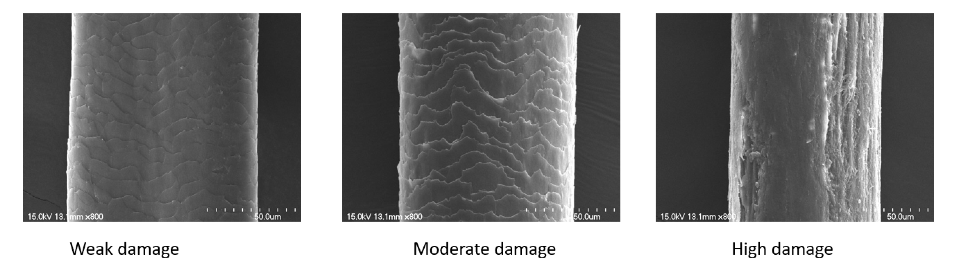
- We created a new hair microscopy data set based on SEM (Scanning Electron Microscope) image data and performed a quantitative analysis to classify the degree of hair damage under the categories: weak damage, moderate damage, and high damage.
- We proposed a novel and effective convolutional network model for hair damage detection: RCSAN-Net (residual channel spatial attention network).
- We designed and introduced a channel and spatial attention mechanism into the hair damage detection model to gather hair features to improve the accuracy of detection and recognition.
2. Related Work
3. Materials and Methods
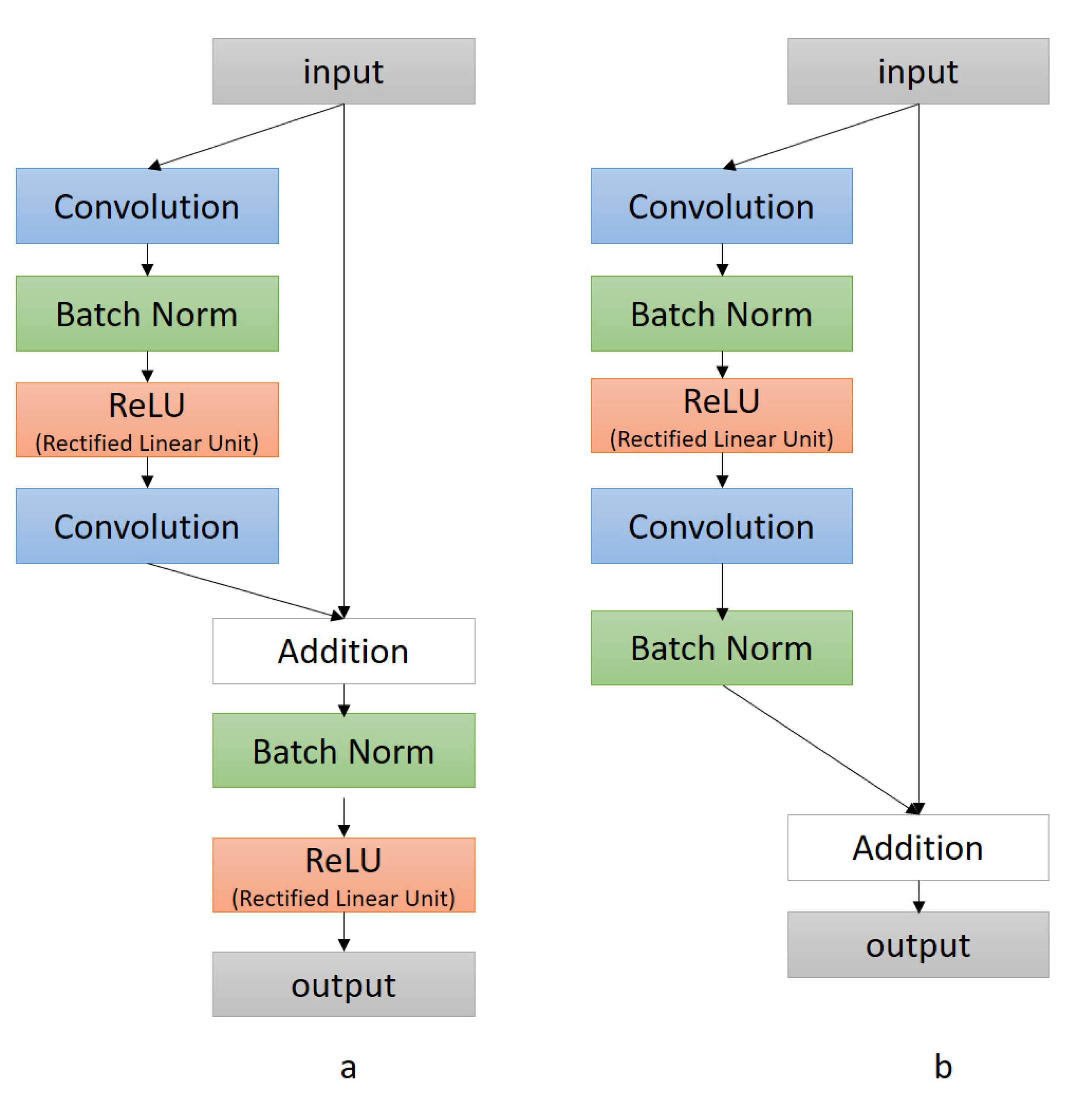
3.1. Convolutional Neural Network
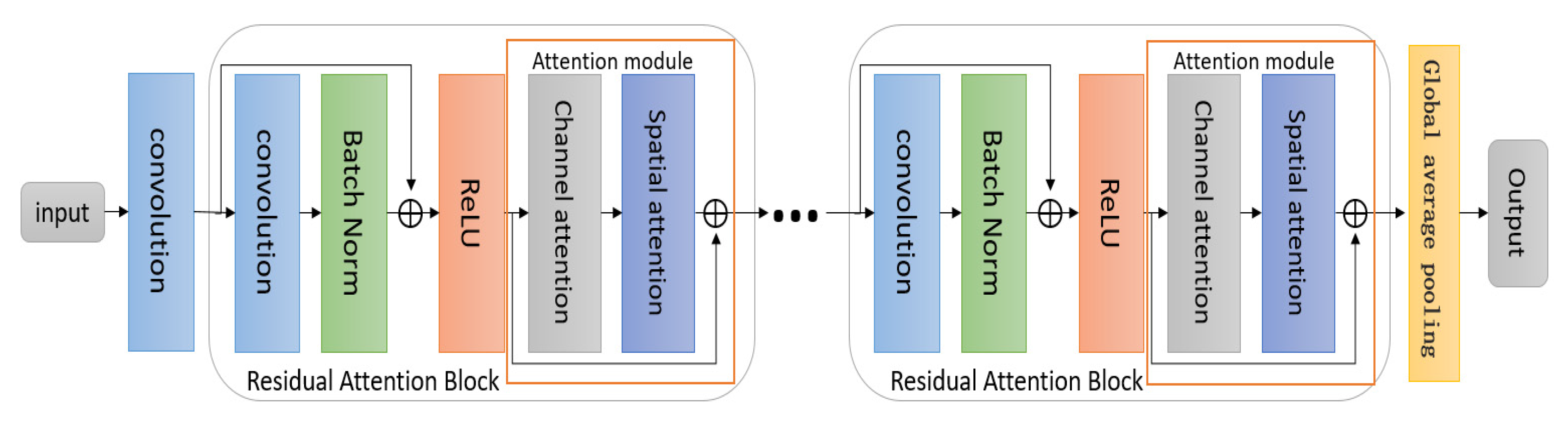
3.2. RCSAN-Net: Residual Channel Spatial Attention Network
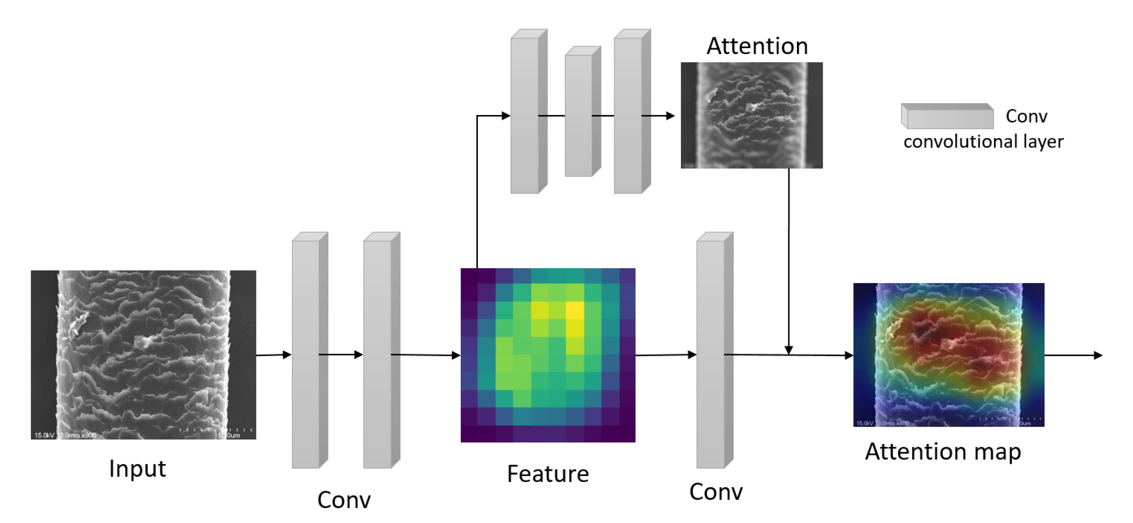
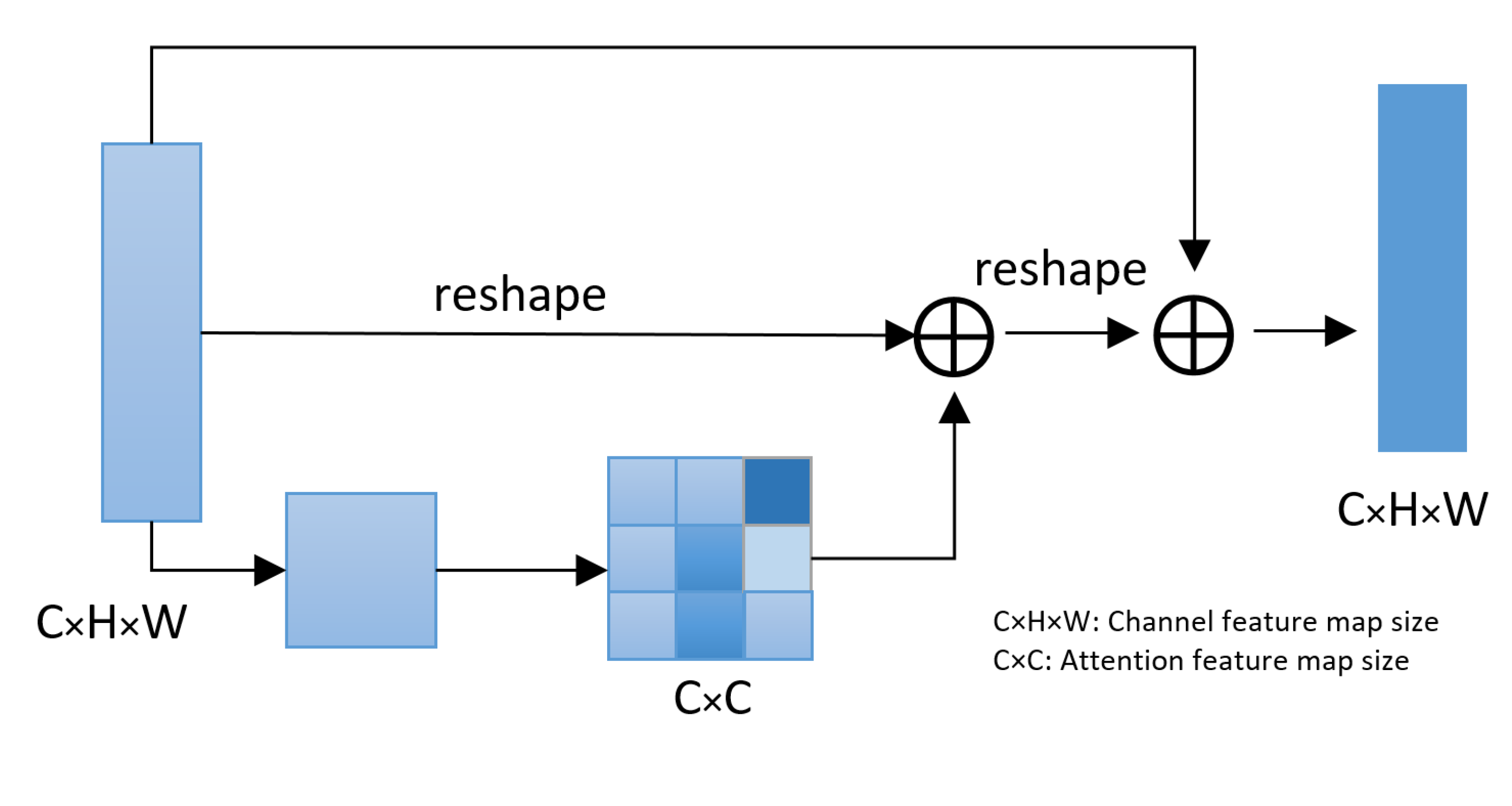
3.3. Attention Mechanism Module
3.3.1. Channel Attention Module
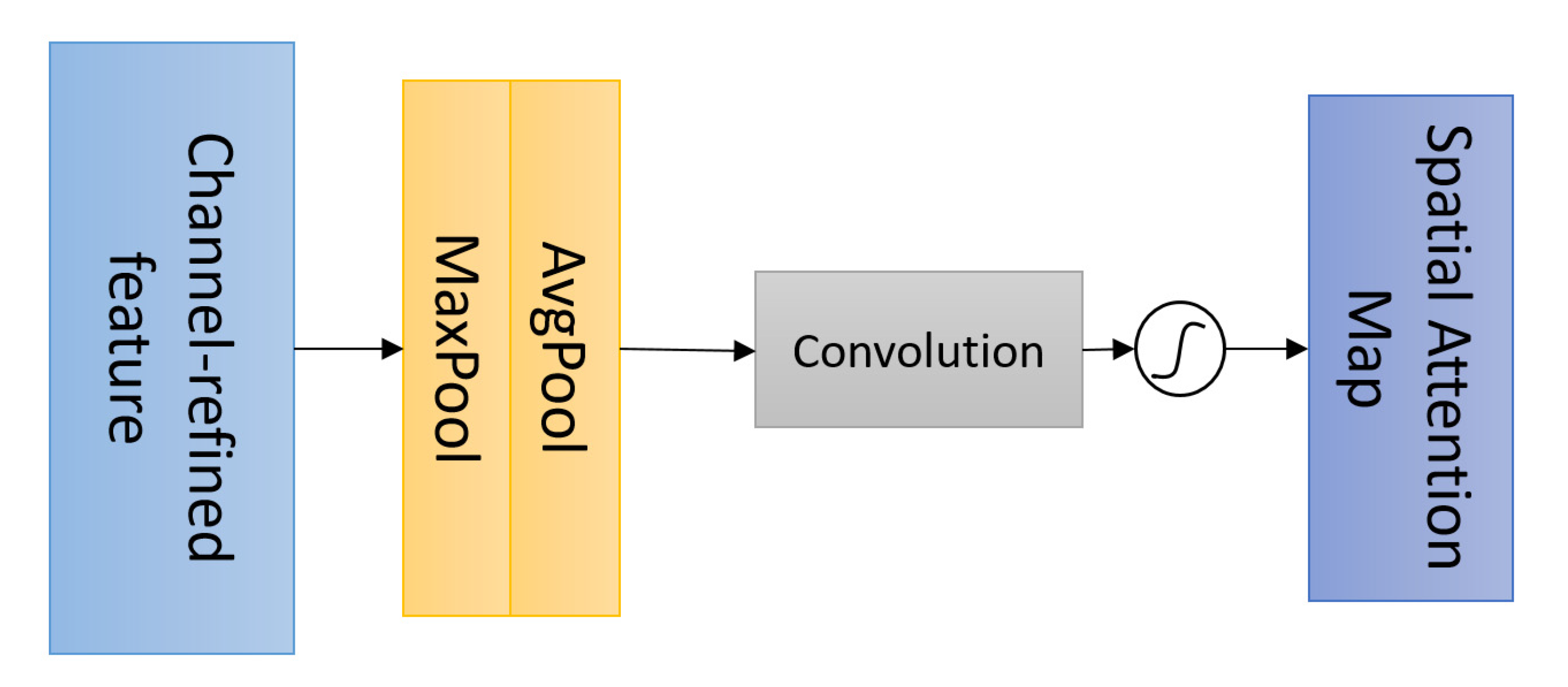
3.3.2. Spatial Attention Module
4. Results
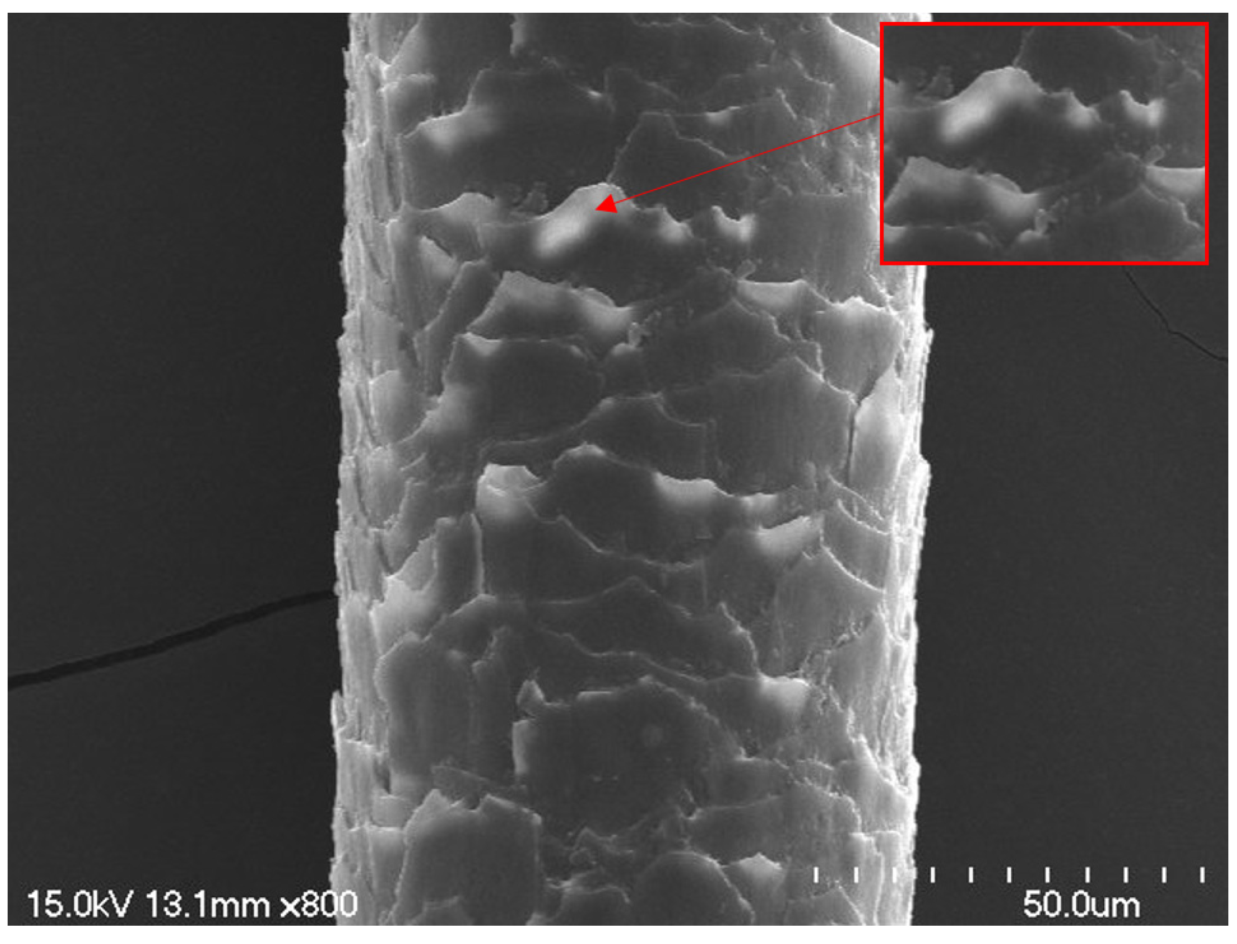
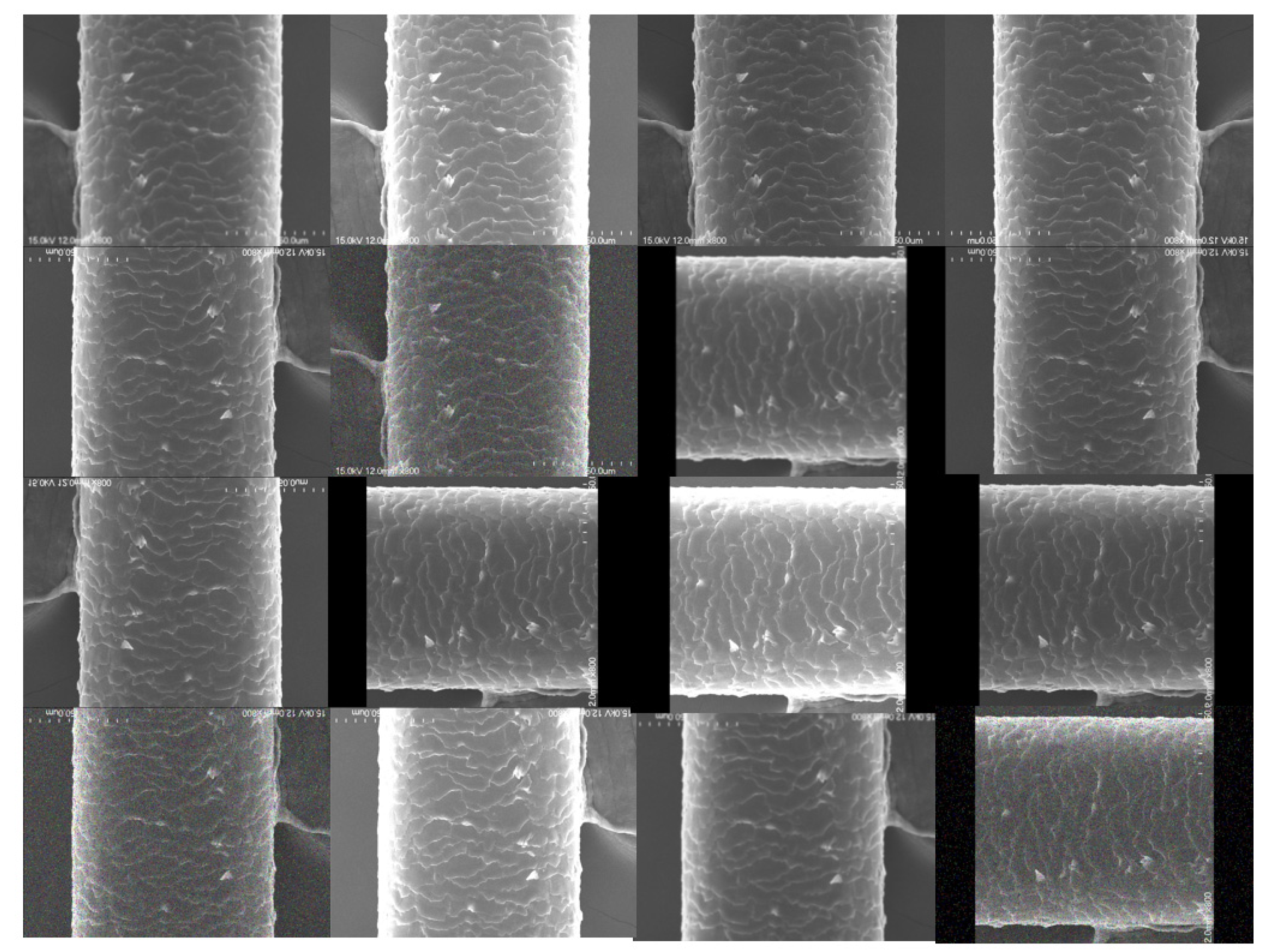
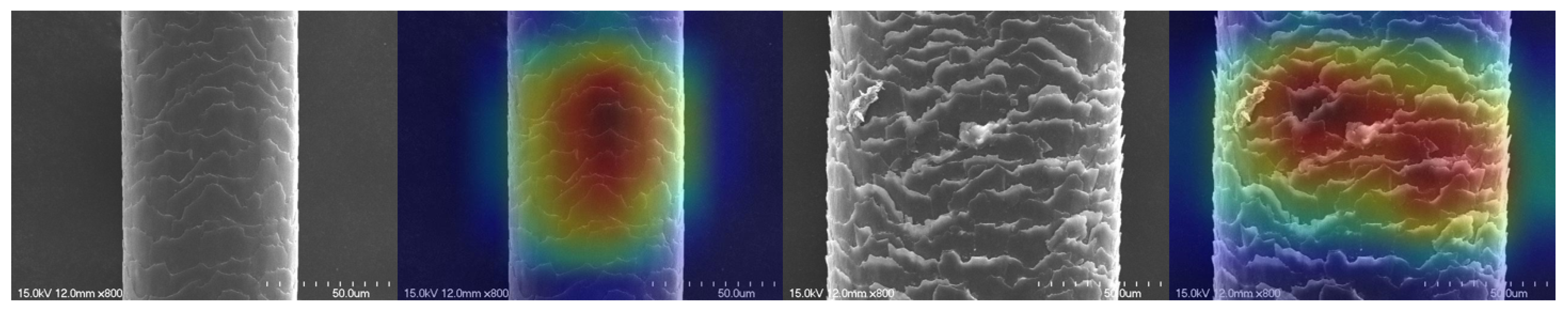
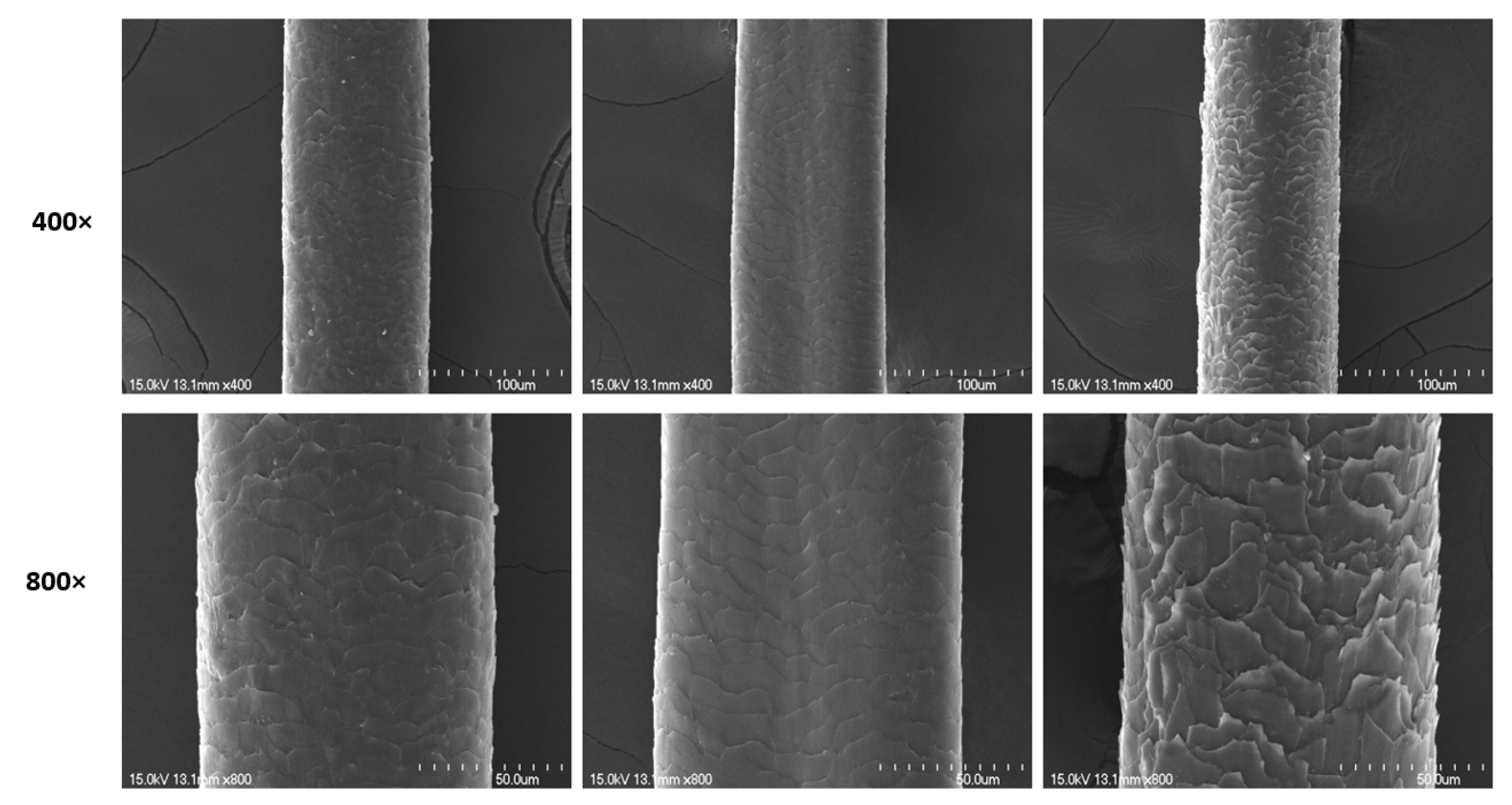
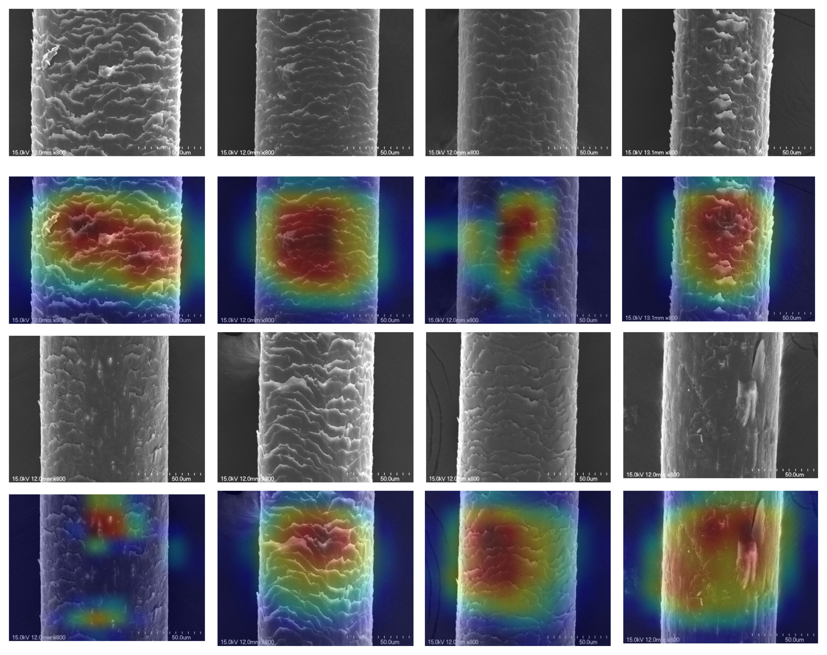
4.1. Datasets
- Five thousand microscopic images of weakly damaged hair.
- Five thousand microscopic images of moderately damaged hair.
- Five thousand microscopic images of highly damaged hair.
4.2. Implementation Details
4.3. Comparison with Other Advanced Methods
5. Discussion
6. Conclusions
Author Contributions
Funding
Institutional Review Board Statement
Informed Consent Statement
Data Availability Statement
Conflicts of Interest
References
- Coroaba, A.; Chiriac, A.E.; Sacarescu, L.; Pinteala, T.; Minea, B.; Ibanescu, S.; Pertea, M.; Moraru, A.; Esanu, I.; Maier, S.S.; et al. New insights into human hair: SAXS, SEM, TEM and EDX for Alopecia Areata investigations. PeerJ 2020, 8, e8376. [Google Scholar] [CrossRef] [Green Version]
- Lima, C.R.R.D.C.; De Couto, R.A.A.; Freire, T.B.; Goshiyama, A.M.; Baby, A.R.; Velasco, M.V.R.; Constantino, V.R.L.; Matos, J.D.R. Heat-damaged evaluation of virgin hair. J. Cosmet. Dermatol. 2019, 18, 1885–1892. [Google Scholar] [CrossRef]
- National Research Council USA. 2009 Strengthening Forensic Science in the United States: A Path Forward; National Academy Press: Wasington, DC, USA, 2009. [Google Scholar]
- Birngruber, C.; Ramsthaler, F.; Verhoff, M.A. The color(s) of human hair—Forensic hair analysis with SpectraCube®. Forensic Sci. Int. 2009, 185, e19–e23. [Google Scholar] [CrossRef]
- Rice, R.H.; Wong, V.J.; Price, V.H.; Hohl, D.; Pinkerton, K.E. Cuticle cell defects in lamellar ichthyosis hair and anomalous hair shaft syndromes visualized after detergent extraction. Anat. Rec. 1996, 246, 433–441. [Google Scholar] [CrossRef]
- Zhang, Y.; Alsop, R.J.; Soomro, A.; Yang, F.-C.; Rheinstädter, M.C. Effect of shampoo, conditioner and permanent waving on the molecular structure of human hair. PeerJ 2015, 3, e1296. [Google Scholar] [CrossRef] [PubMed] [Green Version]
- Richena, M.; Rezende, C.A. Effect of photodamage on the outermost cuticle layer of human hair. J. Photochem. Photobiol. B Biol. 2015, 153, 296–304. [Google Scholar] [CrossRef] [PubMed]
- Takada, K.; Nakamura, A.; Matsuo, N.; Inoue, A.; Someya, K.; Shimogaki, H. Influence of oxidative and/or reductive treatment on human hair (I): Analysis of hair-damage after oxidative and/or reductive treatment. J. Oleo Sci. 2003, 52, 541–548. [Google Scholar] [CrossRef] [Green Version]
- Lee, Y.; Kim, Y.-D.; Hyun, H.-J.; Pi, L.-Q.; Jin, X.; Lee, W.-S. Hair Shaft Damage from Heat and Drying Time of Hair Dryer. Ann. Dermatol. 2011, 23, 455–462. [Google Scholar] [CrossRef] [PubMed] [Green Version]
- Kaliyadan, F.; Gosai, B.B.; Al Melhim, W.N.; Feroze, K.; Qureshi, H.A.; Ibrahim, S.; Kuruvilla, J. Scanning electron microscopy study of hair shaft damage secondary to cosmetic treatments of the hair. Int. J. Trichol. 2016, 8, 94–98. [Google Scholar] [CrossRef]
- McMullen, R.L. Image analysis tools to quantify visual properties of hair fiber assemblies. In Practical Modern Hair Science; Evans, T., Wickett, R.R., Eds.; Allured Publishing: Carol Stream, IL, USA, 2012; pp. 295–332. [Google Scholar]
- Ahn, H.J.; Lee, W.-S. An ultrastuctural study of hair fiber damage and restoration following treatment with permanent hair dye. Int. J. Dermatol. 2002, 41, 88–92. [Google Scholar] [CrossRef]
- Kim, Y.-D.; Jeon, S.-Y.; Ji, J.H.; Lee, W.-S. Development of a classification system for extrinsic hair damage: Standard grading of electron microscopic findings of damaged hairs. Am. J. Dermatopathol. 2010, 32, 432–438. [Google Scholar] [CrossRef]
- Lee, S.Y.; Choi, A.R.; Baek, J.H.; Kim, H.O.; Shin, M.K.; Koh, J.S. Twelve-point scale grading system of scanning electron microscopic examination to investigate subtle changes in damaged hair surface. Skin Res. Technol. 2016, 22, 406–411. [Google Scholar] [CrossRef]
- Verma, M.S.; Pratt, L.; Ganesh, C.; Medina, C. Hair-MAP: A prototype automated system for forensic hair comparison and analysis. Forensic Sci. Int. 2002, 129, 168–186. [Google Scholar] [CrossRef]
- Park, K.H.; Kim, H.J.; Oh, B.; Lee, E.; Ha, J. Assessment of hair surface roughness using quantitative image analysis. Skin Res. Technol. 2018, 24, 80–84. [Google Scholar] [CrossRef] [Green Version]
- Gerace, E.; Veronesi, A.; Martra, G.; Salomone, A.; Vincenti, M. Study of cocaine incorporation in hair damaged by cosmetic treatments. Forensic Chem. 2017, 3, 69–73. [Google Scholar] [CrossRef]
- Muhammad, U.R.; Svanera, M.; Leonardi, R.; Benini, S. Hair detection, segmentation, and hairstyle classification in the wild. Image Vis. Comput. 2018, 71, 25–37. [Google Scholar] [CrossRef] [Green Version]
- Chang, W.-J.; Chen, L.-B.; Chen, M.-C.; Chiu, Y.-C.; Lin, J.-Y. ScalpEye: A Deep Learning-Based Scalp Hair Inspection and Diagnosis System for Scalp Health. IEEE Access 2020, 8, 134826–134837. [Google Scholar] [CrossRef]
- Xiaojia, J.; Mengjing, Y.; Yongzhi, Q.; Ya, H. Hair Microscopic Image Classification Method Based on Convolutional Neural Network. In Proceedings of the 2019 IEEE International Conference on Power, Intelligent Computing and Systems (ICPICS), Shenyang, China, 12–14 July 2019; pp. 433–438. [Google Scholar]
- LeCun, Y.; Bottou, L.; Bengio, Y.; Haffner, P. Gradient-based learning applied to document recognition. Proc. IEEE 1998, 86, 2278–2324. [Google Scholar] [CrossRef] [Green Version]
- Krizhevsky, A.; Sutskever, I.; Hinton, G.E. Imagenet classification with deep convolutional neural networks. Adv. Neural Inf. Process. Syst. 2012, 25, 1097–1105. [Google Scholar] [CrossRef]
- Deng, J.; Dong, W.; Socher, R.; Li, L.; Li, K.; Li, F.-F. ImageNet: A large-scale hierarchical image database. In Proceedings of the 2009 IEEE Conference on Computer Vision and Pattern Recognition, Miami, FL, USA, 20–25 June 2009; pp. 248–255. [Google Scholar] [CrossRef] [Green Version]
- Simonyan, K.; Zisserman, A. Very deep convolutional networks for large-scale image recognition. arXiv 2014, arXiv:1409.1556. [Google Scholar]
- Szegedy, C.; Liu, W.; Jia, Y.; Sermanet, P.; Reed, S.; Anguelov, D.; Erhan, D.; Vanhoucke, V.; Rabinovich, A. Going deeper with convolutions. In Proceedings of the Conference on Computer Vision and Pattern Recognition (CVPR), Honolulu, HI, USA, 21–26 July 2017. [Google Scholar] [CrossRef] [Green Version]
- Targ, S.; Almeida, D.; Lyman, K. Resnet in Resnet: Generalizing Residual Architectures. arXiv 2016, arXiv:abs/1603.08029. [Google Scholar]
- He, K.; Zhang, X.; Ren, S.; Sun, J. Identity Mappings in Deep Residual Networks. In Proceedings of the 14th European Conference on Computer Vision, Amsterdam, The Netherlands, 11–14 October 2016; pp. 630–645. [Google Scholar]
- He, K.; Zhang, X.; Ren, S.; Sun, J. Deep residual learning for image recognition. In Proceedings of the IEEE Conference on Computer Vision and Pattern Recognition (CVPR), Las Vegas, NV, USA, 26 June–1 July 2016. [Google Scholar]
- Chen, L.-C.; Yang, Y.; Wang, J.; Xu, W.; Yuille, A.L. Attention to scale: Scale-aware semantic image segmentation. arXiv 2015, arXiv:1511.03339. [Google Scholar]
- Jaderberg, M.; Simonyan, K.; Zisserman, A. Spatial transformer networks. Adv. Neural Inf. Process. Syst. 2015, 28, 2017–2025. [Google Scholar]
- Zinkevich, M.; Weimer, M.; Li, L.; Smola, A. Parallelized stochastic gradient descent. In Proceedings of the Advances in Neural Information Processing Systems, Vancouver, BC, Canada, 6–9 December 2010. [Google Scholar]
- Sutskever, I.; Martens, J.; Dahl, G.; Hinton, G. On the importance of initialization and momentum in deep learning. In Proceedings of the International Conference on Machine Learning, Atlanta, GA, USA, 16–21 June 2013; pp. 1139–1147. [Google Scholar]


| Layer | Output Size | Attention |
|---|---|---|
| Conv1 | 112 × 112 | 7 × 7, 64, stride 2 |
| Max pooling | 56 × 56 | 3 × 3 stride 2 |
| (1 × 1, 64) | ||
| Residual Unit | 56 × 56 | (3 × 3, 64) × 1 |
| (1 × 1, 256) | ||
| Attention Module | 56 × 56 | Attention × 1 |
| (1 × 1, 128) | ||
| Residual Unit | 28 × 28 | (3 × 3, 128) × 1 |
| (1 × 1, 512) | ||
| Attention Module | 28 × 28 | Attention × 1 |
| (1 × 1, 256) | ||
| Residual Unit | 14 × 14 | (3 × 3, 256) × 1 |
| (1 × 1, 1024) | ||
| Attention Module | 14 × 14 | Attention x1 |
| (1 × 1, 512) | ||
| Residual Unit | 7 × 7 | (3 × 3, 512) × 3 |
| (1 × 1, 2048) | ||
| Global average pooling | 1 × 1 | 7 × 7 stride 1 |
| Mode | Accuracy |
|---|---|
| AlexNet | 0.8310 |
| VGG16 | 0.9053 |
| Inception | 0.9239 |
| MobileNet | 0.9143 |
| ResNet34 | 0.9255 |
| ResNet50 | 0.9474 |
| SACN-Net | 0.9838 |
| Attention Type | Top-1 Err. (%) |
|---|---|
| Channel Attention | 5.03 |
| Spatial Attention | 4.25 |
| Channel & Spatial Attention | 2.92 |
| Spatial & Channel Attention | 2.47 |
Publisher’s Note: MDPI stays neutral with regard to jurisdictional claims in published maps and institutional affiliations. |
© 2021 by the authors. Licensee MDPI, Basel, Switzerland. This article is an open access article distributed under the terms and conditions of the Creative Commons Attribution (CC BY) license (https://creativecommons.org/licenses/by/4.0/).
Share and Cite
Man, Q.; Zhang, L.; Cho, Y. Efficient Hair Damage Detection Using SEM Images Based on Convolutional Neural Network. Appl. Sci. 2021, 11, 7333. https://doi.org/10.3390/app11167333
Man Q, Zhang L, Cho Y. Efficient Hair Damage Detection Using SEM Images Based on Convolutional Neural Network. Applied Sciences. 2021; 11(16):7333. https://doi.org/10.3390/app11167333
Chicago/Turabian StyleMan, Qiaoyue, Lintong Zhang, and Youngim Cho. 2021. "Efficient Hair Damage Detection Using SEM Images Based on Convolutional Neural Network" Applied Sciences 11, no. 16: 7333. https://doi.org/10.3390/app11167333