Article

Cartilage Repair Activity during Joint-Preserving Treatment May Be Accompanied by Osteophyte Formation

by
Mylène P. Jansen
1,*,
Simon C. Mastbergen
1,
Fiona E. Watt
2,
Elske J. Willemse
1,
Tonia L. Vincent
2,
Sander Spruijt
3,
Pieter J. Emans
4,
Roel J. H. Custers
5,
Ronald J. van Heerwaarden
6 and
Floris P. J. G. Lafeber
1
1
Department of Rheumatology & Clinical Immunology, University Medical Center Utrecht, 3584CX Utrecht, The Netherlands
2
Arthritis Research UK Centre for Osteoarthritis Pathogenesis, Kennedy Institute of Rheumatology, Nuffield Department of Orthopaedics, Rheumatology and Musculoskeletal Sciences, University of Oxford, Oxford OX3 7FY, UK
3
Reinier Haga Orthopaedic Centre, 2725NA Zoetermeer, The Netherlands
4
Department of Orthopedics, Maastricht University Medical Center, 6229HX Maastricht, The Netherlands
5
Department of Orthopedic Surgery, University Medical Center Utrecht, 3584CX Utrecht, The Netherlands
6
Centre for Deformity Correction and Joint Preserving Surgery, Kliniek ViaSana, 5451AA Mill, The Netherlands
*
Author to whom correspondence should be addressed.
Appl. Sci. 2021, 11(15), 7156; https://doi.org/10.3390/app11157156
Submission received: 8 July 2021 / Revised: 29 July 2021 / Accepted: 1 August 2021 / Published: 3 August 2021

Abstract

:
Knee joint distraction (KJD) treatment has shown cartilage repair and clinical improvement in patients with osteoarthritis, as has high tibial osteotomy (HTO). Following KJD, TGFβ-1 and IL-6 were increased in synovial fluid (SF), factors related to cartilage regeneration, but also to osteophyte formation. As such, osteophyte formation after both joint-preserving treatments was studied. Radiographic osteophyte size was measured before, one year, and two years after treatment. Changes were compared with natural progression in patients from the CHECK cohort before undergoing total knee arthroplasty. An additional KJD cohort underwent SF aspiration, and one-year Altman osteophyte score changes were compared to SF-marker changes during treatment. After two years, both KJD (n = 58) and HTO (n = 38) patients showed an increase in osteophyte size (+6.2 mm2 and +7.0 mm2 resp.; both p < 0.004), with no significant differences between treatments (p = 0.592). Untreated CHECK patients (n = 44) did not show significant two-year changes (+2.1 mm2; p = 0.207) and showed significant differences with KJD and HTO (both p < 0.044). In SF aspiration patients (n = 17), there were significant differences in TGFβ-1 changes (p = 0.044), but not IL-6 (p = 0.898), between patients with a decrease, no change, or increase in osteophyte Altman score. Since KJD and HTO showed joint space widening and clinical improvement accompanied by osteophyte formation, increased osteophytosis after joint-preserving treatments may be a bystander effect of cartilage repair activity related to intra-articular factors like TGFβ-1 and raises questions regarding osteophyte formation as solely characteristic of the joint degenerative process.

1. Introduction

Osteoarthritis (OA) is characterized by articular cartilage loss, intra-articular inflammation, and osteophyte formation [1]. Osteophytes are often formed at the joint margins, first as cartilage outgrowth and subsequently undergoing ossification [2]. While the exact purpose of osteophytes remains unknown, their presence and size in the knee are associated with joint space width (JSW) decrease, and they are an important radiographic feature used to define the severity of knee OA in classifications like the Altman score and Kellgren–Lawrence grade [3,4,5,6,7]. Osteophytes are frequently present in patients with end-stage knee OA receiving surgical treatment such as total knee arthroplasty (TKA) [8].
TKA is widely used because of its clinical effectiveness, but in younger patients (<65 years) it has a significantly higher risk of failure and revision surgery later in life [9,10]. Therefore, there is a demand for joint-preserving treatments for (severe) knee OA at a younger age. A joint-preserving alternative for patients with unicompartmental knee OA as a result of malalignment is high tibial osteotomy (HTO), which shows good long-term results and clinical improvement and a certain degree of cartilage repair [11,12,13]. Knee joint distraction (KJD) is a relatively new joint-preserving treatment for patients with unicompartmental or generalized severe knee OA, where the tibia and femur are temporarily separated using an external fixation frame [14]. An open prospective study (OPS) has shown good long-term treatment results, and two randomized controlled trials (RCTs), one comparing KJD with HTO and one with TKA, showed that clinical outcome after KJD is comparable to that after HTO or TKA [15,16,17,18,19,20]. Furthermore, cartilage repair has been shown on radiographs and on MRI scans, and systemic biomarker analyses suggest beneficial cartilage and bone turnover after KJD treatment [18,21,22,23].
Cartilage repair activity as a result of treatment could be related to an increase in transforming growth factor-β1 (TGFβ-1), which is generally appreciated to stimulate cartilage repair [24]. During KJD treatment, an increase in synovial fluid TGFβ-1 level was observed [25]. While TGFβ-1 is associated with joint repair, it has also been shown to induce osteophyte formation, predominantly in experimental animal studies, but in ex vivo human studies as well [26,27,28,29,30,31]. Interleukin-6 (IL-6) was also observed to increase intra-articularly as a result of KJD treatment and could be positively associated with osteophyte presence as well, showing increased mRNA expression and protein production in in vitro studies with human osteophyte tissue [25,30,32].
As such, we studied osteophyte formation during KJD and compared this to HTO and natural OA progression, hypothesizing that joint-preserving regenerative treatments demonstrating cartilage repair activity lead to tissue (re)generation in general, including osteophyte formation.

2. Materials and Methods

2.1. Knee Joint Distraction Patients

63 Patients were included for KJD treatment in three different trials. Of these 63 patients, 20 patients with an indication for TKA and age < 60 years old were included in the OPS. Secondly, 20 TKA-indicated patients < 65 years old were treated with KJD in an RCT comparing KJD with TKA. The third and last group of 23 patients with medial compartmental knee OA, an indication for HTO, and age < 65 years were treated with KJD in an RCT comparing KJD with HTO. Inclusion and exclusion criteria have been described before and included radiographic signs of tibiofemoral OA (Kellgren-Lawrence grade > 2, judged by orthopedic surgeon), <10° knee malalignment, body mass index (BMI) < 35, and no presence or history of inflammatory or septic arthritis [33,34] All trials complied with the Declaration of Helsinki, were granted ethical approval by the medical ethical review committee of the University Medical Center Utrecht (protocol numbers 04/086, 10/359/E, and 11/072), and were registered in the Netherlands Trial Register (trial numbers NL419, NL2761 and NL2680). All patients gave written informed consent.
Distraction surgery was performed using an external fixation frame. The knee was distracted 2 mm during surgery and 1 mm every day during a short hospitalization until 5 mm distraction was reached, confirmed on radiographs. Patients were discharged with prophylactic anticoagulant to use during treatment and were allowed full weight-bearing of the treated knee, supported by crutches if necessary. After 6–8 weeks the distraction frame and pins were surgically removed.

2.2. Follow-Up

Patients visited the hospital multiple times, including at baseline and one and two years after treatment, during which standardized weight-bearing, semiflexed posterior–anterior radiographs were performed according to the Buckland-Wright protocol, using an aluminum step wedge as a reference standard for image analysis using ‘knee images digital analysis’ (KIDA) software (described below) [35,36]. Patients completed the Western Ontario and McMaster Universities Osteoarthritis Index (WOMAC, version 3.1) questionnaire as well. Only patients with standardized radiographs at both baseline and 2 years were included.

2.3. High Tibial Osteotomy Patients

HTO patients from the KJD vs. HTO trial were used to study generalizability of the concept of osteophyte formation during regenerative treatments demonstrating endogenous cartilage repair activity. The 46 HTO patients were included in the trial to be treated with biplane medial-based opening-wedge osteotomy and had the same follow-up as described above for KJD patients. Only patients with standardized baseline and 2-year radiographs were included in the analyses. The HTO patients were compared to the 23 KJD patients (KJDHTO) from the RCT comparing KJD and HTO.

2.4. Control Group of Untreated OA Patients

The only relevant OA cohort using the same standardized radiographic analyses, with quantification of osteophyte area, is CHECK (Cohort Hip & Cohort Knee), a cohort of 1002 participants with early symptomatic knee or hip OA who were followed for 10 years and had radiographs of both knees at baseline, 2, 5, 8, and 10 years follow-up [37]. From this cohort, patients that received a TKA during the follow-up period were selected to be compared with KJD patients, since most KJD patients were indicated for TKA but received KJD. For each knee that was treated with TKA in CHECK, all pre-TKA radiographic osteophyte measurements were analyzed to evaluate the linearity of osteophyte formation using a linear regression model, with osteophyte size as a dependent variable and the ‘years before TKA’ and ‘years before TKA squared’ as independent variables. The change in osteophyte area during the last two measurements before TKA, corrected to represent a 2-year period, was used as control for the osteophyte progression rate. WOMAC questionnaires from the last time point before TKA and 2 years prior were used to evaluate 2-year clinical changes.

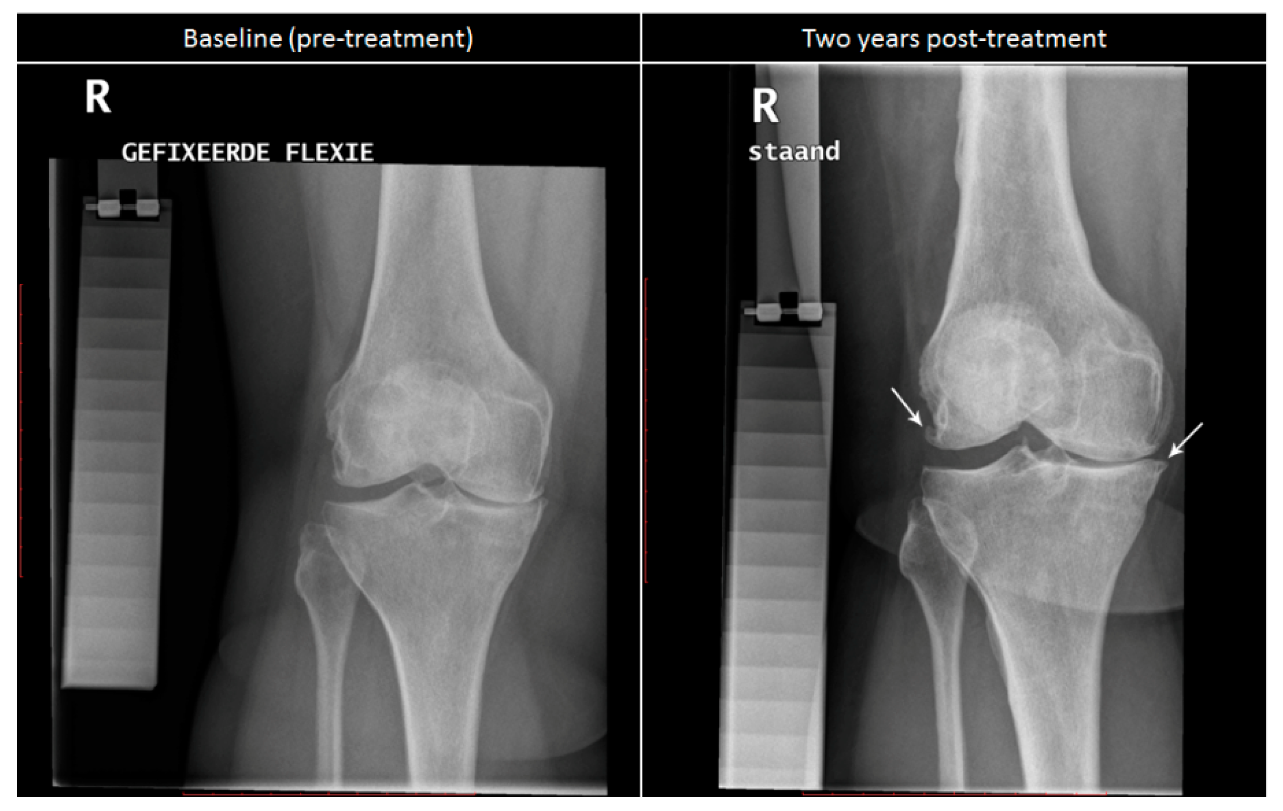
2.5. Radiographic Analysis

The standardized radiographs were analyzed by one experienced observer, blinded to patient characteristics, using KIDA software [38]. The osteophyte size (area on the 2D image) was measured in mm2 for four regions: the lateral and medial femur and tibia. The sum of these regions gives the whole-joint osteophyte size in mm2. The mean JSW of the most affected compartment (MAC; determined pre-treatment) in mm provided by the KIDA measurement was evaluated as a representative of the cartilage-regenerative activity of the treatment. In CHECK, the compartment with the smallest JSW was chosen as MAC.

2.6. Synovial Fluid Aspirations

Between 2014 and 2015, 20 patients treated with KJD in regular care were included for synovial fluid (SF) aspirations in an ethically approved study (protocol number 15/160). The treatment protocol and inclusion and exclusion criteria in regular care were similar, as explained above, and have been described elsewhere [39], with the addition that patients in this study needed to have a successful baseline SF aspiration. At baseline (during frame placement surgery) and after treatment (during frame removal surgery), an SF sample of maximum 2 mL was aspirated from the treated knee. Biomarker levels were measured according to protocols described previously [25]. In short, samples were centrifuged for 20 min at 3000 G and stored in 200 μL aliquots at −80 °C. The supernatants were measured by immunoassay for 10 predefined mechanosensitive molecules; mean analyte concentrations were calculated from duplicate assay reads for each participant and time point. For the present evaluation, only TGFβ-1 and IL-6 were used as predefined potential candidates for association with osteophyte formation as only those have been related to osteophyte formation in literature. For TGFβ-1 analysis, the human TGFβ-1 quantikine ELISA assay (R&D; DB100B) was used, and for IL-6 analysis, the V-PLEX custom human cytokine assay (MSD; K151A0H-1) was used; both were carried out according to the manufacturers’ instructions.
As no standardized (KIDA) radiographs were available in these SF patients, radiographs taken in regular care at baseline and around one year after treatment (range 276–433 days) were used to score osteophytes using the revised Altman score [6]. The correlation between Altman and KIDA in KJD RCT patients was tested and showed to be moderately good (R = 0.669; p < 0.001; Supplementary Table S1). All images were scored for osteophytes in each of the four regions twice by one observer (SM), giving each compartment a grade from 0 (normal) to 3 (severe). The average of both scores was used, and due to the wide follow-up range, the follow-up radiograph was linearly corrected (extrapolated) to 365 days with respect to the baseline radiograph. The separate compartment scores were summarized to obtain a 0–12 whole-joint scoring. Only patients with baseline and 1-year follow-up radiographs were included in the analyses.

2.7. Statistical Analysis

For all continuous parameters, changes over time for separate patient groups were analyzed using paired t-tests or, where more than 2 time points were available, repeated measures ANOVA. The influence of available predefined patient characteristics (age, gender, BMI, and Kellgren-Lawrence grade) on osteophyte formation was tested with linear regression. For comparisons with the control groups, linear regression was used, correcting for baseline values. For comparisons where in both groups more than two time points were available, mixed ANOVA was used instead.
In SF patients, for the categorical Altman score per region, the Wilcoxon Signed Rank test was used to test changes over time The changes in whole-joint osteophyte Altman score and in synovial fluid biomarkers were analyzed with paired t-tests, and the Pearson correlations between total joint osteophyte Altman score and biomarker baseline values and changes over time were calculated. Finally, SF patients were divided into three groups (trichotomized) based on an increase, no change, or decrease in total osteophyte Altman score over time. The change in TGFβ-1 and in IL-6 during the distraction period was compared between these three groups using a Kruskal-Wallis test because of the limited number of patients per group.
Normal distribution was verified for all outcome parameters; in case outcomes were not normally distributed, log transformation was performed. For all tests, a p-value < 0.05 was considered statistically significant. Absolute values are presented with mean ± standard deviation (SD), while changes over time are presented as mean change and 95% confidence interval (95%CI).

3. Results

3.1. Patients

Of all KJD patients, 1 was excluded before surgery due to inoperability, 3 were lost to follow-up after receiving a different surgical treatment during follow-up, and 1 did not have a standardized baseline radiograph, leaving 58 KJD patients for analysis, of whom 20 were in the KJDHTO group. Of the HTO patients, 1 was excluded before treatment due to anxiety while 4 were lost to follow-up due to comorbidities. Five did not have standardized radiographs at both baseline and 2-year follow-up, leaving 36 HTO patients. In CHECK, 30 patients received a TKA during the 10-year follow-up, 14 of whom had a TKA in both knees, giving 44 knees to be compared to the KJD patients. Three of the 20 patients with SF aspirations did not have both a baseline and follow-up radiograph available, leaving 17 SF patients. The baseline characteristics of all groups are shown in Table 1. These characteristics are shown for male and female patients separately in Supplementary Table S2.

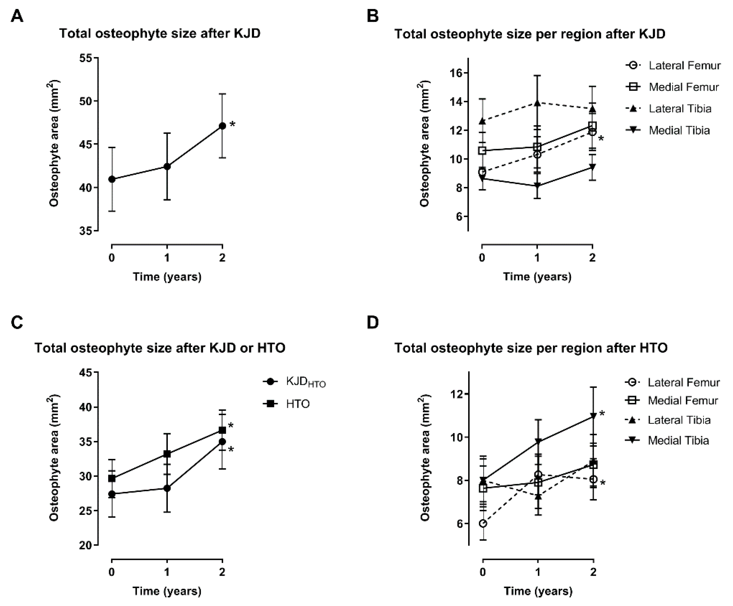
3.2. Changes after Knee Joint Distraction

As shown in Table 2, the total WOMAC showed significant improvement 2 years after KJD (+28.1; 95%CI 22.7-33.4; p < 0.001), as did its subscales. The mean MAC radiographic JSW was significantly increased at 2 years as well (+0.66; 95%CI 0.36–0.97; p < 0.001).
The total osteophyte size showed a statistically significant increase after treatment (p = 0.003), from 40.9 (SD 28.0) mm2 at baseline to 47.1 (28.1) mm2 at 2 years (Figure 1A). Only the lateral femur showed a significant increase (from 9.1 (9.4) mm2 to 11.9 (9.8) mm2; p < 0.001), the other compartments did not (all p ≥ 0.19; Figure 1B).
A representative radiograph of a patient before and 2 years after KJD treatment is shown in Figure 2, here showing an increased osteophyte size on the lateral femur and medial tibia over 2 years.
None of the baseline characteristics, including gender, had a significant influence on the 2-year change in osteophyte size (all p > 0.32; Supplementary Table S3). As such, no separate analyses for both genders, nor for other different groups of patients based on these characteristics, were performed.

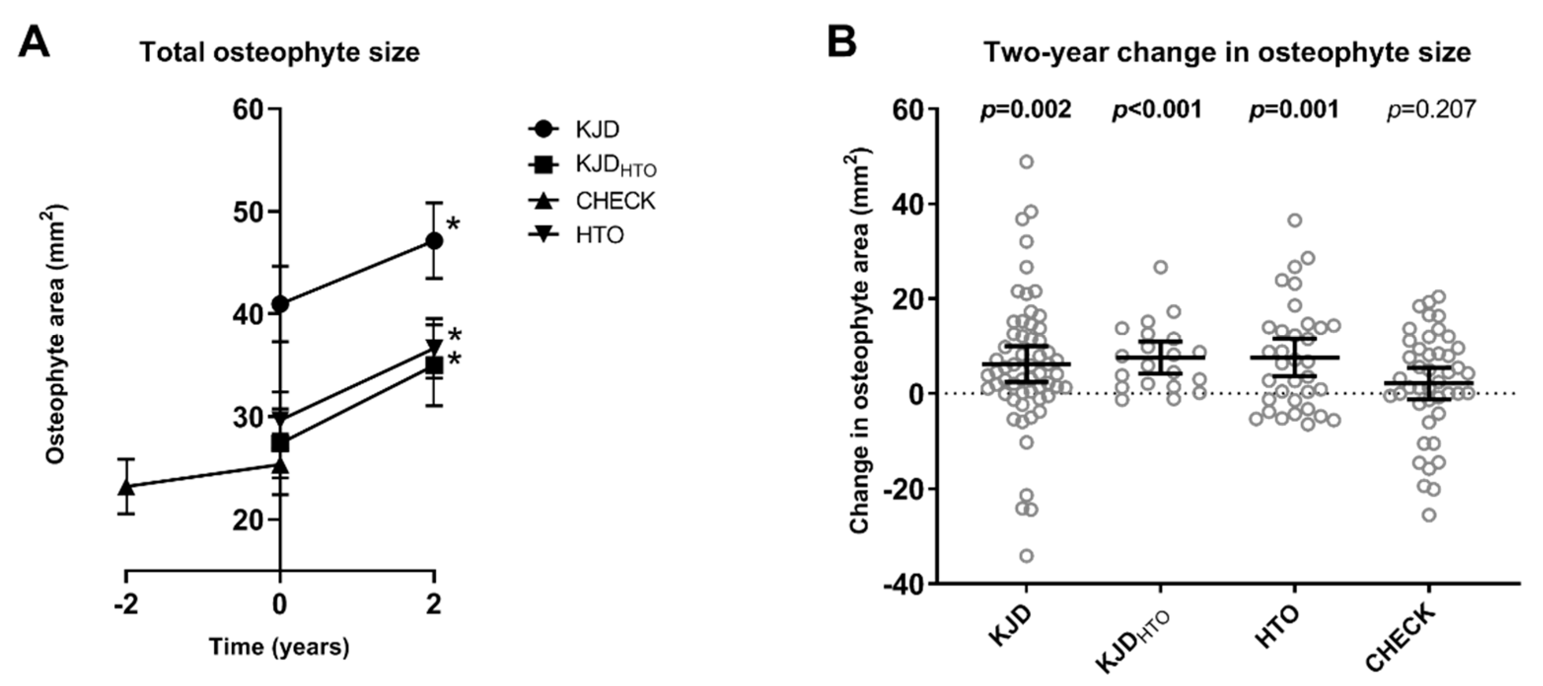
3.3. Comparison with High Tibial Osteotomy

HTO patients showed a significant increase in total WOMAC (+30.8; 95%CI 25.5–36.1; p < 0.001) and in MAC JSW (+0.32; 0.03–0.61; p = 0.034) as shown in Table 2. The WOMAC subscales showed a similar increase.
HTO patients showed a significant osteophyte change after treatment (p < 0.001), increasing from 29.6 (SD 16.5) mm2 at baseline to 36.6 (17.4) mm2 at two years (Figure 1C). The changes in the lateral femur (6.0 (4.6) mm2 to 8.1 (5.7) mm2; p < 0.001) and medial tibia (8.0 (6.7) mm2 to 11.0 (8.2) mm2; p = 0.006) were statistically significant (Figure 1D). Like the entire KJD cohort, the KJDHTO patients showed a significant increase after treatment (from 27.4 (15.0) mm2 to 35.0 (17.6) mm2; p < 0.001), and only the lateral femur showed a significant increase (from 4.6 (SD 3.8) mm2 to 8.1 (4.5) mm2; p = 0.006; Supplementary Figure S1). There was no significant difference between KJDHTO and the other KJD patients for the total osteophyte changes over 2 years (p = 0.566). There was no significant difference in the osteophyte changes between HTO and KJDHTO (p = 0.592; Figure 1C).

3.4. Comparison with Untreated OA Patients

In the 44 knees that received a TKA in CHECK, 124 KIDA measurements were available in the years before the TKA, which were used to confirm a linear approach to osteophyte change over time could be assumed, as the variable ‘years to TKA squared’ did not contribute significantly to the linear regression model predicting osteophyte size (p = 0.759). CHECK patients showed a significant decrease in total WOMAC (−6.3; 95%CI −12.1–−0.5; p = 0.04) and MAC JSW (−0.67; 95%CI −0.86–−0.47; p < 0.001) before undergoing TKA (Table 2).
Before TKA, CHECK knees showed a small nonsignificant increase in osteophyte size (+2.1 mm2; 95%CI −1.2–5.5; p = 0.207; Figure 3A). Correcting for baseline osteophyte size, all KJD patients together (p = 0.027), KJDHTO patients (p = 0.043), and HTO patients (p = 0.027) showed a significantly greater osteophyte increase than CHECK patients prior to TKA. Taking the average of both knees in patients with a TKA in both knees, instead of using the knees separately, did not change significance. Figure 3B displays the 2-year changes in total osteophyte size for the different groups.

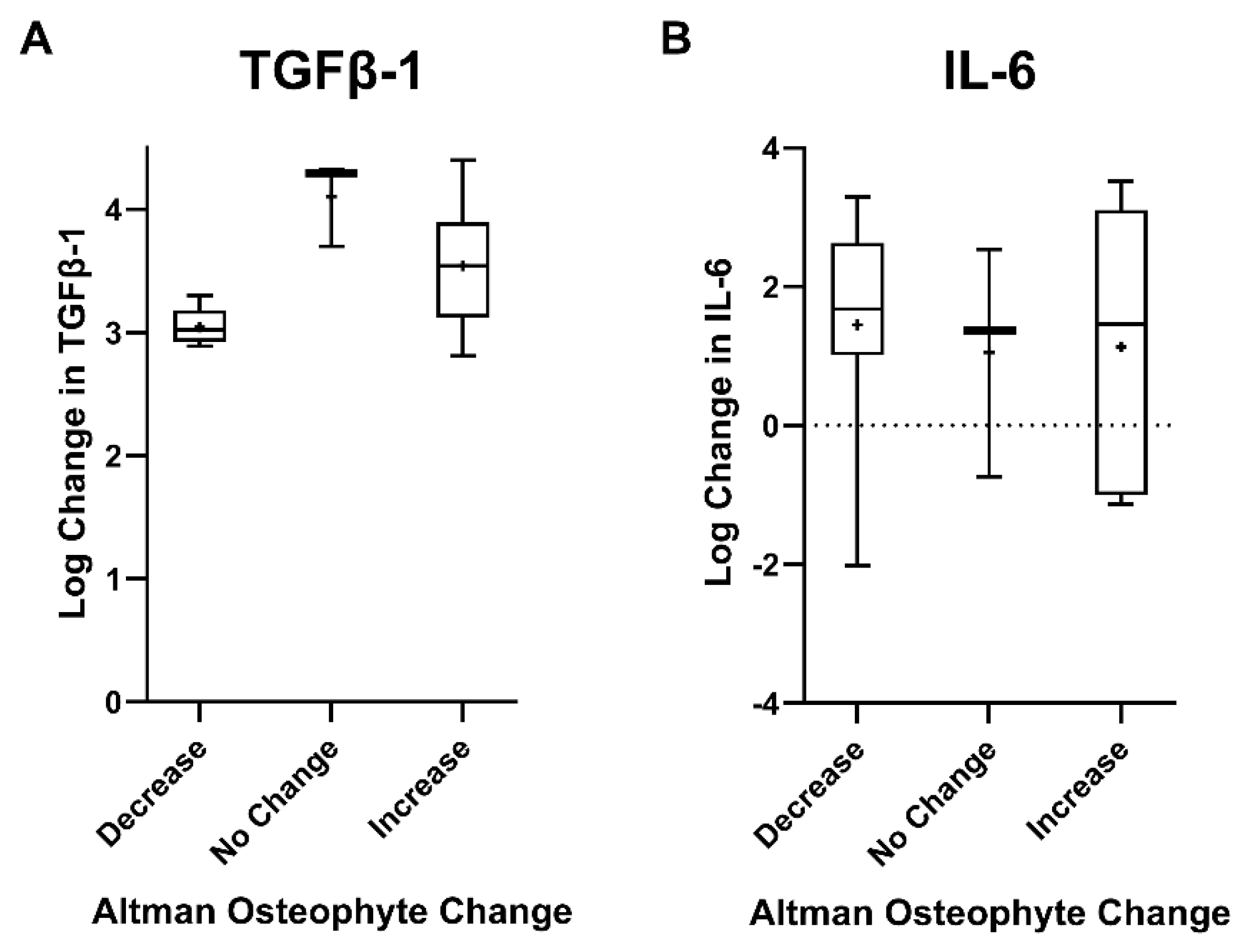
3.5. Relation with Synovial Fluid Markers

None of the four osteophyte locations showed statistically significant 1-year changes in Altman score compared to baseline in the SF patients (all p > 0.074; Supplementary Table S4). The total Altman osteophyte score summarized for the entire joint was at 1 year not different from baseline, increasing with 0.2 points (95%CI −0.6–0.9; p = 0.653). As the biomarker changes were not normally distributed, they were log transformed. In case of negative change values, the log transformation of the absolute change was subtracted from zero. Two patients did not have biomarker results after treatment, and one did not have a baseline value for TGFβ-1, leaving fourteen patients for TGFβ-1 analysis and fifteen for IL-6 analysis. Both biomarkers showed statistically significant changes during the distraction period, as shown previously [25]: TGFβ-1 (1527.9 ± 3346.8 to 8027.9 ± 10,534.8 pg/mL; p < 0.001); IL-6 (24.4 ± 31.3 to 466.3 ± 936.4 pg/mL; p = 0.011). There was no apparent association between baseline values of these biomarkers and the baseline total Altman osteophyte score, or between the changes in these parameters (all p ≥ 0.28; Supplementary Table S5). Trichotomization of patients in groups with a decrease (n = 5), no change (n = 3), or increase (n = 6 for TGFβ-1; n = 7 for IL-6) in total Altman osteophyte score showed there was a statistically significant difference in changes in analyte levels during treatment between the three groups for TGFβ-1 (p = 0.044), but not for IL-6 (p = 0.898), as shown in Figure 4.

4. Discussion

Based on radiographic measurement, using sensitive image analyses like KIDA, KJD seems to induce increased osteophyte formation in the first two years following treatment. This argues against the general assumption that osteophytosis is solely a hallmark of OA worsening or joint degeneration, since this osteophytosis during KJD is combined with a significant increase in clinical benefit and joint space widening (supported in previous studies by MRI cartilage volume measurements [21,22,33]). Increased osteophyte presence has often been associated with increased pain in knee OA patients [40,41,42], but with improvement in clinical outcome for KJD patients, including a significant decrease in pain, parallel with an increase in osteophyte size. No correlation could be found between (changes in) osteophyte size and WOMAC scores or JSW (except between baseline JSW and osteophyte size, see Supplementary Tables S6 and S7), expectedly due to limited numbers.
Treatment-related osteophyte formation is not limited to KJD, but is demonstrated after HTO as well. HTO patients were compared with KJDHTO patients, since those groups were randomized as such in the original RCT, and showed similar osteophyte formation. While KJDHTO patients showed similar results as the entire KJD group, their baseline osteophyte size was smaller and more comparable with the HTO group. This is likely because while KJDHTO patients who were in regular care indicated for a HTO, all other KJD patients were in regular care indicated for TKA and thus likely had further progressed OA. Nevertheless, both treatments showed changes predominantly in the lateral compartment. While in HTO patients this might be explained by an increased load on the lateral side as a result of the medial unloading, such a shift is not necessarily expected in KJD. Since HTO showed an osteophyte increase on the medial side as well, loading may not be directly involved in osteophyte formation after these treatments. Like in KJD, osteophyte formation in HTO accompanied clinical improvement and JSW increase, further questioning the role of osteophytes in OA. Other studies have shown similar findings, showing that lateral osteophyte presence is not associated with lateral cartilage degeneration or with medial knee OA severity [43,44,45]. Our findings suggest that the presence, size and localization of osteophytes may not be such a clear indication of joint degeneration and accompanying symptoms as is generally assumed.
With the analysis of untreated patients from the CHECK cohort it was shown that the increase in osteophytes after KJD was greater than the natural progression that can be expected in knee OA patients. It should be noted however that, despite making a selection of patients that received TKA during follow-up, the CHECK patients differed in baseline characteristics and seemed to have less severe OA at the moment of treatment (TKA) than the KJD patients, as shown by Kellgren-Lawrence grade and osteophyte size. This might be related to the specific characteristics of this CHECK cohort where pain was an essential inclusion criterion, and might be irrelevant for comparison with KJD or HTO, as in none of the groups did the baseline osteophyte size or Kellgren-Lawrence grade have a significant influence on the change in osteophyte size (CHECK: p = 0.391 and p = 0.457, respectively).
For patients who had SF aspirations, the osteophyte formation after KJD seems to be associated with the increase in TGFβ-1 during the six weeks of treatment, based on dividing patients into groups showing an increase, no change, or a decrease in Altman osteophyte score after KJD. However, there were no associations between the (changes in) actual Altman scores and TGFβ-1 values. These results are as such indicative and not conclusive, corroborating the reported role of TGFβ-1 in osteophyte formation. While both TGFβ-1 and IL-6 significantly increased during treatment, the change in IL-6 was not associated with osteophyte formation.
This study has several limitations. First, the different cohorts were not initiated and powered for the presented statistical evaluations and should therefore be considered exploratory. Second, retrospectively comparing patient cohorts that have not been randomized or carefully matched, as was done when comparing KJD patients with CHECK, provides a risk for coincidental findings. Despite selecting the most relevant subgroup from CHECK, there was a clear difference in OA severity with KJD patients. Although the comparison between KJD and CHECK was corrected for baseline osteophyte size and the Kellgren-Lawrence grade was shown to not be an influence on the change in osteophyte size, it could still be that the results in CHECK patients underestimate the natural progression in more severe knee OA patients. CHECK was used since it was a well-established cohort of untreated knee OA patients of which radiographs were evaluated with KIDA, but patients generally had mild OA. Patients with a more comparable severity would make a better comparison, although purposefully not treating severe knee OA patients for multiple years would be ethically unsound.
Another limitation was the fact that no KIDA evaluations were available for the SF patient group. The Altman osteophyte score may not have been sensitive enough to show one-year changes in osteophyte size after KJD, especially in this small group of patients. As TGFβ-1 has previously been associated with both cartilage repair and osteophyte formation, morphometric MRI scans, and/or 3D CT scans in sufficient numbers of patients could be of added value in future studies. The present study provides an indication that a rise in TGFβ-1 might be a mediator in tissue repair activity upon KJD leading to osteophyte formation in addition to cartilage repair, but future studies would have to proof this concept.

5. Conclusions

In conclusion, KJD is accompanied by osteophytosis occurring in parallel with radiographic joint space widening and clinical improvement, including significant pain relief. Similarly, HTO is accompanied by osteophytosis as well. The osteophyte formation during joint-preserving treatments with observed endogenous cartilage repair activity seems to be a bystander effect and may be related to a change in intra-articular anabolic factors such as TGFβ-1. This observation argues against osteophytosis as solely a key parameter in the joint degenerative process.

Supplementary Materials

The following are available online at https://www.mdpi.com/article/10.3390/app11157156/s1, Table S1: Pearson correlation coefficients between the osteophyte Altman scores and the osteophyte size as measured by KIDA, Table S2: Baseline characteristics of the different patient groups for male and female patients separately, Table S3: Influence of baseline characteristics on two-year osteophyte change after knee joint distraction treatment, Table S4: Pearson correlations between baseline osteophyte size and baseline WOMAC and joint space width, Table S5: Pearson correlations between one- and two-year changes in osteophyte size and WOMAC and joint space width, Table S6: Baseline and one-year Altman scores for patients with synovial fluid aspirations, Table S7: Pearson correlations between baseline total Altman score and TGFβ-1 and IL-6, and between one-year changes, Figure S1: Change in osteophyte size in mm2 per region before and one and two years after treatment with knee joint distraction, for patients indicated for high tibial osteotomy (KJDHTO, n = 20).

Author Contributions

Planning and design of (parts of) the study, M.P.J., S.C.M., F.E.W., T.L.V., F.P.J.G.L.; data acquisition, analysis and interpretation, M.P.J., S.C.M., F.E.W., E.J.W., S.S., P.J.E., R.J.H.C., R.J.v.H.; drafting of the article: M.P.J.; critical revision of the article: S.C.M., F.E.W., E.J.W., T.L.V., S.S., P.J.E., R.J.H.C., R.J.v.H., F.P.J.G.L. All authors have read and agreed to the published version of the manuscript.

Funding

Parts of this work were supported by the Dutch Arthritis Association (ReumaNederland, grant numbers LLP-9/ISP14-3-301/16-1-404), ZonMw (The Netherlands Organisation for Health Research and Development, grant number 95110008), Versus Arthritis project (grant number 7837), Centre for OA Pathogenesis Versus Arthritis (grant numbers 20205/21621) and the Kennedy Trust for Rheumatology Research. The funders had no role in the study design, data collection, analysis or interpretation of the data, or in the writing of, or decision to submit the manuscript.

Institutional Review Board Statement

All trials complied with the Declaration of Helsinki and were granted ethical approval by the medical ethical review committee of the University Medical Center Utrecht (protocol numbers 04/086, 10/359/E, and 11/072).

Informed Consent Statement

Written informed consent was obtained from all subjects involved in the study.

Data Availability Statement

Data related to this manuscript cannot be shared publicly because of ethical restrictions related to participant consent. These restrictions are imposed by the institutional review board of the University Medical Center Utrecht, Utrecht, The Netherlands. All relevant data are available upon request by sending an email to the Rheumatology department of the UMC Utrecht (urrci@umcutrecht.nl).

Acknowledgments

The authors wish to thank Benjamin Hamid for his contribution to the synovial fluid biomarker analyses. The CHECK-cohort study is funded by the Dutch Arthritis Foundation. Involved are: Erasmus Medical Center Rotterdam; Kennemer Gasthuis Haarlem; Leiden University Medical Center; Maastricht University Medical Center; Martini Hospital Groningen/Allied Health Care Center for Rheumatology and Rehabilitation Groningen; Medical Spectrum Twente Enschede/Ziekenhuisgroep Twente Almelo; Reade Center for Rehabilitation and Rheumatology; St.Maartens-kliniek Nijmegen; University Medical Center Utrecht and Wilhelmina Hospital Assen.

Conflicts of Interest

FPL was co-founder of ArthroSave BV with no further relation and is consultant for SynerkinePharma BV. The other authors have no potential conflicts of interest to disclose.

References

  1. Woolf, A.; Pfleger, B. Burden of major musculoskeletal conditions. Bull. World Health Organ. 2003, 81, 646–656. [Google Scholar]
  2. Van der Kraan, P.M.; van den Berg, W.B. Osteophytes: Relevance and biology. Osteoarthr. Cartil. 2007, 15, 237–244. [Google Scholar] [CrossRef] [Green Version]
  3. Felson, D.T.; Gale, D.R.; Gale, M.E.; Niu, J.; Hunter, D.J.; Goggins, J.; LaValley, M. Osteophytes and progression of knee osteoarthritis. Rheumatology 2005, 44, 100–104. [Google Scholar] [CrossRef] [Green Version]
  4. Dieppe, P.A.; Cushnaghan, J.; Shepstone, L. The Bristol ‘OA500′ Study: Progression of osteoarthritis (OA) over 3 years and the relationship between clinical and radiographic changes at the knee joint. Osteoarthr. Cartil. 1997, 5, 87–97. [Google Scholar] [CrossRef] [Green Version]
  5. Braun, H.J.; Gold, G.E. Diagnosis of osteoarthritis: Imaging. Bone 2012, 51, 278–288. [Google Scholar] [CrossRef] [Green Version]
  6. Altman, R.; Gold, G. Atlas of individual radiographic features in osteoarthritis, revised. Osteoarthr. Cartil. 2007, 15, A1–A56. [Google Scholar] [CrossRef] [PubMed] [Green Version]
  7. Kellgren, J.H.; Lawrence, J.S. Radiological Assessment of Osteo-Arthrosis. Ann. Rheum. Dis. 1957, 16, 494–502. [Google Scholar] [CrossRef] [PubMed] [Green Version]
  8. Nevalainen, M.T.; Kauppinen, K.; Pylväläinen, J.; Pamilo, K.; Pesola, M.; Haapea, M.; Koski, J.; Saarakkala, S. Ultrasonography of the late-stage knee osteoarthritis prior to total knee arthroplasty: Comparison of the ultrasonographic, radiographic and in-tra-operative findings. Sci. Rep. 2018, 8, 17742. [Google Scholar] [CrossRef] [PubMed]
  9. Lützner, J.; Kasten, P.; Günther, K.-P.; Kirschner, S. Surgical options for patients with osteoarthritis of the knee. Nat. Rev. Rheumatol. 2009, 5, 309–316. [Google Scholar] [CrossRef] [PubMed]
  10. Bayliss, L.E.; Culliford, D.; Monk, A.P.; Glyn-Jones, S.; Prieto-Alhambra, D.; Judge, A.; Cooper, C.; Carr, A.J.; Arden, N.K.; Beard, D.J.; et al. The effect of patient age at interven-tion on risk of implant revision after total replacement of the hip or knee: A population-based cohort study. Lancet 2017, 389, 1424–1430. [Google Scholar] [CrossRef] [Green Version]
  11. Dean, C.S.; Liechti, D.J.; Chahla, J.; Moatshe, G.; LaPrade, R.F. Clinical Outcomes of High Tibial Osteotomy for Knee Instabil-ity: A Systematic Review. Orthop. J. Sport. Med. 2016, 4, 2325967116633419. [Google Scholar]
  12. Kim, C.-W.; Seo, S.-S.; Lee, C.-R.; Gwak, H.-C.; Kim, J.-H.; Jung, S.-G. Factors affecting articular cartilage repair after open-wedge high tibial osteotomy. Knee 2017, 24, 1099–1107. [Google Scholar] [CrossRef] [PubMed]
  13. Kim, J.-H.; Kim, H.-J.; Lee, D.-H. Survival of opening versus closing wedge high tibial osteotomy: A meta-analysis. Sci. Rep. 2017, 7, 1–7. [Google Scholar] [CrossRef] [Green Version]
  14. Lafeber, F.P.J.G.; Intema, F.; Van Roermund, P.M.; Marijnissen, A.C. Unloading joints to treat osteoarthritis, including joint distraction. Curr. Opin. Rheumatol. 2006, 18, 519–525. [Google Scholar] [CrossRef] [PubMed]
  15. Jansen, M.P.; van der Weiden, G.S.; Van Roermund, P.M.; Custers, R.J.; Mastbergen, S.C.; Lafeber, F.P. Initial tissue repair predicts long-term clinical success of knee joint distraction as treatment for knee osteoarthritis. Osteoarthr. Cartil. 2018, 26, 1604–1608. [Google Scholar] [CrossRef] [Green Version]
  16. Van Der Woude, J.A.D.; Wiegant, K.; Van Heerwaarden, R.J.; Spruijt, S.; Emans, P.J.; Mastbergen, S.C.; Lafeber, F.P.J.G. Knee joint distraction compared with total knee arthroplasty a randomised controlled trial. Bone Jt. J. 2017, 99, 51–58. [Google Scholar] [CrossRef]
  17. Van der Woude, J.A.D.; Wiegant, K.; Van Heerwaarden, R.J.; Spruijt, S.; Van Roermund, P.M.; Custers, R.J.H.; Mastbergen, S.C.; Lafeber, F.P.J.G. Knee joint dis-traction compared with high tibial osteotomy: A randomized controlled trial. Knee Surg. Sports Traumatol. Arthrosc. 2017, 25, 876–886. [Google Scholar] [CrossRef] [Green Version]
  18. Jansen, M.P.; Besselink, N.J.; van Heerwaarden, R.J.; Custers, R.J.H.; Emans, P.J.; Spruijt, S.; Mastbergen, S.C.; Lafeber, F.P.J.G. Knee Joint Distraction Compared with High Tibial Osteotomy and Total Knee Arthroplasty: Two-Year Clinical, Radiographic, and Biochemical Marker Outcomes of Two Randomized Controlled Trials. Cartilage 2019, 12, 181–191. [Google Scholar] [CrossRef] [Green Version]
  19. Jansen, M.P.; Boymans, T.A.; Custers, R.J.; Van Geenen, R.C.; Van Heerwaarden, R.J.; Huizinga, M.R.; Nellensteijn, J.M.; Sollie, R.; Spruijt, S.; Mastbergen, S.C. Knee Joint Distraction as Treatment for Osteoarthritis Results in Clinical and Structural Benefit: A Systematic Review and Meta-Analysis of the Limited Number of Studies and Patients Available. Cartilage 2020. submitted. [Google Scholar] [CrossRef]
  20. Hoorntje, A.; Kuijer, P.P.F.M.; Koenraadt, K.L.M.; Waterval-Witjes, S.; Kerkhoffs, G.M.M.J.; Mastbergen, S.C.; Marijnissen, A.C.A.; Jansen, M.; Van Geenen, R.C.I. Return to Sport and Work after Randomization for Knee Distraction versus High Tibial Osteotomy: Is There a Difference? J. Knee Surg. 2020. [Google Scholar] [CrossRef]
  21. Wiegant, K.; van Roermund, P.; Intema, F.; Cotofana, S.; Eckstein, F.; Mastbergen, S.; Lafeber, F. Sustained clinical and structural benefit after joint distraction in the treatment of severe knee osteoarthritis. Osteoarthr. Cartil. 2013, 21, 1660–1667. [Google Scholar] [CrossRef] [PubMed] [Green Version]
  22. Van der Woude, J.T.A.; Wiegant, K.; Van Roermund, P.M.; Intema, F.; Custers, R.J.; Eckstein, F.; van Laar, J.M.; Mastbergen, S.C.; Lafeber, F.P. Five-Year Follow-up of Knee Joint Distraction: Clinical Benefit and Cartilaginous Tissue Repair in an Open Uncontrolled Prospective Study. Cartilage 2017, 8, 263–271. [Google Scholar] [CrossRef]
  23. Jansen, M.P.; Maschek, S.; van Heerwaarden, R.J.; Mastbergen, S.C.; Wirth, W.; Lafeber, F.P.J.G.; Eckstein, F. Changes in Cartilage Thickness and Denuded Bone Area after Knee Joint Distraction and High Tibial Osteotomy—Post-Hoc Analyses of Two Randomized Controlled Trials. J. Clin. Med. 2021, 10, 368. [Google Scholar] [CrossRef]
  24. Tuan, R.S.; Chen, A.F.; Klatt, B.A. Cartilage regeneration. J. Am. Acad. Orthop. Surg. 2013, 21, 303–311. [Google Scholar] [CrossRef] [PubMed]
  25. Watt, F.; Hamid, B.; Garriga, C.; Judge, A.; Hrusecka, R.; Custers, R.; Jansen, M.; Lafeber, F.; Mastbergen, S.; Vincent, T. The molecular profile of synovial fluid changes upon joint distraction and is associated with clinical response in knee osteoarthritis. Osteoarthr. Cartil. 2020, 28, 324–333. [Google Scholar] [CrossRef] [Green Version]
  26. Blaney Davidson, E.N.; van der Kraan, P.M.; van den Berg, W.B. TGF-β and osteoarthritis. Osteoarthr. Cartil. 2007, 15, 597–604. [Google Scholar] [CrossRef] [Green Version]
  27. Bakker, A.C.; Van De Loo, F.A.J.; Van Beuningen, H.M.; Sime, P.; Van Lent, P.L.E.M.; Van Der Kraan, P.M.; Richards, C.D.; Van Den Berg, W.B. Overexpression of ac-tive TGF-beta-1 in the murine knee joint: Evidence for synovial-layer-dependent chondro-osteophyte formation. Osteoarthr. Cartil. 2001, 9, 128–136. [Google Scholar] [CrossRef] [PubMed] [Green Version]
  28. Scharstuhl, A.; Glansbeek, H.L.; van Beuningen, H.M.; Vitters, E.L.; van der Kraan, P.M.; van den Berg, W.B. Inhibition of En-dogenous TGF-β During Experimental Osteoarthritis Prevents Osteophyte Formation and Impairs Cartilage Repair. J. Immunol. 2002, 169, 507–514. [Google Scholar] [CrossRef] [PubMed] [Green Version]
  29. Van Beuningen, H.M.; van der Kraan, P.M.; Arntz, O.J.; van den Berg, W.B. Transforming growth factor-beta 1 stimulates ar-ticular chondrocyte proteoglycan synthesis and induces osteophyte formation in the murine knee joint. Lab. Investig. 1994, 71, 279–290. [Google Scholar]
  30. Dodds, R.A.; Merry, K.; Littlewood, A.; Gowen, M. Expression of mRNA for IL1 beta, IL6 and TGF beta 1 in developing hu-man bone and cartilage. J. Histochem. Cytochem. 1994, 42, 733–744. [Google Scholar] [CrossRef] [PubMed]
  31. Horner, A.; Kemp, P.; Summers, C.; Bord, S.; Bishop, N.; Kelsall, A.; Coleman, N.; Compston, J. Expression and distribution of transforming growth factor-β isoforms and their signaling receptors in growing human bone. Bone 1998, 23, 95–102. [Google Scholar] [CrossRef]
  32. Sakao, K.; Takahashi, K.A.; Arai, Y.; Saito, M.; Honjo, K.; Hiraoka, N.; Asada, H.; Shin-Ya, M.; Imanishi, J.; Mazda, O.; et al. Osteoblasts derived from osteophytes produce in-terleukin-6, interleukin-8, and matrix metalloproteinase-13 in osteoarthritis. J. Bone Miner. Metab. 2009, 27, 412–423. [Google Scholar] [CrossRef] [PubMed]
  33. Intema, F.; Van Roermund, P.M.; Marijnissen, A.C.; Cotofana, S.; Eckstein, F.; Castelein, R.M.; Bijlsma, J.W.; Mastbergen, S.C.; Lafeber, F.P. Tissue structure modifica-tion in knee osteoarthritis by use of joint distraction: An open 1-year pilot study. Ann. Rheum. Dis. 2011, 70, 1441–1446. [Google Scholar] [CrossRef] [PubMed]
  34. Wiegant, K.; van Heerwaarden, R.; van der Woude, J.T.; Custers, R.R.; Emans, P.; Kuchuk, N.; Mastbergen, S.; Lafeber, F.F. Knee Joint Distraction as an Alternative Surgical Treatment for Osteoarthritis: Rationale and Design of two Randomized Controlled Trials (vs High Tibial Osteotomy and Total Knee Prosthesis). Int. J. Orthop. 2015, 2, 353–360. [Google Scholar] [CrossRef]
  35. Buckland-Wright, J.C.; Ward, R.J.; Peterfy, C.; Mojcik, C.F.; Leff, R.L. Reproducibility of the semiflexed (metatarsophalangeal) radiographic knee position and automated measurements of medial tibiofemoral joint space width in a multicenter clinical trial of knee osteoarthritis. J. Rheumatol. 2004, 31, 1588–1597. [Google Scholar]
  36. Buckland-Wright, J.C.; Wolfe, F.; Ward, R.J.; Flowers, N.; Hayne, C. Substantial superiority of semiflexed (MTP) views in knee osteoarthritis: A comparative radiographic study, without fluoroscopy, of standing extended, semiflexed (MTP), and schuss views. J. Rheumatol. 1999, 26, 2664–2674. [Google Scholar]
  37. Wesseling, J.; Boers, M.; Viergever, M.A.; Hilberdink, W.K.; Lafeber, F.P.; Dekker, J.; Bijlsma, J.W. Cohort profile: Cohort Hip and Co-hort Knee (CHECK) study. Int. J. Epidemiol. 2016, 45, 36–44. [Google Scholar] [CrossRef] [PubMed] [Green Version]
  38. Marijnissen, A.; Vincken, K.; Vos, P.; Saris, D.; Viergever, M.; Bijlsma, J.; Bartels, L.; Lafeber, F. Knee Images Digital Analysis (KIDA): A novel method to quantify individual radiographic features of knee osteoarthritis in detail. Osteoarthr. Cartil. 2008, 16, 234–243. [Google Scholar] [CrossRef] [PubMed] [Green Version]
  39. Jansen, M.P.; Mastbergen, S.C.; van Heerwaarden, R.J.; Spruijt, S.; van Empelen, M.D.; Kester, E.C.; Lafeber, F.P.; Custers, R.J. Knee joint distraction in regular care for treatment of knee osteoarthritis: A comparison with clinical trial data. PLoS ONE 2020, 15, e0227975. [Google Scholar] [CrossRef] [PubMed]
  40. O’Reilly, S.C.; Muir, K.R.; Doherty, M.; O’reilly, S.C.; Muir, K.R. Screening for pain in knee osteoarthritis: Which question? Ann Rheum Dis. 1996, 55, 931–933. [Google Scholar] [CrossRef] [Green Version]
  41. Spector, T.D.; Hart, D.J.; Byrne, J.; Harris, P.A.; Dacre, J.E.; Doyle, D.V. Definition of osteoarthritis of the knee for epidemiologi-cal studies. Ann. Rheum. Dis. 1993, 52, 790–794. [Google Scholar] [CrossRef] [PubMed] [Green Version]
  42. Lanyon, P.; O’Reilly, S.; Jones, A.; Doherty, M. Radiographic assessment of symptomatic knee osteoarthritis in the commu-nity: Definitions and normal joint space. Ann. Rheum. Dis. 1998, 57, 595–601. [Google Scholar] [CrossRef]
  43. Hamilton, T.W.; Choudhary, R.; Jenkins, C.; Mellon, S.; Dodd, C.A.F.; Murray, D.W.; Pandit, H.G. Lateral osteophytes do not represent a contraindication to medial unicompartmental knee arthroplasty: A 15-year follow-up. Knee Surg. Sports Traumatol. Arthrosc. 2016, 25, 652–659. [Google Scholar] [CrossRef]
  44. Waldstein, W.; Kasparek, M.F.; Faschingbauer, M.; Windhager, R.; Boettner, F. Lateral-compartment Osteophytes are not As-sociated With Lateral-compartment Cartilage Degeneration in Arthritic Varus Knees. Clin. Orthop. Relat. Res. 2017, 475, 1386–1392. [Google Scholar] [CrossRef] [PubMed] [Green Version]
  45. Teitge, R.A. CORR Insights®: Lateral-compartment Osteophytes are not associated with Lateral-compartment Cartilage Degeneration in Arthritic Varus Knees. Clin. Orthop. Relat. Res. 2017, 475, 1393–1394. [Google Scholar] [CrossRef] [PubMed] [Green Version]
Figure 1. Change in osteophyte size in mm2 before and one and two years after treatment with knee joint distraction (KJD, n = 58) or high tibial osteotomy (HTO, n = 36). (A) The total joint osteophyte area and (B) the osteophyte area per compartment after KJD. (C) Total joint osteophyte area after KJD or HTO and (D) osteophyte area per compartment after HTO. Mean and standard error of the mean (SEM) are shown, * indicates significant changes compared to baseline using repeated measures ANOVA (p < 0.05).
Applsci 11 07156 g001
Figure 2. Representative radiograph of a patient before and two years after knee joint distraction treatment. Note the increase in osteophyte area over the two years as indicated by the arrows.
Applsci 11 07156 g002
Figure 3. Two year changes in total joint osteophyte size in mm2. (A) Osteophyte size after treatment for all patients treated with knee joint distraction (KJD, n = 58), high tibial osteotomy (HTO, n = 36), HTO-indicated KJD patients (KJDHTO, n = 20), and for untreated knee osteoarthritis patients before receiving a total knee arthroplasty (CHECK, n = 44). Mean and standard error of the mean (SEM) are shown, * indicates significant changes (p < 0.05) compared to baseline using paired t-tests. (B) Two-year osteophyte size changes for individual KJD, HTO, KJDHTO and CHECK patients. Mean and 95% confidence interval are shown, p-values above groups indicate significance of two-year changes using paired t-tests (bold values indicating statistical significance).
Applsci 11 07156 g003
Figure 4. Box plots for the changes in synovial fluid concentrations over the course of 6 weeks of knee joint distraction, of (A) transforming growth factor-β1 (TGF-β1) and (B) interleukin-6 (IL-6), categorized into groups of patients with a decrease (n = 5), no change (n = 3) or increase (n = 6 for TGFβ-1; n = 7 for IL-6) in total Altman osteophyte score. The bar represents the median, whiskers represent the minimum and maximum value, the + represents the mean.
Applsci 11 07156 g004
Table 1. Baseline characteristics of the different patient groups.
ParameterKJD
(n = 58)
KJDHTO
(n = 20)
HTO
(n = 36)
CHECK
(n = 44)
SF
(n = 17)
Age, mean (SD)51.4 (8.0)51.2 (5.8)49.1 (6.5)64.0 (4.3)53.8 (4.7)
Male gender, n (%)34 (59)15 (75)23 (64)5 (11)10 (59)
BMI, mean (SD)28.0 (3.4)27.4 (3.3)27.0 (3.5)29.4 (4.6)29.0 (3.3)
Kellgren-Lawrence grade, n (%)
Grade 00 (0)0 (0)1 (3)2 (5)0 (0)
Grade 18 (14)5 (25)4 (11)18 (41)0 (0)
Grade 29 (16)4 (20)10 (28)16 (36)2 (12)
Grade 328 (48)10 (50)18 (50)8 (18)7 (41)
Grade 413 (22)1 (5)3 (8)0 (0)8 (47)
KJD = all knee joint distraction patients with available osteophyte measurements; KJDHTO = subgroup of KJD patients who were included in the KJD vs. HTO clinical trial; HTO = high tibial osteotomy patients from the KJD vs. HTO clinical trial; CHECK = untreated knee osteoarthritis patients from the cohort hip and cohort knee trial who received a total knee arthroplasty during follow-up; SF = KJD patients from a separate clinical study who underwent synovial fluid aspirations.
Table 2. Baseline and two-year WOMAC and JSW for the KJD, HTO and CHECK patient groups.
KJDHTOCHECK
Baseline2 Yearsp-ValueBaseline2 Yearsp-ValueBaseline2 Yearsp-Value
Total WOMAC, mean (SD)50.6 (15.7)78.8 (19.3)<0.00150.7 (14.6)81.5 (14.5)<0.00158.6 (15.9)51.8 (20.3)0.035
JSW, mean (SD)2.36 (1.73)3.03 (1.57)<0.0012.24 (1.28)2.56 (1.37)0.0343.18 (1.76)2.52 (1.72)<0.001
WOMAC = Western Ontario and McMaster Universities Osteoarthritis Index (scale 0–100); JSW = joint space width (mm); KJD = knee joint distraction; HTO = high tibial osteotomy; CHECK = cohort hip & cohort knee. P-values are calculated for two-year changes; bold p-values indicate statistical significance. The mean JSW of the most affected compartment is given.
Publisher’s Note: MDPI stays neutral with regard to jurisdictional claims in published maps and institutional affiliations.

