
Internal Mechanics of a Subject-Specific Wrist in the Sagittal versus Dart-Throwing Motion Plane in Adult and Elder Models: Finite Element Analyses


Abstract
:1. Background
2. Methods
2.1. Wrist Geometry
2.2. Finite Element Modeling and Analysis
2.2.1. Modeling and Meshing
2.2.2. Mechanical Properties
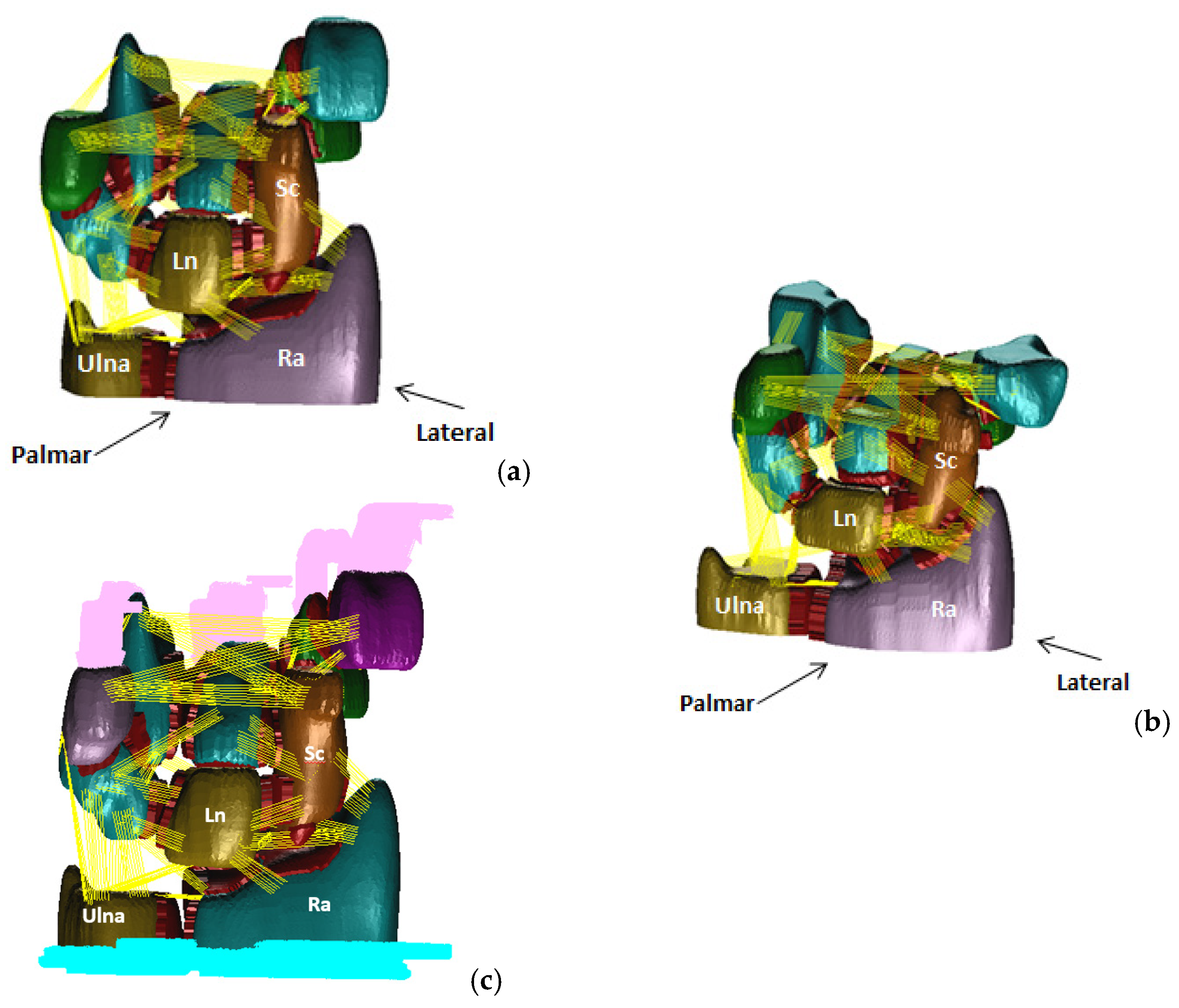
2.2.3. Loading Conditions
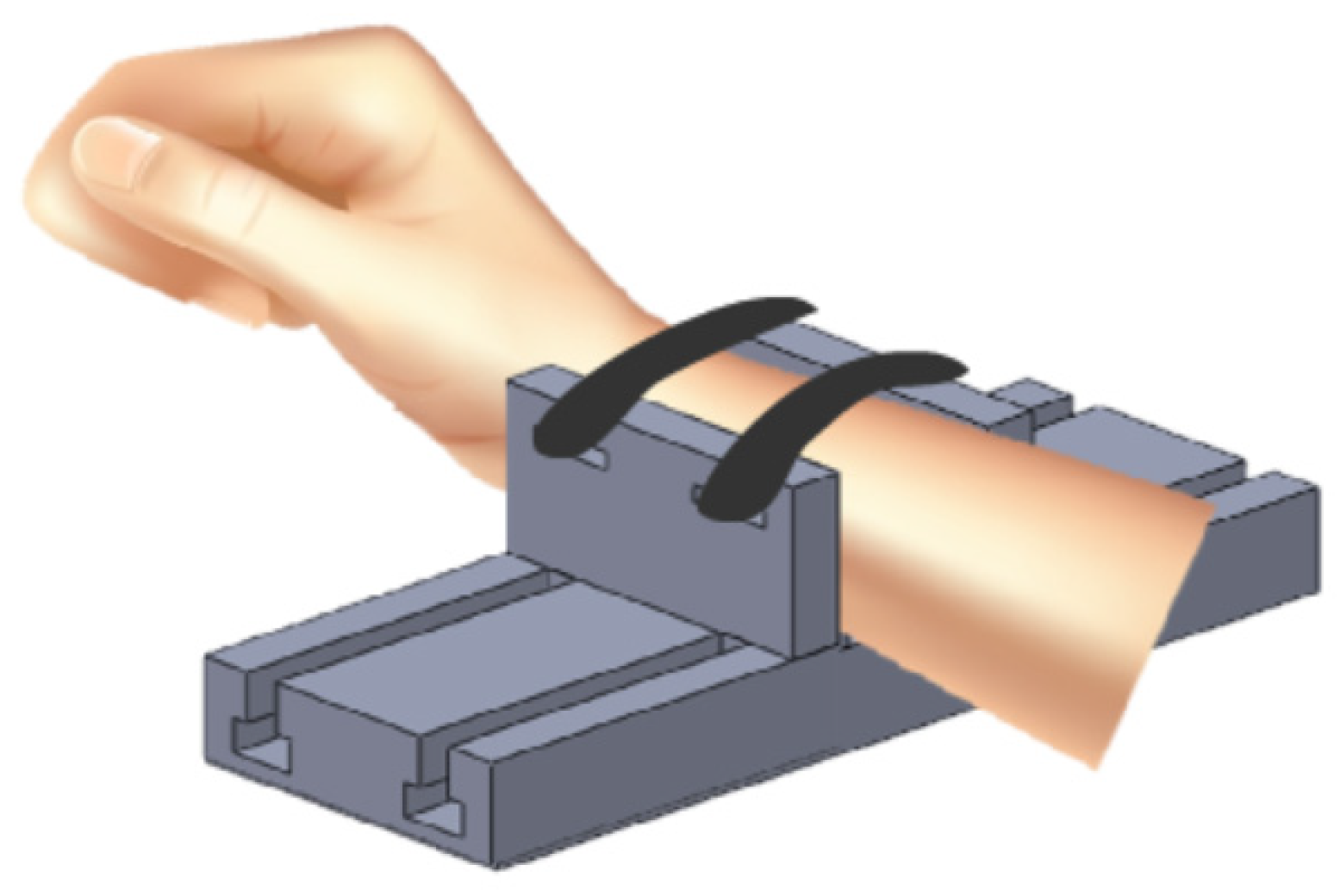
2.2.4. Boundary Conditions
2.2.5. FE Solver and Parameters
3. Results
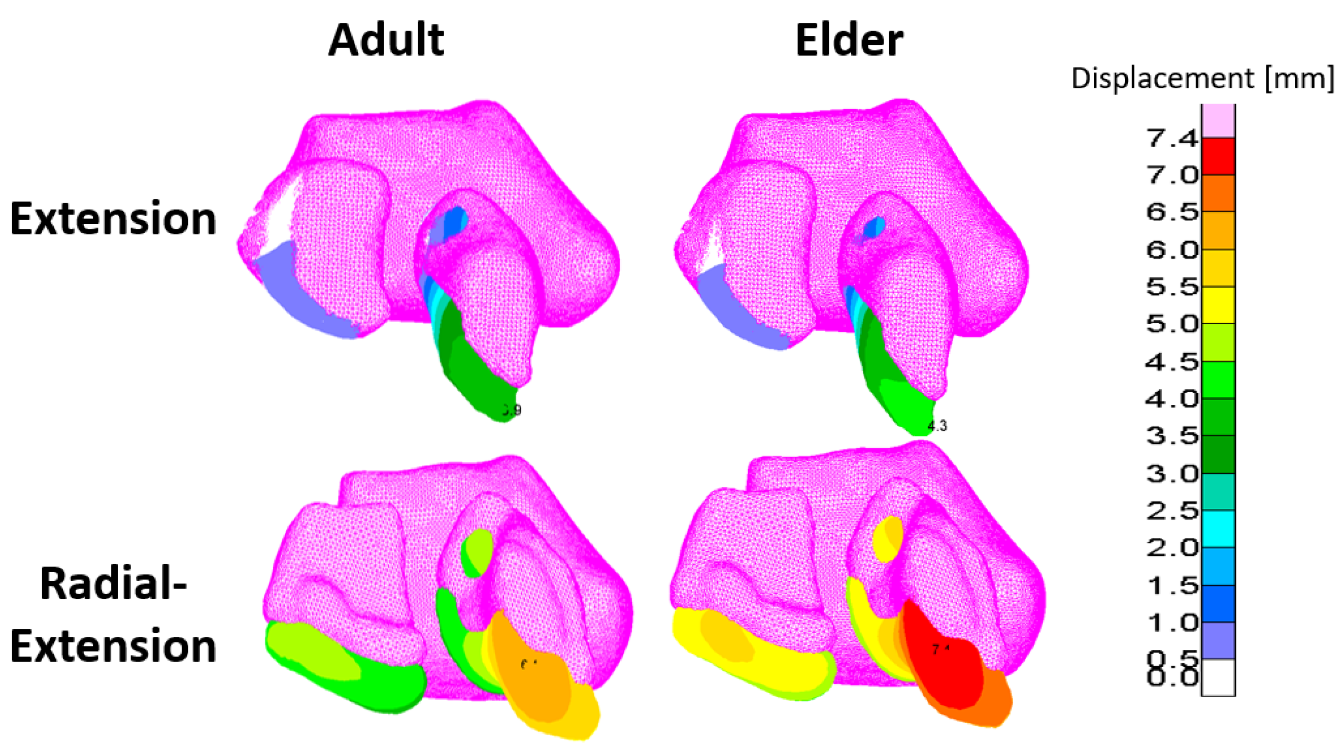
3.1. Displacements
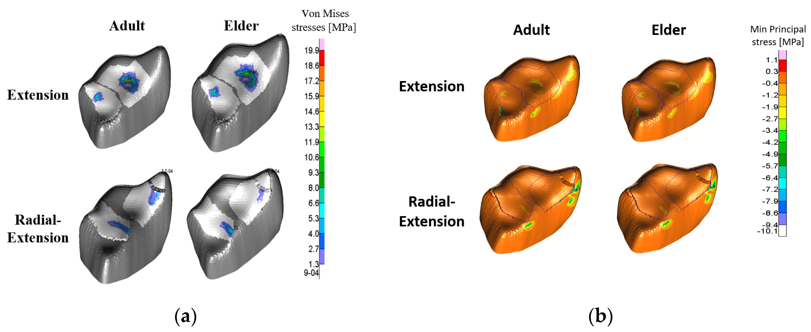
3.2. Stresses
4. Discussions
Author Contributions
Funding
Institutional Review Board Statement
Informed Consent Statement
Data Availability Statement
Acknowledgments
Conflicts of Interest
Appendix A

| Ligament | Origin | Insertion | Stiffness [N/mm] | References |
|---|---|---|---|---|
| TCL * | Scaphoid, Trapezium | Hamate, Pisiform | 64.5 | [25] |
| STT (palmar) | Scaphoid | Trapezium, Trapezoid | 150 | [38] |
| SLIL proximal | Scaphoid | Lunate | 150 | |
| Palmar Capito-hamate | Capitate | Hamate | 325 | [18,38] |
| Capito-trapezial | Capitate | Trapezium | 300 | [18,38] |
| Dorsal Intercarpal | Hamate | Capitate | 325 | [18,38] |
| Dorsal Intercarpal | Capitate | Trapezoid | 300 | [18,38] |
| Dorsal Intercarpal | Hamate | Triquetrum | 300 | [18,38] |
| Dorsal Intercarpal | Hamate | Lunate | 150 | [18,38] |
| Dorsal Intercarpal | Capitate | Lunate | 150 | [18,38] |
| Dorsal Intercarpal | Capitate | Scaphoid | 150 | [18,38] |
| Dorsal Intercarpal | Scaphoid | Trapezium | 150 | [18,38] |
| Dorsal Intercarpal | Trapezoid | Trapezium | 110 | [16] |
| Dorsal Intercarpal (triquetro-trapezial) | Trapezium, Trapezoid | Triquetrum | 128 | [16] |
| Dorsal Intercarpal | Lunate | Triquetrum | 350 | [18,38] |
| Dorsal Radio-ulnar | Radius | Ulna | 50 | [18,38] |
| Dorsal Scapho-lunate (SLIL) | Lunate | Scaphoid | 230 | [18,38] |
| Long Radio-lunate | Lunate | Radius | 75 | [18,38] |
| Short Radio-lunate | Lunate | Radius | 75 | [18,38] |
| Piso-hamate | Hamate | Pisiform | 100 | [18,38] |
| Palmar scapho-capitate | Capitate | Scaphoid | 40 | [18,38] |
| Radial Collateral (radio-scaphoid) | Radius | Scaphoid | 50 | [18,38] |
| Radio-scapho-capitate | Radius | Capitate | 50 | [18,38] |
| Palmar RTq | Radius | Triquetrum | 15 | [39] |
| Dorsal RTq | Radius | Triquetrum | 46 | [39] |
| Scapho-triquetrum | Scaphoid | Triquetrum | 128 | [16] |
| Ulno-lunate | Lunate | Ulna | 40 | [18,38] |
| Triquetro-capitate (TCP) | Capitate | Triquetrum | 36 | [39] |
| Ulnar Collateral | Triquetrum | Ulna | 100 | [18,38] |
| Ulnar Collateral | Pisiform | Ulna | 100 | [18,38] |
| Ulno-triquetral Volar/Dorsal | Triquetrum | Ulna | 40 | [18,38] |
| Volar Radio-ulnar | Radius | Ulna | 50 | [18,38] |
| Volar Triquetro-hamate | Hamate | Triquetrum | 50 | [18,38] |
| Volar Scapho-lunate (SLIL) | Lunate | Scaphoid | 230 | [18,38] |
| Piso-triquetral | Pisiform | Triquetrum | 150 | [18,38] |
| Volar Luno-triquetral | Lunate | Triquetrum | 350 | [18,38] |
| Volar Trapezoid-capitate | Capitate | Trapezoid | 300 | [18,38] |
Appendix B
| Adult Model | Elder Model | |||||
|---|---|---|---|---|---|---|
| Extension | Radial- Extension | % Of Extension | Extension | Radial- Extension | % Of Extension | |
| Dorsal SLIL | 0.16 | 0.27 | 168.8 | 0.13 | 0.37 | 284.6 |
| Volar SLIL | 0 | 0.38 | - | 0 | 0.46 | - |
| Proximal SLIL | 0.16 | 0.14 | 87.5 | 0.17 | 0.16 | 94.1 |
| Volar STT | 0.32 | 0.58 | 181.3 | 0.35 | 0.77 | 220.0 |
| TCL | 0.45 | 0.01 | 2.2 | 1.19 | 0 | - |
| Dorsal Radio-Triquetrum | 0.09 | 0.21 | 233.3 | 0.10 | 0.24 | 240.0 |
| Lunotriq | 0.07 | 0.30 | 428.6 | 0 | 0.40 | - |
| Dorsal Lunotriq | 0.16 | 1.48 | 925.0 | 0.16 | 1.86 | 1162.5 |
| Palmar Radiotriq | 0.01 | 0.12 | 1200.0 | 0 | 0.15 | - |
| Palmar Scaphocapitate | 0.09 | 0.15 | 166.7 | 0.09 | 0.19 | 211.1 |
| Short Radiolunate | 0 | 0 | - | 0 | 0 | - |
| Long Radiolunate | 0 | 0.40 | - | 0 | 0.48 | - |
| Dorsal Radioulnar | 0 | 0 | - | 0 | 0 | - |
| Dorsal Cap Lun | 0.05 | 0.26 | 520.0 | 0 | 0.34 | - |
| Dorsal Ham Lun | 0.04 | 0.02 | 50.0 | 0.05 | 0 | - |
| Radioscaphoid | 0.09 | 0 | - | 0.11 | 0 | - |
| Radioscaphocapitate | 0.07 | 0.21 | 300.0 | 0.09 | 0.26 | 288.9 |
| Dorsal Scaphocapitate | 0.15 | 0 | - | 0.15 | 0 | - |
| Dorsal Scap Trpm | 0.15 | 0.07 | 46.7 | 0.17 | 0.12 | 70.6 |
References
- Moritomo, H.; Apergis, E.P.; Herzberg, G.; Werner, F.W.; Wolfe, S.W.; Garcia-Elias, M. 2007 IFSSH Committee Report of Wrist Biomechanics Committee: Biomechanics of the So-Called Dart-Throwing Motion of the Wrist. J. Hand Surg. Am. 2007, 32, 1447–1453. [Google Scholar] [CrossRef] [PubMed]
- Garcia-Elias, M.; Alomar Serrallach, X.; Monill Serra, J. Dart-throwing motion in patients with scapholunate instability: A dynamic four-dimensional computed tomography study. J. Hand Surg. Eur. Vol. 2014, 39, 346–352. [Google Scholar] [CrossRef] [PubMed]
- Kaufman-Cohen, Y.; Portnoy, S.; Levanon, Y.; Friedman, J. Does Object Height Affect the Dart Throwing Motion Angle during Seated Activities of Daily Living? J. Mot. Behav. 2020, 52, 456–465. [Google Scholar] [CrossRef]
- Kaufman-Cohen, Y.; Friedman, J.; Levanon, Y.; Jacobi, G.; Doron, N.; Portnoy, S. Wrist plane of motion and range during daily activities. Am. J. Occup. Ther. 2018, 72, 7206205080p1–7206205080p10. [Google Scholar] [CrossRef]
- Kaufman-Cohen, Y.; Levanon, Y.; Friedman, J.; Yaniv, Y.; Portnoy, S. Home exercise in the dart-throwing motion plane after distal radius fractures: A pilot randomized controlled trial. J. Hand Ther. 2020. [Google Scholar] [CrossRef]
- Feehan, L.; Fraser, T. Early controlled mobilization using dart-throwing motion with a twist for the conservative management of an intra-articular distal radius fracture and scapholunate ligament injury: A case report. J. Hand Ther. 2016, 29, 191–198. [Google Scholar] [CrossRef] [PubMed]
- Feehan, L.; Fraser, T. Dart-throwing motion with a twist orthoses: Design, fabrication, and clinical tips. J. Hand Ther. 2016, 29, 205–212. [Google Scholar] [CrossRef] [PubMed]
- Schleifenbaum, S.; Prietzel, T.; Hädrich, C.; Möbius, R.; Sichting, F.; Hammer, N. Tensile properties of the hip joint ligaments are largely variable and age-dependent–An in-vitro analysis in an age range of 14–93 years. J. Biomech. 2016, 49, 3437–3443. [Google Scholar] [CrossRef] [PubMed]
- Woo, S.L.Y.; Hollis, J.M.; Adams, D.J.; Lyon, R.M.; Takai, S. Tensile properties of the human femur-anterior cruciate ligament-tibia complex: The effects of specimen age and orientation. Am. J. Sports Med. 1991, 19, 217–225. [Google Scholar] [CrossRef] [PubMed]
- Varga, P.; Schefzig, P.; Unger, E.; Mayr, W.; Zysset, P.K.; Erhart, J. Finite element based estimation of contact areas and pressures of the human scaphoid in various functional positions of the hand. J. Biomech. 2013, 46, 984–990. [Google Scholar] [CrossRef] [PubMed]
- Oflaz, H.; Gunal, I. Maximum loading of carpal bones during movements: A finite element study. Eur. J. Orthop. Surg. Traumatol. 2019, 29, 47–50. [Google Scholar] [CrossRef] [PubMed]
- Alonso Rasgado, T.; Zhang, Q.; Jimenez Cruz, D.; Bailey, C.; Pinder, E.; Mandaleson, A.; Talwalkar, S. Analysis of tenodesis techniques for treatment of scapholunate instability using the finite element method. Int. J. Numer. Method. Biomed. Eng. 2017, 33. [Google Scholar] [CrossRef] [PubMed]
- Guo, X.; Fan, Y.; Li, Z.M. Effects of dividing the transverse carpal ligament on the mechanical behavior of the carpal bones under axial compressive load: A finite element study. Med. Eng. Phys. 2009, 31, 188–194. [Google Scholar] [CrossRef] [PubMed]
- Gosling, J.; Harris, P.; Humpherson, J.; Whitmore, I.; Willan, P. Human Anatomy, Color Atlas and Textbook, 6th ed.; Elsevier Health Sciences: London, UK, 2016. [Google Scholar]
- Gislason, M.; Nash, D. Finite Element Modelling of a Multi Bone Joint: The Human Wrist. In Finite Element Analysis: New Trends and Developments; Ebrahimi, F., Ed.; Intech: Rijeka, Croatia, 2012. [Google Scholar]
- Bajuri, M.N.; Kadir, M.R.A.; Raman, M.M.; Kamarul, T. Mechanical and functional assessment of the wrist affected by rheumatoid arthritis: A finite element analysis. Med. Eng. Phys. 2012, 34, 1294–1302. [Google Scholar] [CrossRef] [PubMed]
- Gíslason, M.K.; Stansfield, B.; Nash, D.H. Finite element model creation and stability considerations of complex biological articulation: The human wrist joint. Med. Eng. Phys. 2010, 32, 523–531. [Google Scholar] [CrossRef] [PubMed] [Green Version]
- Carrigan, S.D.; Whiteside, R.A.; Pichora, D.R.; Small, C.F. Development of a three-dimensional finite element model for carpal load transmission in a static neutral posture. Ann. Biomed. Eng. 2003, 31, 718–725. [Google Scholar] [CrossRef]
- Bajuri, M.N.; Abdul Kadir, M.R.; Murali, M.R.; Kamarul, T. Biomechanical analysis of the wrist arthroplasty in rheumatoid arthritis: A finite element analysis. Med. Biol. Eng. Comput. 2013, 51, 175–186. [Google Scholar] [CrossRef]
- Courtney, A.C.; Hayes, W.C.; Gibson, L.J. Age-related differences in post-yield damage in human cortical bone. Experiment and model. J. Biomech. 1996, 29, 1463–1471. [Google Scholar] [CrossRef]
- Edwards, W.B.; Troy, K.L. Finite element prediction of surface strain and fracture strength at the distal radius. Med. Eng. Phys. 2012, 34, 290–298. [Google Scholar] [CrossRef] [PubMed]
- Brown, C.P.; Nguyen, T.C.; Moody, H.R.; Crawford, R.W.; Oloyede, A. Assessment of common hyperelastic constitutive equations for describing normal and osteoarthritic articular cartilage. Proc. Inst. Mech. Eng. Part H J. Eng. Med. 2009, 223, 643–652. [Google Scholar] [CrossRef] [PubMed]
- Madireddy, S.; Sista, B.; Vemaganti, K. Bayesian calibration of hyperelastic constitutive models of soft tissue. J. Mech. Behav. Biomed. Mater. 2016, 59, 108–127. [Google Scholar] [CrossRef] [PubMed]
- Berger, R.A. The gross and histologic anatomy of the scapholunate interosseous ligament. J. Hand Surg. Am. 1996, 21, 170–178. [Google Scholar] [CrossRef]
- Brett, A.W.; Oliver, M.L.; Agur, A.M.R.; Edwards, A.M.; Gordon, K.D. Quantification of the transverse carpal ligament elastic properties by sex and region. Clin. Biomech. 2014, 29, 601–606. [Google Scholar] [CrossRef]
- Miyamura, S.; Oka, K.; Lans, J.; Sakai, T.; Shiode, R.; Kazui, A.; Tanaka, H.; Shimada, S.; Murase, T. Cartilage and subchondral bone distributions of the distal radius: A 3-dimensional analysis using cadavers. Osteoarthr. Cartil. 2020, 28, 1572–1580. [Google Scholar] [CrossRef]
- Abe, T.; Thiebaud, R.S.; Loenneke, J.P. Age-related change in handgrip strength in men and women: Is muscle quality a contributing factor? Age 2016, 38, 1–7. [Google Scholar] [CrossRef] [PubMed] [Green Version]
- Werner, F.W.; Palmer, A.K.; Somerset, J.H.; Tong, J.J.; Gillison, D.B.; Fortino, M.D.; Short, W.H. Wrist joint motion simulator. J. Orthop. Res. 1996, 14, 639–646. [Google Scholar] [CrossRef] [PubMed]
- Goto, A.; Moritomo, H.; Murase, T.; Oka, K.; Sugamoto, K.; Arimura, T.; Masumoto, J.; Tamura, S.; Yoshikawa, H.; Ochi, T. In vivo three-dimensional wrist motion analysis using magnetic resonance imaging and volume-based registration. J. Orthop. Res. 2005, 23, 750–756. [Google Scholar] [CrossRef] [PubMed]
- Best, G.M.; Zec, M.L.; Pichora, D.R.; Kamal, R.N.; Rainbow, M.J. Does Wrist Laxity Influence Three-Dimensional Carpal Bone Motion? J. Biomech. Eng. 2018, 140. [Google Scholar] [CrossRef] [PubMed]
- McKeon, K.E.; London, D.A.; Osei, D.A.; Gelberman, R.H.; Goldfarb, C.A.; Boyer, M.I.; Calfee, R.P. Ligamentous hyperlaxity and dorsal wrist ganglions. J. Hand Surg. Am. 2013, 38, 2138–2143. [Google Scholar] [CrossRef] [PubMed] [Green Version]
- Pascoletti, G.; Calì, M.; Bignardi, C.; Conti, P.; Zanetti, E.M. Mandible Morphing Through Principal Components Analysis. In Proceedings of the International Conference on Design Tools and Methods in Industrial Engineering, ADM 2019, Modena, Italy, 9–10 September 2019; Springer: Cham, Switzerland, 2020; pp. 15–23. [Google Scholar]
- Anderson, A.E.; Ellis, B.J.; Maas, S.A.; Weiss, J.A. Effects of idealized joint geometry on finite element predictions of cartilage contact stresses in the hip. J. Biomech. 2010, 43, 1351–1357. [Google Scholar] [CrossRef] [PubMed] [Green Version]
- Bajuri, M.N.; Abdul Kadir, M.R.; Amin, I.M.; Öchsner, A. Biomechanical analysis of rheumatoid arthritis of the wrist joint. Proc. Inst. Mech. Eng. Part H J. Eng. Med. 2012, 226, 510–520. [Google Scholar] [CrossRef]
- Cooke, M.E.; Lawless, B.M.; Jones, S.W.; Grover, L.M. Matrix degradation in osteoarthritis primes the superficial region of cartilage for mechanical damage. Acta Biomater. 2018, 78, 320–328. [Google Scholar] [CrossRef]
- Llopis, E.; Restrepo, R.; Kassarjian, A.; Cerezal, L. Overuse Injuries of the Wrist. Radiol. Clin. N. Am. 2019, 57, 957–976. [Google Scholar] [CrossRef] [PubMed]
- Terzini, M.; Zanetti, E.M.; Audenino, A.L.; Putame, G.; Gastaldi, L.; Pastorelli, S.; Panero, E.; Sard, A.; Bignardi, C. Multibody modelling of ligamentous and bony stabilizers in the human elbow. Muscles. Ligaments Tendons J. 2017, 7, 493–502. [Google Scholar] [CrossRef] [PubMed] [Green Version]
- Schuind, F.; Cooney, W.P.; Linscheid, R.L.; An, K.N.; Chao, E.Y.S. Force and pressure transmission through the normal wrist. A theoretical two-dimensional study in the posteroanterior plane. J. Biomech. 1995, 28, 587–601. [Google Scholar] [CrossRef]
- Savelberg, H.H.C.M.; Kooloos, J.G.M.; Huiskes, R.; Kauer, J.M.G. Stiffness of the ligaments of the human wrist joint. J. Biomech. 1992, 25, 369–376. [Google Scholar] [CrossRef] [Green Version]


| Tendon Force [N] | ||||||
|---|---|---|---|---|---|---|
| Wrist position | ECU | ECRB | ECRL | APL | FCR | FCU |
| Radial-Extension | 58.2 | 50.5 | 38.4 | 38.2 | 16.0 | 65.7 |
| 20° Extension | 54.2 | 44.1 | 31.8 | 37.6 | 12.5 | 86.8 |
| ECU = Extensor Carpi Ulnaris ECRB = Extensor Carpi Radialis Brevis ECRL = Extensor Carpi Radialis Longus | APL = Abductor Pollicis Longus FCR = Flexor Carpi Radialis FCU = Extensor Carpi Radialis Longus | |||||
| Adult Model | Elder Model | |||||
|---|---|---|---|---|---|---|
| Extension | Radial- Extension | % Of Extension | Extension | Radial- Extension | % Of Extension | |
| Radius-Scaphoid interface | ||||||
| Maximal von Mises stress [MPa] | 19.9 | 8.2 | 41.1 | 16.7 | 3.8 | 22.5 |
| Maximal shear stress [MPa] | 11.2 | 4.7 | 41.5 | 9.4 | 2.1 | 21.7 |
| Maximal contact stress [MPa] | 29.7 | 10.4 | 35.1 | 29.1 | 5.9 | 20.2 |
| Average contact stress [MPa] | 8.4 | 2.8 | 33.6 | 8.5 | 1.0 | 11.5 |
| Contact area [mm2] | 6.4 | 6.5 | 100.9 | 7.3 | 3.7 | 50.2 |
| Radius-Lunate interface | ||||||
| Maximal von Mises stress [MPa] | 9.5 | 6.0 | 62.7 | 10.9 | 7.5 | 68.8 |
| Maximal shear stress [MPa] | 5.4 | 3.4 | 63.0 | 6.1 | 4.2 | 68.8 |
| Maximal contact stress [MPa] | 15.5 | 10.7 | 68.7 | 18.6 | 10.7 | 57.5 |
| Average contact stress [MPa] | 5.3 | 3.1 | 57.2 | 5.1 | 4.1 | 81.0 |
| Contact area [mm2] | 3.4 | 7.6 | 222.2 | 3.9 | 5.0 | 127.9 |
Publisher’s Note: MDPI stays neutral with regard to jurisdictional claims in published maps and institutional affiliations. |
© 2021 by the authors. Licensee MDPI, Basel, Switzerland. This article is an open access article distributed under the terms and conditions of the Creative Commons Attribution (CC BY) license (https://creativecommons.org/licenses/by/4.0/).
Share and Cite
Mahpari, V.; Levanon, Y.; Kaufman-Cohen, Y.; Zilberman, M.; Portnoy, S. Internal Mechanics of a Subject-Specific Wrist in the Sagittal versus Dart-Throwing Motion Plane in Adult and Elder Models: Finite Element Analyses. Appl. Sci. 2021, 11, 5275. https://doi.org/10.3390/app11115275
Mahpari V, Levanon Y, Kaufman-Cohen Y, Zilberman M, Portnoy S. Internal Mechanics of a Subject-Specific Wrist in the Sagittal versus Dart-Throwing Motion Plane in Adult and Elder Models: Finite Element Analyses. Applied Sciences. 2021; 11(11):5275. https://doi.org/10.3390/app11115275
Chicago/Turabian StyleMahpari, Vered, Yafa Levanon, Yael Kaufman-Cohen, Meital Zilberman, and Sigal Portnoy. 2021. "Internal Mechanics of a Subject-Specific Wrist in the Sagittal versus Dart-Throwing Motion Plane in Adult and Elder Models: Finite Element Analyses" Applied Sciences 11, no. 11: 5275. https://doi.org/10.3390/app11115275