
Polydopamine-Mediated Ag and ZnO as an Active and Recyclable SERS Substrate for Rhodamine B with Significantly Improved Enhancement Factor and Efficient Photocatalytic Degradation

 , and
, and 
Abstract
:1. Introduction
2. Materials and Methods
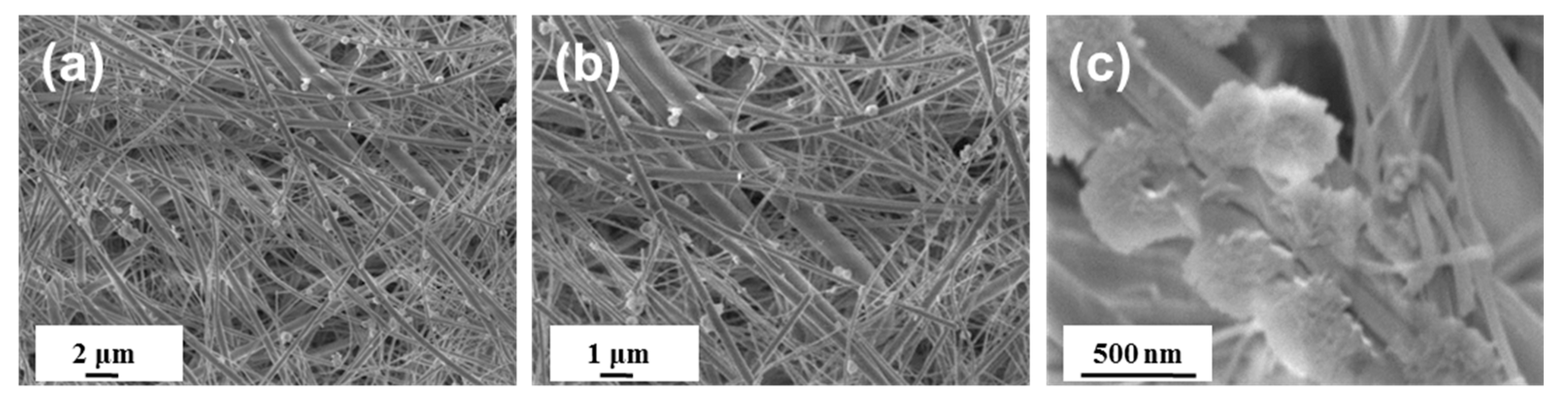
2.1. Preparation of Zinc Oxide Substrate (ZnO@GMF)
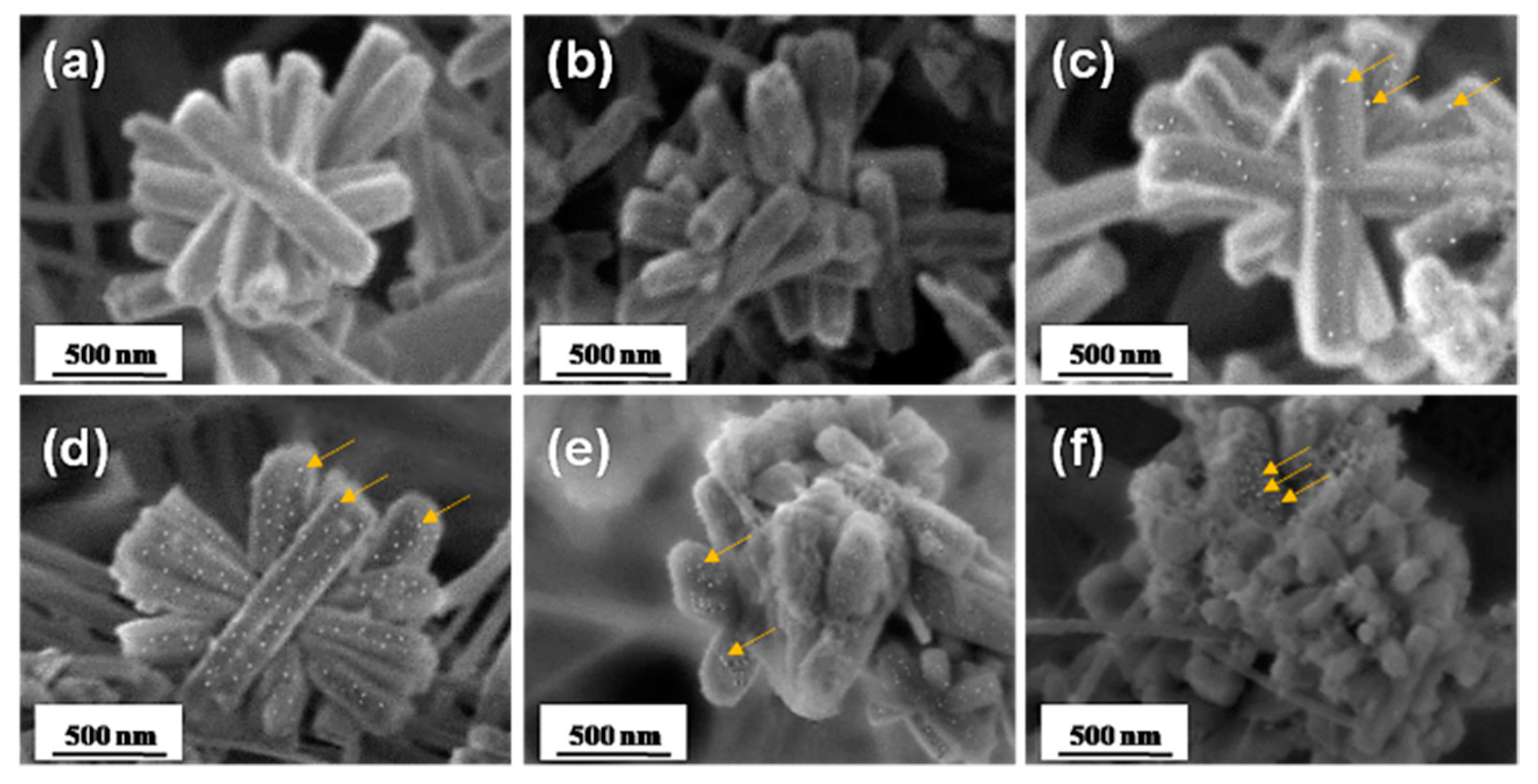
2.2. Preparation of PDA-Reduced Silver on Zinc Oxide Substrate (Ag/PDA/ZnO@GMF)
2.3. Preparation of Polydopamine-Reduced Silver Nanoparticles in Glass Fiber Filter Paper (Ag/PDA@GMF)
2.4. Surface-Enhanced Raman Spectroscopy for Detection Limit Test
2.5. Surface-Enhanced Raman Spectroscopy for Monitoring the Reaction Degradation of Rhodamine B
2.6. ICP-MS Sample Preparation
3. Results
4. Conclusions
Author Contributions
Funding
Institutional Review Board Statement
Informed Consent Statement
Data Availability Statement
Conflicts of Interest
Appendix A


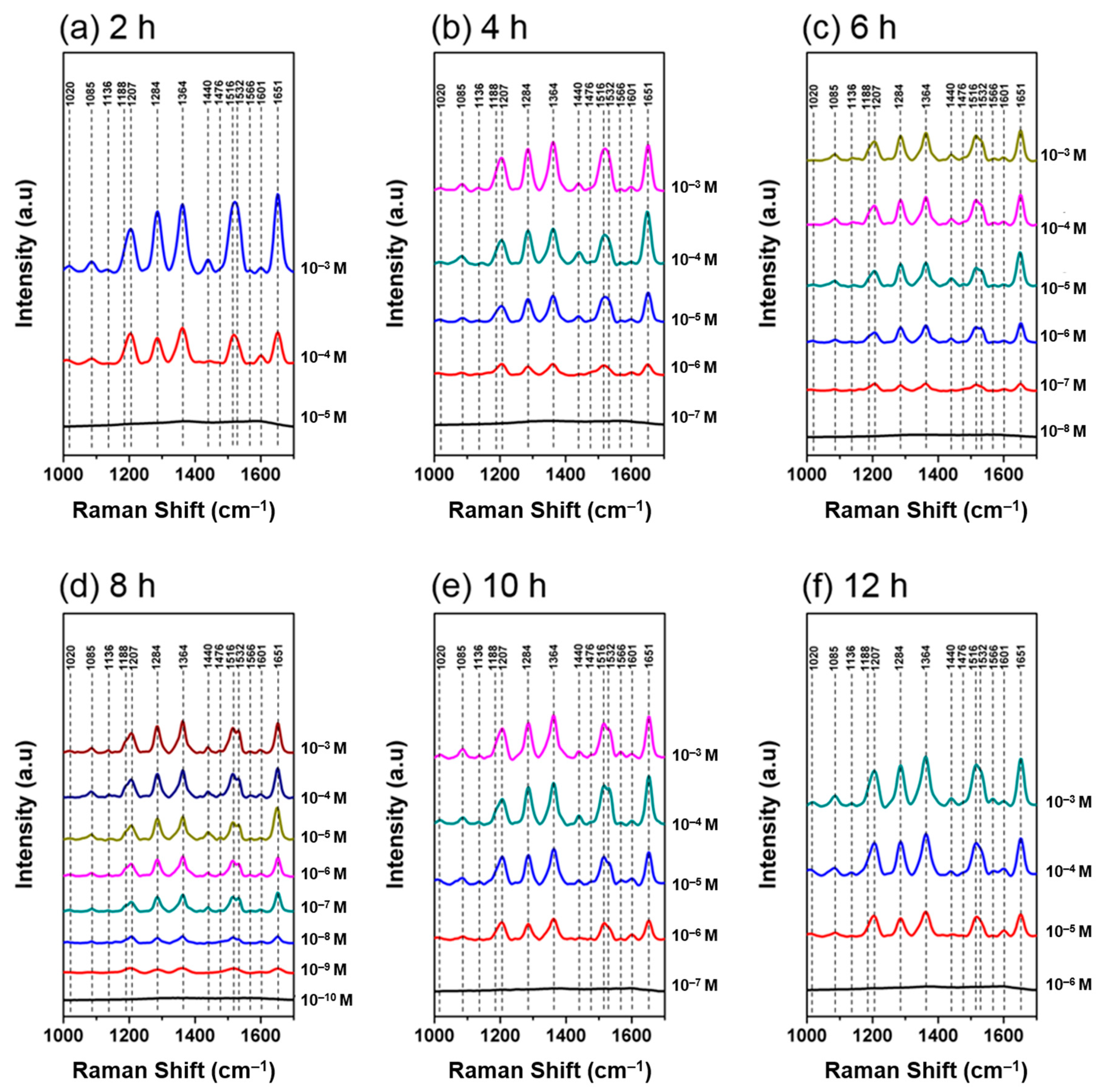
| 2 h | 4 h | 6 h | 8 h | 10 h | 12 h | |
|---|---|---|---|---|---|---|
| 10−3 M | 4.72 × 10⁴ | 5.36 × 10⁴ | 7.14 × 10⁴ | 8.94 × 10⁴ | 6.42 × 10⁴ | 6.67 × 10⁴ |
| 10−4 M | 2.50 × 10⁵ | 4.69 × 10⁵ | 7.21 × 10⁵ | 7.30 × 10⁵ | 4.51 × 10⁵ | 6.46 × 10⁵ |
| 10−5 M | 2.86 × 10⁶ | 6.11 × 10⁶ | 6.41 × 10⁶ | 3.64 × 10⁶ | 5.63 × 10⁶ | |
| 10−6 M | 4.75 × 10⁷ | 5.71 × 10⁷ | 1.64 × 10⁷ | 3.43 × 10⁷ | ||
| 10−7 M | 2.39 × 10⁸ | 5.02 × 10⁸ | ||||
| 10−8 M | 2.31 × 10⁹ | |||||
| 10−9 M | 1.95 × 10¹⁰ |
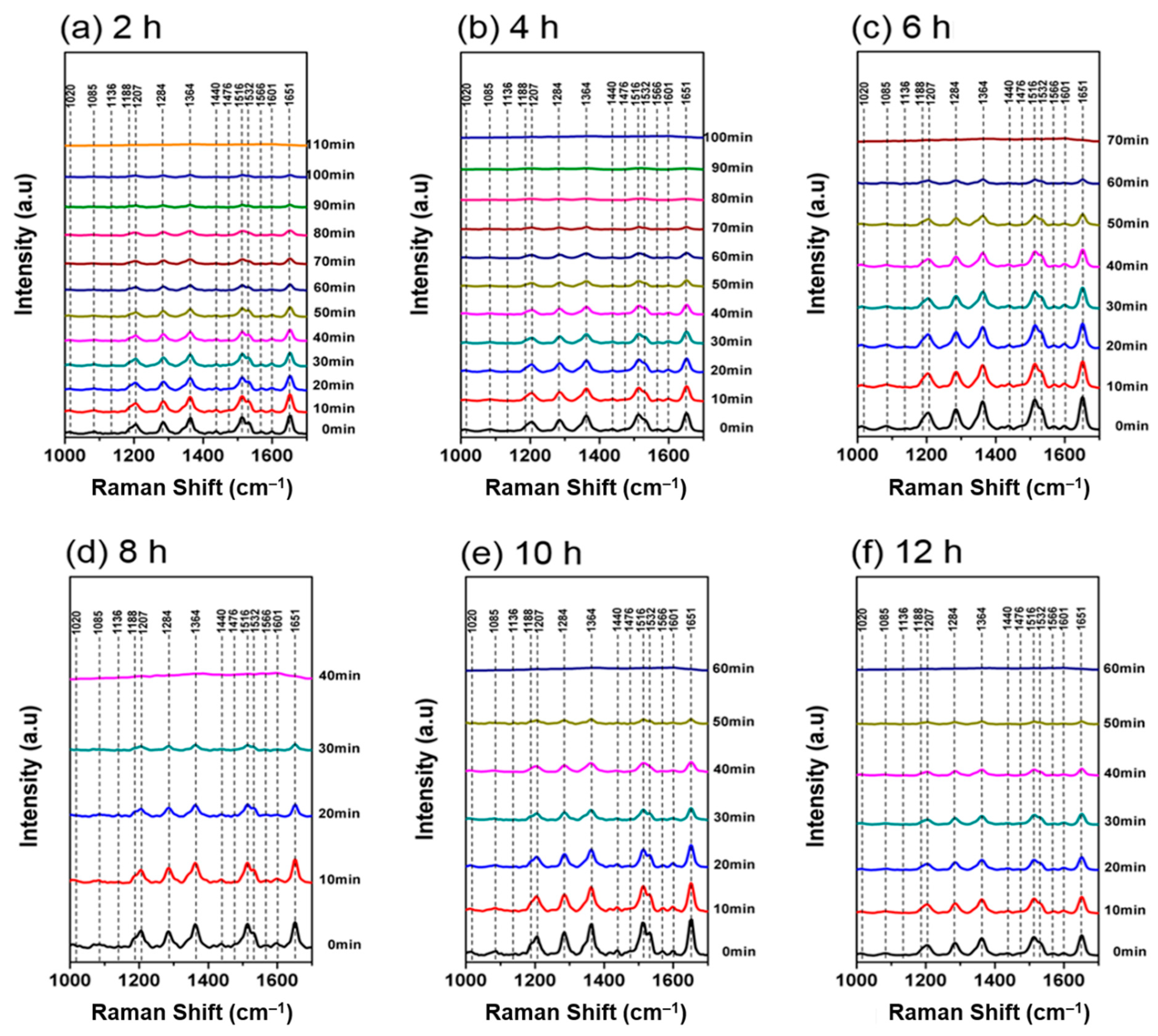
| Time (h) | Reaction Rate Constant (min−1) | R Squared |
|---|---|---|
| 2 | 0.0134 | 0.9866 |
| 4 | 0.0187 | 0.9637 |
| 6 | 0.0251 | 0.9796 |
| 8 | 0.0391 | 0.9680 |
| 10 | 0.0316 | 0.9813 |
| 12 | 0.0294 | 0.9717 |
References
- Keshavarz, M.; Kassanos, P.; Tan, B.; Venkatakrishnan, K. Metal-oxide surface-enhanced Raman biosensor template towards point-of-care EGFR detection and cancer diagnostics. Nanoscale Horiz. 2020, 5, 294–307. [Google Scholar] [CrossRef]
- Huang, Z.H.; Peng, S.W.; Hsieh, S.L.; Kirankumar, R.; Huang, P.F.; Chang, T.M.; Dwivedi, A.K.; Chen, N.F.; Wu, H.M.; Hsieh, S. Polydopamine ultrathin film growth on mica via in-situ polymerization of dopamine with applications for silver-based antimicrobial coatings. Materials 2021, 14, 671. [Google Scholar] [CrossRef]
- Saha, A.; Jana, N.R. Paper-based microfluidic approach for surface-enhanced raman spectroscopy and highly reproducible detection of proteins beyond picomolar concentration. ACS Appl. Mater. Interfaces 2015, 7, 996–1003. [Google Scholar] [CrossRef] [PubMed]
- Xia, L.; Wu, S.; Wang, J.; Ma, C.; Song, P. Spectral proof for the 4-aminophenyl disulfide plasma assisted catalytic reaction. Sci. Rep. 2017, 7, 4358. [Google Scholar] [CrossRef] [PubMed] [Green Version]
- Huang, Q.; Liu, S.; Wei, W.; Yan, Q.; Wu, C. Selective synthesis of different ZnO/Ag nanocomposites as surface enhanced Raman scattering substrates and highly efficient photocatalytic catalysts. RSC Adv. 2015, 5, 27075–27081. [Google Scholar] [CrossRef]
- Tarakeshwar, P.; Palma, J.L.; Finkelstein-Shapiro, D.; Keller, A.; Urdaneta, I.; Calatayud, M.; Atabek, O.; Mujica, V. SERS as a probe of charge-transfer pathways in hybrid dye/molecule–metal oxide complexes. J. Phys. Chem. C 2014, 118, 3774–3782. [Google Scholar] [CrossRef]
- Zhang, X.; Liu, Y.; Soltani, M.; Li, P.; Zhao, B.; Cui, B. Probing the interfacial charge-transfer process of uniform ALD semiconductor–molecule–metal models: A SERS study. J. Phys. Chem. C 2017, 121, 26939–26948. [Google Scholar] [CrossRef]
- Zhang, X.; Yu, Z.; Ji, W.; Sui, H.; Cong, Q.; Wang, X.; Zhao, B. Charge-transfer effect on surface-enhanced raman scattering (SERS) in an Ordered AgNPs/4-mercaptobenzoic Acid/TiO2 system. J. Phys. Chem. C 2015, 119, 22439–22444. [Google Scholar] [CrossRef]
- Keshavarz, M.; Chowdhury, A.K.M.R.H.; Kassanos, P.; Tan, B.; Venkatakrishnan, K. Self-assembled N-doped Q-dot carbon nanostructures as a SERS-active biosensor with selective therapeutic functionality. Sens. Actuators B Chem. 2020, 323, 128703. [Google Scholar] [CrossRef]
- Lin, P.-Y.; He, G.; Chen, J.; Dwivedi, A.K.; Hsieh, S. Monitoring the photoinduced surface catalytic coupling reaction and environmental exhaust fumes with an Ag/PDA/CuO modified 3D glass microfiber platform. J. Ind. Eng. Chem. 2020, 82, 424–432. [Google Scholar] [CrossRef]
- Fang, H.-P.; Chiang, I.H.; Chu, C.-W.; Yang, C.-C.; Lin, H.-C. Applications of novel dithienothiophene- and 2,7-carbazole-based conjugated polymers with surface-modified ZnO nanoparticles for organic photovoltaic cells. Thin Solid Films 2011, 519, 5212–5218. [Google Scholar] [CrossRef]
- Lin, C.-M.; Li, M.-S.; Dwivedi, A.K.; Lin, H.-C. Synthesis and enhanced electron transfer of supramolecular nano-composite containing dendritic dye and surface-modified ZnO nano-rods. Dye. Pigment. 2018, 157, 179–189. [Google Scholar] [CrossRef]
- Gaiardo, A.; Fabbri, B.; Giberti, A.; Guidi, V.; Bellutti, P.; Malagù, C.; Valt, M.; Pepponi, G.; Gherardi, S.; Zonta, G.; et al. ZnO and Au/ZnO thin films: Room-temperature chemoresistive properties for gas sensing applications. Sens. Actuators B Chem. 2016, 237, 1085–1094. [Google Scholar] [CrossRef]
- Panda, D.; Tseng, T.-Y. One-dimensional ZnO nanostructures: Fabrication, optoelectronic properties, and device applications. J. Mater. Sci. 2013, 48, 6849–6877. [Google Scholar] [CrossRef]
- Shi, X.-F.; Xia, X.-Y.; Cui, G.-W.; Deng, N.; Zhao, Y.-Q.; Zhuo, L.-H.; Tang, B. Multiple exciton generation application of PbS quantum dots in ZnO@PbS/graphene oxide for enhanced photocatalytic activity. Appl. Catal. B Environ. 2015, 163, 123–128. [Google Scholar] [CrossRef]
- Wang, C.-A.; Ho, H.-C.; Hsueh, C.-H. Periodic ZnO-elevated gold dimer nanostructures for surface-enhanced raman scattering applications. J. Phys. Chem. C 2018, 122, 27016–27023. [Google Scholar] [CrossRef]
- Chen, J.S.; Tan, Y.L.; Li, C.M.; Cheah, Y.L.; Luan, D.; Madhavi, S.; Boey, F.Y.C.; Archer, L.A.; Lou, X.W. Constructing hierarchical spheres from large ultrathin anatase TiO2 nanosheets with nearly 100% exposed (001) facets for fast reversible lithium storage. J. Am. Chem. Soc. 2010, 132, 6124–6130. [Google Scholar] [CrossRef] [Green Version]
- Han, X.; Kuang, Q.; Jin, M.; Xie, Z.; Zheng, L. Synthesis of titania nanosheets with a high percentage of exposed (001) facets and related photocatalytic properties. J. Am. Chem. Soc. 2009, 131, 3152–3153. [Google Scholar] [CrossRef] [PubMed]
- Lee, Y.; Lee, J.; Lee, T.K.; Park, J.; Ha, M.; Kwak, S.K.; Ko, H. Particle-on-film gap plasmons on antireflective ZnO nanocone arrays for molecular-level surface-enhanced raman scattering sensors. ACS Appl. Mater. Interfaces 2015, 7, 26421–26429. [Google Scholar] [CrossRef] [PubMed]
- Kim, W.; Lee, S.H.; Kim, S.H.; Lee, J.-C.; Moon, S.W.; Yu, J.S.; Choi, S. Highly reproducible Au-decorated ZnO nanorod array on a graphite sensor for classification of human aqueous humors. ACS Appl. Mater. Interfaces 2017, 9, 5891–5899. [Google Scholar] [CrossRef]
- Cheng, C.; Yan, B.; Wong, S.M.; Li, X.; Zhou, W.; Yu, T.; Shen, Z.; Yu, H.; Fan, H.J. Fabrication and SERS performance of silver-nanoparticle-decorated Si/ZnO nanotrees in ordered arrays. ACS Appl. Mater. Interfaces 2010, 2, 1824–1828. [Google Scholar] [CrossRef] [PubMed]
- Macias-Montero, M.; Peláez, R.J.; Rico, V.J.; Saghi, Z.; Midgley, P.; Afonso, C.N.; González-Elipe, A.R.; Borras, A. Laser treatment of Ag@ZnO nanorods as long-life-span SERS surfaces. ACS Appl. Mater. Interfaces 2015, 7, 2331–2339. [Google Scholar] [CrossRef] [PubMed] [Green Version]
- Huang, J.; Chen, F.; Zhang, Q.; Zhan, Y.; Ma, D.; Xu, K.; Zhao, Y. 3D silver nanoparticles decorated zinc Oxide/Silicon heterostructured nanomace arrays as high-performance surface-enhanced raman scattering substrates. ACS Appl. Mater. Interfaces 2015, 7, 5725–5735. [Google Scholar] [CrossRef]
- Chatterjee, A.; Gale, D.J.G.; Grebennikov, D.; Whelan, L.D.; Merschrod, S.E.F. Surface potential and morphology mapping to investigate analyte adsorption effects on surface enhanced Raman scattering (SERS). Chem. Commun. 2017, 53, 12024–12027. [Google Scholar] [CrossRef] [PubMed]
- Tang, F.; Zhang, M.; Li, Z.; Du, Z.; Chen, B.; He, X.; Zhao, S. Hexagonally arranged arrays of urchin-like Ag-nanoparticle decorated ZnO-nanorods grafted on PAN-nanopillars as surface-enhanced Raman scattering substrates. Cryst. Eng. Comm. 2018, 20, 3550–3558. [Google Scholar] [CrossRef]
- Zhu, Q.; Xu, C.; Wang, D.; Liu, B.; Qin, F.; Zhu, Z.; Liu, Y.; Zhao, X.; Shi, Z. Femtomolar response of a plasmon-coupled ZnO/graphene/silver hybrid whispering-gallery mode microcavity for SERS sensing. J. Mater. Chem. C 2019, 7, 2710–2716. [Google Scholar] [CrossRef]
- Yao, J.; Quan, Y.; Gao, M.; Gao, R.; Chen, L.; Liu, Y.; Lang, J.; Shen, H.; Zhang, Y.; Yang, L.; et al. AgNPs decorated Mg-doped ZnO heterostructure with dramatic SERS activity for trace detection of food contaminants. J. Mater. Chem. C 2019, 7, 8199–8208. [Google Scholar] [CrossRef]
- Zhou, J.; Zhang, J.; Yang, H.; Wang, Z.; Shi, J.-a.; Zhou, W.; Jiang, N.; Xian, G.; Qi, Q.; Weng, Y.; et al. Plasmon-induced hot electron transfer in Au–ZnO heterogeneous nanorods for enhanced SERS. Nanoscale 2019, 11, 11782–11788. [Google Scholar] [CrossRef]
- Yin, D.; Wang, M.-L.; Wang, Y.-Z.; Hu, X.; Liu, B.; Liu, H.; Ma, L.; Gao, G.-G. A ternary ZnO/ZnS/MoS2 composite as a reusable SERS substrate derived from the polyoxomolybdate/ZIF-8 host–guest framework. J. Mater. Chem. C 2019, 7, 9856–9864. [Google Scholar] [CrossRef]
- Chen, L.; Luo, L.; Chen, Z.; Zhang, M.; Zapien, J.A.; Lee, C.S.; Lee, S.T. ZnO/Au Composite nanoarrays as substrates for surface-enhanced raman scattering detection. J. Phys. Chem. C 2010, 114, 93–100. [Google Scholar] [CrossRef]
- Zhang, Z.; Si, T.; Liu, J.; Han, K.; Zhou, G. Controllable synthesis of AgNWs@PDA@AgNPs core–shell nanocobs based on a mussel-inspired polydopamine for highly sensitive SERS detection. RSC Adv. 2018, 8, 27349–27358. [Google Scholar] [CrossRef] [Green Version]
- Rosman, C.; Prasad, J.; Neiser, A.; Henkel, A.; Edgar, J.; Sönnichsen, C. Multiplexed plasmon sensor for rapid label-free analyte detection. Nano Lett. 2013, 13, 3243–3247. [Google Scholar] [CrossRef] [PubMed]
- Guo, L.; Zhang, X.; Li, P.; Han, R.; Liu, Y.; Han, X.; Zhao, B. Surface-enhanced Raman scattering (SERS) as a probe for detection of charge-transfer between TiO2 and CdS nanoparticles. New J. Chem. 2019, 43, 230–237. [Google Scholar] [CrossRef]
- Cong, S.; Liu, X.; Jiang, Y.; Zhang, W.; Zhao, Z. Surface enhanced raman scattering revealed by interfacial charge-transfer transitions. Innovation 2020, 1. [Google Scholar] [CrossRef]
- Du, J.; Jing, C. One-step fabrication of dopamine-inspired Au for SERS sensing of Cd2+ and polycyclic aromatic hydrocarbons. Anal. Chim. Acta 2019, 1062, 131–139. [Google Scholar] [CrossRef] [PubMed]
- Kaspar, T.C.; Droubay, T.; Chambers, S.A.; Bagus, P.S. Spectroscopic evidence for Ag(III) in highly oxidized silver films by X-ray photoelectron spectroscopy. J. Phys. Chem. C 2010, 114, 21562–21571. [Google Scholar] [CrossRef]
- Feng, S.; dos Santos, M.C.; Carvalho, B.R.; Lv, R.; Li, Q.; Fujisawa, K.; Elías, A.L.; Lei, Y.; Perea-López, N.; Endo, M.; et al. Ultrasensitive molecular sensor using N-doped graphene through enhanced Raman scattering. Sci. Adv. 2016, 2, e1600322. [Google Scholar] [CrossRef] [Green Version]
- Le Ru, E.C.; Blackie, E.; Meyer, M.; Etchegoin, P.G. Surface enhanced raman scattering enhancement factors: A comprehensive study. J. Phys. Chem. C 2007, 111, 13794–13803. [Google Scholar] [CrossRef]
- Yan, Z.-X.; Zhang, Y.-L.; Wang, W.; Fu, X.-Y.; Jiang, H.-B.; Liu, Y.-Q.; Verma, P.; Kawata, S.; Sun, H.-B. Superhydrophobic SERS substrates based on silver-coated reduced graphene oxide gratings prepared by two-beam laser interference. ACS Appl. Mater. Interfaces 2015, 7, 27059–27065. [Google Scholar] [CrossRef]


| SERS Substrate Based on ZnO | SERS Reporters | EF | Ref. |
|---|---|---|---|
| Ag/ZnO nano arrays | benzenethiol, Rhodamine 6 G, adenine | 1010–1011 | [19] |
| Au/ZnO Nano Rods/Graphite | Rhodamine 6 G | 2.3 × 10⁶ | [20] |
| Si/ZnO nanotrees | Rhodamine 6 G | 106 | [21] |
| Ag@ZnO Nanorods | Rhodamine 6 G | 1.6 × 10⁶ | [22] |
| Ag/ZnO/Si | Rhodamine 6 G | 8.7 × 10⁷ | [23] |
| Au/ZnO | phenanthrene | 106 | [24] |
| Ag/ZnO-nanorods | 4-aminothiophenol | 3.5 × 10⁷ | [25] |
| ZnO/graphene/Ag | Rhodamine 6 G | 0.95 × 10¹² | [26] |
| Mg/ZnO heterostructure | 4-MPY | 1.36 × 10⁷ | [27] |
| Au/ZnO nanorods | Dopamine | 1.2 × 10⁴ | [28] |
| ZnO/ZnS/MoS2 | Rhodamine 6 G | 1.4 × 10⁸ | [29] |
| ZnO/Au Nanoarrays | Rhodamine 6 G | 1.2 × 10⁷ | [30] |
| Ag/PDA/ZnO@GMF | Rhodamine B | 1010 | Our substrate |
| Sample | Zn (μg/mL) | Ag (μg/mL) |
|---|---|---|
| Blank | 5.12 × 10⁻⁴ | 0.07 × 10⁻⁴ |
| 0 h | 42.5 | 1.86 × 10⁻⁴ |
| 2 h | 34.5 | 2.00 |
| 4 h | 44.9 | 4.49 |
| 6 h | 41.8 | 7.46 |
| 8 h | 41.1 | 10.2 |
| 10 h | 37.8 | 10.8 |
| 12 h | 39.3 | 12.8 |
Publisher’s Note: MDPI stays neutral with regard to jurisdictional claims in published maps and institutional affiliations. |
© 2021 by the authors. Licensee MDPI, Basel, Switzerland. This article is an open access article distributed under the terms and conditions of the Creative Commons Attribution (CC BY) license (https://creativecommons.org/licenses/by/4.0/).
Share and Cite
Chin, H.-K.; Lin, P.-Y.; Chen, J.; Kirankumar, R.; Wen, Z.-H.; Hsieh, S. Polydopamine-Mediated Ag and ZnO as an Active and Recyclable SERS Substrate for Rhodamine B with Significantly Improved Enhancement Factor and Efficient Photocatalytic Degradation. Appl. Sci. 2021, 11, 4914. https://doi.org/10.3390/app11114914
Chin H-K, Lin P-Y, Chen J, Kirankumar R, Wen Z-H, Hsieh S. Polydopamine-Mediated Ag and ZnO as an Active and Recyclable SERS Substrate for Rhodamine B with Significantly Improved Enhancement Factor and Efficient Photocatalytic Degradation. Applied Sciences. 2021; 11(11):4914. https://doi.org/10.3390/app11114914
Chicago/Turabian StyleChin, Hsien-Kuo, Pei-Ying Lin, Jyunde Chen, Rajendranath Kirankumar, Zhi-Hong Wen, and Shuchen Hsieh. 2021. "Polydopamine-Mediated Ag and ZnO as an Active and Recyclable SERS Substrate for Rhodamine B with Significantly Improved Enhancement Factor and Efficient Photocatalytic Degradation" Applied Sciences 11, no. 11: 4914. https://doi.org/10.3390/app11114914