1. Introduction
Many consumers associate the fruit of the olive tree (
Olea europaea L.) mainly with the resulting oil, the precious olive oil that is considered particularly healthy compared to other oils. The raw olive fruit contains several types of phenols, thecontents of which vary with the olive cultivar, mainly tyrosol and its derivatives, phenolic acids, and flavonoids [
1]. However, many of these substances that the olive has to offer from a health perspective are water-soluble, and thus remain largely in the residue of the olive pressing and the oil contains only a small part. This is the case of hydroxytyrosol, a polar phenol slightly soluble in fats, which can be found in olives as a simple phenol, or either esterified with elenolic acid to form oleuropein aglycone, and which is naturally present in significantly higher concentrations in the olive fruit’s aqueous fraction.
The vegetation water, resulting from the pressing of the olive fruits duringthe production of the olive oil, is rich in bioactive compounds, particularly polar phenols, and typically contains 98% of the total phenols of the olive fruit [
2].
The positive health effects of olive polyphenols are already known; in particular, hydroxytyrosol has potential antioxidant, anti-inflammatory, and health benefits mainly related with cardiovascular diseases [
3,
4,
5,
6].
Bioavailability and pharmacokinetic analyses, which were mainly reported with pure hydroxytyrosol and with olive oil, suggest that hydroxytyrosol can be rapidly absorbed from blood and distributed in the human body [
7], metabolized, and quickly eliminated in urine mainly as glucuronide and sulfate [
8]. Currently, hydroxytyrosol from different sources is available on the market. Its absorption and subsequent urine excretion may be dependent on the vehicle of administration [
9]. Thus, the bioavailability of hydroxytyrosol and its precursors (oleuropein and tyrosol) from those specific sources would be a prerequisite for its health effects in humans.
The present study addresses the bioactivity of hydroxytyrosol-rich extracts, obtained from the vegetation water resulting from olive oil production. Herein, hydroxytyrosol is present as both a simple phenol and as oleuropein aglycone.
For the study, olive-derived concentrates combined with 6% lemon juice or 70% grape juice and marketed as liquid supplements were characterized. Bioactivities, mainly related to the antioxidant potential, were evaluated in cultured cells by means of the antioxidant capacity (cellular antioxidant activity assay, superoxide dismutase and catalase activities), the protection against lipoxidation (inhibition of F2-isoprostanes formation) and glycation (inhibition of AGEs formation). In addition, preliminary data on the bioavailability and urinary recovery of free hydroxytyrosol through acute administration of the food supplements are presented from an open-label cross-over study with four volunteers.
Despite the difference in the composition of both formulations, the main phytochemical in the ones that were investigated was hydroxytyrosol, present in both olive fruit and to a lesser extent in grapes. The treatment of the cells with the supplements gave positive results, although these were slightly different in magnitude, through antioxidant actions. The high bioactivity observed suggests a possible application in the maintenance of the cellular redox state and for related health benefits.
2. Materials and Methods
2.1. Standards and Reagents
2,2′-azobis [2-methylpropionamide] dihydrochloride (AAPH), quercetin dihydrate, and 2′-7′-dichlorodihydrofluorescin diacetate (DCFH2-DA) were purchased from Sigma-Aldrich (Milan, Italy). Hydroxytyrosol and oleuropein were procured from Cayman Chemical (Ann Arbor, MI, USA). Resveratrol was purchased from Sigma-Aldrich (Steinheim, Germany). Dulbecco’s modified Eagle’s medium (DMEM) high-glucose culture media, L-glutamine, trypan blue solution, and trypsin-EDTA solution 10X were culture grade and purchased from Merck (Milan, Italy). Fetal bovine serum (FBS), Dulbecco’s phosphate buffered saline (PBS) without Mg2+ and Ca2+, and Hank’s balanced salts solution (HBSS) were culture grade and purchased from Euroclone SpA (Milan, Italy). Water, acetonitrile, formic acid, and methanol (all LC-MS-grade) were purchased from VWR Chemicals (Darmstadt, Germany). All other chemicals were analytical grade and purchased from common sources.
2.2. Sample Material
The food supplements analyzed are derived from olive fruit (Olea europaea L.) vegetation water subjected to filtration and concentration, and were supplied by Fattoria La Vialla (Castiglion Fibocchi, Arezzo, Italy). The commercial brands are Oliphenolia bitter™ and Oliphenolia™, hereinafter referred to as P-1 and P-2, respectively. P-1 consists of 94% concentrated olive aqueous fraction and 6% lemon juice (Citrus limon L. fructus); while P-2 is characterized by 30% concentrated olive extract and 70% grape juice (Vitis vinifera L. fructus).
2.3. Analysis of Polyphenols
The samples were diluted with methanol (50:50 v/v), ultrasonicated, centrifuged, and filtrated through 0.45 µm regenerated cellulose filters prior to measurement by UHPLC-MS, with an Acquity UPLC I-Class system coupled to a XEVO-TQS micro mass spectrometer (bothWaters, Milford, MA, USA). The instrument consisted of a sample manager cooled at 10 °C, a binary pump, a column oven, and a diode array detector measuring at 280 nm. The column oven temperature was set at 40 °C. The gradient started with 2% A and raised linearly to 15% within 5.5 min, then to 100% A within 1 min before holding for 1.5 min as a washing step; it then decreased back to 2% B within 1 min and was equilibrated for 2 min. Eluent B was water with 0.1% formic acid, eluent A was acetonitrile with 0.1% formic acid, the flow was 0.4 mL/min on an HSS T3 RP column (150 mm x 2.1 mm, 1.7 µm particle size) combined with a precolumn (Acquity UPLC HSS T3 VanGuard, 100 Å, 2.1 mm × 5 mm, 1.8 µm), both from Waters (Milford, MA, USA). The injection volume was 2 µL.
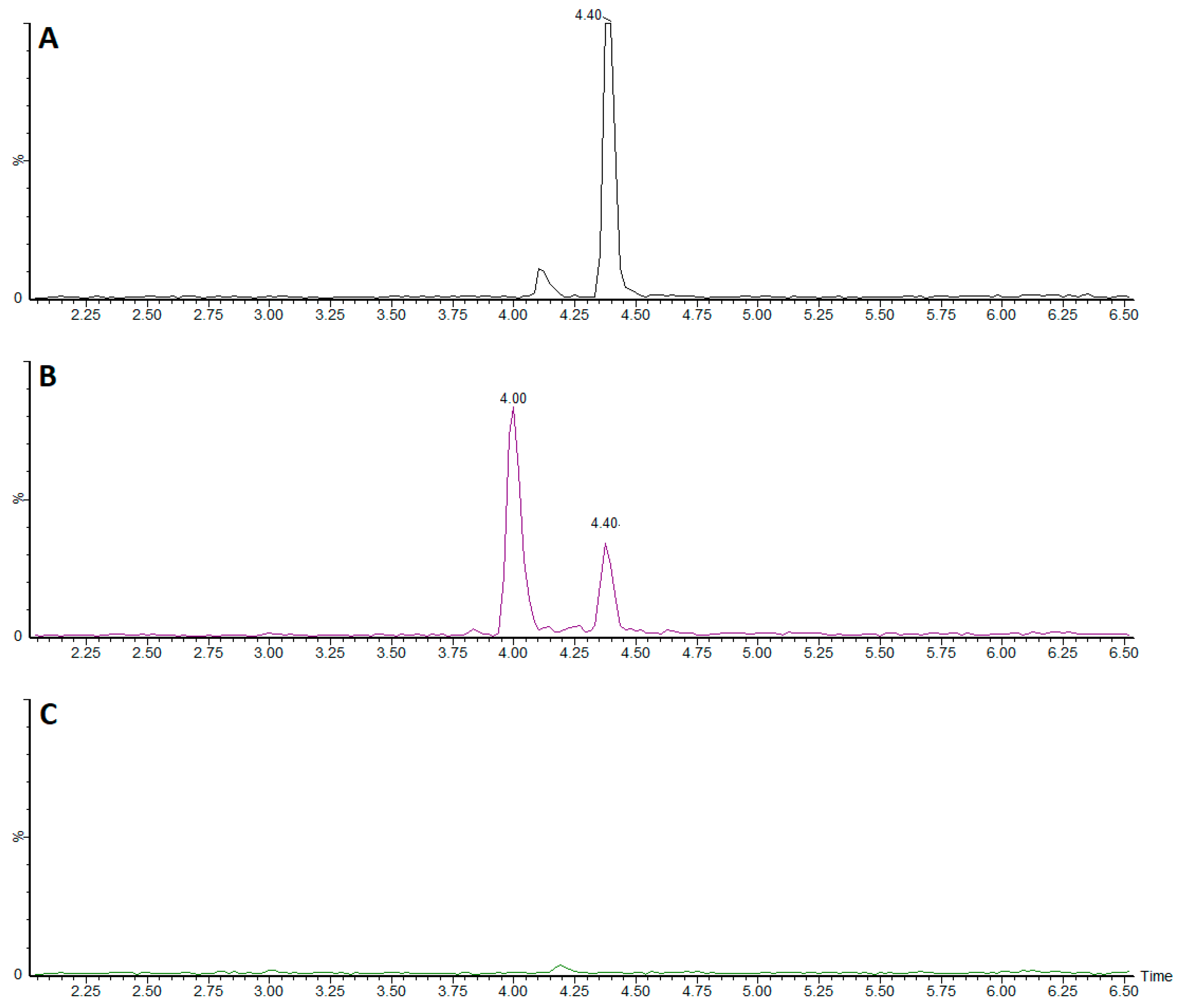
The peaks were identified by MS/MS (MRM 153 > 123 for hydroxytyrosol, 539 > 377 for oleuropein and SIR 137 for tyrosol in negative mode and MRM 229 > 135 for resveratrol operating in positive ion mode). The source voltage was kept at 1.5 kV, and the cone voltage was 20 V. The source temperature was set at 150 °C and the desolvation temperature at 350 °C with a desolvation gas flow of 650 L/h and a cone gas flow of 50 L/h. Standard substances were used as reference.
Proanthocyanidin monomers were determined according to Kelm et al. [
10].
Data were acquired and processed using MassLynx (Waters, Milford, MA, USA).
2.4. Cell Cultures
Human hepatocellular carcinoma (HepG2) and human keratinocytes (HaCat) cell lines were obtained from CLS (Cell Lines Service GmbH, Germany) and cultured at 37 °C under a humidified atmosphere of 5% CO2 in DMEM containing 2 mM L-Glutamine, 4.5 g/L glucose, and 10% of heat-inactivated FBS. Experiments were performed with DMEM low-glucose (Lonza Ltd., Morristown, NJ, USA) supplemented with 2 mM L-Glutamine without FBS in either, 6-well culture plates for AGEs, catalase, and superoxide dismutase (SOD), 12-well plates for F2-isoprostanes, or 96-well black plates for the cellular antioxidant activity (CAA) and vitality assays. For each cell-based test, P-1 and P-2 samples were centrifuged, sterile-filtered and directly diluted into culture media before testing.
2.5. Cellular Viability Assay
To determine the optimal growth conditions of cells following 4 h treatment with the sample material, five serial dilutions were evaluated for P-1 and P-2 (range 1:150 to 1:750 and 1:250 to 1:1250 for HaCat and HepG2 cells, respectively), and the metabolic activity was monitored using the resazurin toxicity assay according to the manufacturer’s instructions (Tox-8 kit, Sigma-Aldrich, Italy). Fluorescence was read at 37 °C (Em. 590 nm/Ex. 540 nm) in a multiwell fluorescence reader (Fluostar Optima, BMG LabTech, Offenburg, Germany). Data were processed using Mars 2.0 Optima Data Analysis software (BMG LabTech GmbH, Germany).
2.6. Cellular Antixidant Activity (CAA)
The intracellular reactive oxygen species (ROS) formation was detected with the CAA method by spectrofluorimetry using the cell-permeable probe DCFH2-DA, as previously described [
11,
12]. Briefly, HepG2 cells were cultured until confluence and pre-incubated for an hour with DCFH2-DA and increasing concentrations of the sample dilutions (1:750 to 1:250
v/
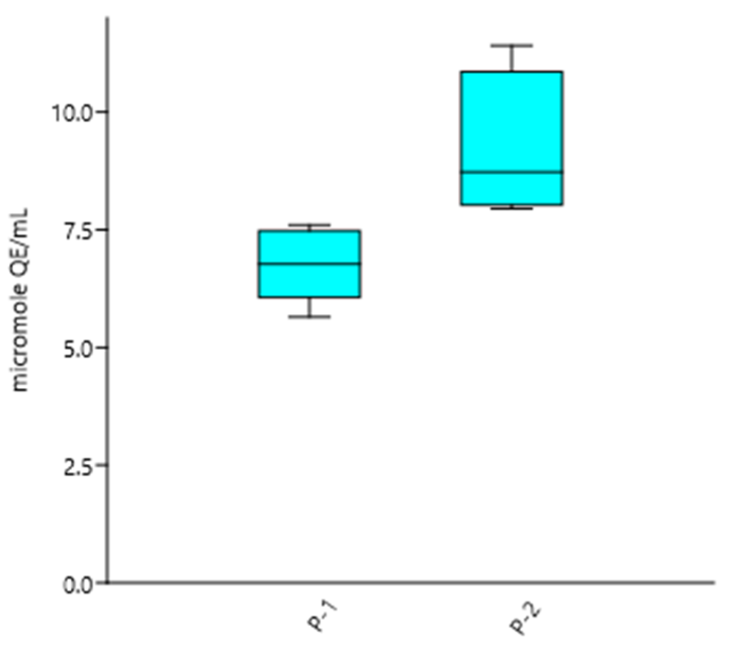
v) or the quercetin standard. After the addition of AAPH, the absorbed probe was oxidized to a high fluorescent molecule within the cytoplasm, which was measured at 37 °C for an hour at excitation 485 nm and emission 540 nm (Fluostar Optima, BMG LabTech, Germany). Raw data were analyzed using MARS 2.0 Optima Data Analysis software (BMG LabTech, Germany). Results are expressed as µmol of quercetin equivalency (QE) per mL of product and as the mean of five independent measurements ± standard deviation.
2.7. Cellular Extract Preparation
At the indicated time points, the cells were harvested in ice-cold PBS and collected in 2 mL centrifuge tubes before homogenization for the AGEs, catalase and SOD experiments. For whole lysates preparation, samples were homogenized using the Cell Disruptor Genie
® (Scientific Industries Inc., Bohemia, NY, USA) with 0.5 mm glass beads, according to the manufacturer’s instructions. Whole protein lysates were obtained by centrifugation for 10 min at 10,000×
g, clear supernatants were transferred to clean tubes and their total protein content was determined according to the Bradford method [
13], and they were then preservedat −80 °C for further analysis.
2.8. Catalase Activity Assay
HepG2 cells were cultured without (untreated control) or with the specific sample material (dilution 1:750 v/v) for 72 h. After treatment cells were harvested and lysed, the catalase activity was immediately measured by fluorescence using a commercial assay (Arbor Assays Ltd., Ann Arbor, MI, USA, Cat. No: K033-F1) according to manufacturer’s instructions. Raw data were analyzed using Mars 2.0 Optima Data Analysis software (BMG LabTech GmbH, Germany), and the results were normalized with the total protein content, expressed as the mean of two experiments and as units of catalase activity per mg of protein ± standard deviation, and then compared with the untreated control.
2.9. Superoxide Dismutase (SOD)
HepG2 cells were incubated for 72 h with 1:750 v/v dilution of the samples.After treatment cells were harvested and lysed for further absolute quantification of SOD activity using a commercial kit (Sigma-Aldrich, Italy; SOD assay, Cat. No: 19160) following the manufacturer’s recommendations. In the presence of oxygen, xanthine oxidase generates O2•−, which in turn converts a colorless substrate into a yellow product. Samples with increasing levels of SOD cause a decrease in the O2•− concentration, reducing the yellow color, which is read at 450 nm. Raw data were analyzed using Mars 2.0 Optima Data Analysis software (BMG LabTech GmbH, Germany), and the results were normalized, expressed as mean of three experiments in terms of units of SOD activity per mg of protein ± standard deviation, and compared with the untreated cells.
2.10. Endogenous F2-Isoprostanes Measurement
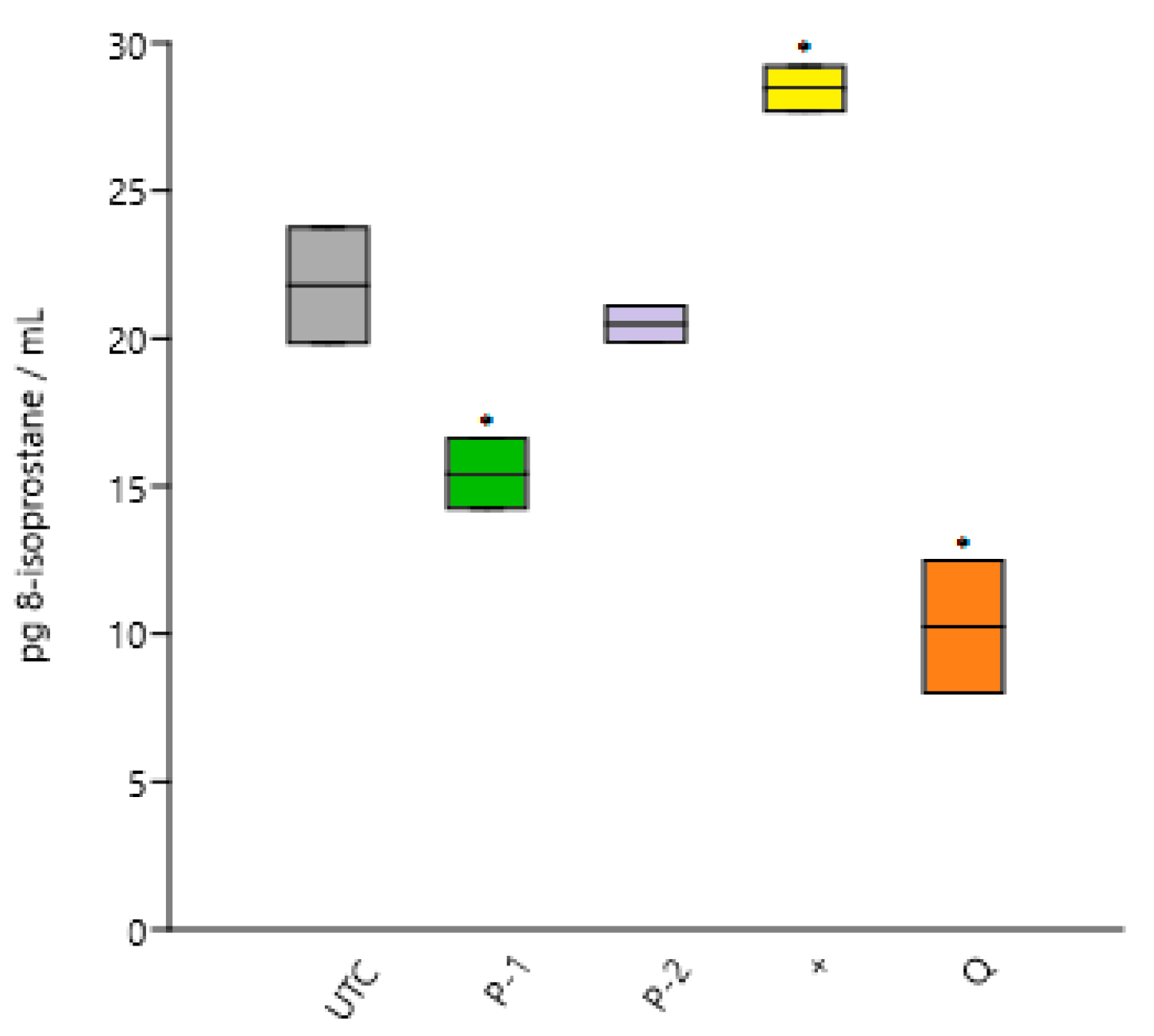
HaCat cells were seeded in 12-well plates (500,000 cells/mL) and pre-incubated overnight without (untreated control) or with diluted samples (1:750 v/v). After replacing the culture media, lipoxidation was provoked by incubating with AAPH 1 mM for 2.5 h. Supernatants were then removed, centrifuged and immediately investigated for 8-epi PGF2α concentrations using a commercial ELISA kit (item n. 516360, Cayman Chemical, USA) following manufacturer’s protocol. Results, expressed as the mean of three experiments ± standard deviation, were determined using Mars 2.0 Optima Data Analysis software (BMG LabTech GmbH, Germany).
2.11. Endogenous AGEs Measurement
HaCat cells were plated and incubated overnight in complete culture medium. For the experiments the culture medium was replaced with serum-free medium without (untreated control) or with diluted samples (1:750 v/v). After an hour of incubation, the medium was replaced with an AGEs-inducer solution containing glyoxal and/or S-p-bromobenzylglutathione cyclopentyl diester at increasing concentrations and incubated for 4 h. Whole protein lysates were processed for quantitative determination of AGEs with a commercial ELISA kit (Cusabio Ltd., Wilmington, DE, USA, Cat. No: CSB-E09412h) according to the manufacturer’s recommendations. Spectrophotometric measurements were recorded with a multiwell reader (Fluostar Optima, BMG Labtech, Germany), and the raw data were analyzed using Mars 2.0 Optima Data Analysis software (BMG LabTech GmbH, Germany) and expressed as the mean of two experiments ± standard deviation and as AGEs concentrations relative to protein content.
2.12. Bioavailability
A pilot, open-label, single-dose, two-period, cross-over design study was conducted in our laboratory to test the urinary excretion of free hydroxytyrosol and trans-resveratrol in self-reported healthy volunteers. In the investigation, two males and two females received, after an overnight fast, a single dose (50 mL) of one food supplement with 200 mL of water separated by one week wash-out period before administration of a single dose of the second food supplement. Urine samples were collected immediately before intake (baseline) and after 30 min of intake. The samples were centrifuged and filtered before being measured by LC-MS/MS, as described above. Freshly prepared urine-blank samples spiked with standards were used for the hydroxytyrosol calibration.
4. Discussion
Our study explores the antioxidant potential of two food supplements derived from olive vegetation water, mainly characterized by a high content of hydroxytyrosol. The antioxidant activity was determined by measuring the cellular antioxidant activity, the catalase and SOD activities in the HepG2 cell line, as well as the inhibition of lipid peroxidation and glycoxidation in the HaCat cell line. The tests carried out have shown that both olive-derived products have a strong positive influence on cells; this influence is complex and not one-dimensional. This reflects the complex nature of the sample materials and suggests a powerful synergy of hydroxytyrosol with other olive phenols, which is further potentiated in the presence of the grape phytocomplex. In particular, both supplements are able to reduce oxidative parameters in vitro.
On one hand, P-1, containing 94% olive water concentrate, showed a good capacity in the CAA and SOD assays, but a better performance in preventing isoprostanes formation in vitro when compared to P-2 (
Table 6). On the other hand, P-2, containing olive and grape concentrates, showed a greater antioxidant potential for scavenging reactive species, as indicated by its greater potential in removing the hydroxyl radicals and by its higher SOD and catalase activities. Regarding the prevention of AGEs accumulation, both products showed an excellent capacity in vitro. The overall better antioxidant performance of P-2 in vitro could be explained by the higher concentration of olive-derived polyphenols, as well as the presence of grape-derived antioxidants, which include, among others, the trans-resveratrol, anthocyanins and proanthocyanidin monomers.
Table 6 describes the cellular effects measured, and their links to the attributed in vivo effects.
The mechanism of this positive influence needs to be better understood and has led us to further investigations on the mechanisms and dynamics of the effects of food supplements derived from olive vegetation water on human cells. However, understanding the absorption and bioavailability of these key molecules after oral administration remains a prerequisite before any potential health effect can be derived. In this sense, the pilot trial shows that the hydroxytyrosol supplied with ahydrophilic matrix combining olive fruit concentrate and lemon or grape juices is effectively absorbed, and then urinarily excreted as hydroxytyrosol in its free form.
In subsequent studies, the exact excreted fraction will be determined, and further focus will be placed on the metabolites to obtain a broader picture of the entire ADME properties.
Overall, preliminary data obtained in vitro indicate that the aqueous extracts of olives can actually improve the cellular redox status and related markers, and that their main active ingredient is bioavailable to the human body. Aqueous olive concentrates, with or without grape concentrate, are valid candidates for the prevention of cellular oxidative damage, and thus merit further attention.


