Article

Isolation of Circulating Tumor Cells from Glioblastoma Patients by Direct Immunomagnetic Targeting

1
Centre for Circulating Tumour Cell Diagnostics & Research, Ingham Institute for Applied Medical Research, 1 Campbell St, Liverpool, NSW 2170, Australia
2
Western Sydney University Clinical School, Elizabeth St, Liverpool, NSW 2170, Australia
3
Liverpool Hospital, Elizabeth St & Goulburn St, Liverpool, NSW 2170, Australia
4
South Western Sydney Clinical School, University of New South Wales, Goulburn St, Liverpool, NSW 2170, Australia
5
School of Medicine, University of Wollongong, Wollongong, NSW 2522, Australia
*
Author to whom correspondence should be addressed.
Appl. Sci. 2020, 10(9), 3338; https://doi.org/10.3390/app10093338
Submission received: 8 April 2020 / Revised: 30 April 2020 / Accepted: 8 May 2020 / Published: 11 May 2020
(This article belongs to the Special Issue Circulating Biomarkers)

Abstract

:
Glioblastoma (GBM) is the most common form of primary brain cancer in adults and tissue biopsies for diagnostic purposes are often inaccessible. The postulated idea that brain cancer cells cannot pass the blood–brain barrier to form circulating tumor cells (CTCs) has recently been overthrown and CTCs have been detected in the blood of GBM patients albeit in low numbers. Given the potential of CTCs to be analyzed for GBM biomarkers that may guide therapy decisions it is important to define methods to better isolate these cells. Here, we determined markers for immunomagnetic targeting and isolation of GBM-CTCs and confirmed their utility for CTC isolation from GBM patient blood samples. Further, we identified a new marker to distinguish isolated GBM-CTCs from residual lymphocytes.

1. Introduction

Glioblastoma (GBM) is the most common form of primary brain cancer in adults and one of the most lethal forms of cancer; only 5% of patients survive five years [1]. Diagnosis requires surgery to obtain tissue for standard histopathological processing, but the location of some tumors makes access challenging [2]. Moreover, follow-up MRI scans can be difficult to interpret as to whether patients have chemoradiation-related treatment changes such as pseudo-progression versus recurrent disease, and timely clinical decision making may thus be hampered. Therefore, there is a critical need for new methods to determine prognosis and identify biomarkers that guide therapy decisions in GBM.
Circulating tumor cells (CTCs) are cancer cells that detach from the main tumor mass and enter the bloodstream which may be associated with the metastatic process. Over the past 15 years, clinical studies suggest that capture and enumeration of CTCs can provide important prognostic information [3,4,5]; moreover, these cells can be isolated and analyzed for molecular biomarkers [6]. For many years, it had been argued that CTCs were not present in GBM patients, due to the rarity of the development of the distant metastatic disease, and the belief that the inability of the cells to egress across the blood-brain barrier created an insurmountable difficulty. However, in 2014, CTCs were discovered in the bloodstream of GBM patients by three research teams. The proportion of CTC-positive patients ranged from 21% to 72% (29/141, 13/33 and 8/11), generally with low CTC counts. On this basis, it was proposed that CTCs could provide an alternative method of accessing GBM cells, compared to surgery, for analysis of relevant biomarkers [7].
The methods for detecting GBM-CTCs in these seminal studies involved separation from red blood cells by gradient centrifugation, alone or followed by CD45-targeted cell depletion to exclude white blood cells, combined with immunocytostaining for GBM specific markers, mainly glial fibrillary acidic protein (GFAP), or fluorescence in situ hybridization (FISH) probing of the chromosome 8 centromere to determine aneuploidy. Alternatively, viral transduction of green fluorescent protein expressed under the regulation of the telomerase (hTERT) promoter to identify cancer cells was used for CTC detection [8,9,10,11]. However, these methods of CTC detection may not be ideal, as millions of cells need to be screened for GBM markers by microscopy, or negative enrichment may cause loss of already rare CTCs due to potential antibody cross-reactions or physical co-segregation of CTCs amongst blood cells in the process. Further, current markers used to identify GBM-CTCs lack specificity, and clearly distinguishing CTCs from residual rare or common blood cells may either be associated with lack of specificity causing relatively high background staining, or potential of false-positive detection [10]; or lack of sensitivity due to marker heterogeneity in GBM cells, meaning some CTCs may be missed [8,12]. A more recent study used size exclusion to successfully detect GBM-CTCs and less frequently GBM-CTC clusters [13]; size exclusion CTC enrichment relies on tumor cells being larger than residual blood cells which is common, but not always the case.
Here, we tested a range of antibodies against candidate GBM cell surface proteins to develop a positive immunomagnetic GBM-CTC isolation protocol. We further screened for other GBM-CTC identification markers which established one additional marker useful for brain cancer CTC identification. Using this novel method, we have successfully isolated GBM-CTCs from GBM patient blood samples.

2. Materials and Methods

2.1. Cell Lines

The GBM cell lines A172, LN-229, T98G, U251 and U87 were cultured in DMEM media (Lonza, Basel, Switzerland) containing 10% foetal bovine serum (FBS) (Interpath, Melbourne, Australia), 4 mM L-glutamine (Sigma) and 20mM HEPES (Sigma) at 37 °C with 5% atmospheric CO2. All cell lines were STR-authenticated (AGRF, Melbourne, Australia) and tested as free of mycoplasma using the MycoAlert kit (Lonza, Basel, Switzerland).

2.2. Patients

Thirteen patients with a diagnosis of primary brain cancer were recruited from Liverpool Cancer Therapy Centre. Clinical information was sourced from patient medical records. Patient characteristics are summarized in Table 1. Per sample, 3x 9ml EDTA vacutubes (Greiner Bio-One, Frickenhausen, Germany) of peripheral blood were drawn. Blood samples from healthy individuals were analyzed as controls. The study was undertaken with written consent and approval of the South Western Sydney Human Ethics Committee (HREC/13/LPOOL/158).

2.3. Immunocytostaining

Cell lines: 1 × 10⁵ cells were seeded on glass coverslips and grown for 72 hours at 37 °C before immunocytostaining. For cell-surface protein probing, cells were fixed using 3.7% formaldehyde (VWR, Radnor, USA), blocked in 10% FBS in PBS for 10min, and incubated with primary anti-human MCSP antibody (αMCSP) and anti-human MCAM antibody (αMCAM) for 1 hour (see Table S1 for antibody details and dilutions). The cells were washed three times with PBS and incubated for 30 min with secondary anti-mouse IgG AlexaFluor488 antibodies (Thermo Fisher), washed twice with PBS and once with distilled H2O. The coverslips were mounted on slides with mounting media containing Hoechst-dye (Fluxion, San Francisco, CA, USA ).
For cytoplasmic protein detection cells were permeabilized after fixing using 0.2% Triton-X in PBS for 10 minutes (Sigma-Aldrich, St. Louis, USA). The cells were then incubated for 1 hour with a primary antibody, anti-human GFAP (αGFAP), followed by AF555 conjugated anti-rabbit-IgG antibody probing (Thermo Fisher), FITC-conjugated anti-human glutamate aspartate transporter (GLAST) (αGLAST) (Novus Biologicals, Centennial, CA, USA) or both (see Table S2 for dilutions). The coverslips were mounted using Hoechst-dye containing mounting media (Fluxion) and were visualized on a LSM 800 laser-scanning confocal microscope with AiryScan (Carl Zeiss, Oberkochen, Germany) or Olympus BX53 microscope (Olympus, Notting Hill, Australia).

2.4. Flowcytometry

Cells were seeded at 50% confluency in tissue culture flasks cultured for 2 days and harvested using 0.5mM EDTA in PBS to maintain cell surface proteins. Cells were blocked with 10% FBS in PBS and probed sequentially with primary and secondary antibodies for 30 and 20 minutes respectively (Table S1) and resuspended in 300 µl of PBS for flowcytometry (FACS) analysis (FACS Canto II Cell Analyzer, BD Biosciences). Flowing Software 2.5.1 was used for analysis (Turku Centre for Biotechnology, Turku, Finland). Antigen detectability of ≤ 3 ± 2% of a cell line’s cell population was considered negative in regards to suitability as the isolation target.

2.5. Immunomagnetic Cell Isolation

Immunomagnetic beads, with a Rare Cell Isolation Kit (Fluxion, San Francisco, USA), were incubated with αMCAM or αMCSP antibodies for conjugation according to the manufacturer’s protocol (see Table S1 for antibody concentrations). For basic confirmation of suitability for GBM cell isolation n = 100 LN-229 cells were suspended in 800 µL binding buffer (Fluxion), and, after addition of 40 µL FC buffer (Fluxion) and either 30 µL αMCAM antibody coupled beads, 30 µL αMCSP antibody coupled beads or 30 µL of each, incubated for 90 minutes at 4 °C on a rotating platform. Cells were then isolated by magnetic force using a magnetic rack, washed three times with binding buffer, fixed with 4% formaldehyde in PBS, washed, immunocytostained for GBM markers and Hoechst and enumerated using the CellCelector (ALS) for fluorescent imaging.
Patient and healthy donor blood samples were processed within 24-hours using lymphoprep and Sepmate tubes (Stemcell Technologies, Vancouver, Canada) to separate the peripheral blood mononuclear cells (PBMCs) according to the manufacturer’s instructions. PBMCs derived from 27 mL blood each were washed once in PBS and resuspended in 800 µL binding buffer and incubated with 30 µL αMCAM coupled and 30 µL αMCSP coupled magnetic beads. Cells were incubated for 90 minutes at 4 °C on a a rotating platform and then loaded into primed IsoFlux cartridges for CTC enrichment using the IsoFlux CTC isolation platform with the standard isolation protocol (Fluxion). Enriched CTCs samples were fixed with 3.7% formaldehyde in PBS before immunocytostaining.

2.6. Immunocytostaining of Circculating Tumor Cells

Anti-human CD45-AF647 conjugate (Thermo Fisher) was used to stain residual blood cells; before permeabilization (0.2% Triton X-100 in PBS) and incubation with rabbit αGFAP combined with AF555 anti-rabbit IgG secondary probing and FITC-conjugated αGLAST antibody, after two washes cells were placed on slides with Hoechst dye in the mounting media (See Table S2 for antibody dilutions). Stained CTCs were visualized and enumerated using the CellCelector fluorescent microscope (ALS, Jena Germany).

3. Results

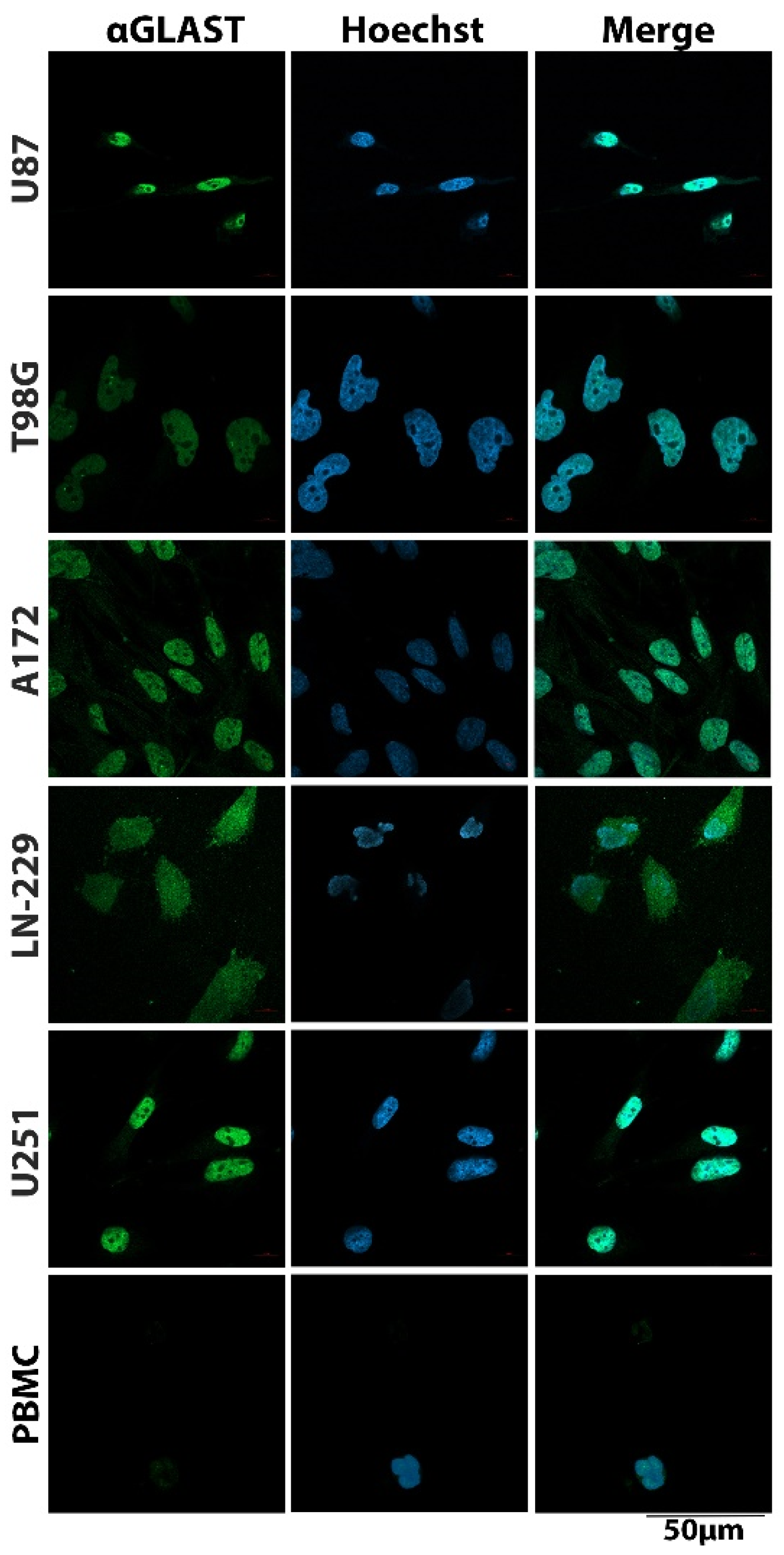
3.1. Cell Surface Protein Expression on Glioblastoma Cells

To identify appropriate cell surface targets for immunomagnetic isolation of GBM cells, a number of cell surface proteins identified from literature searches were screened with appropriate antibodies for the presence on a cohort of GBM cell lines by FACS analysis (Table 2, Table S1). Antibodies that demonstrated limited reactivity to GBM cells (≤3% ± 2% of the cell population positive) were disregarded for isolation. Those that reacted with GBM cells were further tested for antibody cross-reactivity with healthy donor peripheral blood mononucleated cells (PBMCs); considerable PBMC cross-reactivity suggested unsuitability as potential tools for immunomagnetic GBM-CTC isolation due to expected and undesirable co-capture of PBMCs. The remaining antibodies against promising target proteins were further evaluated by immunocytostaining.
Since neural cells and melanocytes are embryologically related (both originate developmentally from the neural crest) [14], we included antibodies against the melanoma-associated chondroitin sulfate proteoglycan (MCSP) and melanoma cell adhesion molecule (MCAM) in our screen. We have previously shown both proteins to be suitable targets for immunomagnetic melanoma CTC isolation [15]. After the exclusion of other candidates, MCSP and MCAM were identified as the most promising cell surface protein targets for immunomagnetic isolation of GBM cells. MCSP was detected on up to 92% of the cell populations of GBM cell lines tested with high proportions of LN-229 (92%), U87 (89%) and A172 (80%) cells expressing the antigen. However, both T98G and U251 cells were MCSP negative. MCAM was expressed on 43%–95% of the cells of all cell lines. Very high proportions of LN-229 (95%), most U87 (68%) and T98G (64%) cells were positive for MCAM, while around half of A172 (51%) and U251 (43%) cell populations expressed MCAM (Table 2, Figure 1). Importantly, the anti-MCSP (αMCSP) and anti-MCAM (αMCAM) antibodies showed negligible reactivity with PBMCs from healthy donors (Table 2).
Cell surface expression of MCAM (left) and MCSP (right) was screened on the indicated cell lines by immunocytostaining (specific protein detected in green FITC channel, while Hoechst staining detects cell nuclei, merged images included) and flow cytometry. Proportions of cell populations expressing the indicated protein are stated from at least n = 2 independent experiments (as gated on flowcytometry data comparing the light grey protein-specific peak to isotype-matched IgG probing (dark grey peak)). Representative histograms included.

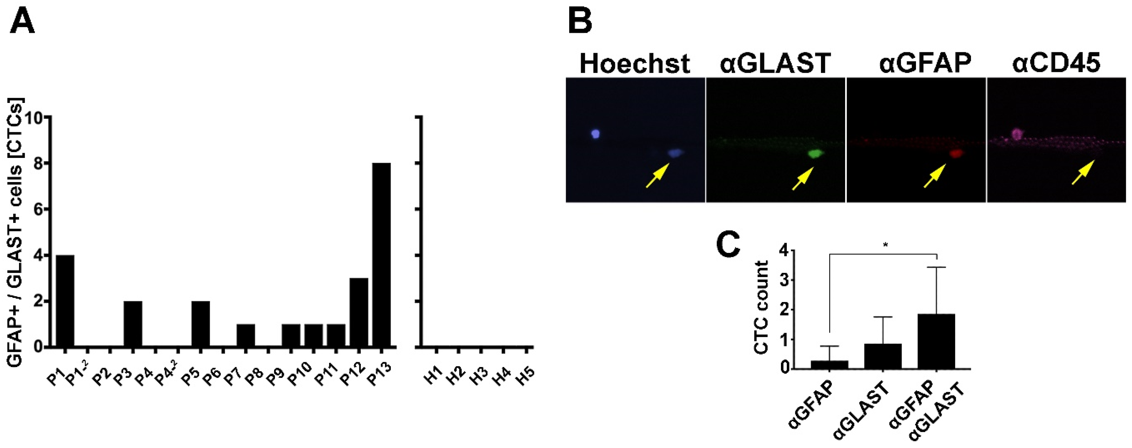
3.2. GBM-CTC Identification Markers

It is important to emphasize that CTC isolation methods usually deliver enrichment of CTCs rather than complete isolation, therefore further CTC identification to distinguish them from residual lymphocytes is generally required. Therefore, we also screened for alternate markers for the identification of GBM-CTCs in addition to GFAP, which is known to be heterogeneously expressed in GBM [12]. Immunocytostaining was used on our cohort of brain cancer cell lines for a range of proteins expressed in GBM (Table S2). The GLAST was found to be expressed in all GBM cell lines (~98–100% of the cell populations). Moreover, only negligible cross-reactivity was found between the αGLAST antibody and healthy donor PBMCs (Figure 2), suggesting GLAST may be a suitable identification marker for brain cancer CTCs. Other potential markers had either poor expression in GBM cell lines or strong cross-reactivity of the corresponding antibodies with PBMCs (Table S2, Figure S1). Notably, GFAP was in our hands not detected in any GBM cell line (data not shown).
Representative images of the indicated cell lines and healthy donor PBMCs stained using FITC conjugated αGLAST antibody (green, left panel), Hoechst (blue, center panel) with merged images shown (right panel) are shown. Note, the nuclear localization of GLAST in most GBM cell lines, only LN-229 cells show ubiquitous GLAST localization.

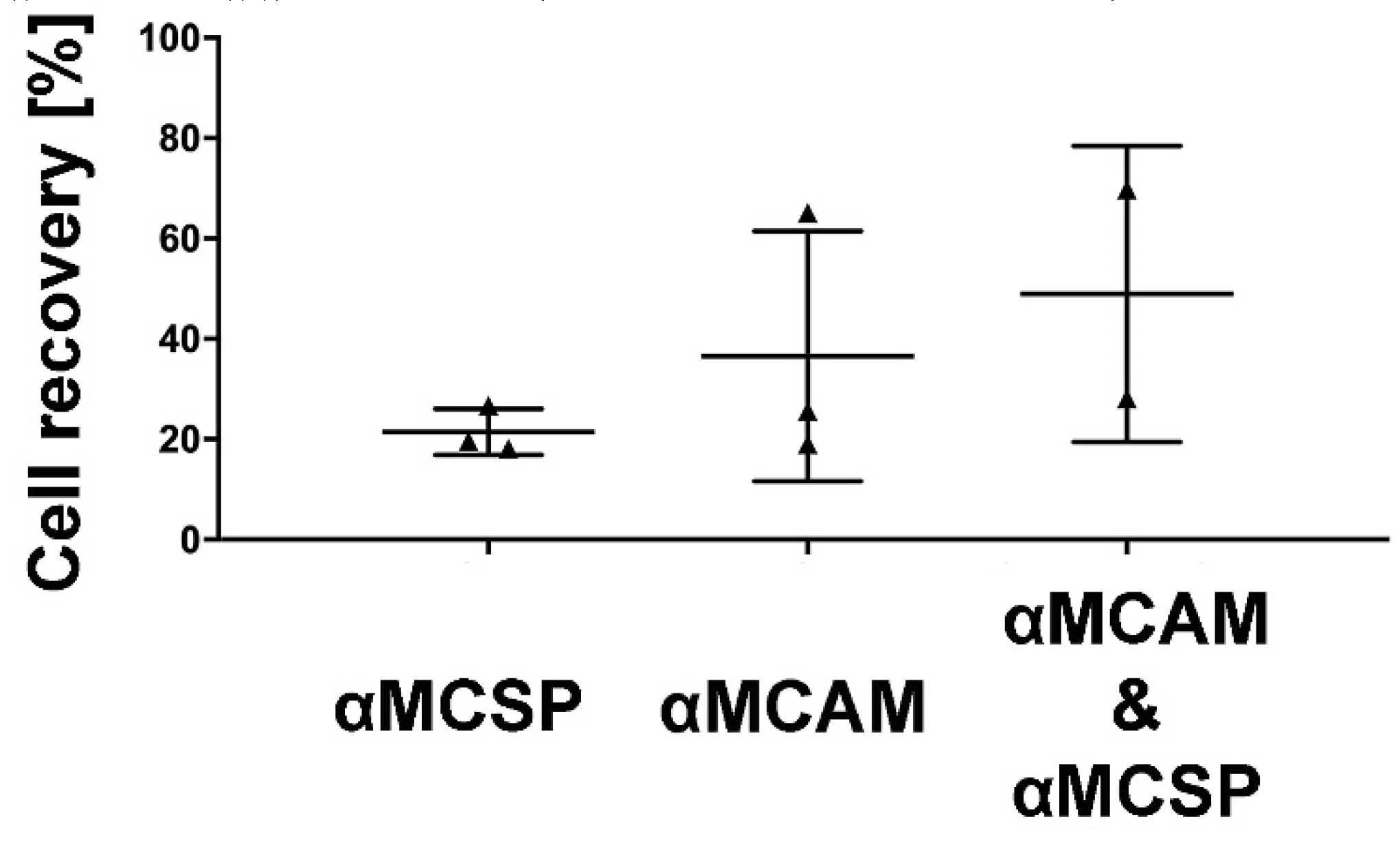
3.3. Immunomagnetic Capture of GBM Cells

To confirm that both MCAM and MCSP were suitable immunomagnetic targets for GBM cell isolation, we used LN-229 cells, due to MCAM and MCSP co-expression as determined by flow cytometry. Individually, targeting MCAM for cell isolation performed slightly better than MCSP targeting (26% versus 20% recovery) but there was a clear advantage of targeting both antigens in combination, which captured approximately 50% LN-229 cells (Figure 3).
The indicated antibodies were used to immunomagnetically recover LN-229 cells. Data from at least n = 2 independent experiments are presented. Horizontal error bars: mean range.
We then tested our method for GBM-CTC isolation on 15 blood samples from 13 patients (samples from two patients were included at 2 independent time points approximately 3 months apart). The amount of blood available for CTC isolation was 27 mL (3 × 9 mL) per patient. We successfully isolated 1-8 CTCs from 60% (9/15) of GBM patient blood draws when targeting the combination of MCAM and MCSP for CTC isolation (Figure 4A). Two-sided Fisher’s exact test established significance (p-value = 0.0379) in isolating cells that are positive for GLAST and/or GFAP from GBM patients vs healthy blood donors. Importantly, a comparison of CTC identification by GFAP and GLAST staining across all patient samples confirmed that the combination of both identification antibodies increased CTC detection (Figure 4B,C).
No correlation between CTC positivity and patient progression-free survival or overall survival was found in the small patient cohort (Figure 5).
CTC numbers were quantified for 13 patients and follow up progression-free survival (PFS) and overall survival (OS) were determined for each patient and are presented for CTC positive (red) and CTC negative patients (blue) as Kaplan–Meier graphs.

4. Discussion

The aim of this study was to develop a positive immunomagnetic enrichment strategy for GBM-CTCs and improve identification markers for GBM-CTCs.
A cohort of neurological cell-specific, cell surface markers were considered less useful for CTC isolation since there was no, the low or variable expression on cultured GBM cells or when the expression on common blood cells was detected with the validated antibodies used in our study. While most of the markers we screened had previously been detected by others on cultured GBM cells or tissue (Tables S1 and S2), our inability to replicate some of these findings in our cultured GBM cells may be due to factors such as different antibodies used, different protein levels between cultured cells and GBM tissue or increasing heterogeneity of long term established cell cultures used in the various laboratories. The most promising markers, MCAM and MCSP, have been successfully used by us and others for the isolation of melanoma CTCs previously [15,16]. Since melanocytes originate from the neural crest [14], the detection of GBM cells is not surprising. It is important to note however that MCAM and MCSP were not equally well expressed on all tested GBM cell lines (proportion of positive cell population, Figure 1). If such heterogeneity translates to patient-derived GBM-CTCs, GBM-CTCs expressing low antigen would be captured less efficiently.
While immunomagnetic targeting of MCAM and MCSP proved independently successful for the capture of LN-229 cells, the combination of both antibodies was superior for cell isolation. Additionally, our previous data clearly show that the strategy of targeting both proteins together improved melanoma CTC capture from patient blood [15]. Therefore, we used the same strategy for successful GBM-CTCs enrichment from patient samples. CTC counts for our study were low, which is consistent with reported GBM-CTC counts in other studies [8,9,10,13] and our immunomagnetic targeted CTC isolation produces better CTC enrichment (ratio CTCs: residual blood cells) than some previous studies likely to make certain down-stream analyses easier. This is the first method positively immune-targeting GBM-CTCs for enrichment. Recently, another study did achieve GBM-CTC enrichment by conjugating a malaria protein, VAR2CSA, to magnetic beads. VAR2CSA has a high affinity to chondroitin sulfate proteoglycans proposed to be uniquely expressed on cancer cells. The study isolated CTCs in comparable numbers to our study [17]. Of note, MCSP belongs to the chondroitin sulfate proteoglycan protein family and our data confirm high expression on some GBM cell lines but total lack on others, highlighting that targeting more than one cell surface protein should be considered advantageous for GBM-CTC isolation.
In our hands, markers such as nestin, previously suggested as suitable for GBM-CTC detection, were less satisfactory [9]; this may have been due to a different anti-nestin antibody (raised in rabbit) used here, due to compatibility with our isolation antibodies. Since our isolation antibodies were raised in mice, a mouse-anti-nestin antibody would have produced strong cross-reactivity with magnetic beads. Our nestin probing showed positivity in GBM cells but also PBMCs (Figure S1). Nevertheless, we identified one marker to better distinguish GBM-CTCs from residual lymphocytes. GLAST, supposedly present on all brain cells, is a transporter protein, and is normally cell-membrane localized, but can be internalized and predominantly found in the nucleus in GBM tissue and cultured GBM cells [18]. Our GBM cell staining confirmed nuclear GLAST for all but LN-229 cells (Figure 2). The recognition of anti-GLAST as an additional identification antibody is important, since the commonly used GFAP identification marker protein is not present in all GBM cells [12]. Interestingly, in our hands, immunocytostaining did not detect GFAP in any of the GBM cell lines tested, whereas GLAST was readily detected in all five GBM cell lines. However, GFAP expression is reportedly often lost in cultured cells, likely via epigenetic silencing [19]; thus, our inability to detect it in cell lines may be an artifact of cell culture. Since GFAP probing was previously used to successfully identify GBM-CTCs by others [8], we retained it as a CTC identification marker and successfully detected GFAP in patient GBM-CTCs. Here, we confirmed heterogeneity for GFAP and GLAST expression in patient CTCs; and showed that probing for both identification markers is significantly superior to only probing for GFAP to identify GBM-CTCs (Figure 4C). Combining GFAP with GLAST for GBM-CTC detection can, therefore, function as an additional validation marker for the GBM origin of these cells, as well as aid in identifying more CTCs overall.
The major limitation of our study is the small patient cohort, which makes it premature to try to establish a correlation between GBM-CTC counts and disease outcomes. To date, the overall small investigated patient numbers in all relevant studies that tested for correlations of GBM-CTC counts to sub-type of glioma, histological grade, pre- and post-surgical tumour burden, treatment response, survival or pseudoprogression were challenging [8,9,11]. However, the advent of novel techniques such as single-cell methodologies has enabled successful interrogation of even single CTCs for clinical biomarkers, thus detecting even small CTC numbers that may provide a source of biomarker detection [6]. Given the rarity of primary brain cancer CTCs, obtaining larger blood volumes such as 50 mL would be desirable and still remain less invasive than surgical biopsy or lumbar puncture cerebral spinal fluid (CSF) collection. Another, technical limitation of this study is that we based screening for potential immunomagnetic cell surface targets on cultured monolayer cells only. As discussed, cell culture may change protein expression patterns and thus reflect less truly expression of proteins for cancer tissue or, important for this study, CTCs. While including examination of cell surface markers in tissue and potentially cultured cells that grow in spheres may have revealed additional immunomagnetic targets, their true value for CTC isolation may only be shown by direct comparison for CTC isolation from patient samples; and the limitation is the amount of patient blood that can be drawn to allow for multiple comparisons. Here we tested two immunomagnetic isolation target candidates and have shown that GBM CTCs are captured targeting these markers. This could become a benchmark to test other isolation target candidates against in the future.

5. Conclusions

We have established an immunomagnetic method for targeted isolation of GBM-CTCs and identified a novel GBM-CTC identification marker. GLAST probing can complement GFAP probing for improved GBM-CTC identification. Our GBM-CTC isolation and identification methods are a viable, relatively simple strategy for detecting GBM-CTCs from patient samples. Since repeated brain cancer biopsies from surgery or even repeated CSF collection are not clinically justified, it is possible that blood-based CTC analysis may become a feasible way to access biomarker information for optimal GBM patient management.

Supplementary Materials

Supplementary materials can be found at https://www.mdpi.com/2076-3417/10/9/3338/s1. Figure S1: Nestin expression in GBM cells and PBMCs. Table S1: Isolation Marker Candidates.; Table S2: Identification Marker Candidates.

Author Contributions

Conceptualization: D.L., T.M.B., T.L.R., P.d.S.; methodology: D.L., B.P., J.W.P., C.G., formal analysis: D.L., B.P., J.W.P., C.G., T.M.B., T.L.R., P.d.S.; resources: T.M.B., P.d.S., A.C., E.-S.K., M.S., J.v.G., B.D., S.M., R.B., M.J.; writing—original draft preparation: T.M.B., D.L., B.P.; writing—review and editing: D.L., B.P., J.W.P., A.C., E.S.K., M.S., J.v.G., B.D., S.M., R.B., M.J., C.G., T.L.R., P.d.S., T.M.B., supervision: T.M.B., T.L.R., P.d.S.; project administration: T.B., P.d.S. All authors have read and agreed to the published version of the manuscript.

Funding

This work was supported by the Cancer Institute New South Wales through the Centre for Oncology Education and Research Translation (CONCERT, grant ID: 13/TRC/1-01) and Western Sydney University. D.L. is recipient of an Ingham Research Institute Director’s PhD Scholarship. J.P. is recipient of an Australian Rotary Health PhD Scholarship.

Acknowledgments

Human ethics approval, HREC/13/LPOOL/158, was obtained and managed by the CONCERT Biobank.

Conflicts of Interest

The authors declare no conflict of interest.

References

  1. Ostrom, Q.T.; Gittleman, H.; Liao, P.; Rouse, C.; Chen, Y.; Dowling, J.; Wolinsky, Y.; Kruchko, C.; Barnholtz-Sloan, J. CBTRUS statistical report: Primary brain and central nervous system tumors diagnosed in the United States in 2007–2011. Neuro-Oncology 2014, 16, iv1–iv63. [Google Scholar] [CrossRef] [PubMed]
  2. Omuro, A.; DeAngelis, L.M. Glioblastoma and other malignant gliomas: A clinical review. JAMA 2013, 310, 1842–1850. [Google Scholar] [CrossRef] [PubMed]
  3. de Bono, J.S.; Scher, H.I.; Montgomery, R.B.; Parker, C.; Miller, M.C.; Tissing, H.; Doyle, G.V.; Terstappen, L.W.; Pienta, K.J.; Raghavan, D. Circulating tumor cells predict survival benefit from treatment in metastatic castration-resistant prostate cancer. Clin. Cancer Res. 2008, 14, 6302–6309. [Google Scholar] [CrossRef] [PubMed] [Green Version]
  4. Alva, A.; Friedlander, T.; Clark, M.; Huebner, T.; Daignault, S.; Hussain, M.; Lee, C.; Hafez, K.; Hollenbeck, B.; Weizer, A. Circulating tumor cells as potential biomarkers in bladder cancer. J. Urol. 2015, 194, 790–798. [Google Scholar] [CrossRef] [PubMed]
  5. Hayes, D.F.; Cristofanilli, M.; Budd, G.T.; Ellis, M.J.; Stopeck, A.; Miller, M.C.; Matera, J.; Allard, W.J.; Doyle, G.V.; Terstappen, L.W. Circulating tumor cells at each follow-up time point during therapy of metastatic breast cancer patients predict progression-free and overall survival. Clin. Cancer Res. 2006, 12, 4218–4224. [Google Scholar] [CrossRef] [PubMed] [Green Version]
  6. Becker, T.M.; Caixeiro, N.J.; Lim, S.H.; Tognela, A.; Kienzle, N.; Scott, K.F.; Spring, K.J.; Souza, P. New frontiers in circulating tumor cell analysis: A reference guide for biomolecular profiling toward translational clinical use. Int. J. Cancer 2014, 134, 2523–2533. [Google Scholar] [CrossRef] [PubMed] [Green Version]
  7. Best, M.G.; Sol, N.; Zijl, S.; Reijneveld, J.C.; Wesseling, P.; Wurdinger, T. Liquid biopsies in patients with diffuse glioma. Acta Neuropathol. 2015, 129, 849–865. [Google Scholar] [CrossRef] [PubMed] [Green Version]
  8. Müller, C.; Holtschmidt, J.; Auer, M.; Heitzer, E.; Lamszus, K.; Schulte, A.; Matschke, J.; Langer-Freitag, S.; Gasch, C.; Stoupiec, M. Hematogenous dissemination of glioblastoma multiforme. Sci. Transl. Med. 2014, 6, ra101–ra247. [Google Scholar] [CrossRef] [PubMed]
  9. MacArthur, K.M.; Kao, G.D.; Chandrasekaran, S.; Alonso-Basanta, M.; Chapman, C.; Lustig, R.A.; Wileyto, E.P.; Hahn, S.M.; Dorsey, J.F. Detection of brain tumor cells in the peripheral blood by a telomerase promoter-based assay. Cancer Res. 2014, 74, 2152–2159. [Google Scholar] [CrossRef] [PubMed] [Green Version]
  10. Sullivan, J.P.; Nahed, B.V.; Madden, M.W.; Oliveira, S.M.; Springer, S.; Bhere, D.; Chi, A.S.; Wakimoto, H.; Rothenberg, S.M.; Sequist, L.V. Brain tumor cells in circulation are enriched for mesenchymal gene expression. Cancer Discov. 2014, 4, 1299–1309. [Google Scholar] [CrossRef] [PubMed] [Green Version]
  11. Gao, F.; Cui, Y.; Jiang, H.; Sui, D.; Wang, Y.; Jiang, Z.; Zhao, J.; Lin, S. Circulating tumor cell is a common property of brain glioma and promotes the monitoring system. Oncotarget 2016, 7, 71330. [Google Scholar] [CrossRef] [PubMed]
  12. Schiffer, D.; Giordana, M.T.; Germano, I.; Mauro, A. Anaplasia and heterogeneity of GFAP expression in gliomas. Tumori 1986, 72, 163–170. [Google Scholar] [CrossRef] [PubMed]
  13. Krol, I.; Castro-Giner, F.; Maurer, M.; Gkountela, S.; Szczerba, B.M.; Scherrer, R.; Coleman, N.; Carreira, S.; Bachmann, F.; Anderson, S.; et al. Detection of circulating tumour cell clusters in human glioblastoma. Br. J. Cancer 2018, 119, 487–491. [Google Scholar] [CrossRef] [PubMed] [Green Version]
  14. Yaar, M.; Park, H.Y. Melanocytes: A window into the nervous system. J. Investig. Dermatol. 2012, 132, 835–845. [Google Scholar] [CrossRef] [PubMed] [Green Version]
  15. Po, J.W.; Ma, Y.; Balakrishna, B.; Brungs, D.; Azimi, F.; de Souza, P.; Becker, T.M. Immunomagnetic isolation of circulating melanoma cells and detection of PD-L1 status. PLoS ONE 2019, 14, e0211866. [Google Scholar] [CrossRef] [PubMed]
  16. Gray, E.S.; Reid, A.L.; Bowyer, S.; Calapre, L.; Siew, K.; Pearce, R.; Cowell, L.; Frank, M.H.; Millward, M.; Ziman, M. Circulating Melanoma Cell Subpopulations: Their Heterogeneity and Differential Responses to Treatment. J. Investig. Dermatol. 2015, 135, 2040–2048. [Google Scholar] [CrossRef] [PubMed] [Green Version]
  17. Bang-Christensen, S.R.; Pedersen, R.S.; Pereira, M.A.; Clausen, T.M.; Loppke, C.; Sand, N.T.; Ahrens, T.D.; Jorgensen, A.M.; Lim, Y.C.; Goksoyr, L.; et al. Capture and Detection of Circulating Glioma Cells Using the Recombinant VAR2CSA Malaria Protein. Cells 2019, 8, 998. [Google Scholar] [CrossRef] [PubMed] [Green Version]
  18. Ye, Z.-C.; Rothstein, J.D.; Sontheimer, H. Compromised glutamate transport in human glioma cells: Reduction–mislocalization of sodium-dependent glutamate transporters and enhanced activity of cystine–glutamate exchange. J. Neurosci. 1999, 19, 10767–10777. [Google Scholar] [CrossRef] [PubMed] [Green Version]
  19. Restrepo, A.; Smith, C.A.; Agnihotri, S.; Shekarforoush, M.; Kongkham, P.N.; Seol, H.J.; Northcott, P.; Rutka, J.T. Epigenetic regulation of glial fibrillary acidic protein by DNA methylation in human malignant gliomas. Neuro-Oncology 2011, 13, 42–50. [Google Scholar] [CrossRef] [PubMed] [Green Version]
Figure 1. Melanoma cell adhesion molecule (MCAM) and MCSP on glioblastoma (GBM) cells.
Applsci 10 03338 g001
Figure 2. Glutamate aspartate transporter (GLAST) expression in GBM cells.
Applsci 10 03338 g002
Figure 3. GBM cell capture.
Applsci 10 03338 g003
Figure 4. CTC isolation from GBM patient blood. Fifteen 27 mL patient blood samples from 13 GBM patients (P1-P13) were analyzed for GBM-circulating tumor cells (CTCs) using combined immunomagnetic targeting of MCAM and MCSP. CTCs were detected by αGFAP and αGLAST probing. (A) CTC counts for 13 patients/15 samples (patients with repeat samples are indicated -2). The same isolation protocol was used for 5 healthy donor blood samples (H1-H5) and detected no false positive “CTCs”. (B) Typical identification of a CTC (arrow) by nuclear staining (Hoechst), GLAST (green) and GFAP (red) positivity as well as lack of CD45 (pseudo color purple). (C) For the nine GBM-CTC positive patient blood draws, the mean of CTCs detected by either αGFAP or αGLAST probing alone or detected by double positivity is presented. Columns indicate the average and error bars standard deviations. * indicates significance (p ≤ 0.05).
Applsci 10 03338 g004
Figure 5. CTC status and survival.
Applsci 10 03338 g005
Table 1. Patient characteristics.
Patient GenderAge #GradeHistologyTumour LocationECOG 1ECOG 2
1M614GBMLeft tempoparietal region10
2M474GBMRight parietal lobe01
3M324GBMLeft frontal lobe01
4F304GSM *Right frontoparietal region00
5F474GBMRight temporal lobe10
6M754GBMCerebellum11
7M364GBMRight parietal lobe01
8M334GBMRight frontal lobe00
9M684GBMRight temporal lobe12
10M604GBMLeft frontal lobe22
11F504GBMBifrontal region03
12F714GBMLeft parietal lobe12
13M834GBMRight frontal lobe11
M male; F female; * re-diagnosed as GSM (gliosarcoma); # at diagnosis; ECOG1 Eastern Cooperative Oncology Group score pre-surgery, ECOG2 Eastern Cooperative Oncology Group score post-surgery.
Table 2. Cell surface protein detection on cell lines by flowcytometry.
Detectable [% of Cells]
AntigenA172LN-229T98GU87U251PBMCs
MCAM 51.0 ± 3.694.6 ± 1.764.1 ± 5.467.8 ± 4.942.7 ± 0.1-
MCSP80.4 ± 4.192.1 ± 2.6- 89.3 ± 13.7- -
N-Cadherin70.2 ± 13.855.3 ± 0.617.1 ± 0.6-94.3 ± 3.3-
EGFR96.8 ± 0.378.1 ± 1.329.2 ± 0.411.2 ± 3.234.5 ± 8.3-
CD2715.9 ± 0.157.3 ± 0.48.6 ± 10.5---
A2B5- - - - n/dn/d
-: Values ≤ 3.0 ± 2% were considered negative for purpose of isolation target candidates; n/d: not determined due to early elimination of isolation candidate.

