Article

Evaluation of Aloe Vera Coated Polylactic Acid Scaffolds for Bone Tissue Engineering

by
Ricardo Donate
1,*,
María Elena Alemán-Domínguez
1,
Mario Monzón
1,
Jianshu Yu
2,3,
Francisco Rodríguez-Esparragón
4 and
Chaozong Liu
2
1
Departamento de Ingeniería Mecánica, Grupo de Investigación en Fabricación Integrada y Avanzada, Universidad de Las Palmas de Gran Canaria, Campus Universitario de Tafira s/n, 35017 Las Palmas, Spain
2
Institute of Orthopaedic & Musculoskeletal Science, University College London, Royal National Orthopaedic Hospital, Stanmore HA4 4LP, London, UK
3
School of Materials Science and Engineering, Shanghai University, Shanghai 200444, China
4
Unidad de Investigación, Hospital Universitario de Gran Canaria Doctor Negrín, Barranco de la Ballena s/n, 35010 Las Palmas, Spain
*
Author to whom correspondence should be addressed.
Appl. Sci. 2020, 10(7), 2576; https://doi.org/10.3390/app10072576
Submission received: 17 February 2020 / Revised: 26 March 2020 / Accepted: 7 April 2020 / Published: 9 April 2020
(This article belongs to the Special Issue Patient-Tailored Biomimetic Scaffold Constructs for Bone Regeneration)

Abstract

:
3D-printed polylactic acid (PLA) scaffolds have been demonstrated as being a promising tool for the development of tissue-engineered replacements of bone. However, this material lacks a suitable surface chemistry to efficiently interact with extracellular proteins and, consequently, to integrate into the surrounding tissue when implanted in vivo. In this study, aloe vera coatings have been proposed as a strategy to improve the bioaffinity of this type of structures. Aloe vera coatings were applied at three different values of pH (3, 4 and 5), after treating the surface of the PLA scaffolds with oxygen plasma. The surface modification of the material has been assessed through X-ray photoelectron spectroscopy (XPS) analysis and water contact angle measurements. In addition, the evaluation of the enzymatic degradation of the structures showed that the pH of the aloe vera extracts used as coating influences the degradation rate of the PLA-based scaffolds. Finally, the cell metabolic activity of an in vitro culture of human fetal osteoblastic cells on the samples revealed an improvement of this parameter on aloe vera coated samples, especially for those treated at pH 3. Hence, these structures showed potential for being applied for bone tissue regeneration.

Graphical Abstract

1. Introduction

Polylactic acid (PLA)-based scaffolds have been extensively used for bone regeneration [1,2,3,4,5,6], as they have a suitable degradation rate and mechanical properties to replace the tissue in non-load-bearing applications. However, PLA is a hydrophobic material and, as a consequence, its interaction with the extracellular matrix (ECM) is low [7]. As the adsorption of adhesion-signaling proteins from the ECM is a key step to promote a good osseointegration of the scaffolds [8], the limited affinity of this biomaterial to these substances in the ECM is one of the main drawbacks of PLA-based structures.
Different strategies have been proposed to overcome this limitation and increase the number of applications of PLA-based structures in the field of tissue engineering. An important approach comprises the incorporation of additives, including bioceramics [9], graphene oxide [10] and natural-derived polymers [11]. Another strategy is the surface modification of the scaffold [12] by changing the roughness, wettability and/or surface chemistry of the structures [8]. The processes to change the properties of the surface can be classified into two groups: the functionalization of PLA or the coating of the structure with a bioactive compound able to interact effectively with the proteins in the ECM. In the first group of techniques, it is possible to mention the plasma [13,14] or alkaline treatment [15] of the parts. These surface treatments can be used to provide anchoring points to link bioactive compounds to the surface of PLA-based scaffolds. Different substances have been proposed with this aim, including chitosan [16], alginate [17], collagen [7,12,18] or calcium phosphates [19].
In this work, the authors have tested the effect of applying an innovative coating based on extracts of aloe vera to 3D-printed PLA-based scaffolds. Aloe vera is a plant from the Liliaceal family with demonstrated healing properties [20]. In the tissue engineering field, it has been tested mainly for skin regeneration [21,22]. However, the use of this natural substance in bone tissue engineering remains limited [23] to the best of our knowledge and no references have been found regarding its use to improve the affinity of PLA and promote the attachment and proliferation of osteoblastic cells.
According to the state of the art on the chemical characterization of aloe vera extracts [24,25], acemannan is the main bioactive compound in the gel of the leaves. It is a mannose-containing polysaccharide that has demonstrated the promotion of collagen synthesis during wound healing [21,26]. This characteristic could have positive implications for enhancing the osseointegration process in aloe vera coated scaffolds. Besides, there is currently a high availability of this product at affordable prices, which makes it a viable strategy from the economic point of view compared with other options, such as the use of growth factors [27]. For these reasons, this strategy has been proposed in this work as a promising approach to improve the performance of 3D-printed PLA scaffolds in vitro. Plasma treatment was used as a first step to functionalize the surface of the materials and promote aloe vera adhesion.

2. Materials and Methods

2.1. Materials

PLA L105 (melt flow index of 65 g/10 min, molecular weight of approximately 105,000 g·mol−1) was purchased from Corbion Purac (Diemen, The Netherlands) in powder form. Aloe vera juice was purchased from aloe vera Costa Canaria (Spain). The quality protocols from the company indicated that the content in aloin is below 0.0007% w/w (analysis by high performance liquid chromatography). Sodium hydroxide (NaOH) in pellets form was purchased from Honeycomb (30620, FlukaTM), while hydrochloric acid (HCl) was purchased from Merck (1090571000, Supelco).

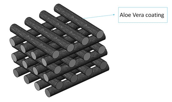
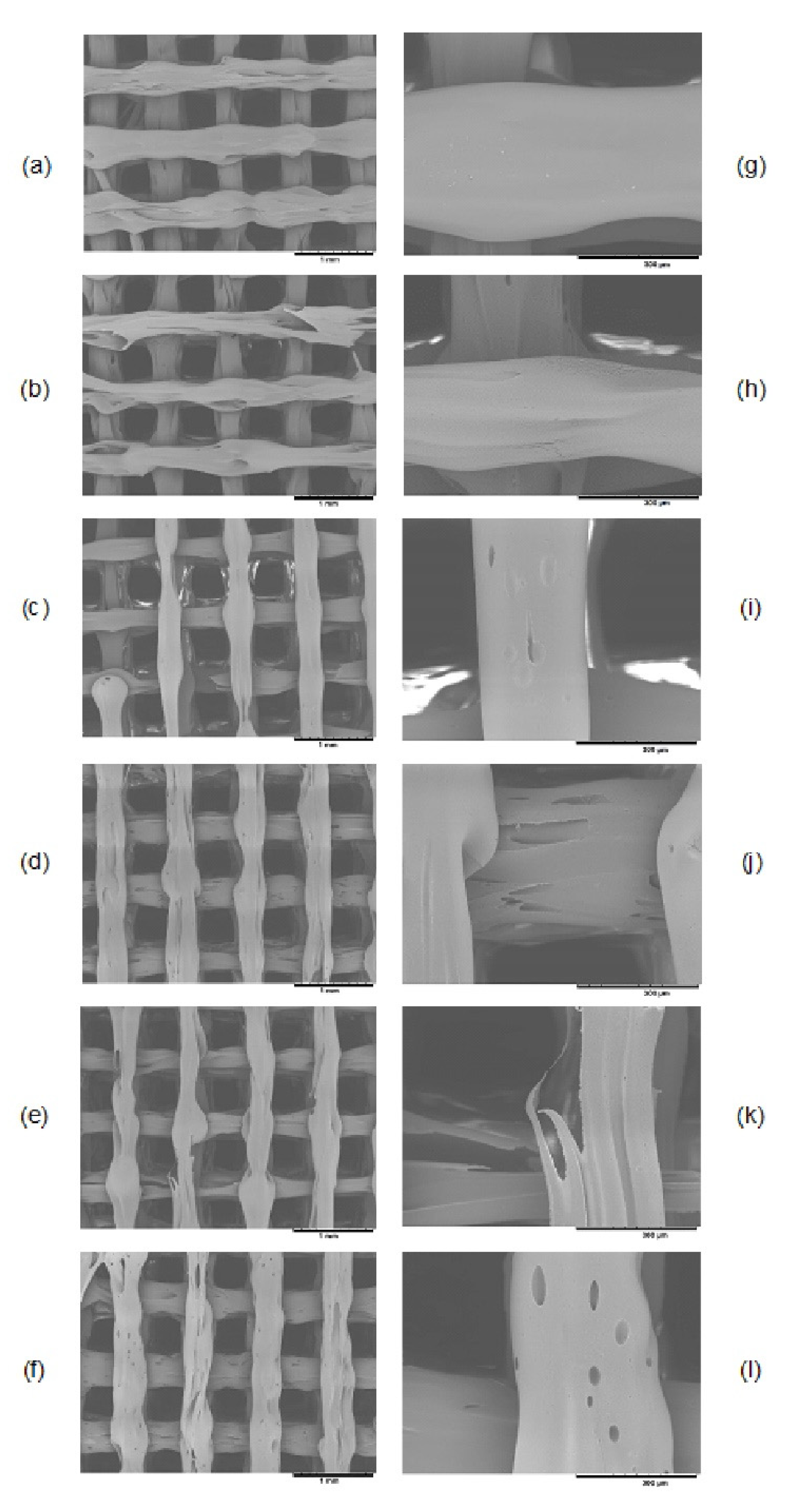
2.2. Scaffolds Manufacturing

Firstly, continuous PLA filaments were obtained using a lab prototype extruder consisting of an 8 mm screw, a cylinder with an L/D ratio of 10 and a 1.6 mm diameter nozzle tip. The extrusion was carried out at a rotating speed of 7 rpm, at a temperature of 180 °C and with a final air-cooling stage. These filaments were used to print the scaffolds by using a BQ Hephestos 2 3D printer (Spain). This additive manufacturing technique is based on the extrusion of heated material, so it can be classified as a “material extrusion” process (ISO/ASTM 52900:2015), commonly known as fused deposition modelling (FDM). Structures with a rectangular 0/90° pattern, square-shaped pores and a theoretical porosity of 50%, were manufactured. The printing settings included a nozzle diameter of 0.4 mm, a layer height of 0.3 mm, a speed of extrusion of 40 mm·s−1 and a printing temperature of 215 °C.
In order to better characterize the PLA surface after applying the plasma treatment or the different coatings proposed, flat-surface samples with dimensions of 80 × 10 × 1 mm were also manufactured by compression molding, using a Collin P 200 P/M press and a cycle comprising of the following steps: heating up to 190 °C at a heating rate of 20 °C·min-1; maintaining the temperature at that constant value for 90 s at 10 bar of pressure; and finally cooling at a rate of 20 °C·min−1 while maintaining the pressure applied in the second step.

2.3. Surface Treatment and Coating Process

The plasma treatment was carried out in a low-pressure device (Zepto Diener electronic GmbH, Ebhause, Germany) using oxygen as a carrier gas (Carburos metálicos, Spain). The oxygen supply contains less than 500 ppb of H20 and less than 400 ppb of N2. The oxygen pressure inside the chamber was fixed at 1.8 mbar. The treatment was applied at a power of 30 W for 1 minute.
The commercial aloe vera juice was centrifuged at 3000 rpm for 15 min in a Mixtasel BL centrifuge (JP Selecta), in order to separate the water-soluble fraction (rich in acetylated polysaccharides) from the cellulose-rich solid fraction. The water-soluble fraction (the supernatant) was used to carry out the coating of the structures and it contains, apart from the bioactive polysaccharides, phenolic compounds, soluble carbohydrates, proteins and minerals [24,28]. The supernatant has a pH value of around 4 (measured with a sensIONTM+PH1 pHmeter ±0.01, HACH). The slightly acid behavior of the extracts can be explained by the presence of different organic acids (acetic acid, lactic acid, succinic acid, etc.) [29] Three types of samples of aloe vera extracts were obtained through pH adjustment: a pH 3 solution (adjusted with HCl 0.018 M); a pH 5 solution (adjusted with NaOH 0.5 M); and the as-obtained gel with a pH of 4 (pH was adjusted when needed).
Each scaffold to be tested was individually immersed in 1 mL of aloe vera extract solution and stirred for 3 h, allowing the interaction between the functional groups created on the surface of the plasma-treated scaffolds and the functional groups in the polysaccharides’ structures to take place. Afterwards, the samples were rinsed with 70% ethanol to remove the excess of aloe vera which was not able to attach to the surface. Furthermore, as polysaccharides are not soluble in ethanol [30], their precipitation helps to fix the coating to the surface of the 3D-printed PLA scaffolds.

2.4. Water Contact Angle (WCA)

In order to analyze the effect of the coating on the hydrophilicity of the surface, the WCA was measured through the sessile drop method. For this technique, as it is recommended to use flat-surface samples [31], the samples obtained by compression molding described in Section 2.2 were used. The different group of samples tested were PLA, PLA plasma, PLA aloe vera pH 3, PLA aloe vera pH 4 and PLA aloe vera pH 5. The WCA measurements were carried out at room temperature using a Krüss DSA100 water contact angle measuring device (KRÜSS GmbH, Germany) and the open source software ImageJ was used to measure the static contact angle of 2 µL distilled water droplets placed onto the surface of the samples. Reported contact angles are the average of six measurements per group of samples.

2.5. X-ray Photoelectron Spectroscopy (XPS) Analysis

The assessment of the chemical modifications induced into the PLA surface was carried out by the XPS analysis of non-treated, plasma-treated and coated samples, using a Thermo Scientific K-Alpha XPS system (Thermo Fisher Scientific, UK) and the CasaXPS software (version 2.3.19PR1.0, Casa Software Ltd., Teignmouth, UK) for data analysis. Flat-surface samples obtained by compression molding (as detailed in Section 2.2.) were used for this test and three samples per group were analyzed. The oxygen/carbon (O/C) ratio of the surface was evaluated from the peak areas of the XPS survey using a U2 Tougaard background for peak fitting. In a first step, the hydrocarbon component of the C1s spectrum (284.80 eV) was used to calibrate the energy scale.

2.6. Mechanical Characterization

Plasma treatment is the most common technique to modify the surface chemistry of different biomaterials, since it does not alter their bulk properties [14]. However, this golden standard depends on the operating conditions that are used. Two mechanisms have been reported about the chemical reactions during the plasma treatment of polylactic acid: an oxidative one and a destructive one [32]. The interaction of oxygen and free radicals could lead to the formation of hydroxyl and peroxide groups, while the mechanism of destruction of plasma-treated PLA generates free radicals, accompanied with the formation of volatile gases. If the treatment is too aggressive, the chemical reaction may take place preferentially through the destruction mechanism rather than the oxidation one and, as a consequence, the mechanical properties of the structure could be affected. Therefore, it is recommended to confirm the lack of modification on the mechanical properties of the structure after plasma treatment. For this reason, non-treated and plasma-treated samples were subjected to mechanical characterization in the compression mode. In addition, aloe vera coated scaffolds after plasma treatment were tested to assess the effect of the different coatings (pH 3, 4 and 5) on the mechanical properties of the structures. Four replicas per group were used. Scaffolds with a diameter of 9.8 mm and a height of 5 mm were tested in a LIYI (LI-1065, Dongguan Liyi Environmental Technology Co., Ltd., China) testing machine in displacement control mode. Crosshead speed was set at 1 mm·min−1. The compressive modulus and the offset compressive yield strength (2% deviation from linearity) were calculated according to ASTM D695-15.

2.7. Degradation Analysis

In order to confirm if the presence of the aloe vera coating increases the degradation rate of the base material, an enzymatic degradation test was carried out. The enzymatic degradation test was preferred over a more common hydrolytic degradation study in PBS, since the first one offers the possibility of accelerating the test while obtaining comparative results. The enzymatic degradation test was carried out using proteinase K enzymes from Tritirachium album (30 units per mg of protein, Merck) diluted in Tris-HCl buffer pH 8.6 (BioReagent, Merck), at a concentration of 0.2 mg·mL−1. Sodium azide (ReagentPlus®, ≥99.5%, Merck) was added at the same concentration, in order to diminish the probability of bacterial contamination. Several examples can be found in the literature about the use of this type of enzymes to accelerate the degradation study of PLA samples from several months to a few days or even hours [33,34,35,36]. Under enzymatic degradation and according to the literature, PLA structures are able to retain their properties almost unchanged for up to 8 months [37].
Scaffolds with a diameter of 9.8 mm and a height of 7 mm were used for the enzymatic degradation test. Four replicas were analyzed per group: PLA, PLA plasma, PLA aloe vera pH 3, PLA aloe vera pH 4 and PLA aloe vera pH 5. The samples were placed in a non-treated 24-well plate and 2 mL of degradation media were added to each well. In addition, non-treated PLA samples immersed in Tris–HCl solution without enzymes were used as control samples. The buffer-enzyme solution was replaced daily to maintain a high enzymatic activity, as the reduction of the pH value due to the release of lactic acid would cause the denaturation of the enzyme [37]. The weight loss of the structures after 5 days of degradation at 37 °C was assessed by using an analytical balance (±0.1 mg, A&D Scales Gemini Series, GR-200, Germany), to measure the weight of the scaffolds before and after the test. In addition, the pH value of the media was measured (±0.01) in order to follow up the evolution of this parameter during the enzymatic degradation process. The morphology of the scaffolds’ surface before and after degradation was evaluated by scanning electron microscopy (SEM; Hitachi TM 3030 at an acceleration voltage of 15 kV).

2.8. Cell Seeding and Culture

A human fetal osteoblastic cell line (hFOB 1.19, from ATCC® CRL-11372TM) was used to analyze the influence of the surface modifications proposed on the bioactivity of the scaffold’s surface. The cell culture medium used was a mixture 1:1 of DMEM and Ham’s F-12 without fenol red (Dulbecco’s Modified Eagle Medium/Nutrient Mixture F-12, high glucose, L-glutamine, Gibco), supplemented with 10% of FBS (Gibco) and 0.3 mg·mL−1 of G-148 antibiotic solution (Geneticin, Biowest). The cells were cultured in 75 cm2 flasks (T75, Sarstedt) until reaching an 80–90% confluence. At this level, they were trypsinized with trypsin-EDTA 0.5% (no phenol red, HyClone).
The scaffolds to be seeded were 9.8 mm in diameter and 5 mm in height. Four replicas were used per group of samples: PLA plasma, PLA aloe vera pH 3, PLA aloe vera pH 4 and PLA aloe vera pH 5. The scaffolds were sterilized by soaking them in ethanol 75% v/v followed by later exposure to UV light under the fume hood for 30 min. Afterwards, they were washed three times with PBS (59321C Dulbecco’s Phosphate Buffered Saline, Merck) and with the culture medium in order to remove any trace of ethanol. Then, for cell seeding, the scaffolds were placed individually in sterilized centrifuge tubes (CFT011150, 15 mL sterile tubes, Jet Biofil) and immersed in 2 mL of culture media containing 110,000 hFOB cells. The constructs were kept in an incubator at 37 °C and 5% CO2 for 3 h before adding 2 mL of fresh culture medium to the tubes, in order to provide enough nutrients for cell culture during the first 24 h.

2.9. Cell Metabolic Activity Evaluation

Cell metabolic activity was evaluated through the CCK-8 protocol (Cell Counting Kit-8, Dojindo Molecular Technologies). At day 1 of cell culture, the samples were transferred from the tubes to a non-treated 24-well plate (Thermo Scientific™ Nunc™), where they were kept until the end of the experiment. The scaffolds were washed with 1 mL of PBS and afterwards 1 mL of a solution of the CCK-8 reagent in supplemented media was added (10% v/v). The same procedure was followed with 4 empty wells, so the data from these replicas could be used as negative controls. The plate was incubated at 37 °C and 5% CO2 for 4 h. After incubation, two 100 µL-aliquots were transferred from each well of the culture plate to a 96-well reader plate (Thermo Scientific™ Nunc™ MicroWell™). The absorbance of the samples was read at an excitation wavelength of 450 nm with a BioTek ELx800 reader (Bio Tek Instruments Inc., Winooski, VT, USA). After sampling, the solution containing the CCK-8 was removed from the culture, the samples were rinsed with PBS three times and 2 mL of fresh supplement media was added, before placing the plate into the incubator again. This procedure was repeated at days 3, 7 and 10 of cell culture, while the culture medium was changed every 2–3 days.

2.10. Statistical Analysis

Statistical analysis was performed using MATLAB software (MATLAB and Statistics Toolbox Release 2017a, The MathWorks, Inc., Natick, USA). The data obtained during this study were analyzed by the Wilcoxon two-sided rank sum test when comparing two groups and by the Kruskal–Wallis test when more than two groups were compared. The significance level was set to * p < 0.05, ** p < 0.01 and *** p < 0.001, for statistically significant, highly statistically significant and very highly statistically significant differences, respectively. All the figures show the mean values of each group, and their standard deviations are represented with error bars.

3. Results

3.1. Water Contact Angle (WCA) Measurement

A water contact angle of 79 ± 4° was obtained for the PLA samples, while the treated and coated samples showed a value around 50° (Figure 1). As expected, the plasma treatment of PLA surfaces decreases their WCA significantly (p < 0.01) compared to the non-treated samples. The aloe vera coating of the samples at pH 4 and 5 does not reduce this effect, as these groups provide similar results to the ones obtained for the samples that were only plasma-treated. However, a very highly statistically significant difference (p < 0.001) was obtained for the PLA samples coated with aloe vera at pH 3, when compared to the non-treated samples.

3.2. X-ray Photoelectron Spectroscopy (XPS) Analysis

The oxygen and carbon composition (%), as well as the O/C ratio of each group of samples, are shown in Table 1. Non-significant differences were obtained between the non-treated and plasma-treated samples. On the other hand, the aloe vera coating of the plasma-treated samples promoted the reduction of the O/C ratio. Highly statistically significant (p < 0.01) and statistically significant (p < 0.05) differences were obtained between the plasma-treated samples and the ones coated with aloe vera at pH 3 and 4, respectively. In contrast, a non-significant difference (p > 0.05) was obtained between the PLA group and the group coated with aloe vera at pH 5. Therefore, in the case of the coated samples, the reduction of the O/C ratio could be an indicator of the attachment efficiency of the aloe vera extracts to the plasma-treated surface.

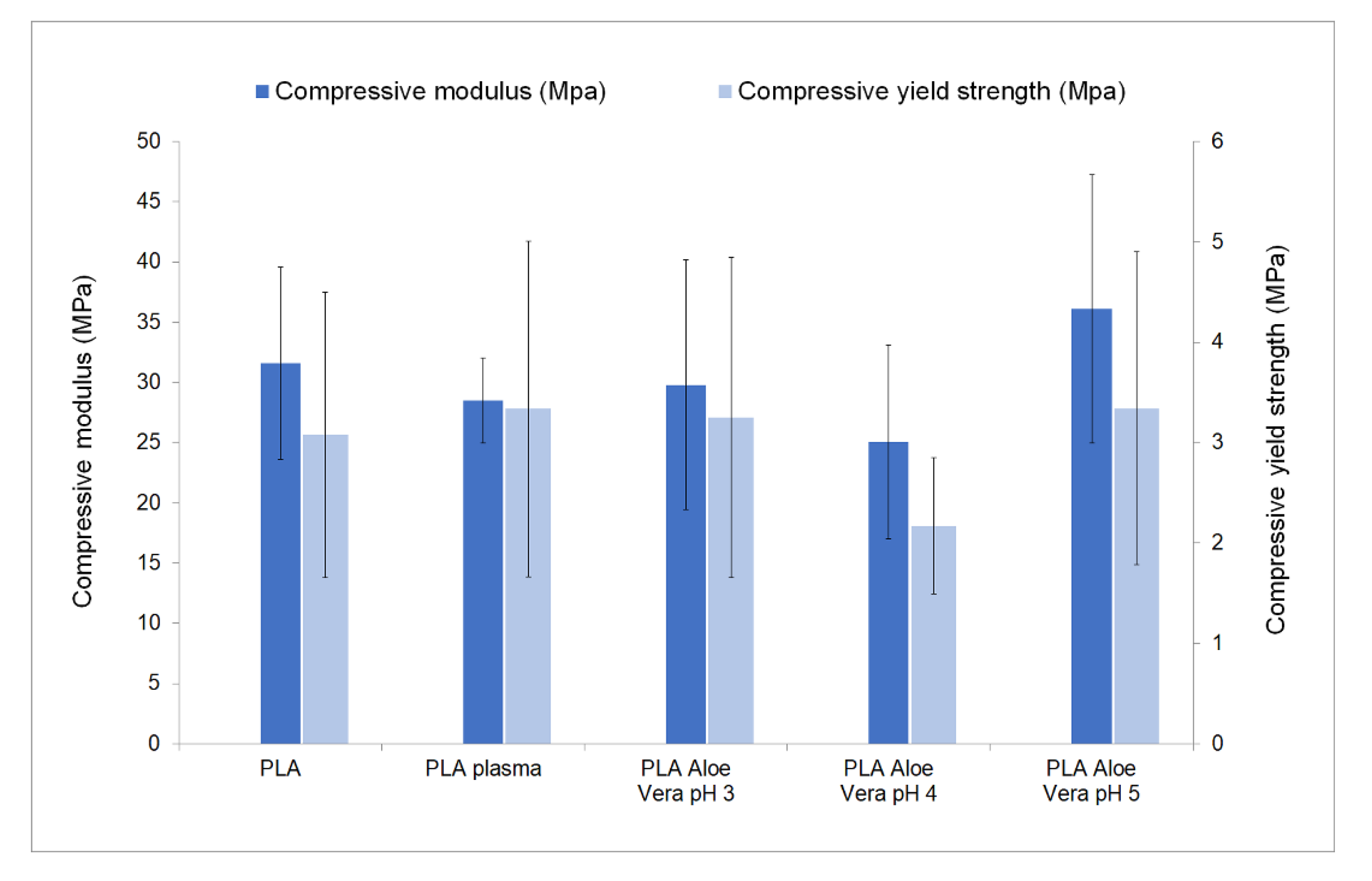
3.3. Mechanical Characterization

Prior to the compression test, the scaffolds’ morphology was assessed in terms of porosity and pore size by the methods described in our previous work [38]. Non-significant differences were obtained between the non-treated, plasma-treated and aloe vera coated samples in regard to these parameters. The average pore size and porosity of all groups of samples were 480 ± 66 µm and 61 ± 5%, respectively. As shown in Figure 2, no significant differences were obtained in terms of compressive modulus or compressive yield strength between the different groups of scaffolds tested. Regarding the compressive modulus, the values obtained were 31.60 ± 7.97 MPa for non-treated scaffolds and 28.52 ± 3.53 MPa for the plasma-treated ones. The compressive modulus of aloe vera coated samples at pH 3, 4 and 5 were 29.79 ± 10.36 MPa, 25.07 ± 8.04 MPa and 36.11 ± 11.16 MPa, respectively. These values are in the range of values reported for cancellous bone (20–500 MPa) [39].

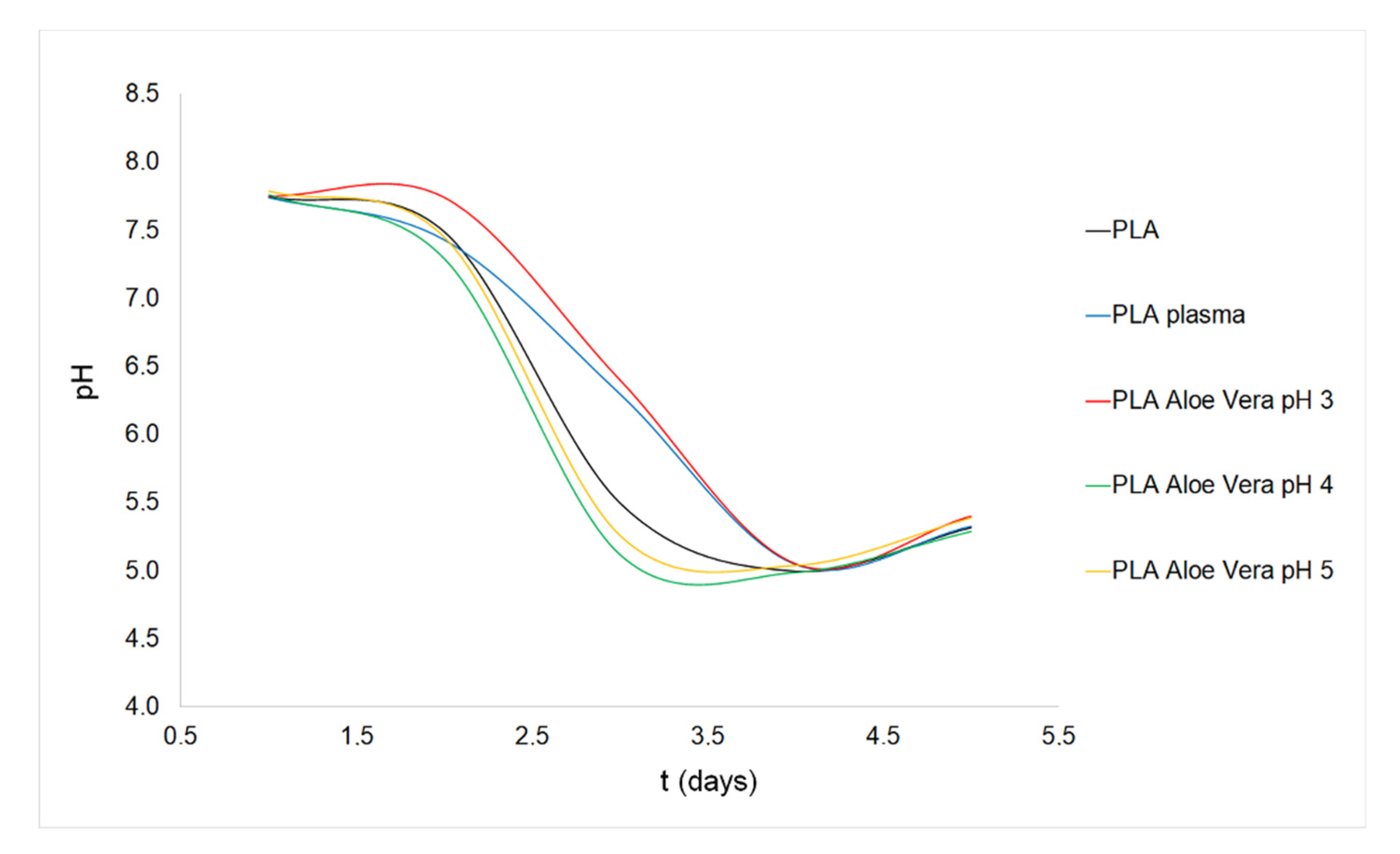
3.4. Degradation Study

The mean weight loss of each group of samples after five days of enzymatic degradation is shown in Table 2. Non-significant differences (p > 0.05) were obtained among the groups tested, resulting in a total mean value of around 5.5%. The weight of the PLA scaffolds used as control (immersed in Tris-HCl buffer solution with sodium azide but without enzymes) showed no variation during the degradation test.
Regarding the pH of the degradation media during the experiment, its value decreased remarkably from day 2 for all the group of scaffolds tested (Figure 3). This reduction was less drastic for the plasma-treated samples and for the samples coated with aloe vera at pH 3, which maintained a pH value greater than 6 at day 3. However, at days 4 and 5, similar results were obtained for all the groups of samples tested. While the coating at pH 3 seems to influence the maintenance of the pH at a higher level, the samples coated with aloe vera at pH 4 or 5 showed a degradation profile that nearly matched the one of the non-treated PLA scaffolds.
In Figure 4, SEM images obtained for each type of scaffold tested are shown, including non-degraded PLA samples for comparison (Figure 4a,g). A slight reduction of the diameter of the filaments is observed after 5 days of enzymatic degradation. There is also an evident increment of the microporosity of the scaffolds’ surface after the degradation test. This was an expected result, as while immersed in the buffer solution the hydrolytic degradation of PLA takes place via a bulk erosion mechanism; in the presence of the proteinase K enzymes the degradation of the structure is enhanced via a surface-erosion mechanism [40]. The evaluation of the high magnification SEM images (Figure 4g–l) suggests that the enzymatic degradation of the PLA samples followed a different mechanism than the plasma-treated or coated samples, as for the latter, the presence of newly formed voids over the surface can be observed (especially in Figure 4l), which correspond to the scaffolds coated with aloe vera at pH 5). In contrast, the non-treated PLA scaffolds surface showed some small fractures, from which the degradation of the structure progresses.

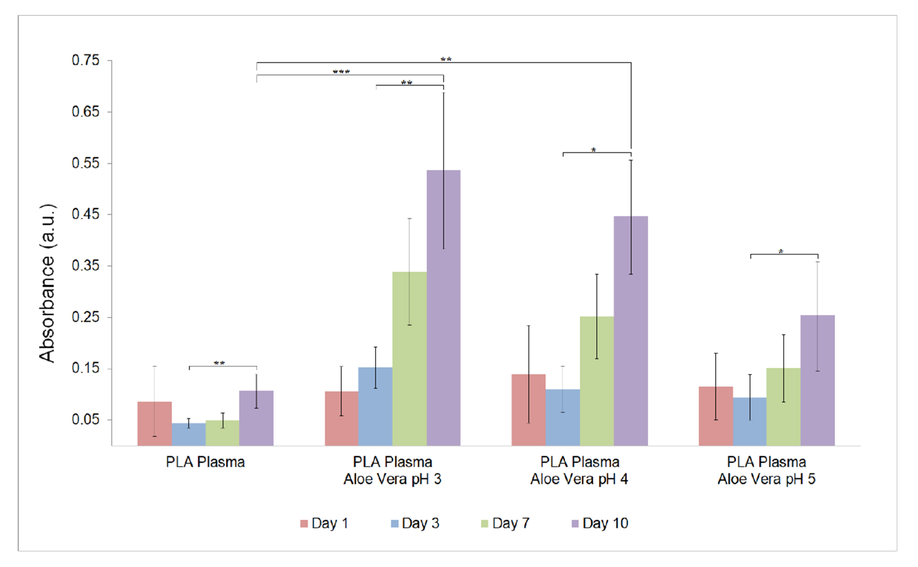
3.5. Cell Metabolic Activity Evaluation

The values of absorbance obtained from the CCK-8 assay are shown in Figure 5. All the groups of coated samples showed higher values of absorbance from day 3 until the end of the experiment (day 10). It is remarkable that for the plasma-treated samples and the coated samples at pH 4 and 5 there was a decrease between day 1 and day 3. One possible explanation for this result may be that some of the seeded cells were able to attach and survive until day 1, but the attachment was not steady enough to live until day 3 or to proliferate. Only the group coated at pH 3 was able to support the growth of cells from day 1 to day 3. Furthermore, this group offered the best results for the overall experiment, as it provides a very highly statically significant difference (p < 0.001) compared to the plasma-treated group of samples.

4. Discussion

The aloe vera coating method proposed introduces several changes in the physicochemical properties of the PLA surface and influences its biological activity. The increase of the hydrophilicity of plasma-treated PLA scaffolds enhance the affinity of the surface to support the aloe vera coating in an effective way. The functional groups created during plasma treatment of PLA are mainly hydroxyl and carbonyl ones [13,41]. These groups are not reactive with the functional groups in the structure of the polysaccharides in aloe vera extracts (mainly hydroxyl and acetyl) [42]. Nevertheless, they provide an increase in the polarity of the surface which is more favorable for attaching the extract by intermolecular forces.
The similarity of the WCA results between coated and non-coated groups indicates that the hydrophilicity increases similarly (compared to untreated PLA samples) whether the surface modification consisted only in the application of the plasma treatment or if it is followed by the aloe vera coating. Therefore, any change in the biological properties of the scaffolds needs to be explained by the change in the chemical composition of the surfaces, not by their surface energy.
Regarding the surface chemistry, despite being a non-significant difference (p > 0.05), the modification in the O/C ratio of the PLA surface before and after the plasma treatment suggests the prevalence of the oxidation mechanism over the destructive one [32]. It is important to take into account that both mechanisms may take place simultaneously and the predominance of one of them depends on the treatment time, as demonstrated by Izdebska-Podsiadly et al. [43] The prevalence of the oxidation route during the plasma treatment in the present study is also supported by the results from the mechanical characterization, as a non-significant difference (p > 0.05) was detected when the plasma treated samples and the non-treated ones were compared (Figure 2). These results also evidenced that the aloe vera coating does not play a role on the mechanical properties of the final structure. The low thickness of the coating and the absence of etching compounds in the aloe vera extract justify this lack of modification on the mechanical properties of the scaffolds after the coating.
With respect to the coated samples, it is worth mentioning that there is a significant decrease of the O/C ratio (p < 0.05) for the samples coated at pH 3 and pH 4, but not for those coated at pH 5. This trend could be related to the amount and type of bioactive compounds that are able to be fixed on the PLA surface. The O/C ratio expected from the coating depends on the mannose:glucose:galactose ratio of the polysaccharides in the extracts [25]. The composition of aloe vera extracts is complex and it depends on different parameters, such as the geographical location of the plants or the season when the leaves are harvested, as the carbohydrates have a storage biological function in the living plants [25], or the extraction and processing procedures used; so it is difficult to predict a clear expected trend of this parameter.
Regarding the enzymatic degradation of the scaffolds, the results confirm an influence of the coating on the degradation rate of the material. As the adsorption of the enzymes to the surface is the first step of any enzymatic degradation process, the modifications introduced on the surface chemistry can potentially modify the enzymatic degradation mechanism of PLA. In fact, the adsorption process has been demonstrated to be a key element to understand the enzymatic degradation of PLA and, in general, of any poly(hydroxyalkanoates), according to Yamashita et al. [44]. These authors stated that the adsorption of proteinase K on PLA surfaces is an irreversible process that could be related to a hydrophobic interaction between the PLA and the enzymes. This conclusion can explain why the degradation of the scaffolds is delayed (during the first steps of the test) when they are plasma-treated or coated. As demonstrated by the WCA results (Figure 1), any of the strategies applied lead to a decrease of the hydrophobicity. Consequently, according to the conclusions of Yamashita et al., this trend would hinder the adsorption of the enzymes on the PLA surface and delay the degradation process. The coating applied at pH 3 seems to limit this adsorption step especially, as this group of samples shows the lowest levels of degradation measured as mass loss (Table 2). The protein adsorption on solid surfaces is a complex process affected by different parameters [45], so the reason why the coating at this pH value limits the adsorption process needs to be further analyzed, although it is out of the scope of the present work.
The trend evidenced by the mass loss results is also supported by the pH measurements of the degradation media (Figure 3), as the decrease of this parameter is explained by the presence of carboxylic acids released during the degradation process [46]. If the degradation is delayed (as it is especially for plasma-treated samples and pH 3 coated samples), the pH value of the media will be higher during the process, as confirmed in this experiment. The results obtained from the enzymatic degradation test open the possibility of controlling the degradation rate of the proposed constructs intended for bone regeneration, aiming to match this rate to the specific tissue growth rate.
Finally, the results of the cell viability evaluation prove that the aloe vera coating offers an improvement on the ability of the bone cells to proliferate and remain viable on the scaffolds compared to the results from the structures that were plasma-treated but not coated. As plasma treatment is the most common method to modify the surface of PLA scaffolds [14,47,48], the results from this work confirm the potential of the coating strategy to increase the biological performance of PLA-based scaffolds for bone regeneration.
In addition, the pH of the aloe vera extracts has been demonstrated to be a parameter that strongly affects the bioaffinity of the surface coating, as the best results were obtained for the aloe vera coating applied at pH 3. The adjustment of the pH may change the chemistry of the bioactive polysaccharides in the aloe vera extracts. According to different studies, the presence of the acetyl group in the acemannan structure (one of the most important components in the aloe vera extracts) seems to be a key parameter to explain its bioactivity [42]. Chokboribal et al. [49] have already proven that the deacetylation in alkaline conditions of acemannan decreases its bioactivity. Although in this work, the pH level is not aggressive enough to produce the deacetylation of the bioactive molecules, it is possible that the introduction of an alkali (NaOH used for pH adjustment of the extract solutions) changed the conformation of these active functional groups. This modification could hinder the effectiveness of the aloe vera attachment to the surface, leading to a lower bioactivity of the coating at pH 5 compared to the coating applied at pH 3.

5. Conclusions

This study shows the feasibility of improving the biological response of PLA-based scaffolds by applying a coating of aloe vera extracts. The pH value used during the coating process has demonstrated to be a parameter that strongly influences the characteristics of the final surface, as the pH 3 level offers better results in terms of cell viability during the cell culture of human osteoblasts. Regarding the effect of the coating on the enzymatic degradation of the structures, the coating applied at pH 3 seems to hinder the enzyme adsorption and, consequently, delays the degradation of the scaffold. Therefore, the pH level of the aloe vera coating can be used as an important parameter, in order to tune the degradation process of the structures intended for bone regeneration.

Author Contributions

Conceptualization, R.D. and M.E.A.-D.; Methodology, R.D. and M.E.A.-D.; Formal analysis, R.D.; Investigation, R.D. and J.Y.; Resources, M.M., F.R.-E. and C.L.; Writing—Original Draft, R.D. and M.E.A.-D; Writing—Review and Editing, M.M., J.Y., F.R.-E. and C.L.; Visualization, R.D.; Supervision, M.M.; Project administration, M.M.; Funding acquisition, M.M. All authors have read and agreed to the published version of the manuscript.

Funding

This contribution is part of the BioAM project (DPI2017-88465-R) funded by the Science, Innovation and Universities Spanish Ministry. Additionally, the authors would like to thank the H2020-MSCA-RISE program for providing funding for mobility through the BAMOS project, funded from the European Union’s Horizon 2020 research and innovation program, under grant agreement Nº 734156. Ricardo Donate express his gratitude for the funding through the PhD grant program co-financed by the Canarian Agency for Research, Innovation and Information Society of the Canary Islands Regional Council for Employment, Industry, Commerce and Knowledge and by the European Social Fund (ESF) Canary Islands Integrated Operational Program 2014-2020, Axis 3 Priority Theme 74 (85%). Grant code: TESIS2017010036.

Conflicts of Interest

The authors declare no conflict of interest. The funders had no role in the design of the study; in the collection, analyses, or interpretation of data; in the writing of the manuscript, or in the decision to publish the results.

References

  1. Grémare, A.; Guduric, V.; Bareille, R.; Heroguez, V.; Latour, S.; L’heureux, N.; Fricain, J.C.; Catros, S.; Le Nihouannen, D. Characterization of printed PLA scaffolds for bone tissue engineering. J. Biomed. Mater. Res. Part A 2018, 106, 887–894. [Google Scholar] [CrossRef] [PubMed]
  2. Kao, C.T.; Lin, C.C.; Chen, Y.W.; Yeh, C.H.; Fang, H.Y.; Shie, M.Y. Poly(dopamine) coating of 3D printed poly(lactic acid) scaffolds for bone tissue engineering. Mater. Sci. Eng. C 2015, 56, 165–173. [Google Scholar] [CrossRef] [PubMed]
  3. Savioli Lopes, M.; Jardini, A.L.; Maciel Filho, R. Poly (lactic acid) production for tissue engineering applications. Procedia Eng. 2012, 42, 1402–1413. [Google Scholar] [CrossRef] [Green Version]
  4. Teixeira, B.N.; Aprile, P.; Mendonça, R.H.; Kelly, D.J.; Thiré, R.M.D.S.M. Evaluation of bone marrow stem cell response to PLA scaffolds manufactured by 3D printing and coated with polydopamine and type I collagen. J. Biomed. Mater. Res. Part B Appl. Biomater. 2019, 107, 37–49. [Google Scholar] [CrossRef] [PubMed] [Green Version]
  5. Martin, V.; Ribeiro, I.A.; Alves, M.M.; Gonçalves, L.; Claudio, R.A.; Grenho, L.; Fernandes, M.H.; Gomes, P.; Santos, C.F.; Bettencourt, A.F. Engineering a multifunctional 3D-printed PLA-collagen-minocycline-nanoHydroxyapatite scaffold with combined antimicrobial and osteogenic effects for bone regeneration. Mater. Sci. Eng. C 2019, 101, 15–26. [Google Scholar] [CrossRef]
  6. Esposito Corcione, C.; Scalera, F.; Gervaso, F.; Montagna, F.; Sannino, A.; Maffezzoli, A. One-step solvent-free process for the fabrication of high loaded PLA/HA composite filament for 3D printing. J. Therm. Anal. Calorim. 2018, 134, 575–582. [Google Scholar] [CrossRef]
  7. Ma, Z.; Gao, C.; Ji, J.; Shen, J. Protein immobilization on the surface of poly-L-lactic acid films for improvement of cellular interactions. Eur. Polym. J. 2002, 38, 2279–2284. [Google Scholar] [CrossRef]
  8. Chen, S.; Guo, Y.; Liu, R.; Wu, S.; Fang, J.; Huang, B.; Li, Z.; Chen, Z.; Chen, Z. Tuning surface properties of bone biomaterials to manipulate osteoblastic cell adhesion and the signaling pathways for the enhancement of early osseointegration. Colloids Surf. B Biointerfaces 2018, 164, 58–69. [Google Scholar] [CrossRef]
  9. Niaza, K.V.; Senatov, F.S.; Kaloshkin, S.D.; Maksimkin, A.V.; Chukov, D.I. 3D-printed scaffolds based on PLA/HA nanocomposites for trabecular bone reconstruction. J. Phys. Conf. Ser. 2016, 741, 012068. [Google Scholar] [CrossRef] [Green Version]
  10. Belaid, H.; Nagarajan, S.; Teyssier, C.; Barou, C.; Barés, J.; Balme, S.; Garay, H.; Huon, V.; Cornu, D.; Cavaillès, V.; et al. Development of new biocompatible 3D printed graphene oxide-based scaffolds. Mater. Sci. Eng. C 2020, 110, 110595. [Google Scholar] [CrossRef]
  11. Wei, Q.; Cai, X.; Guo, Y.; Wang, G.; Guo, Y.; Lei, M.; Song, Y.; Yingfeng, Z.; Wang, Y. Atomic-scale and experimental investigation on the micro-structures and mechanical properties of PLA blending with CMC for additive manufacturing. Mater. Des. 2019, 183, 108158. [Google Scholar] [CrossRef]
  12. Chen, Y.; Mak, A.F.T.; Wang, M.; Li, J.; Wong, M.S. PLLA scaffolds with biomimetic apatite coating and biomimetic apatite/collagen composite coating to enhance osteoblast-like cells attachment and activity. Surf. Coat. Technol. 2006, 201, 575–580. [Google Scholar] [CrossRef]
  13. Nakagawa, M.; Teraoka, F.; Fujimoto, S.; Hamada, Y.; Kibayashi, H.; Takahashi, J. Improvement of cell adhesion on poly(L-lactide) by atmospheric plasma treatment. J. Biomed. Mater. Res. Part A 2006, 77, 112–118. [Google Scholar] [CrossRef]
  14. Jacobs, T.; Morent, R.; De Geyter, N.; Dubruel, P.; Leys, C. Plasma Surface Modification of Biomedical Polymers: Influence on Cell-Material Interaction. Plasma Chem. Plasma Process. 2012, 32, 1039–1073. [Google Scholar] [CrossRef]
  15. Guo, C.; Xiang, M.; Dong, Y. Surface modification of poly (lactic acid) with an improved alkali-acid hydrolysis method. Mater. Lett. 2015, 140, 144–147. [Google Scholar] [CrossRef]
  16. Zeng, S.; Cui, Z.; Yang, Z.; Si, J.; Wang, Q.; Wang, X.; Peng, K.; Chen, W. Characterization of highly interconnected porous poly(lactic acid) and chitosan-coated poly(lactic acid) scaffold fabricated by vacuum-assisted resin transfer molding and particle leaching. J. Mater. Sci. 2016, 51, 9958–9970. [Google Scholar] [CrossRef]
  17. Fernández-Cervantes, I.; Morales, M.A.; Agustín-Serrano, R.; Cardenas-García, M.; Pérez-Luna, P.V.; Arroyo-Reyes, B.L.; Maldonado-García, A. Polylactic acid/sodium alginate/hydroxyapatite composite scaffolds with trabecular tissue morphology designed by a bone remodeling model using 3D printing. J. Mater. Sci. 2019, 54, 9478–9496. [Google Scholar] [CrossRef]
  18. Li, J.; Chen, Y.; Mak, A.F.T.; Tuan, R.S.; Li, L.; Li, Y. A one-step method to fabricate PLLA scaffolds with deposition of bioactive hydroxyapatite and collagen using ice-based microporogens. Acta Biomater. 2010, 6, 2013–2019. [Google Scholar] [CrossRef]
  19. Kim, S.H.; Oh, S.A.; Lee, W.K.; Shin, U.S.; Kim, H.W. Poly(lactic acid) porous scaffold with calcium phosphate mineralized surface and bone marrow mesenchymal stem cell growth and differentiation. Mater. Sci. Eng. C 2011, 31, 612–619. [Google Scholar] [CrossRef]
  20. Liu, P.; Chen, D.; Shi, J. Chemical Constituents, Biological Activity and Agricultural Cultivation of Aloe vera. Asian J. Chem. 2013, 25, 6477–6485. [Google Scholar] [CrossRef]
  21. Suganya, S.; Venugopal, J.; Ramakrishna, S.; Lakshmi, B.S.; Dev, V.R.G. Naturally derived biofunctional nanofibrous scaffold for skin tissue regeneration. Int. J. Biol. Macromol. 2014, 68, 135–143. [Google Scholar] [CrossRef]
  22. Garcia-Orue, I.; Gainza, G.; Gutierrez, F.B.; Aguirre, J.J.; Evora, C.; Pedraz, J.L.; Hernandez, R.M.; Delgado, A.; Igartua, M. Novel nanofibrous dressings containing rhEGF and Aloe vera for wound healing applications. Int. J. Pharm. 2017, 523, 556–566. [Google Scholar] [CrossRef]
  23. Shanmugavel, S.; Reddy, V.J.; Ramakrishna, S.; Lakshmi, B.S.; Dev, V.G. Precipitation of hydroxyapatite on electrospun polycaprolactone/aloe vera/silk fibroin nanofibrous scaffolds for bone tissue engineering. J. Biomater. Appl. 2014, 29, 46–58. [Google Scholar] [CrossRef]
  24. Femenia, A.; Sánchez, E.S.; Simal, S.; Rosselló, C. Compositional features of polysaccharides from Aloe vera (Aloe barbadensis Miller) plant tissues. Carbohydr. Polym. 1999, 39, 109–117. [Google Scholar] [CrossRef]
  25. Hamman, J.H. Composition and applications of Aloe vera leaf gel. Molecules 2008, 13, 1599–1616. [Google Scholar] [CrossRef] [Green Version]
  26. Tabandeh, M.R.; Oryan, A.; Mohammadalipour, A. Polysaccharides of Aloe vera induce MMP-3 and TIMP-2 gene expression during the skin wound repair of rat. Int. J. Biol. Macromol. 2014, 65, 424–430. [Google Scholar] [CrossRef]
  27. Blackwood, K.A.; Bock, N.; Dargaville, T.R.; Ann Woodruff, M. Scaffolds for growth factor delivery as applied to bone tissue engineering. Int. J. Polym. Sci. 2012. [Google Scholar] [CrossRef] [Green Version]
  28. Maan, A.A.; Nazir, A.; Khan, M.K.I.; Ahmad, T.; Zia, R.; Murid, M.; Abrar, M. The therapeutic properties and applications of Aloe vera: A review. J. Herb. Med. 2018, 12, 1–10. [Google Scholar] [CrossRef]
  29. Bozzi, A.; Perrin, C.; Austin, S.; Arce Vera, F. Quality and authenticity of commercial aloe vera gel powders. Food Chem. 2007, 103, 22–30. [Google Scholar] [CrossRef]
  30. Xu, J.; Yue, R.Q.; Liu, J.; Ho, H.M.; Yi, T.; Chen, H.B.; Han, Q. Bin Structural diversity requires individual optimization of ethanol concentration in polysaccharide precipitation. Int. J. Biol. Macromol. 2014, 67, 205–209. [Google Scholar] [CrossRef] [Green Version]
  31. Yuan, Y.; Lee, T.R. Contact angle and wetting properties. In Springer Series Surface Sciences; Springer: Berlin/Heidelberg, Germany, 2013; Volume 51, pp. 3–34. [Google Scholar]
  32. Laput, O.; Vasenina, I.; Salvadori, M.C.; Savkin, K.; Zuza, D.; Kurzina, I. Low-temperature plasma treatment of polylactic acid and PLA/HA composite material. J. Mater. Sci. 2019, 54, 11726–11738. [Google Scholar] [CrossRef]
  33. Zeng, J.; Chen, X.; Liang, Q.; Xu, X.; Jing, X. Enzymatic Degradation of Poly(L-lactide) and Poly(ε-caprolactone) Electrospun Fibers. Macromol. Biosci. 2004, 4, 1118–1125. [Google Scholar] [CrossRef]
  34. Tsuji, H.; Muramatsu, H. Blends of aliphatic polyesters: V. Non-enzymatic and enzymatic hydrolysis of blends from hydrophobic poly(L-lactide) and hydrophilic poly(vinyl alcohol). Polym. Degrad. Stab. 2001, 71, 403–413. [Google Scholar] [CrossRef]
  35. Liu, L.; Li, S.; Garreau, H.; Vert, M. Selective enzymatic degradations of poly(L-lactide) and poly(∈-caprolactone) blend films. Biomacromolecules 2000, 1, 350–359. [Google Scholar] [CrossRef]
  36. De Tayrac, R.; Chentouf, S.; Garreau, H.; Braud, C.; Guiraud, I.; Boudeville, P.; Vert, M. In vitro degradation and in vivo biocompatibility of poly(lactic acid) mesh for soft tissue reinforcement in vaginal surgery. J. Biomed. Mater. Res. Part B Appl. Biomater. 2008, 85, 529–536. [Google Scholar] [CrossRef]
  37. Hegyesi, N.; Zhang, Y.; Kohári, A.; Polyák, P.; Sui, X.; Pukánszky, B. Enzymatic degradation of PLA/cellulose nanocrystal composites. Ind. Crops Prod. 2019, 141. [Google Scholar] [CrossRef] [Green Version]
  38. Donate, R.; Monzón, M.; Ortega, Z.; Wang, L.; Ribeiro, V.; Pestana, D.; Joaquim, M.; Reis, R.L. Comparison between calcium carbonate and β -tricalcium phosphate as additives of 3D printed scaffolds with polylactic acid matrix. J. Tissue Eng. Regen. Med. 2019, 14, 272–283. [Google Scholar] [CrossRef]
  39. Leong, K.F.; Chua, C.K.; Sudarmadji, N.; Yeong, W.Y. Engineering functionally graded tissue engineering scaffolds. J. Mech. Behav. Biomed. Mater. 2008, 1, 140–152. [Google Scholar] [CrossRef]
  40. Tsuji, H.; Ishida, T. Poly(l-lactide). X. Enhanced surface hydrophilicity and chain-scission mechanisms of poly(l-lactide) film in enzymatic, alkaline, and phosphate-buffered solutions. J. Appl. Polym. Sci. 2002, 87, 1628–1633. [Google Scholar] [CrossRef]
  41. Scaffaro, R.; Lopresti, F.; Sutera, A.; Botta, L.; Fontana, R.M.; Gallo, G. Plasma modified PLA electrospun membranes for actinorhodin production intensification in Streptomyces coelicolor immobilized-cell cultivations. Colloids Surf. B Biointerfaces 2017, 157, 233–241. [Google Scholar] [CrossRef]
  42. Liu, C.; Cui, Y.; Pi, F.; Cheng, Y.; Guo, Y.; Qian, H. Extraction, purification, structural characteristics, biological activities and pharmacological applications of acemannan, a polysaccharide from aloe vera: A review. Molecules 2019, 24, 1554. [Google Scholar] [CrossRef] [Green Version]
  43. Izdebska-Podsiadły, J.; Dörsam, E. Effects of argon low temperature plasma on PLA film surface and aging behaviors. Vacuum 2017, 145, 278–284. [Google Scholar] [CrossRef]
  44. Yamashita, K.; Kikkawa, Y.; Kurokawa, K.; Doi, Y. Enzymatic degradation of poly(L-lactide) film by proteinase K: Quartz crystal microbalance and atomic force microscopy study. Biomacromolecules 2005, 6, 850–857. [Google Scholar] [CrossRef]
  45. Fang, F.; Szleifer, I. Kinetics and thermodynamics of protein adsorption: A generalized molecular theoretical approach. Biophys. J. 2001, 80, 2568–2589. [Google Scholar] [CrossRef] [Green Version]
  46. Elsawy, M.A.; Kim, K.H.; Park, J.W.; Deep, A. Hydrolytic degradation of polylactic acid (PLA) and its composites. Renew. Sustain. Energy Rev. 2017, 79, 1346–1352. [Google Scholar] [CrossRef]
  47. Yamaguchi, M.; Shinbo, T.; Kanamori, T.; Wang, P.C.; Niwa, M.; Kawakami, H.; Nagaoka, S.; Hirakawa, K.; Kamiya, M. Surface modification of poly(L-lactic acid) affects initial cell attachment, cell morphology, and cell growth. J. Artif. Organs 2004, 7, 187–193. [Google Scholar] [CrossRef]
  48. Wang, M.; Favi, P.; Cheng, X.; Golshan, N.H.; Ziemer, K.S.; Keidar, M.; Webster, T.J. Cold atmospheric plasma (CAP) surface nanomodified 3D printed polylactic acid (PLA) scaffolds for bone regeneration. Acta Biomater. 2016, 46, 256–265. [Google Scholar] [CrossRef]
  49. Chokboribal, J.; Tachaboonyakiat, W.; Sangvanich, P.; Ruangpornvisuti, V.; Jettanacheawchankit, S.; Thunyakitpisal, P. Deacetylation affects the physical properties and bioactivity of acemannan, an extracted polysaccharide from Aloe vera. Carbohydr. Polym. 2015, 133, 556–566. [Google Scholar] [CrossRef]
Figure 1. Water contact angle values for the non-treated, plasma-treated and coated samples obtained by compression molding (** p < 0.01 and *** p < 0.001), compared to the group of non-treated polylactic acid (PLA) samples.
Applsci 10 02576 g001
Figure 2. Mechanical properties of non-treated, plasma-treated and aloe vera-coated PLA 3D-printed scaffolds under compression testing.
Applsci 10 02576 g002
Figure 3. Variation of the pH level of the degradation medium during the enzymatic degradation study of the 3D-printed scaffolds.
Applsci 10 02576 g003
Figure 4. SEM images (scale bar: 1 mm) of the 3D-printed PLA scaffolds analyzed: (a) non-degraded PLA; (b) PLA; (c) PLA plasma; (d) PLA aloe vera pH 3; (e) PLA aloe vera pH 4 and (f) PLA aloe vera pH 5. Additionally, SEM images with higher magnification (scale bar: 300 µm) are shown for (g) non-degraded PLA; (h) PLA; (i) PLA plasma; (j) PLA aloe vera pH 3; (k) PLA aloe vera pH 4 and (l) PLA aloe vera pH 5.
Applsci 10 02576 g004
Figure 5. Metabolic activity of human fetal osteoblastic cell line (hFOB) cells on the plasma-treated and coated scaffolds determined by the CCK-8 assay (* p < 0.05, ** p < 0.01 and *** p < 0.001).
Applsci 10 02576 g005
Table 1. XPS results, including the oxygen and carbon content of the surface and the O/C ratio of the non-treated, plasma-treated and coated samples analyzed.
Group of ScaffoldsO %C %O/C
PLA33.89 ± 1.8766.11 ± 1.870.51 ± 0.04
PLA plasma35.44 ± 1.0561.89 ± 2.530.57 ± 0.03
PLA aloe vera pH 317.13 ± 1.9982.14 ± 1.400.21 ± 0.03
PLA aloe vera pH 428.83 ± 0.7970.60 ± 1.780.41 ± 0.02
PLA aloe vera pH 534.09 ± 1.3065.91 ± 1.300.52 ± 0.03
Table 2. Average weight loss (%) of the scaffolds after five-day enzymatic degradation.
Group of ScaffoldsWeight Loss %
PLA5.62 ± 0.37
PLA plasma5.37 ± 0.85
PLA aloe vera pH 34.81 ± 0.79
PLA aloe vera pH 46.01 ± 0.90
PLA aloe vera pH 55.85 ± 0.75

