Review

Determining the Effects of Eugenol on the Bond Strength of Resin-Based Restorative Materials to Dentin: A Meta-Analysis of the Literature

by
Isadora M. Garcia
1,
Vicente C. B. Leitune
1,
Maria S. Ibrahim
2,
Mary Anne S. Melo
3,
Vicente Faus Matoses
4,
Salvatore Sauro
5,6,* and
Fabrício M. Collares
1
1
Dental Materials Laboratory, Department of Conservative Dentistry, School of Dentistry, Federal University of Rio Grande do Sul, Rua Ramiro Barcelos, 2492, Rio Branco, Porto Alegre 90035-003, RS, Brazil
2
Department of Preventive Dental Sciences, College of Dentistry, Imam Abdulrahman Bin Faisal University, Dammam 34212, Saudi Arabia
3
Dental Biomedical Sciences, University of Maryland School of Dentistry, Baltimore, MD 21201, USA
4
Department of Stomatology, Faculty of Medicine and Dentistry, University of Valencia, 46010 Valencia, Spain
5
Departamento de Odontología, Facultad de Ciencias de la Salud, Universidad CEU-Cardenal Herrera, C/Del Pozos/n, Alfara del Patriarca, 46115 Valencia, Spain
6
Department of Therapeutic Dentistry, Sechenov University of Moscow, 119435 Moscow, Russia
*
Author to whom correspondence should be addressed.
Appl. Sci. 2020, 10(3), 1070; https://doi.org/10.3390/app10031070
Submission received: 1 January 2020 / Revised: 24 January 2020 / Accepted: 3 February 2020 / Published: 5 February 2020
(This article belongs to the Special Issue Permanent and Long-Term Biodegradable Biomaterials)

Abstract

:
The aim of this study was to determine whether the residual presence of eugenol in coronal dentin may compromise the bond strength of resin-based restorative materials. A search was performed on MEDLINE/Pubmed, Scopus, and by hand search for relevant papers. No restriction was applied for language and publication date. The studies selected for analysis tested specimens with reduced size (micro-shear bond strength (µSBS) and micro-tensile bond strength (µTBS)) of adhesive systems and resin-based restorative materials applied to coronary dentin “contaminated” with eugenol-based materials. The search provided 335 articles, but only 10 studies met the inclusion criteria. The pooled global analysis showed a significant influence of eugenol, as it negatively influenced the bond strength of resin-based restorations (5.79 (3.31–8.28) MPa, p < 0.00001). The subgroup analyses for conventional etch-and-rinse (p = 0.003) and self-etch (p < 0.0004) adhesive systems, as well as for µSBS (p = 0.01) and µTBS (p < 0.0001), showed a negative influence of eugenol on the bond strength. Data were statistically heterogeneous. However, it was possible to observe that eugenol could negatively affect the bonding of resin-based restorative materials to dentin. Further evidence is necessary in order to acquire more accurate information about this issue and confirm that the residual presence of eugenol in dentin compromises the bond strength of resin-based materials.

1. Introduction

Eugenol, in combination with zinc oxide (ZOE), is one of the most common materials used as a root canal sealer in endodontics [1,2,3,4,5], as well as a pulp sedative in cases of pulpotomies [6,7,8,9] and an impression material for edentulous patients [10,11,12,13]. In restorative dentistry, ZOE cements are frequently applied as temporary materials during indirect restorations [14]. It is also known that ZOE cements can perform as anodynes for pulpal pain [15], especially in those patients presenting dentin hypersensitivity after tooth preparation and temporary restorations [16].
ZOE cements used in temporary luting procedures are usually characterized by reasonable retention and compression strength and they can be removed quite easily [17]. Several techniques to eliminate residual provisional cements from dental preparations have been advocated. These include grating the tooth with a hand instrument (usually scalers or spoon excavators), cleaning the tooth preparation with a prophylaxis cup in combination with a water–pumice paste slurry, and/or using an intraoral air abrasion system commonly in combination with alumina or sodium bicarbonate. Although such temporary cements may be considered user-friendly materials in the clinic, there is evidence that the residual eugenol remaining on the dentin surface after setting can jeopardize the bonding performance of resin-based materials due to polymerization inhibition [1,18,19,20]. The current literature shows conflicting results and a distinct lack of robust evidence about this issue.
The remaining presence of eugenol-containing cements on dentin can act as a physical barrier [21,22], impairing the degree of monomer conversion of resin-based materials [23] due to the existence of radical-scavenging molecules in such phenolic compound [24]. Indeed, the reactivity of monomers is reduced due to the hydroxyl groups within the eugenol molecule, which can protonate free radicals during the polymerization reaction [25]. Hence, an optimal removal of provisional eugenol-containing cements must be achieved in order to accomplish a suitable adhesion of permanent resin-based restorations to dentin [1,26].
Dentin hybridization through the use of adhesive systems is an essential mechanism for tooth/material adhesion [27]. Adhesive systems are made of photocurable resin monomers, which are responsible for micromechanical interlocking with the dental tissues. Such adhesive systems can be classified according to their mode of interaction with the bonding substrate as (i) etch-and-rinse (ER) or (ii) self-etch (SE) systems [28]. Studies confirmed that eugenol can jeopardize their bonding performance, although it is still unclear which type of adhesive (ER or SE) is more at risk of debonding in case of contact with eugenol. A recent meta-analysis has shown that sealers with eugenol may be able to reduce the push-out strength of fiber posts applied in the root canal in combination with resin-based cements [29]. Although many studies tried to elucidate the effect of eugenol on the bond strength of resin adhesives to coronal dentin, there is still no consensus about this issue [18,19,20,30,31,32,33,34,35].
Therefore, this study aimed to analyze whether the residual presence of eugenol in the coronal dentin may compromise the bond strength of resin-based restorative materials. The hypothesis tested in this study was that the presence of residual eugenol on dentin surface would potentially interfere with SE and/or ER adhesive systems.

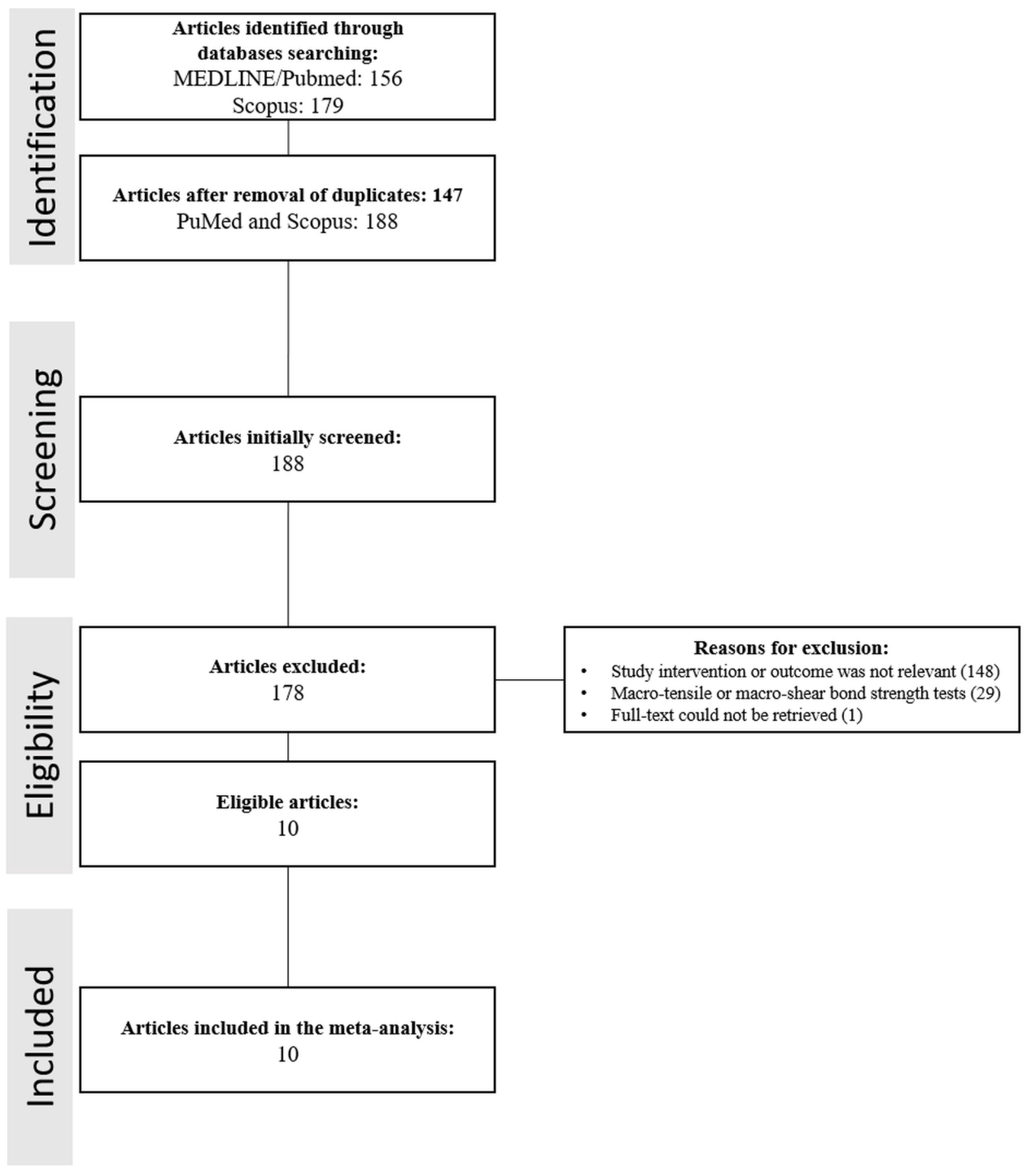
2. Material and Methods
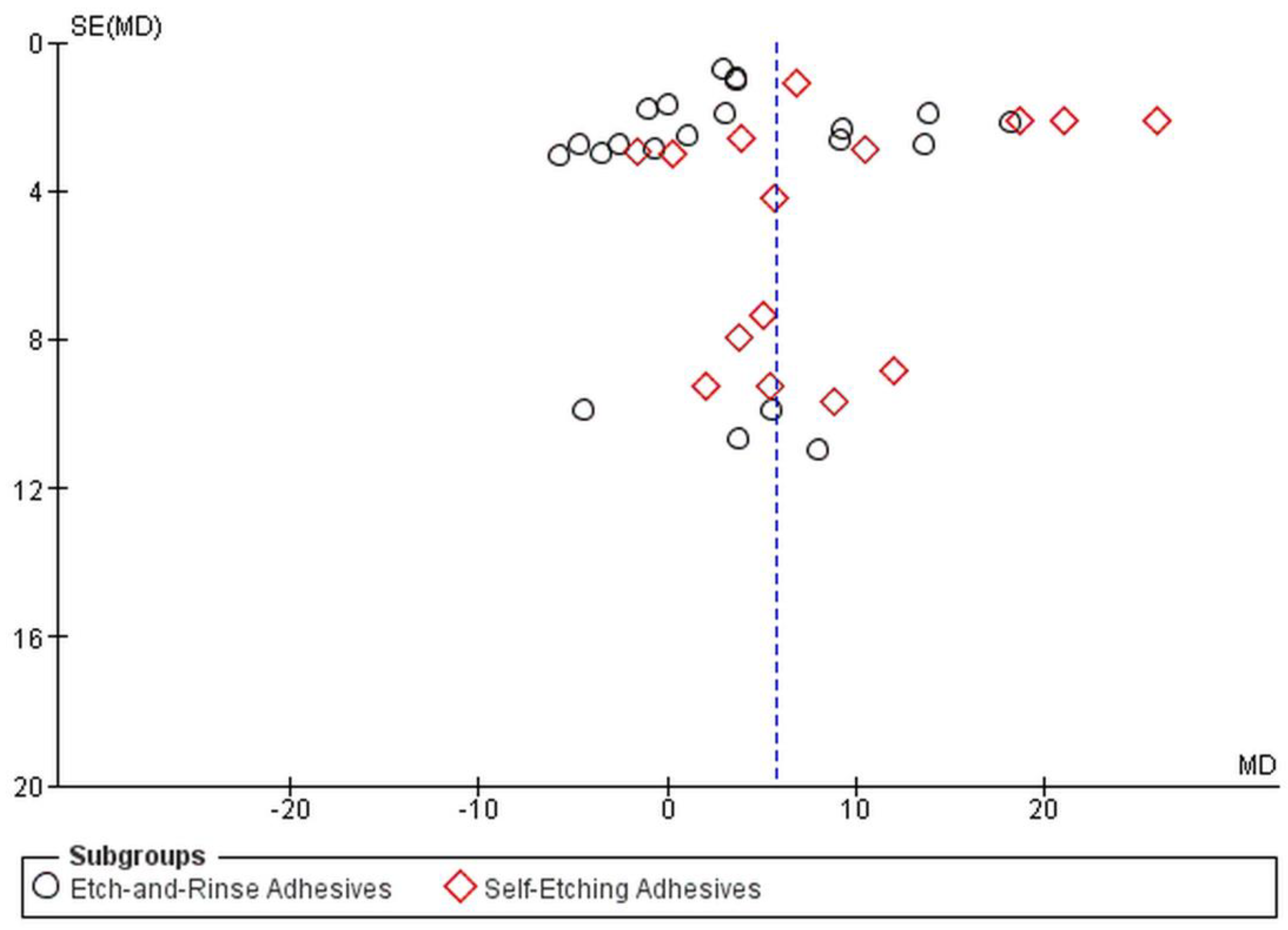
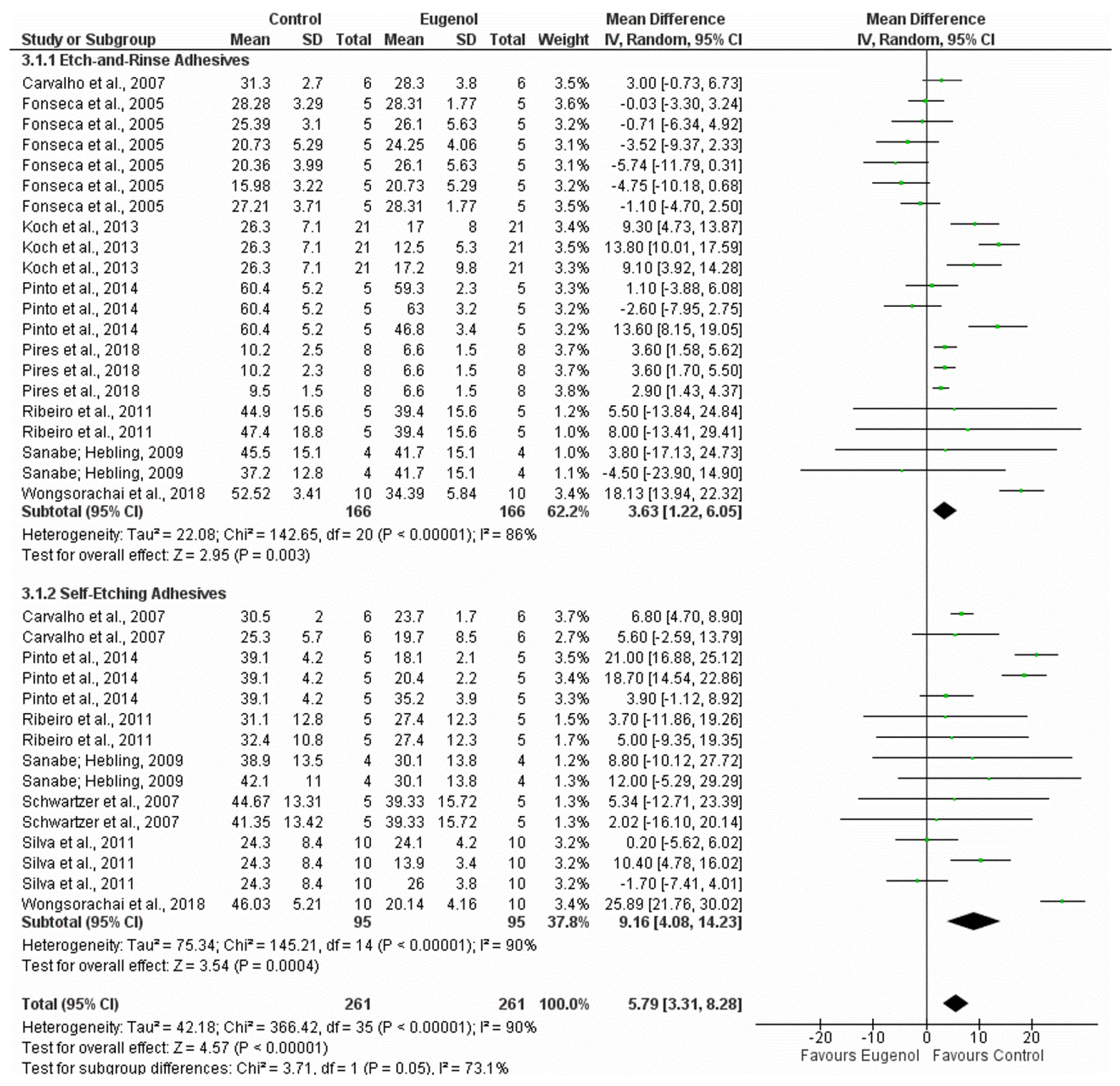
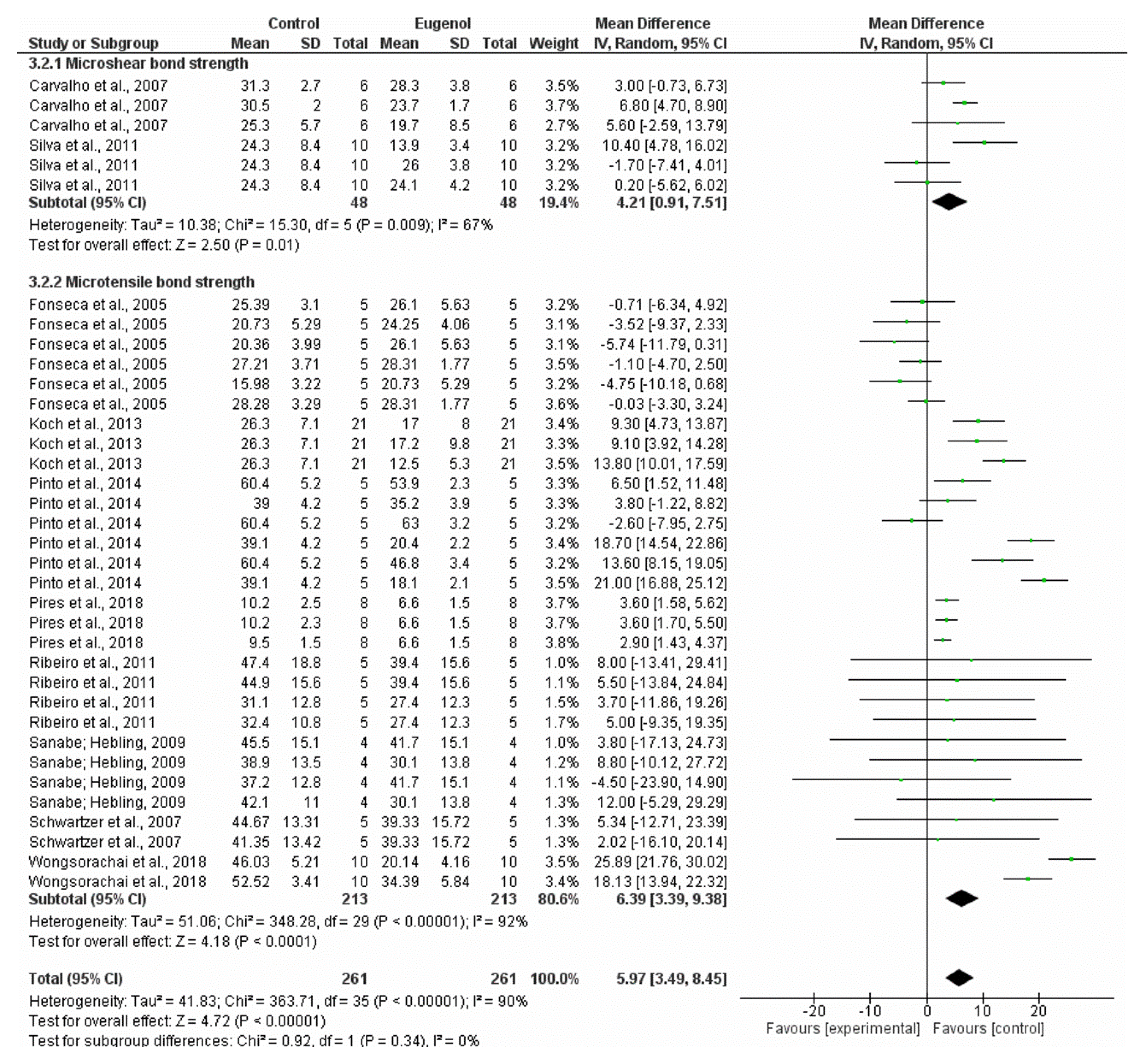
This review was carried out following the guidelines of the PRISMA Statement (Preferred Reporting Items for Systematic Review and Meta-Analysis) [36,37]. A systematic electronic search was performed in MEDLINE/Pubmed and Scopus databases using the keywords “(eugenol) AND (bond strength)” (no restriction was applied for language and publication dates). Eligible studies were those that assessed immediate bond strength of adhesive systems to coronal dentin after contact with eugenol-containing materials by testing specimens with a reduced size (micro-shear bond strength (µSBS) or micro-tensile bond strength (µTBS)). Inclusion criteria comprised the presence of a comparison of bond strength values with and without (control group) eugenol, with mean and standard deviation values presented in megapascal (MPa).
The searched titles and abstracts were checked carefully, and noneligible articles were excluded. Subsequently, full texts of the selected studies were obtained, and data were extracted to gather information about (1) method, (2) teeth type, (3) adhesive system, (4) eugenol-based material, (5) eugenol contact time, (6) storage time of samples prior the bond strength test, (7) sample size, (8) mean bond strength, and (9) standard deviation.
To assess the individual risk of bias of each study, six methodological items were analyzed: (1) screening for caries and cracks on teeth, (2) blindness of the researcher, (3) teeth randomization, (4) use of materials according to manufacturers’ instructions, (5) time of storage before debonding, and (6) presence of pretest failure that was included in statistical analyses. Such studies were classified s having high, moderate, or low risk of bias: studies with high risk failed to report four items or more, those with moderate risk failed to report 3 or 2 items, and those with low risk failed to report one item or less. The bias risk of the articles was evaluated by plotting the effect by the inverse of its standard error. The symmetry of the resulting funnel plot was visually assessed.
Meta-analysis was conducted using the “Review Manager Software (RMS)” (version 5.3) from the Cochrane Collaboration (Copenhagen, Denmark), and the mean difference with 95% confidence interval (CI) was calculated for mean bond strength of all groups of the studies included using the inverse variance method. The p-value for significance was set at p < 0.05. Statistical homogeneity (I2) of the treatment effect was assessed by the modified chi-square test (Cochran’s Q). The assessment indicated that there was statistical homogeneity when p > 0.10. Global and subgroup analyses were carried out (subgroups were arranged in ER or SE adhesive system and µSBS or µTBS).

3. Results

The latest search was performed on 9 November 2019. Figure 1 displays a flowchart representing the selection process of the studies involved in our work. Initially, 335 manuscripts were obtained (156 from PubMed and 179 from Scopus). Of those 335 studies, 147 were excluded as they were present in both databases. The title and abstract of each article were inspected, and 224 manuscripts were excluded because they were not relevant to the topic of this study or they did not meet the selection criteria (i.e., studies not related to dentistry or to dental restorations or based on macro-mechanical bonding tests; the latter were 29 studies). One study was excluded because the access to the manuscript was not possible, even after several attempts to contact the corresponding author [38]. A hand search was performed by checking the references in relevant papers, although no additional studies were found. Ten studies were included in the analysis, and their full texts were carefully examined.
Among the studies selected, nine were published in English and one in Portuguese. Eight studies assessed µTBS, and two assessed µSBS. Only two studies were performed using bovine teeth, while the other eight studies were performed using human teeth (one used the primary molar, and seven used the third molar). Five studies used only a eugenol-based material as a control group, whereas the other five also had a non-eugenol-based material group as a control. The adhesive systems tested in the studies were Adper Single Bond 2 (3M ESPE, Saint Paul, MN, USA) and Optibond FL (Kerr, Orange, CA, USA); the SE adhesive systems were Clearfil SE Bond (Kuraray, Okayama, Japan), iBond (Heraeus Kulzer, Hanau, Germany), One-Up Bond F (Tokuyama, Tokyo, Japan), Adper SE Plus (3M ESPE, St Paul, MN, USA), and Adper Prompt (3M ESPE, Saint Paul, MN, USA). The descriptive data of the 10 included studies are shown in Table 1.
The provisional eugenol-based materials used were Provy (Herpo/Dentsply, São Paulo, Brazil), IRM (Dentsply, Milford, USA), TempCem (Vigodent, Rio de Janeiro, Brazil), TempBond (Kerr, Orange, CA, USA), and ZOE and eugenol (Biodinâmica, Ibiporá, Brazil and Maquira, Maringá, Brazil). The provisional non-eugenol-based materials were Dycal (Dentsply/Caulk, Milfort, DE, USA), TempCem NE (Vigodent, Rio de Janeiro, Brazil), Freegenol (GC, Kasugai, Japan), iodoform-based Guedes-Pinto paste (Biodinâmica, Ibiporá, Brazil; Maquira, Maringá, Brazil; Fórmula&Ação, compounding pharmacy, São Paulo, Brazil), and calcium hydroxide paste thickened with ZOE (S. S. White Artigos Dentários Ltd.a., Rio de Janeiro, RJ, Brazil and Biodinâmica, Ibiporá, Brazil). In several studies, the effect of different eugenol contact times was tested, being the bond strength normally tested 24 h after the bonding step. In all studies, the primary outcome was bond strength expressed in MPa. The lowest mean for bond strength was 6.6 MPa, and the highest was 63 MPa (Table 1). During the qualitative analysis (Table 2), three studies were classified as having a high individual risk of bias, six as having a moderate individual risk of bias, and one as having a low individual risk of bias. The funnel plot (Figure 2) indicates the asymmetry among the studies.
The meta-analysis conducted on 10 studies with subgroups in ER and SE adhesive systems resulted in 36 comparisons (Figure 3): 21 in the ER adhesive system and 15 in the SE adhesive system. The random effects model was used, and the pooled global analysis showed a significant influence of eugenol (p < 0.00001). Eugenol had a negative effect on the bond strength of resin-based restorations (5.79 (3.31–8.28) MPa). Within the subgroup analyses, a previous contact with eugenol-based materials also negatively affected the bond strength (ER: 3.63 MPa (1.22–6.05), p = 0.003; SE: 9.16 (4.08–14.23) MPa, p = 0.0004). The heterogeneity was calculated via Tau2, Cochrane’s Q value, and I2. For the ER subgroup, Tau2 was 22.08 (Cochrane’s Q value p = 0.00001), and I2 was 86%. For the SE subgroup, Tau2 was 75.34 (Cochrane’s Q value p < 0.00001), and I2 was 90%. For the global analysis, Tau2 was 42.18 (Cochrane’s Q value p < 0.00001), and I2 was 90%. Therefore, significant heterogeneity was detected within comparisons and in the global analysis. The test for subgroup difference showed I2 = 73.1%.
Besides the meta-analysis comparing ER and SE adhesive systems, the differences between subgroups for µSBS and µTBS were compared (Figure 4). Starting from 36 comparisons, only 6 were used for µSBS and 30 for µTBS. For both subgroups, the presence of eugenol before bonding negatively affected the bond strength of the tested adhesives (µSBS: 4.21 (0.91–7.51) MPa; µTBS: 6.39 (3.39–9.38) MPa). Intragroup heterogeneity was observed for µSBS(Tau2 = 10.38,(Cochrane’s Q value p = 0.009) and I2 = 67%) and for µTBS (Tau2 = 51.06 (Cochrane’s Q value p < 0.00001) and I2 = 92%. The test for subgroup differences showed no heterogeneity between them (I2 = 0%, Cochrane’s Q value = 0.34).
Table 3 shows a qualitative description of the methods applied to remove the cements and to clean the surfaces before performing any bonding procedure. There was high variability among the techniques. For instance, some of them used Al2O3 sandblasting, and others a pumice–water slurry, a scaler, or a combination of these methods. Moreover, these techniques were based on different parameters, such as times of application, distance, and presence or absence of water irrigation, in different studies.

4. Discussion

The bonding performance of dental adhesives and luting agents to dentin “contaminated” with eugenol [1,17,18,19,30,31,39] has been the subject of a plethora of investigations during the last two decades. Nevertheless, the controversial results, inconsistencies, and inaccuracies of these studies have not provided the dentists with any evidence to support a judicious clinical decision considering the adverse effect of eugenol on adhesive dentistry. In order to increase the quality of the current systematic review and meta-analysis, we systematized all the selected studies into subgroups: (1) conventional ER adhesive system or SE adhesive system and, (2) evaluation through µSBS or µTBS. For both subgroups analyses as well as for the global analysis, it was found that the presence of residual eugenol jeopardized the bond strength of resin-based materials.
Previous in vitro studies highlighted the detrimental effect of eugenol-containing materials on dental bonding outcomes of resin-based materials [1,20], although there was no general agreement in the literature about such issue. Several studies have suggested that the effect of eugenol on the bond strength depended on the adhesive system employed [17]. While ER adhesive systems require the use of phosphoric acid [43], SE adhesives act through acid monomers, which can demineralize and consecutively infiltrate the dentin surface. Accordingly, the conventional ER bonding approach involves a water-rinsing step after the application of phosphoric acid to remove the residual acid containing a dissolved smear layer [43], while SE adhesives modify and incorporate the smear layer within the adhesive layer [44]. Therefore, after the removal of eugenol-containing provisional cement, the following cleaning steps for dentinal surface treatment can change the concentration of eugenol present on dentin and affect the final bond strength of resin-based materials [17,18,19]. In a previous study [18], it was emphasized that the use of phosphoric acid during the application of the conventional ER system could remove the smear layer contaminated with eugenol. Nevertheless, the pooled outcome of the meta-analysis performed here showed that the eugenol-contaminated dentin presented lower bond strength, regardless of the adhesive system applied.
Surface treatments such as orthophosphoric acid or ethylenediaminetetraacetic acid (EDTA) were investigated, and both significantly reduced the quantity of eugenol on the dentin surface [1]. However, it was previously observed that, even after acid application, residues of provisional cements could remain on the dentin surface [22]; hence, this justifies the outcomes of the current study. The phenolic component of eugenol can react with free radicals in the monomeric conversion from C = C bonds to C–C bonds, reducing the reactivity of the resin monomers and their polymerization. In this way, the remaining eugenol can bind to hydroxyl groups of the monomers [23,24,45,46] and reduce the degree of conversion of the adhesive system [47]. A reliable degree of conversion is essential for polymer-based materials, as it is related to the achievement of proper physical properties (e.g., high tensile strength [48] and microhardness [49]) and lower water sorption and solubility [48,50]. Thus, resin adhesive systems that show a high polymerization rate achieve better bonding performance and stability over time [51]. Here, by analyzing only immediate µTBS and µSBS, it was possible to observe that eugenol could reduce the adhesion values significantly. Therefore, it is possible to consider that, in terms of long-term results, this scenario could be even worse due to water sorption and degradation of the hybrid layer over time, mainly for a system with its monomer conversion impaired.
Bonding tests, mainly those through µTBS, are important for the study of resin-based restorative materials. The µTBS results have been associated with in vivo outcomes, mainly when the specimens are subjected to storage and longitudinal assessment [52]. The longer the specimens of bonding procedures are stored, the higher is the correlation with in vivo results [53]. Indeed, µTBS after six months of storage in water correlates with marginal discoloration of restorations. Unfortunately, during literature screening, we have noticed that there was no longitudinal assessment of bonding after eugenol application on dentin. The most extended time of specimens’ storage was 45 days [41] but this study did not perform a longitudinal analysis for dentin bonding strength. Thus, a further in vitro longitudinal analysis, possibly including in vivo outcomes, would be beneficial in order to avoid overestimating the results found in immediate µTBS. However, to the best of our knowledge, the present review provides the most complete view of the impact of eugenol in resin restorations on coronal dentin. In this context, bond strength tests using a reduced specimen size were preferred based on Griffith’s defect theory, which suggests a decrease in the tensile strength of large materials in comparison to small ones [54]. The bond strength of large specimens could be low or involve too many defects. Such test could not show the proper performance of the tested materials [54,55,56]. The subgroup analysis considered for the type of method applied (µTBS or µSBS) showed I2 = 0 in the test for subgroup difference and supported the findings of the pooled results. Thus, eugenol jeopardized the bonding to coronal dentin regardless of the adhesive systems used and the method of application.
Through this meta-analysis, it was possible to combine data across multiple studies and better estimate the differences between single studies. The percentage value defined by I2 showed the magnitude of the difference among the studies, which can be explained by heterogeneity and not by chance [57]. Although eugenol reduced the bond strength in all meta-analyses performed here, high heterogeneity could be observed in the subgroups and in the pooled data analysis. Therefore, other factors presented here should explain the model and the obtained results. Among 10 studies included, 4 showed a high risk of bias, and 5 a moderate risk of bias. The resulting bias can be accredited to the lack of a proper report of methodologies. It can also be hypothesized that a link exists between the lack of a proper report of methodologies and the high heterogeneity found here.
Moreover, the studies differed mainly regarding the method used for the removal of the temporary eugenol-based material (Table 3). The different instruments applied and the duration of each step (e.g., time for cleaning with a pumice–water slurry) may have influenced the results. Some studies reported similar cleaning steps for the control group (no contact with eugenol prior to bonding), while other papers did not mention cleaning (Table 3).
Variation in the cleaning steps was also found among several studies. Different cleaning steps can lead to changes in the dentin’s surface, which might impact the bonding performance. Finally, the type of ZOE-based material could also be an influencing factor for high heterogeneity. Different powder/liquid ratios for the ZOE cements classifications could increase eugenol release if a higher concentration of liquid with respect to that of powder is used [18]. One of the studies included in our analysis tested the effect of a root canal filling paste with eugenol instead of provisional eugenol–cement on the µSBS of an adhesive system [6]. We have included this study because its methodological design was very similar to those applied for testing eugenol-containing cements. Regardless of the high heterogeneity observed, it is possible to conclude that this study corroborates a previous one showing that eugenol jeopardizes not only the bonding to root dentin [29] but also the bonding to coronal dentin.

5. Conclusions

Within the limitations of an in vitro systematic review, the included studies have an acceptable methodological quality, contributing to a fact-based pathway of information regarding this subject. We found moderate-quality evidence supporting the adverse effect of eugenol on the bonding of resin-based materials (e.g., adhesive systems) to coronal dentin. Finally, we believe that further clinical investigations can improve the quality of the evidence of the detrimental effect of eugenol and guide dentists to better treatment decisions that foster the longevity of adhesive restorations.

Author Contributions

I.M.G.: Investigation, Formal analysis, Writing- Original draft preparation. V.C.B.L.: Conceptualization, Writing-Reviewing and Editing. M.S.I.: Writing-Reviewing and Editing. M.A.S.M.: Supervision, Writing-Reviewing and Editing. V.F.M.: Writing-Reviewing and Editing. S.S.: Conceptualization, Data curation, Writing-Reviewing and Editing. F.M.C.: Conceptualization, Data curation, Supervision, Formal analysis, Writing-Reviewing and Editing. All authors have read and agreed to the published version of the manuscript.

Funding

This research received no external funding.

Conflicts of Interest

The authors declare no conflict of interest.

References

  1. Koch, T.; Peutzfeldt, A.; Malinovskii, V.; Flury, S.; Haner, R.; Lussi, A. Temporary zinc oxide-eugenol cement: Eugenol quantity in dentin and bond strength of resin composite. Eur. J. Oral. Sci. 2013, 121, 363–369. [Google Scholar] [CrossRef] [PubMed]
  2. Taraslia, V.; Anastasiadou, E.; Lignou, C.; Keratiotis, G.; Agrafioti, A.; Kontakiotis, E.G. Assessment of cell viability in four novel endodontic sealers. Eur. J. Dent. 2018, 12, 287–291. [Google Scholar] [CrossRef] [PubMed]
  3. Ricucci, D.; Rocas, I.N.; Alves, F.R.; Loghin, S.; Siqueira, J.F., Jr. Apically Extruded Sealers: Fate and Influence on Treatment Outcome. J. Endod. 2016, 42, 243–249. [Google Scholar] [CrossRef] [PubMed]
  4. Thosar, N.R.; Chandak, M.; Bhat, M.; Basak, S. Evaluation of Antimicrobial Activity of Two Endodontic Sealers: Zinc Oxide with Thyme Oil and Zinc Oxide Eugenol against Root Canal Microorganisms-An in vitro Study. Int. J. Clin. Pediatr. Dent. 2018, 11, 79–82. [Google Scholar]
  5. AlShwaimi, E.; Bogari, D.; Ajaj, R.; Al-Shahrani, S.; Almas, K.; Majeed, A. In Vitro Antimicrobial Effectiveness of Root Canal Sealers against Enterococcus faecalis: A Systematic Review. J. Endod. 2016, 42, 1588–1597. [Google Scholar] [CrossRef]
  6. Pires, C.W.; Lenzi, T.L.; Soares, F.Z.M.; Rocha, R.O. Zinc oxide eugenol paste jeopardises the adhesive bonding to primary dentine. Eur. Arch. Paediatr. Dent. 2018, 19, 163–169. [Google Scholar] [CrossRef]
  7. Schwendicke, F.; Brouwer, F.; Schwendicke, A.; Paris, S. Different materials for direct pulp capping: Systematic review and meta-analysis and trial sequential analysis. Clin. Oral Investig. 2016, 20, 1121–1132. [Google Scholar] [CrossRef]
  8. Gonzalez-Lara, A.; Ruiz-Rodriguez, M.S.; Pierdant-Perez, M.; Garrocho-Rangel, J.A.; Pozos-Guillen, A.J. Zinc Oxide-Eugenol Pulpotomy in Primary Teeth: A 24-Month Follow-up. J. Clin. Pediatr. Dent. 2016, 40, 107–112. [Google Scholar] [CrossRef]
  9. Smail-Faugeron, V.; Glenny, A.M.; Courson, F.; Durieux, P.; Muller-Bolla, M.; Fron Chabouis, H. Pulp treatment for extensive decay in primary teeth. Cochrane Database Syst. Rev. 2018, 5. [Google Scholar] [CrossRef]
  10. Chebib, N.; Kalberer, N.; Srinivasan, M.; Maniewicz, S.; Perneger, T.; Muller, F. Edentulous jaw impression techniques: An in vivo comparison of trueness. J. Prosthet. Dent. 2019, 121, 623–630. [Google Scholar] [CrossRef]
  11. De Resende, G.P.; Nogueira, T.E. Effectiveness of a simplified method for final impression in complete denture treatment: A pragmatic clinical trial. Gerodontology 2019, 36, 365–373. [Google Scholar] [CrossRef] [PubMed]
  12. Shin, J.O.; Ko, K.H. Displacement of Simulated Flabby Tissue by Different Tray Designs and Impression Materials. J. Prosthodont. 2019, 28, 444–451. [Google Scholar] [CrossRef] [PubMed]
  13. Jayaraman, S.; Singh, B.P.; Ramanathan, B.; Pazhaniappan Pillai, M.; MacDonald, L.; Kirubakaran, R. Final-impression techniques and materials for making complete and removable partial dentures. Cochrane Database Syst. Rev. 2018, 4. [Google Scholar] [CrossRef] [PubMed]
  14. Hill, E.E.; Lott, J. A clinically focused discussion of luting materials. Aust. Dent. J. 2011, 56 (Suppl. S1), 67–76. [Google Scholar] [CrossRef]
  15. Barcelos, R.; Santos, M.P.; Primo, L.G.; Luiz, R.R.; Maia, L.C. ZOE paste pulpectomies outcome in primary teeth: A systematic review. J. Clin. Pediatr. Dent. 2011, 35, 241–248. [Google Scholar] [CrossRef]
  16. Hashimoto, S.; Uchiyama, K.; Maeda, M.; Ishitsuka, K.; Furumoto, K.; Nakamura, Y. In vivo and in vitro effects of zinc oxide-eugenol (ZOE) on biosynthesis of cyclo-oxygenase products in rat dental pulp. J. Dent. Res. 1988, 67, 1092–1096. [Google Scholar] [CrossRef]
  17. Schwartzer, E.; Collares, F.M.; Ogliari, F.A.; Leitune, V.C.B.; Samuel, S.M.W. Influence of zinc oxide-eugenol temporary cement on bond strength of an all-in-one adhesive system to bovine dentin. Braz. J. Oral. Sci. 2007, 6, 1423–1427. [Google Scholar]
  18. Carvalho, C.N.; de Oliveira Bauer, J.R.; Loguercio, A.D.; Reis, A. Effect of ZOE temporary restoration on resin-dentin bond strength using different adhesive strategies. J. Esthet. Restor. Dent. 2007, 19, 144–152. [Google Scholar] [CrossRef]
  19. Ribeiro, J.C.; Coelho, P.G.; Janal, M.N.; Silva, N.R.; Monteiro, A.J.; Fernandes, C.A. The influence of temporary cements on dental adhesive systems for luting cementation. J. Dent. 2011, 39, 255–262. [Google Scholar] [CrossRef]
  20. Silva, J.P.; Queiroz, D.M.; Azevedo, L.H.; Leal, L.C.; Rodrigues, J.L.; Lima, A.F.; Marchi, G.M.; Brito-Junior, M.; Faria-e-Silva, A.L. Effect of eugenol exposure time and post-removal delay on the bond strength of a self-etching adhesive to dentin. Oper. Dent. 2011, 36, 66–71. [Google Scholar] [CrossRef] [Green Version]
  21. Terata, R.; Nakashima, K.; Obara, M.; Kubota, M. Characterization of enamel and dentin surfaces after removal of temporary cement—Effect of temporary cement on tensile bond strength of resin luting cement. Dent. Mater. J. 1994, 13, 148–154. [Google Scholar] [CrossRef] [PubMed] [Green Version]
  22. Watanabe, E.K.; Yatani, H.; Ishikawa, K.; Suzuki, K.; Yamashita, A. Pilot study of conditioner/primer effects on resin-dentin bonding after provisional cement contamination using SEM, energy dispersive x-ray spectroscopy, and bond strength evaluation measures. J. Prosthet. Dent. 2000, 83, 349–355. [Google Scholar] [CrossRef]
  23. Bayindir, F.; Akyil, M.S.; Bayindir, Y.Z. Effect of eugenol and non-eugenol containing temporary cement on permanent cement retention and microhardness of cured composite resin. Dent. Mater. J. 2003, 22, 592–599. [Google Scholar] [CrossRef] [PubMed] [Green Version]
  24. Taira, J.; Ikemoto, T.; Yoneya, T.; Hagi, A.; Murakami, A.; Makino, K. Essential oil phenyl propanoids. Useful as OH scavengers? Free Radical Res. Commun. 1992, 16, 197–204. [Google Scholar] [CrossRef] [PubMed]
  25. Hansen, E.K.; Asmussen, E. Influence of temporary filling materials on effect of dentin-bonding agents. Scand. J. Dent. Res. 1987, 95, 516–520. [Google Scholar] [CrossRef] [PubMed]
  26. Yap, A.U.; Shah, K.C.; Loh, E.T.; Sim, S.S.; Tan, C.C. Influence of eugenol-containing temporary restorations on bond strength of composite to dentin. Oper. Dent. 2001, 26, 556–561. [Google Scholar]
  27. Nakabayashi, N.; Kojima, K.; Masuhara, E. The promotion of adhesion by the infiltration of monomers into tooth substrates. J. Biomed. Mater. Res. 1982, 16, 265–273. [Google Scholar] [CrossRef]
  28. Van Landuyt, K.L.; Snauwaert, J.; De Munck, J.; Peumans, M.; Yoshida, Y.; Poitevin, A.; Coutinho, E.; Suzuki, K.; Lambrechts, P.; Van Meerbeek, B. Systematic review of the chemical composition of contemporary dental adhesives. Biomaterials 2007, 28, 3757–3785. [Google Scholar] [CrossRef]
  29. Altmann, A.S.; Leitune, V.C.; Collares, F.M. Influence of Eugenol-based Sealers on Push-out Bond Strength of Fiber Post Luted with Resin Cement: Systematic Review and Meta-analysis. J. Endodont. 2015, 41, 1418–1423. [Google Scholar] [CrossRef]
  30. Carvalho, E.M.; Carvalho, C.N.; Loguercio, A.D.; Lima, D.M.; Bauer, J. Effect of temporary cements on the microtensile bond strength of self-etching and self-adhesive resin cement. Acta Odontol. Scand. 2014, 72, 762–769. [Google Scholar] [CrossRef]
  31. Leirskar, J.; Nordbo, H. The effect of zinc oxide-eugenol on the shear bond strength of a commonly used bonding system. Endod. Dent. Traumatol. 2000, 16, 265–268. [Google Scholar] [CrossRef] [PubMed]
  32. Peutzfeldt, A.; Asmussen, E. Influence of eugenol-containing temporary cement on efficacy of dentin-bonding systems. Eur. J. Oral. Sci. 1999, 107, 65–69. [Google Scholar] [CrossRef] [PubMed]
  33. Peutzfeldt, A.; Asmussen, E. Influence of eugenol-containing temporary cement on bonding of self-etching adhesives to dentin. J. Adhes. Dent. 2006, 8, 31–34. [Google Scholar] [PubMed]
  34. Al-Wazzan, K.A.; al-Harbi, A.A.; Hammad, I.A. The effect of eugenol-containing temporary cement on the bond strength of two resin composite core materials to dentin. J. Prosthodont. 1997, 6, 37–42. [Google Scholar] [CrossRef]
  35. Macchi, R.L.; Capurro, M.A.; Herrera, C.L.; Cebada, F.R.; Kohen, S. Influence of endodontic materials on the bonding of composite resin to dentin. Endod. Dent. Traumatol. 1992, 8, 26–29. [Google Scholar] [CrossRef]
  36. Moher, D.; Liberati, A.; Tetzlaff, J.; Altman, D.G. Preferred reporting items for systematic reviews and meta-analyses: The PRISMA statement. PLoS Med. 2009, 6, e1000097. [Google Scholar] [CrossRef] [Green Version]
  37. Macaskill, P.; Gatsonis, C.; Deeks, J.J.; Harbord, R.M.; Takwoingi, Y. Analysing and presenting results. In Cochrane Handbook for Systematic Reviews of Diagnostic Test Accuracy; Deeks, J.J., Bossuyt, P.M., Gatsonis, C., Eds.; The Cochrane Collaboration: London, UK, 2010. [Google Scholar]
  38. Paul, S.J.; Scharer, P. The dual bonding technique: A modified method to improve adhesive luting procedures. Int. J. Periodont. Restor. Dent. 1997, 17, 536–545. [Google Scholar]
  39. Fonseca, R.B.; Martins, L.R.; Quagliatto, P.S.; Soares, C.J. Influence of provisional cements on ultimate bond strength of indirect composite restorations to dentin. J. Adhes. Dent. 2005, 7, 225–230. [Google Scholar]
  40. Sanabe, M.E.; Bossolan, A.P.O.G.; da Cruz, A.R.; Hebling, J. Influence of dentin contamination by temporary cements on the bond strengh of adhesive systems. RGO 2009, 57, 33–39. [Google Scholar]
  41. Pinto, K.T.; Stanislawczuk, R.; Loguercio, A.D.; Grande, R.H.M.; Bauera, J. Effect of exposure time of zinc oxide eugenol restoration on microtensile bond strength of adhesives to dentin. Rev. Port. Estomatol. Med. Dent. Cir. Maxilofac. 2014, 55, 83–88. [Google Scholar] [CrossRef] [Green Version]
  42. Wongsorachai, R.N.; Thanatvarakorn, O.; Prasansuttiporn, T.; Jittidecharaks, S.; Hosaka, K.; Foxton, R.M.; Nakajima, M. Effect of Polymerization Accelerator on Bond Strength to Eugenol-Contaminated Dentin. J. Adhes. Dent. 2018, 20, 541–547. [Google Scholar] [PubMed]
  43. Pashley, D.H.; Tay, F.R.; Breschi, L.; Tjaderhane, L.; Carvalho, R.M.; Carrilho, M.; Tezvergil-Mutluay, A. State of the art etch-and-rinse adhesives. Dent. Mater. 2011, 27, 1–16. [Google Scholar] [CrossRef] [Green Version]
  44. Van Meerbeek, B.; Yoshihara, K.; Yoshida, Y.; Mine, A.; De Munck, J.; Van Landuyt, K.L. State of the art of self-etch adhesives. Dent. Mater. 2011, 27, 17–28. [Google Scholar] [CrossRef] [PubMed]
  45. Watanabe, E.K.; Yatani, H.; Ishikawa, K.; Suzuki, K.; Yamashita, A. Action of eugenol as a retarder against polymerization of methyl methacrylate by benzoyl peroxide. J. Prosthet. Dent. 2000, 83, 349–355. [Google Scholar] [CrossRef]
  46. Fujisawa, S.; Kadoma, Y. Action of eugenol as a retarder against polymerization of methyl methacrylate by benzoyl peroxide. Biomaterials 1997, 18, 701–703. [Google Scholar] [CrossRef]
  47. Bohrer, T.C.; Fontana, P.E.; Wandscher, V.F.; Morari, V.H.C.; Dos Santos, S.S.; Valandro, L.F.; Kaize, O.B. Endodontic Sealers Affect the Bond Strength of Fiber Posts and the Degree of Conversion of Two Resin Cements. J. Adhes. Dent. 2018, 20, 165–172. [Google Scholar]
  48. Collares, F.M.; Ogliari, F.A.; Zanchi, C.H.; Petzhold, C.L.; Piva, E.; Samuel, S.M. Influence of 2-hydroxyethyl methacrylate concentration on polymer network of adhesive resin. J. Adhes. Dent. 2011, 13, 125–129. [Google Scholar]
  49. Ferracane, J. Correlation between hardness and degree of conversion during the setting reaction of unfilled dental restorative resins. Dent. Mater. 1985, 1, 11–14. [Google Scholar] [CrossRef]
  50. Ferracane, J.L. Hygroscopic and hydrolytic effects in dental polymer networks. Dent. Mater. 2006, 22, 211–222. [Google Scholar] [CrossRef]
  51. De Munck, J.; Van Meerbeek, B.; Yoshida, Y.; Inoue, S.; Vargas, M.; Suzuki, K.; Lambrechts, P.; Vanherle, G. Four-year water degradation of total-etch adhesives bonded to dentin. J. Dent. Res. 2003, 82, 136–140. [Google Scholar] [CrossRef]
  52. Van Meerbeek, B.; Peumans, M.; Poitevin, A.; Mine, A.; Van Ende, A.; Neves, A.; De Munck, J. Relationship between bond-strength tests and clinical outcomes. Dent. Mater. 2010, 26, e100–e121. [Google Scholar] [CrossRef] [PubMed]
  53. Braga, R.R.; Meira, J.B.; Boaro, L.C.; Xavier, T.A. Adhesion to tooth structure: A critical review of "macro" test methods. Dent Mater. 2010, 26, e38–e49. [Google Scholar] [CrossRef] [PubMed]
  54. Griffith, A.A. The phenomena of rupture and flow in solid. Phil. Trans. Roy. Soc. Lon. 1920, A221, 168–198. [Google Scholar]
  55. Sano, H.; Shono, T.; Sonoda, H.; Takatsu, T.; Ciucchi, B.; Carvalho, R.; Pashley, D.H. Relationship between surface area for adhesion and tensile bond strength–evaluation of a micro-tensile bond test. Dent. Mater. 1994, 10, 236–240. [Google Scholar] [CrossRef]
  56. Pashley, D.H.; Sano, H.; Ciucchi, B.; Yoshiyama, M.; Carvalho, R.M. Adhesion testing of dentin bonding agents: A review. Dent. Mater. 1995, 11, 117–125. [Google Scholar] [CrossRef]
  57. Higgins, J.P.; Thompson, S.G.; Deeks, J.J.; Altman, D.G. Measuring inconsistency in meta-analyses. BMJ 2003, 327, 557–560. [Google Scholar] [CrossRef] [Green Version]
Figure 1. Systematic review flow chart of studies comparing the bond strength of adhesive systems/composite resin to coronal dentin after contact with eugenol-based materials and without contact.
Applsci 10 01070 g001
Figure 2. Funnel plot of the included studies.
Applsci 10 01070 g002
Figure 3. Forest plot of global and subgroups (etch-and-rinse (ER) or self-etch (SE) adhesive system) meta-analyses. The bond strengths of adhesive systems/resin-based restorative materials to coronal dentin after contact with eugenol-based materials and without contact were compared.
Applsci 10 01070 g003
Figure 4. Forest plot of global and subgroups (µSBS or µTBS) meta-analyses. The bond strength of adhesive systems/resin-based restorative materials to coronal dentin after contact with eugenol-based materials and without contact were compared via two tests used to classify the studies as µSBS or µTBS.
Applsci 10 01070 g004
Table 1. Identification and characteristics of the included studies.
AuthorMethodologySubstrateAdhesiveEugenol MaterialEugenol Contact TimeStorage TimeGroup Sample SizeBond Strength (MPa)SD
Fonseca et al. 2005 [39]µTBS
(sticks of 1 mm2)
Bovine TeethSingle BondDycal7 days-527.21
(Al2O3 sandblasting)
3.71
-520.36
(Pumice-water slurry)
3.99
-515.98
(Hand scaler)
3.22
Provy7 days-528.31
(Al2O3 sandblasting)
1.77
-526.10
(Pumice-water slurry)
5.63
-520.73
(Hand scaler)
5.29
TempBond NE7 days-528.28
(Al2O3 sandblasting)
3.29
-525.39
(Pumice-water slurry)
3.10
-524.25
(Hand scaler)
4.06
Carvalho et al., 2007 [18]µSBS
(Cylinders - H:0.5 mm × D:0.75 mm)
Human Teeth
(third molar)
Clearfil SE BondIRM24 h24 h623.71.7
NoneN/A630.52.0
iBondIRM24 h619.78.5
NoneN/A625.35.7
Single BondIRM24 h628.33.8
NoneN/A631.32.7
Schwartzer, et al., 2007 [17]µTBS
(sticks of 0.5 mm2 Aprox. 0.7 mm × 0.7 mm)
Bovine TeethOne-UP Bond FTempCem7 days24 h539.315.72
NoneN/A544.6713.31
TempCem NE7 days541.3513.42
Sanabe; Hebling,
2009 [40]
µTBS
(sticks of 0.81 mm2)
Human Teeth
(third molar)
Adper Single BondCavit7 days24 h437.212.8
IRM7 days441.715.1
NoneN/A445.515.1
Clearfil SE BondCavit7 days442.111.0
IRM7 days430.113.8
NoneN/A438.913.5
Ribeiro, et al., 2011 [19]µTBS
(rectangular beams of 0.9 mm2)
Human Teeth
(third molars)
Single Bond TempBond7 days24 h539.415.6
NoneN/A544.915.6
Freegenol7 days547.418.8
Adper PromptTempBond7 days527.412.3
NoneN/A532.410.8
Freegenol7 days531.112.8
Silva et al., 2011 [20]µSBS
(Cylinders -H:2mmxD:1mm)
Human Teeth
(third molars)
Adper SE PlusIRM24 hImmediate1013.93.4
7 days1026.03.8
14 days1024.14.2
NoneN/A1024.38.4
Koch, et al., 2013 [1]µTBS
(sticks of 1.07 mm2)
Human Teeth
(molars)
Optibond FLIRM1 day7 days2112.55.3
7 days2117.29.8
28 days2117.08.0
NoneN/A2126.37.1
Pinto et al., 2014 [41]µTBS
(sticks of 0.8 mm2)
Human Teeth
(third molars)
Adper Single Bond 2IRM24 hImmediate546.83.4
7 days563.03.2
45 days559.32.3
NoneN/A560.45.2
Clearfil S3 BondIRM24 h520.42.2
7 days518.12.1
45 days535.23.9
NoneN/A539.14.2
Pires et al., 2018 [6]µTBS
(sticks of 1 mm2)
Human Teeth
(primary molar)
Adper Single
Bond 2
Zinc oxide and eugenol15 min24 h86.61.5
Iodoform-based Guedes-Pinto paste15 min810.22.5
Calcium hydroxide paste thickened
with zinc oxide
15 min89.51.5
NoneN/A810.22.3
Wongsorachai et al., 2018 [42]µTBS
(sticks of 1 mm2)
Human Teeth
(third molar)
Optibond FLIRM24 h24 h1034.395.84
NoneN/A1052.523.41
Clearfil SE BondIRM24 h1020.144.16
NoneN/a1046.035.21
Table 2. Individual risk of bias of the included studies.
StudyTeeth Free of CariesBlinded ResearcherTeeth RandomizationManufacturer’s InstructionsStorage TimePre-Test FailureClassification
Fonseca et al., 2005 [39]+--+?-High
Carvalho et al., 2007 [18]+--++-Moderate
Schwartzer et al., 2007 [17]+--++-Moderate
Sanabe; Hebling, 2009 [40]+---+-High
Ribeiro et al., 2011 [19]+-+++?Moderate
Silva et al., 2011 [20]+-++--Moderate
Koch et al., 2013 [1]--+-+-High
Pinto et al., 2014 [41] ---++-High
Pires et al., 2018 [6]+++++-Low
Wongsorachai et al., 2018 [42]+-+++-Moderate
+ reported; - not reported; ? unclear.
Table 3. Identification of the study and detailed method apply to remove the eugenol-containing cement and to clean the dentin surfaces prior to the bonding procedures.
AuthorAdhesiveEugenol MaterialDetails of Cleanness Method Applied
Fonseca et al. 2005 [39]Single BondDycal
  • Hand scaler for 10 s, or
  • Pumice-water slurry for 10 s, or
  • Sandblasting with 50 µm aluminum oxide particles for 5 s at a pressure of 4 bars and source-to-samples distance of 2 cm.
Provy
  • Hand scaler for 10 s, or
  • Pumice-water slurry for 10 s, or
  • Sandblasting with 50 µm aluminum oxide particles for 5 s at a pressure of 4 bars and source-to-samples distance of 2 cm.
TempBond NE
  • Hand scaler for 10 s, or
  • Pumice-water slurry for 10 s, or
  • Sandblasting with 50 µm aluminum oxide particles for 5 s at a pressure of 4 bars and source-to-samples distance of 2 cm.
Carvalho et al., 2007 [18] Clearfil SE BondIRM
  • Mechanically removed with a scaler until the dentin surfaces were visually macroscopically free of material;
  • Cleaned with a pumice-water slurry in a slow-speed handpiece for 60 s and rinsed off with air-water stream 60 s.
None
  • Not reported
iBondIRM
  • Mechanically removed with a scaler until the dentin surfaces were visually macroscopically free of material;
  • Cleaned with a pumice-water slurry in a slow-speed handpiece for 60 s and rinsed off with air-water stream 60 s.
None
  • Not reported
Single BondIRM
  • Mechanically removed with a scaler until the dentin surfaces were visually macroscopically free of material;
  • Cleaned with a pumice-water slurry in a slow-speed handpiece for 60 s and rinsed off with air-water stream 60 s.
None
  • Not reported
Schwartzer, et al., 2007 [17]One-UP Bond FTempCem
  • Mechanically removed using periodontal instruments and Robinson brushes equipped with pumice;
  • Rinsing with distilled water.
None
  • Not reported
TempCem NE
  • Mechanically removed using periodontal instruments and Robinson brushes equipped with pumice;
  • Rinsing with distilled water.
Sanabe; Hebling,
2009 [40]
Adper Single BondCavit
  • Mechanically removed with dentin spoon up to a visually clean surface;
  • Pumice-water and running water.
IRM
  • Mechanically removed with dentin spoon up to a visually clean surface;
  • Pumice-water and running water.
None
  • Not reported
Clearfil SE BondCavit
  • Mechanically removed with dentin spoon up to a visually clean surface;
  • Pumice-water and running water.
IRM
  • Mechanically removed with dentin spoon up to a visually clean surface;
  • Pumice-water and running water.
None
  • Not reported
Ribeiro et al., 2011 [19]Single Bond TempBond
  • Scraped off with a dental instrument until unaided visual inspection indicated that the surface was free of cement;
  • Rinsed with water spray, cleaned with cotton pellets soaked with pumice-water slurry for 20 s;
  • Rinsed again and air-dried.
None
  • Not reported
Freegenol
  • Scraped off with a dental instrument until unaided visual inspection indicated that the surface was free of cement;
  • Rinsed with water spray, cleaned with cotton pellets soaked with pumice-water slurry for 20 s;
  • Rinsed again and air-dried.
Adper PromptTempBond
  • Scraped off with a dental instrument until unaided visual inspection indicated that the surface was free of cement;
  • Rinsed with water spray, cleaned with cotton pellets soaked with pumice-water slurry for 20 s/
  • Rinsed again and air-dried.
None
  • Not reported
Freegenol
  • Scraped off with a dental instrument until unaided visual inspection indicated that the surface was free of cement;
  • Rinsed with water spray, cleaned with cotton pellets soaked with pumice-water slurry for 20 s;
  • Rinsed again and air-dried.
Silva et al., 2011 [20]Adper SE PlusIRM
  • Mechanically removed with a scaler until the dentin surface was visually (macroscopically) free of the material;
  • Cleaned with pumice-water slurry in a slow-speed handpiece and rinsed with an air-water stream.
  • Mechanically removed with a scaler until the dentin surface was visually (macroscopically) free of the material;
  • Cleaned with pumice-water slurry in a slow-speed handpiece and rinsed with an air-water stream.
  • Mechanically removed with a scaler until the dentin surface was visually (macroscopically) free of the material;
  • Cleaned with pumice-water slurry in a slow-speed handpiece and rinsed with an air-water stream.
None
  • Not reported.
Koch et al., 2013 [1]Optibond FLIRM
  • Macroscopically cleaned of the ZOE using a dental scaler.
  • Macroscopically cleaned of the ZOE using a dental scaler.
  • Macroscopically cleaned of the ZOE using a dental scaler.
None
  • Not reported.
Pinto et al., 2014 [41]Adper Single Bond 2IRM
  • Removed using a stainless-steel spatula;
  • Cleaned with pumice- water slurry using a slow speed handpiece for 60 s and rinsed with an air-water stream (60 s).
  • Removed using a stainless-steel spatula;
  • Cleaned with pumice- water slurry using a slow speed handpiece for 60 s and rinsed with an air-water stream (60 s).
  • Removed using a stainless-steel spatula;
  • Cleaned with pumice- water slurry using a slow speed handpiece for 60 s and rinsed with an air-water stream (60 s).
None
  • Cleaned with pumice- water slurry using a slow speed handpiece for 60 s and rinsed with an air-water stream (60 s).
Clearfil S3 BondIRM
  • Removed using a stainless-steel spatula;
  • Cleaned with pumice- water slurry using a slow speed handpiece for 60 s and rinsed with an air-water stream (60 s).
  • Removed using a stainless-steel spatula;
  • Cleaned with pumice- water slurry using a slow speed handpiece for 60 s and rinsed with an air-water stream (60 s).
  • Removed using a stainless-steel spatula;
  • Cleaned with pumice- water slurry using a slow speed handpiece for 60 s and rinsed with an air-water stream (60 s).
None
  • Cleaned with pumice- water slurry using a slow speed handpiece for 60 s and rinsed with an air-water stream (60 s).
Pires et al., 2018 [6]Adper Single
Bond 2
Zinc oxide and eugenol
  • Mechanically removed with a spatula and the dentine surfaces rubbed with a dry cotton pellet until visually free of material;
  • Water rinsing for 15 s and drying with oil-free airstream for 5 s.
Iodoform-based Guedes-Pinto paste
  • Mechanically removed with a spatula and the dentine surfaces rubbed with a dry cotton pellet until visually free of material;
  • Water rinsing for 15 s and drying with oil-free airstream for 5 s.
Calcium hydroxide paste thickened
with zinc oxide
  • Mechanically removed with a spatula and the dentine surfaces rubbed with a dry cotton pellet until visually free of material;
  • Water rinsing for 15 s and drying with oil-free airstream for 5 s.
None
  • Not reported.
Wongsorachai et al., 2018 [42]Optibond FLIRM
  • Mechanically removed with an ultrasonic scaler at the frequency of 28 kHz until the dentin surfaces were visually free of material;
  • cleaned with pumice and water slurry using a slow-speed handpiece for 60 s and rinsed off with an air-water stream for 30 s.
None
  • cleaned with pumice and water slurry using a slow-speed handpiece for 60 s and rinsed off with an air-water stream for 30 s.
Clearfil SE BondIRM
  • Mechanically removed with an ultrasonic scaler at the frequency of 28 kHz until the dentin surfaces were visually free of material.
  • Cleaned with pumice and water slurry using a slow-speed handpiece for 60 s and rinsed off with an air-water stream for 30 s.
None
  • cleaned with pumice and water slurry using a slow-speed handpiece for 60 s and rinsed off with an air-water stream for 30 s.

