1. Introduction
Splinting is a part of the management of the teeth after traumatic injuries. The splinting protocol has changed over the years. The trend to ligate or splint the injured teeth using rigid, prolonged fixation, the same as in jaw fractures, was maintained for many years. However, these splinting principles, which are still necessary for the treatment of broken jaws, have proven to be inadequate for the management of dental trauma [
1]. The effect of splinting on healing has for many years been well documented in animal studies [
2,
3]. Striving to improve the quality of splinting, to optimize the healing of damaged dental tissues, to simplify techniques, and the availability of materials, caused the devices like arch bars, wire ligatures, or cap splints to no longer be recommended. They have been replaced by flexible splints fixed with adhesive techniques [
4].
A dental trauma splint is defined as a device used to protect and stabilize a traumatized tooth in its natural position. The wide variety of dental trauma splints available allows a dentist to select such material that meets specific requirements of an individual patient. The materials that are currently used to stabilize the teeth are mostly wires (with the diameter being no greater than 0.4 mm), titanium trauma splint (TTS), and orthodontic brackets. A nylon fishing line fixed with a composite resin is also a good alternative to use as a splinting device. Fiber, composite resin, multiphase resin, and removable splints are also employed by dentists. For review, see Andreasen and Oikarinen 2019 [
4].
An optimal splint should be commonly accessible in the dental office and easy to apply and remove. It should ensure proper fixation of the tooth during the healing period. Splinting materials should be passive and elastic, allowing for physiological tooth mobility. The comfort of wearing trauma splints is also important. This is why the device used must not impair oral hygiene and speech, interfere with occlusal movements, or traumatize oral soft tissues [
4].
Among all of the requirements for trauma splints, the rigidity of splinting materials has been the focus of most studies. In only two of these studies, the rigidity was evaluated in vivo in healthy [
5] and injured [
6] patients. Most authors conducted research using a human cadaveric model [
7], an animal [
8], and artificial models [
9,
10,
11,
12,
13,
14,
15,
16,
17,
18,
19].
Recently, in an experimental study, the power chain has been shown to be a suitable device for splitting [
18]. Before using this material in trauma patients, it is advisable to evaluate its properties in a clinical study on healthy volunteers and to compare the results with those obtained for commonly used splints.
The main objective of this study was to evaluate and compare three different materials used for splinting in human volunteers. Two of the splints tested known as a wire-composite splint and a titanium trauma splint (TTS) are commonly used in case of dental injuries. The third material, known as the power chain, is made of connected elastic ligatures, which are used in orthodontics mainly for space closure.
2. Materials and Methods
The study was conducted in 10 volunteers who were recruited from the students of our university. Four of them were females and six were males with a mean age of 25 years old (±9.26). The study was positively evaluated and approved by the Independent Bioethics Commission for Research at the Medical University of Gdansk (study number: NKBBN/197/2016). All the participants received written information about the study and were asked to sign a consent form.
The inclusion criteria for volunteers were as follows: no medical contraindications for planned procedures, the presence of four maxillary incisors free from caries, periodontal diseases, and without fixed orthodontic appliances or retainers.
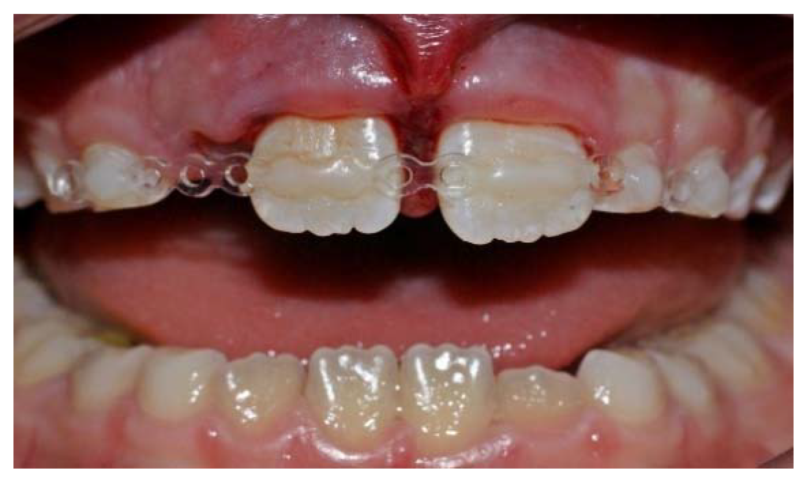
Three different splinting materials were tested in each person: a wire with a diameter of 0.5 mm (wire-composite splint) (
Figure 1), a titanium trauma splint (TTS) (
Figure 2), and a power chain-composite splint (PCS) (
Figure 3). Each splint was applied for four days. The sequence of splint application was determined at random. After the removal, we waited at least four days before the fixation of the next splint. The splints were fixed to four maxillary incisors of which both central incisors simulated the teeth after trauma, while the lateral incisors served as uninjured ones firmly encased in the socket.
2.1. Splints Application
All splints were bonded to the labial surface of maxillary incisors. The enamel was etched with 35% phosphoric acid gel for 30 s, then rinsed, and dried prior to bond application. The bonding agent was applied and polymerized for 40 s. Each splinting material was cut to the required length, contoured to dental arch, and fixed using a flowable composite.
2.2. Splints Removal
A high-speed bur was used to grind the composite down to a splinting material, which was then removed. Any residual material was eliminated with composite removal burs and Sof-Lex disks.
2.3. Parameters Evaluated in the Study
2.3.1. Teeth Mobility
The Periotest M (Medizintechnik Gulden, Modautal, Germany) device was used to measure vertical and horizontal tooth mobility. The Periotest measurements were taken three times and the average value was calculated for each tooth. The Periotest values (PTVs) were recorded immediately before and after splint application and removal. To define the changes of tooth mobility, a splint effect (ΔPTV = PTV pre-application − PTV post-application) was calculated.
2.3.2. Periodontal Parameters
As the indicators of periodontal and oral hygiene status, an Approximal Plaque Index (API) and Sulcus Bleeding Index (SBI) were used. The indices were recorded before splint application and before its removal. Mesio-buccal and disto-buccal sites were assessed and the sign (+) indicated the bleeding or the presence of dental plaque.
2.3.3. Chair Time
A time needed for application and removal of each splint was recorded.
2.3.4. Comfort and Discomfort
All volunteers were asked to complete a special form with a visual analogue scale (VAS, length 10 cm) to assess their subjective feelings about the splints. The evaluated subjective parameters were as follows: an aesthetics of the splint, sensitiveness of splinted teeth, irritation of lips and cheeks, impairment of the oral hygiene, speech and eating, pain during application, and removal of the splint. All these parameters, except pain, were assessed daily for every splinting method. After completing the form, the numerical values for each parameter were measured in centimeters. While assessing the esthetic value of materials, the VAS anchors represented “the most esthetic” at 0 cm and “the least esthetic” at 10 cm. For the remaining parameters, 0 cm was for “not at all” and 10 cm meant “the worst possible.”
2.3.5. Data Acquisition and Statistical Analysis
For each volunteer, a special form for recording data was used. The analysis was conducted in IBM SPSS Statistics software. First, the data exploration was provided in order to analyze distributions of each measurement. Since they were not normally distributed, only non-parametric tests were used, such as the Kruskal-Wallis one-way analysis of variance, the Mann-Whitney U-test, and the Wilxocona signed-rank test. When conducting multiple comparisons, the p-values were adjusted using the Bonferroni method. The significance level in this research was equal to 0.05.
3. Results
The total of 30 splints were evaluated in 10 participants. None of the volunteers withdraw from the study. The time needed for the splint fixation was the shortest for the power chain (4.82 min) and it was significantly shorter than time needed for application of the wire-composite splint (WCS) (6.52 min). As for the removal, the power chain method also needed the shortest time to remove the material (3.50 min) and the WCS needed the longest time (5.04 min). Working time for all methods’ application and removal is given in
Table 1.
According to the participants, the power chain splint was significantly more esthetic compared to the other two materials (
Table 2). However, based on the results from the questionnaire, all splints can be considered as esthetic ones.
When asked about the sensitiveness of splinted teeth, which was defined in the questionnaire as pain during chewing and at rest or any other sensitivity to stimuli, the volunteers pointed to only slight discomfort of the splinted teeth in every method used (
Table 3).
None of the materials tested severely irritated the surrounding soft tissues. The greatest irritation was marked for the TTS. However, regarding this parameter, there were no statistical differences between the three splints (
Table 4).
The VAS results for parameters “impairment of speech and eating” were similar for all methods. Speaking and eating were impaired by the splint slightly. The highest VAS values were marked on the first day and they diminished with each passing day (
Table 5).
The participants did not find it more difficult to maintain oral hygiene while wearing splints. The mean VAS values were very similar in all three kinds of materials (WCS = 2.18, PCS = 2.53, TTS = 2.10) and there were no statistical differences between them. Moreover, the results of API and SBI measurements reveal that the volunteers were able to maintain very good oral hygiene (
Table 6,
Table 7 and
Table 8).
According to the respondents, neither the application nor the removal of the splints was painful.
The decreased lateral tooth mobility was significantly marked for the WCS (p = 0.037) and the TTS (p = 0.009). There were no significant changes of tooth mobility reduction between the three splints. Significantly decreased mobility was observed for the PCS (p = 0.037) and for the TTS (p = 0.005) after splints removal compared to baseline values. For the PCS, statistically significant differences were also seen for pre-removal and post-removal readings (p = 0.009).
The lowest horizontal ΔPTV was observed for the PCS. For the two other methods splint effects were similar and amounted to less than two Periotest units.
For all three treatment methods vertical tooth mobility increased before (PTV pre-removal) and after (PTV post-removal) splints removal compared to baseline mobility (PTV pre-application). No significant differences were observed across evaluated splinting methods.
The change of vertical tooth mobility was pronounced very slightly in all techniques. However, for the TTS the loosening of teeth was observed after splint application. There were no statistical differences of vertical tooth mobility within and across the various methods.
- 2.
Lateral incisors
Comparing PTV pre- and post-application, the reduction of lateral tooth mobility was significantly pronounced only for the WCS (p = 0.013).
For all techniques horizontal splint effect was small and remained below one Periotest unit. No statistical differences were found across three splints.
Vertical Periotest values did not change significantly in all materials. Comparing PTVs after removal of each splint to the initial values, a minimal increase in the mobility of the lateral incisors was noted.
The changes of vertical tooth mobility after splint application were very slight in all splints and there were no statistical differences within and across various methods.
4. Discussion
The severity of the injury is considered one of the most important factors affecting the healing of damaged tooth structures [
20]. Apart from that, the stage of root development, compression of tissues (pulp, PDL, bone), patient’s age, and bacterial contamination are also found to have a significant impact on regeneration/repair potential.
The value of splinting in healing appears questionable. An evidence-based appraisal of splinting luxated, avulsed, and root-fractured teeth revealed that the healing outcomes are determined by the type of the injury rather than factors associated with splinting, such as type of the splint or the fixation period [
21]. Another study suggests that the likelihood of successful periodontal healing after replantation of avulsed teeth is unaffected by splinting duration [
22]. However, both of these papers were based upon studies not providing the highest level of evidence and the authors recommend that dentists should follow the current guidelines while splinting injured teeth.
Although the role of splints in promoting optimal healing of injured dental tissues remains unclear, their use is still accepted and recommended. In terms of a medico-legal aspect, applying a splint is relevant for creating a safe environment that prevents additional trauma, accidental ingestion, and aspiration of replanted or a repositioned tooth.
From a biomechanical point of view, there are two factors that promote tissues healing: (1) low-magnitude strains induction to the tissues and (2) a controlled micromovement of the tooth in the traumatized socket (approximately 50 µm) [
13,
23]. The micro tension across the healing wound has a positive effect on the activity of fibroblasts responsible for the production of procollagen and on the production and maturation of collagen [
23]. However, excessive loading should be avoided. Both types of splints, rigid and flexible ones, reduce stresses in comparison with non-splinted teeth. This is due to the lack of load partitioning between teeth via a splint in the un-splinted group [
23]. Moreover, the splinting materials with three different flexibility patterns (rigid, semi-rigid, and flexible) used for tooth replantation elicit similar biomechanical behavior of bone structures and PDL [
24]. That means that even the splint which is classified as rigid displays some amount of flexibility allowing for some tension to be transferred to traumatized structures. However, the greatest stress concentration is observed in the alveolus of a traumatized tooth when rigid material is used [
24]. It has been proven that masticatory stimulation during the healing period partially prevents or reduces the development of ankylosis in replanted teeth [
25] and promotes regeneration of the PDL [
26]. That is why flexible splints that allow for physiological tooth mobility are recommended. Excessive forces, especially during the initial period of healing, cause occlusal trauma, which may lead to severe root and bone resorption [
26]. According to researchers, the teeth need protection from such traumatic forces for one week after replantation to let the PDL heal [
26].
Most of the materials used as dental traumatic splints (e.g., ligature wires, orthodontic wires) are commonly found in dental offices and are used for other purposes on a daily basis. Others, such as the TTS, have been specifically designed for splinting. As an alternative to wire, some clinicians also use a nylon fishing line [
27] or a whipper snipper nylon purchased from a hardware store [
28]. We focused on three different materials in our study: the wire with a diameter of 0.5 mm, the TTS, and the power chain.
The wire-composite splint (WCS) is one of the most widely used in trauma treatment. It is cheap and easily available in dental offices. According to different research studies, the WCS with a wire diameter up to 0.4 mm is classified as a flexible one [
5,
7,
8,
9,
12,
14,
15,
16]. A 0.5-mm diameter wire is assessed in our study with its law horizontal splint effect (below two Periotest units), which should be also considered as flexible.
The titanium trauma splint (TTS) with its unique rhomboid mesh structure is designed to ease the application and removal of the device and its adaptation to the contour of the dental arch [
29]. Since the values of a horizontal splint effect obtained for the TTS and the WCS were very similar, we may also classify the TTS as a flexible splint. It is in accordance with results from another studies where splinting with the TTS was presented as a method allowing for physiological tooth mobility [
5,
10,
12,
13,
18].
A significant decrease in lateral mobility of central incisors was observed after the application of the WCS and the TTS. However, based on the results of the physiological Periotest values (PTV) obtained for central incisors in individuals in a very similar age group [
30] to this in our study, we could assume that PTV values recorded for central incisors after splinting with both materials were within the limits of physiological mobility. With a significantly marked decrease in the horizontal mobility of the central incisors and only slight changes in vertical mobility of these teeth following splinting, we revealed similar findings to those presented in the previous studies [
5,
8]. Such results indicated that splinting materials ensure adequate lateral teeth stabilization, while allowing for vertical physiological mobility.
The power chain (PCS) is made of connected strings of elastics used in orthodontic commonly for space closure. The idea of using it as a dental trauma splint was presented a few years ago. It was chosen due to its availability in dental offices, its flexibility and design, similar to the TTS [
18]. A definite advantage of both materials are the holes that defines a small area of bonding, reducing the amount of composite to be used and preventing it from flowing out and from creating a bulk of composite around the splint [
18,
29]. Such fixation of the splints clearly simplifies their application and removal. It is important to bond the PCS in a passive state without activating the elastic.
The evaluation of the parameters of the power chain carried out so far in an experimental situation has provided promising results [
18]. It was found to be flexible due to its low stiffness, ensuring sufficient mobility of the teeth. The splint was also characterized by a very good esthetic, an easiness of application, and a low material cost [
18]. Our study had very similar findings on the basis of which we can consider the power chain as a material that fulfilled most of the requirements for dental trauma splints. The PCS was easy and fast to apply and remove. The participants found the PCS significantly more esthetic compared to other materials assessed. It was very well tolerated, causing, like two other materials, only a slight discomfort of the teeth while wearing and a mild impairment of speaking and eating mainly on the first day after application [
31]. The power chain did not traumatize surrounding soft tissues, which is important for healing of injured structures as well as the maintenance of proper oral hygiene. Both the volunteers’ subjective findings and SBI/API results reveled that all three materials did not impair the oral hygiene. Dental trauma splints, due to their designs, may be the retention factors for plaque accumulation and compromises plaque control. The oral cavity, next to the external environment at the site of the injury, is the main source of bacteria that may act as an inflammatory stimulus. The bacteria have the ability to migrate down the blood clot that forms in the periodontal ligament [
32]. The constructions of the splints we tested did not hinder proper oral hygiene and did not contribute to the development of an inflammatory process.
It is noticeable that, in case of all three splints, we recorded more pronounced reduction of the central incisors’ mobility following splint application compared to the lateral incisors. As the authors of another study have already mentioned, such results are clinically relevant as the central incisors, representing the injured teeth, required stability after splinting while the lateral incisors served as neighboring intact, firmly fixed in the socket teeth [
5].
On the basis of the assessment of the power chain splint conducted in both studies, the one done in a laboratory situation [
18] and ours, we can conclude that the power chain is a good alternative for injured teeth splinting. Application of the power chain benefits both the patient and the dentist. With its characteristics, such as stiffness, good esthetics, or low price, it was compared to a monofilament nylon fishing line [
18]. However, due to its small diameter, the application of a nylon splint can sometimes be problematic. According to these authors, a splinting technique with monofilament nylon line cannot be used in case of the presence of edentulous spaces [
27]. We had some doubts as to whether the power chain is suitable for using in such situations. However, applying it clinically in injured patients with some missing teeth has resulted in good treatment effects so far (
Figure 8). However, further research on this aspect is needed.
One limitation of this study is that the mobility was measured on the teeth with healthy periodontal ligaments, bone, and other teeth structures not damaged due to trauma. Nevertheless, the definite advantage of this study is the use of vital teeth with their complex structures of the biological tissues. The results of the present study will justify using it in trauma patients.


