Technical Note

Easy, Fast, and Accurate Method of 3-Dimensional Mirror Plane Creation for Actual Clinical Users

1
Department of Oral and Maxillofacial Surgery, School of Dentistry, Kyung Hee University, Seoul 02447, Korea
2
Department of Oral and Maxillofacial Surgery, Dental Clinic, National Medical Center, Seoul 04564, Korea
*
Author to whom correspondence should be addressed.
Appl. Sci. 2020, 10(17), 6141; https://doi.org/10.3390/app10176141
Submission received: 30 July 2020 / Revised: 29 August 2020 / Accepted: 31 August 2020 / Published: 3 September 2020
(This article belongs to the Special Issue Application of CAD/CAM and 3D Printing Technologies in Dentistry)

Abstract

:
The first thing to do before planning the surgical treatment of unilateral defects or the asymmetry of the facial area is probably establishing adequate mid-plane for comparison and analysis of the normal and affected side. In such cases, a mirror image can be useful to obtain customized and optimized mid-plane for specific individuals considering the degree of the defect and asymmetry. With the concept of the iterative closest point (ICP) algorithm, the surface-based registration of the initial structure and mirrored structure allow us to generate a mirror plane that bisects the mid planes of each structure. This mirror plane would improve the quality of pre-operative evaluation and provide an appropriate start point for the treatment plan with as few errors as possible. Hence, the aim of this article is to introduce a method to create a mirror plane that can be assisting in increasing the accuracy of evaluation and analysis so a precise treatment plan would follow consequently.

1. Introduction

Surgeons might have a difficult time when they encounter patients who require comprehensive and reconstructive surgeries due to anatomical asymmetry or large facial defects. [1,2,3]. For this reason, there is a rise in interest in establishment of the facial mid-plane with accurate measurement of asymmetry deviation in the field of oral and maxillofacial surgery [4]. Many techniques have been introduced in the last few decades to help improve the precision of evaluation and pre-operative surgical planning; computer tomography, three-dimensional (3D) scanning, CAD/CAM, image processing software, tissue engineering, and even bio-printing [5]. Among these advances, medical images from the CT database and commercially available medical image software allow us to construct 3D facial models of the actual patients. In addition, useful engineering algorithms, such as iterative closest point (ICP) could extend the utilization of currently growing resources in the medical field by improving the accuracy and rate of convergence. From this technological advance, virtually mirrored 3D images of normal anatomic structures can provide an ideal image of desired structure for reconstruction of the defects or correction of the asymmetries [6,7]. By using these mirror images, surgeons can obtain a lot of useful information for treatment plans, such as volume, form, location, and dimension measures of the affected sides [8].
Despite the many advantages of surgical simulation using mirror images, until now, mirror planes have been mostly produced using facial bone landmarks. It is easy to make mirror planes using landmarks, but it is difficult to set the exact landmarks. For this reason, an accurate mirror image should be created through additional manual manipulation. This manual manipulation process is time-consuming and requires familiarity with dealing with medical CAD software. Therefore, we would like to introduce a method to create an easy, fast, and accurate mirror plane.

2. Materials and Methods

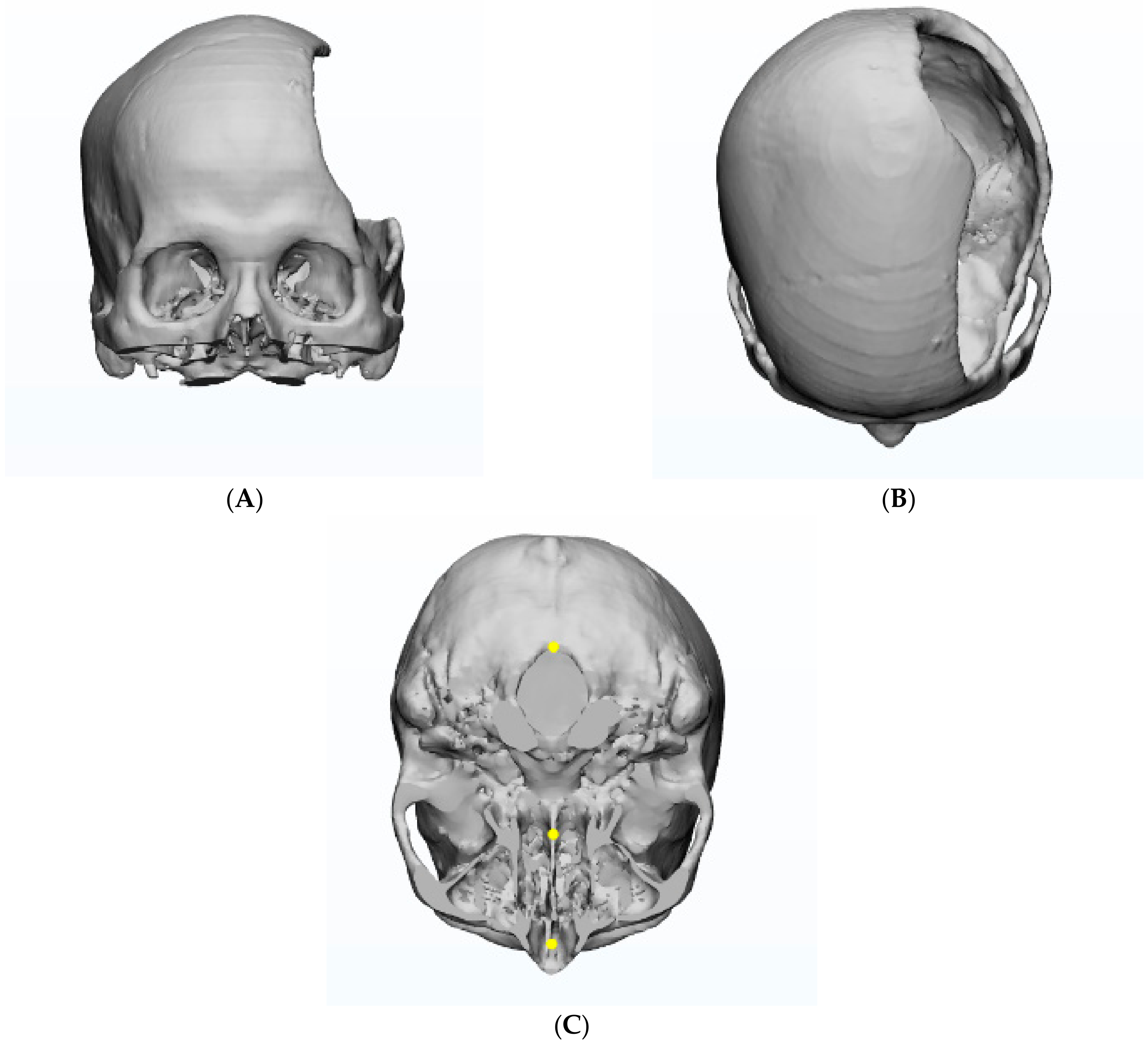
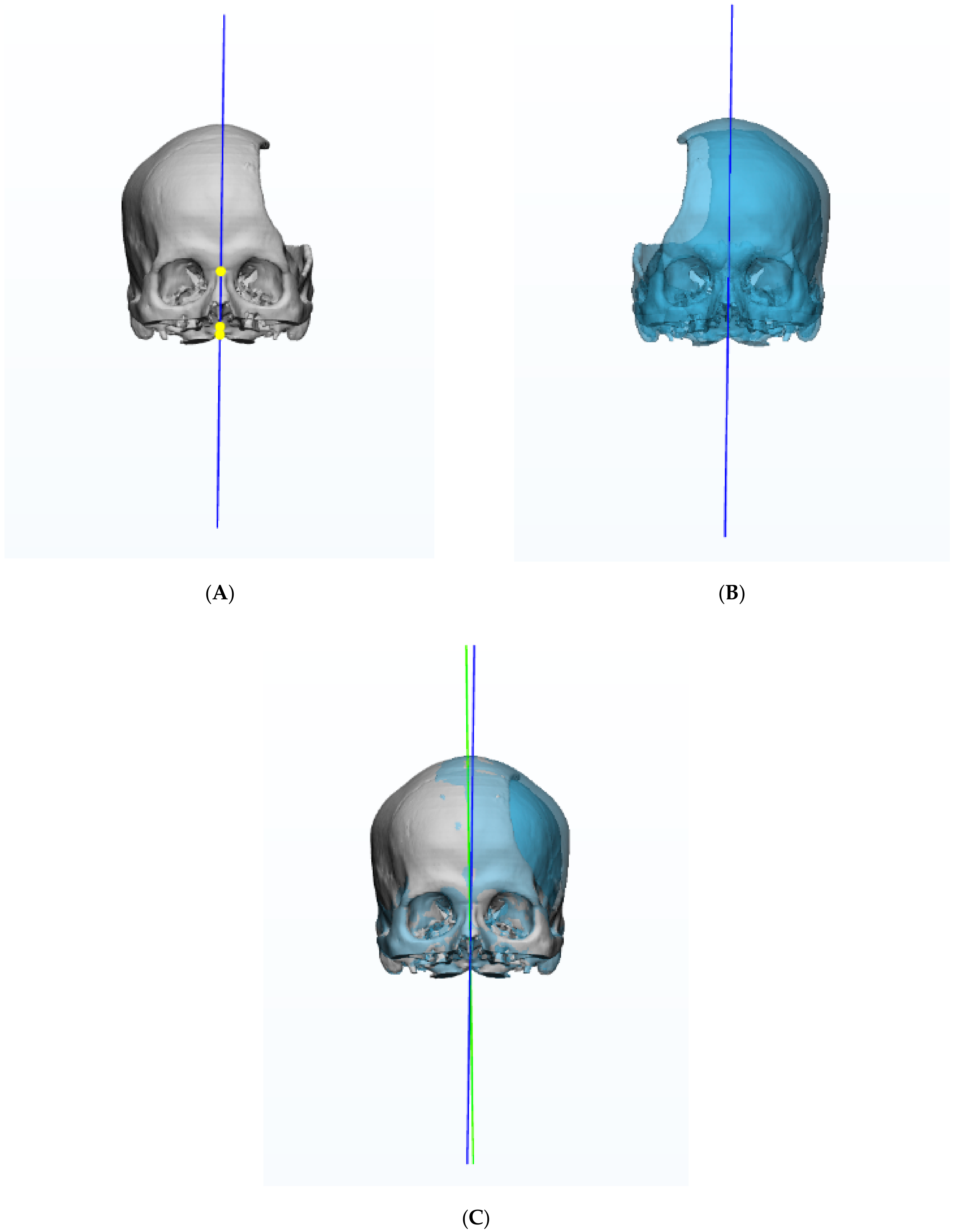
2.1. Preparation of 3-Dimensional Skull Model

The 3D skull model was created from facial CT data of randomly selected individuals among patients who visited the Department of Oral and Maxillofacial Surgery of Kyung Hee University Dental Hospital between December 2016 and November 2017. Ethical approval was received from the institutional review board (KHD IRB 1806-2) and written informed consent was obtained from the patient. The datasets of CT were imported into Mimics as Digital Imaging and Communications in Medicine (DICOM) files. The DICOM datasets of CT were segmented as the craniofacial regions and reconstructed into 3D skull models by using Mimics 20.0 software (Materialise LV, Leuven, Belgium).

2.2. Creation of Mirror Plane
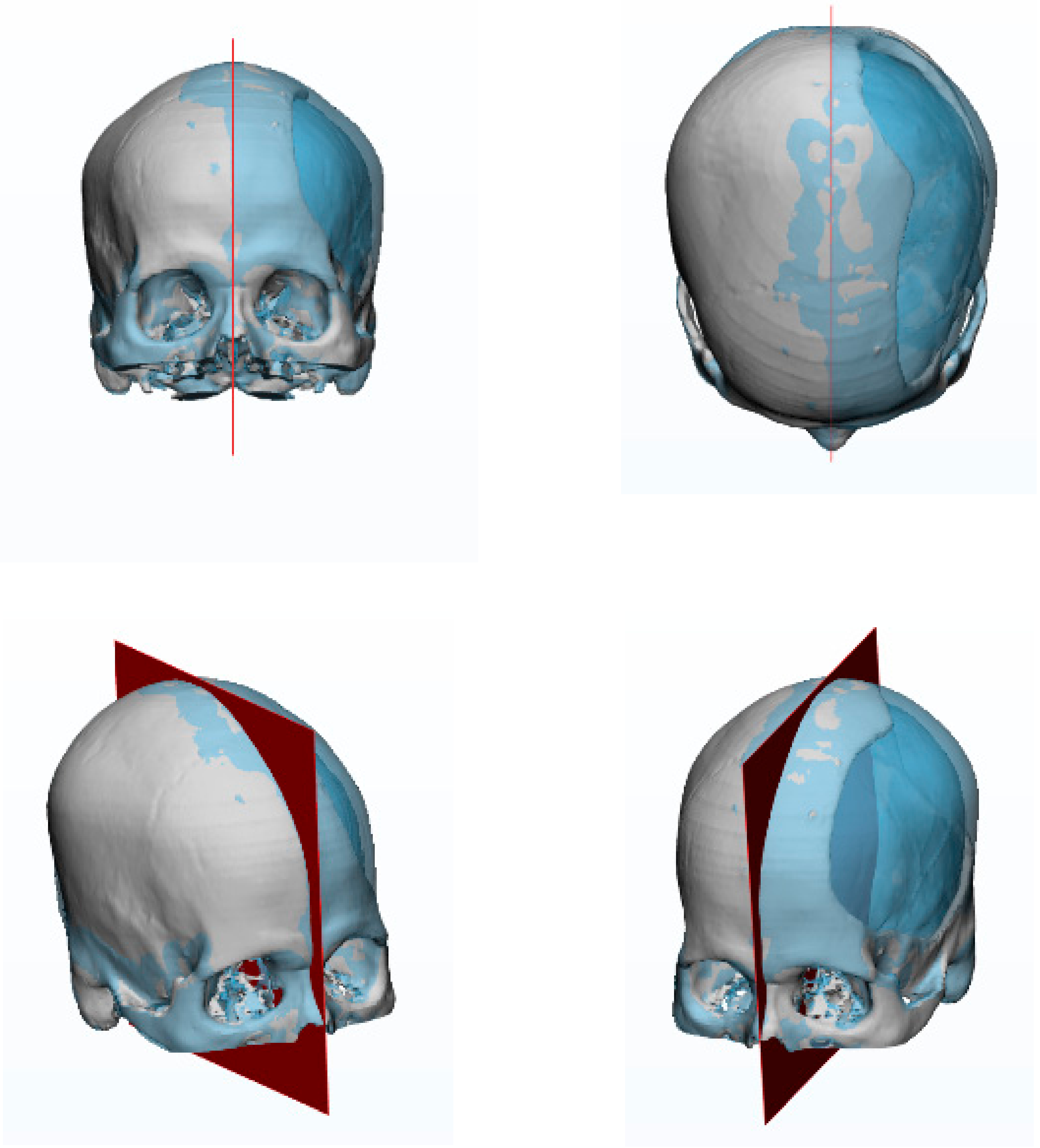
Additional medical image software (3-matic®, Materialize NV, Leuven, Belgium) was used to create the mirror plane, including functions of mirroring and surface-based registration. The creation of a unilaterally defected skull model was performed on the left side of the temporal area on the skull (Figure 1A,B). Three points were plotted on the defected skull model according to the commonly used reference landmarks from recent literature; Nasion, PNS, and Basion [9] (Figure 1C).
These reference points were connected to establish an initial mirror plane as a temporary mid-plane to begin the mirroring process (Figure 2A). Reflecting the original defect model by the initial mid-plane created a mirrored defect mode (Figure 2B). Then, the original skull model and mirror skull model underwent surface base registration by a conjunctive scale of 5, 1, 0.5, 0.1, 0.05, 0.01, respectively (Figure 2C).
The three reference points of the mirror model moved along during the registration procedure and were used to construct a moved mirror plane. Bisecting the initial mid-plane and moved mirror plane enabled us to achieve a final mirror plane that was customized and optimized for the initial defect model (Figure 3).

3. Results

A final mirror plane was obtained which evenly bisected the initial and mirrored models. Precise measurements and further treatment plans will be possible for the area to be reconstructed or corrected by using this final mirror plane.

4. Discussion

Unilateral defects or asymmetrical figures on facial area may result from congenital abnormality, trauma, or neoplastic lesions. It is essential to set a proper mid-plane to begin evaluation and analysis for the patients with such facial conditions. Three reference points are usually used to establish the facial mid-plane; however, the plotting error of the points cannot be neglected in order to carry out precise evaluation and accurate analysis. There has been an enormous advance in technology in the medical field from data collection using computer assisted devices to software enabling 3D image processes [10,11,12]. Engineering algorithms, such as ICP could also enhance the application of currently progressing medical resources by improving precision and rate of overlap. Although the algorithms are very effective, it could be very challenging to apply them in actual clinical use. Accordingly, this study attempted to combine the commercially available medical software and concept of ICP to bring them into practical use at the genuine clinical settings. Taking advantage of these highly developed current technologies, the mid-plane generated by using mirror images can be a beneficial solution for the interest in decreasing errors of primary setup. This mirroring technique not only helps decrease such errors during the initial steps of evaluation and analysis, but also provides an ideal image of the structure to be reconstructed or reshaped. As a consequence, surgeons can come up with meticulous treatment plans which are more patient specific and suitable. Traditional free flap techniques may exhibit limitations of positioning the flaps and bones properly because large defects and congenital asymmetry can cause dramatic deviations on the normal structures [13]. On the other hand, reconstruction or asymmetry correction by using computer aided mirror images can allow the surgeons to overcome such problems and achieve better outcomes. The benefits of using mirror images are expected to be exceedingly productive when they are combined with bio-printing and even tissue engineering. In addition, this kind of application of current technologies would lead surgeons to the next level of oral and maxillofacial surgery, such as the fabrication of bio-reactive scaffolds that can be an alternative for the traditional free flap surgeries [6,14]. Donor site morbidity would thereby no longer be an issue, eventually pursuing a decreased duration of surgery and recovery time. Therefore, this method could help actual users as an appropriate start point to plan comprehensive treatment for reconstruction or asymmetry correction surgeries in an easier, fast, and accurate way.

Author Contributions

Conceptualization, J.-W.L. and M.-S.K.; methodology, B.-Y.H. and H.L., software, B.-Y.H., J.-W.L. and M.-S.K.; writing—original draft preparation, J.-W.L. and M.-S.K.; writing—review and editing, J.-W.L.; project administration, J.-W.L.; funding acquisition, J.-W.L. All authors have read and agreed to the published version of the manuscript.

Funding

This work was supported by the National Research Foundation of Korea Grant funded by the Korean Government (NRF-2017R1D1A1B04030398)

Conflicts of Interest

The authors declare no conflict of interest. The funders had no role in the design of the study; in the collection, analyses, or interpretation of data; and in the writing of the manuscript, or in the decision to publish the results.

References

  1. Chai, G.; Zhang, Y.; Ma, X.; Zhu, M.; Yu, Z.; Mu, X. Reconstruction of fronto-orbital and nasal defects with compound epoxied maleic acrylate/hydroxyapatite implant prefabricated with a computer design program. Ann. Plast. Surg. 2011, 67, 493–497. [Google Scholar] [CrossRef] [PubMed]
  2. Kelly, C.P.; Moreira-Gonzalez, A.; Ali, M.A.; Topf, J.; Persiani, R.J.; Jackson, I.T.; Wiens, J. Vascular iliac crest with inner table of the ilium as an option in maxillary reconstruction. J. Craniofac. Surg. 2004, 15, 23–28. [Google Scholar] [CrossRef] [PubMed]
  3. Baliarsing, A.S.; Kumar, V.V.; Malik, N.A.; B, D.K. Reconstruction of maxillectomy defects using deep circumflex iliac artery-based composite free flap. Oral Surg. Oral Med. Oral Pathol. Oral Radiol. Endod. 2010, 109, e8–e13. [Google Scholar] [CrossRef] [PubMed]
  4. Dobai, A.; Markella, Z.; Vizkelety, T.; Fouquet, C.; Rosta, A.; Barabas, J. Landmark-based midsagittal plane analysis in patients with facial symmetry and asymmetry based on CBCT analysis tomography. J. Orofac. Orthop. 2018, 79, 371–379. [Google Scholar] [CrossRef] [PubMed]
  5. Guibert, M.; Franchi, G.; Ansari, E.; Billotet, B.; Diner, P.A.; Cassier, S.; Vazquez, M.P.; Picard, A.; Kadlub, N. Fat graft transfer in children’s facial malformations: A prospective three-dimensional evaluation. J. Plast. Reconstr. Aesthet. Surg. 2013, 66, 799–804. [Google Scholar] [CrossRef] [PubMed]
  6. Jang, W.H.; Lee, J.M.; Jang, S.; Kim, H.D.; Ahn, K.M.; Lee, J.H. Mirror Image Based Three-Dimensional Virtual Surgical Planning and Three-Dimensional Printing Guide System for the Reconstruction of Wide Maxilla Defect Using the Deep Circumflex Iliac Artery Free Flap. J. Craniofac. Surg. 2019, 30, 1829–1832. [Google Scholar] [CrossRef] [PubMed]
  7. Zonneveld, F.W.; Lobregt, S.; van der Meulen, J.C.; Vaandrager, J.M. Three-dimensional imaging in craniofacial surgery. World J. Surg. 1989, 13, 328–342. [Google Scholar] [CrossRef] [PubMed]
  8. English, J.D.; Akyalcin, S.; Peltomaki, T.; Litschel, K. Three-Dimensional Update on Clinical Orthodontic Issues In Mosby’s Orthodontic Review, 2nd ed.; Mosby, an imprint of Elsevier Inc.: St. Louis, MO, USA, 2015; p. 335. [Google Scholar]
  9. Arias, E.; Huang, Y.H.; Zhao, L.; Seelaus, R.; Patel, P.; Cohen, M. Virtual Surgical Planning and Three-Dimensional Printed Guide for Soft Tissue Correction in Facial Asymmetry. J. Craniofac. Surg. 2019, 30, 846–850. [Google Scholar] [CrossRef] [PubMed]
  10. Rotaru, H.; Stan, H.; Florian, I.S.; Schumacher, R.; Park, Y.T.; Kim, S.G.; Chezan, H.; Balc, N.; Baciut, M. Cranioplasty with custom-made implants: Analyzing the cases of 10 patients. J. Oral Maxillofac. Surg. 2012, 70, e169–e176. [Google Scholar] [CrossRef] [PubMed]
  11. Turgut, G.; Ozkaya, O.; Kayali, M.U. Computer-aided design and manufacture and rapid prototyped polymethylmethacrylate reconstruction. J. Craniofac. Surg. 2012, 23, 770–773. [Google Scholar] [CrossRef] [PubMed]
  12. Farronato, G.; Giannini, L.; Galbiati, G.; Mortellaro, C.; Maspero, C. Presurgical virtual three-dimensional treatment planning. J. Craniofac. Surg. 2015, 26, 820–823. [Google Scholar] [CrossRef] [PubMed]
  13. Su, T.; Fernandes, R. Microvascular reconstruction of the mandible: An argument for the fibula osteocutaneous free flap. Revista Española de Cirugía Oral y Maxilofacial 2014, 36, 1–8. [Google Scholar] [CrossRef] [Green Version]
  14. Jacobs, C.A.; Lin, A.Y. A New Classification of Three-Dimensional Printing Technologies: Systematic Review of Three-Dimensional Printing for Patient-Specific Craniomaxillofacial Surgery. Plast. Reconstr. Surg. 2017, 139, 1211–1220. [Google Scholar] [CrossRef] [PubMed]
Figure 1. Unilateral defect skull model (A). front view (B). top view (C). bottom view with three reference points; Nasion, PNS, Basion.
Applsci 10 06141 g001
Figure 2. (A). initial mirror plane (in blue) created from the three reference points. (B). Mirror image of the original defect model (C). Surface based registration was performed and moved mirror plane was obtained (in green).
Applsci 10 06141 g002
Figure 3. Final mirror plane (in red) was created by bisecting the two planes; initial and moved mirror planes. Complete reconstruction of the defected areas of each model was achieved after the registration.
Applsci 10 06141 g003

