Evaluation of Two Extraction Methods for the Analysis of Hydrophilic Low Molecular Weight Compounds from Ganoderma lucidum Spores and Antiproliferative Activity on Human Cell Lines

 , , , , , ,
  and
and 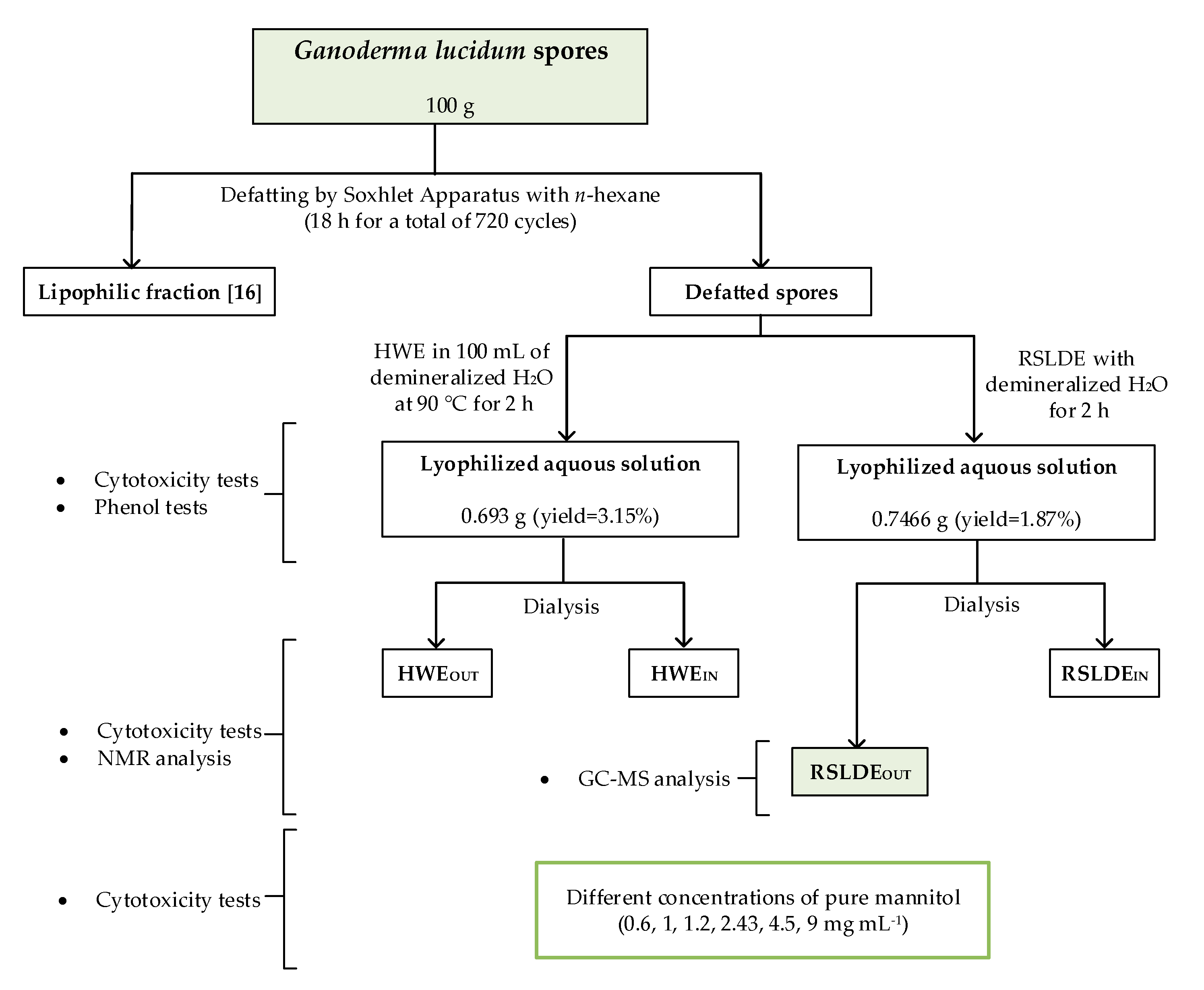
Abstract
:Featured Application
Abstract
1. Introduction
2. Materials and Methods
2.1. General Experimental Procedures
2.2. Fungal Material
2.3. Extraction of the Hydrophilic Component
2.3.1. Defatting of Ganoderma lucidum Spores
2.3.2. Hot Water Extraction (HWE)
2.3.3. Rapid Solid-Liquid Dynamic Extraction (RSLDE) by Naviglio Extractor
2.3.4. Dialysis of Aqueous Extracts
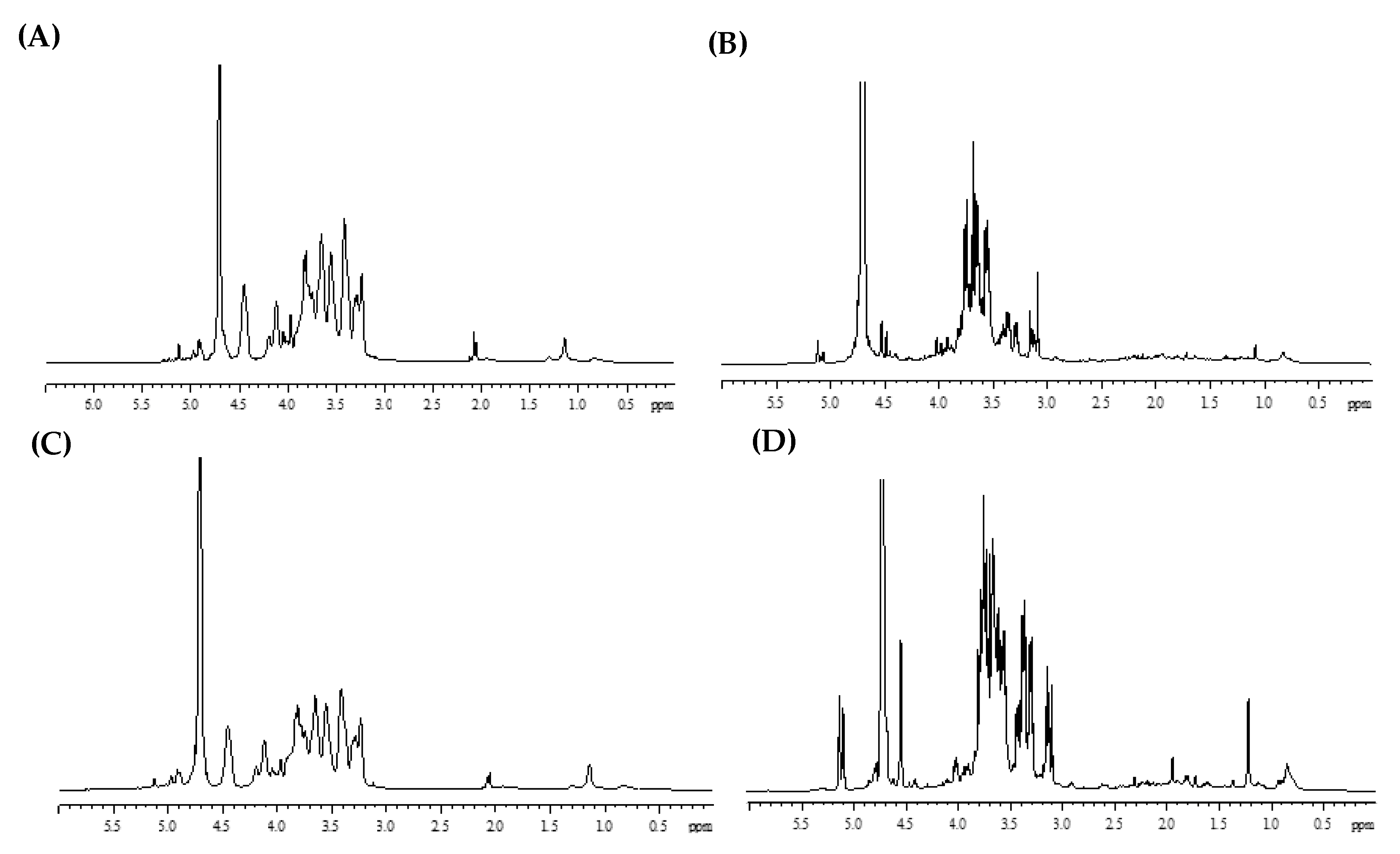
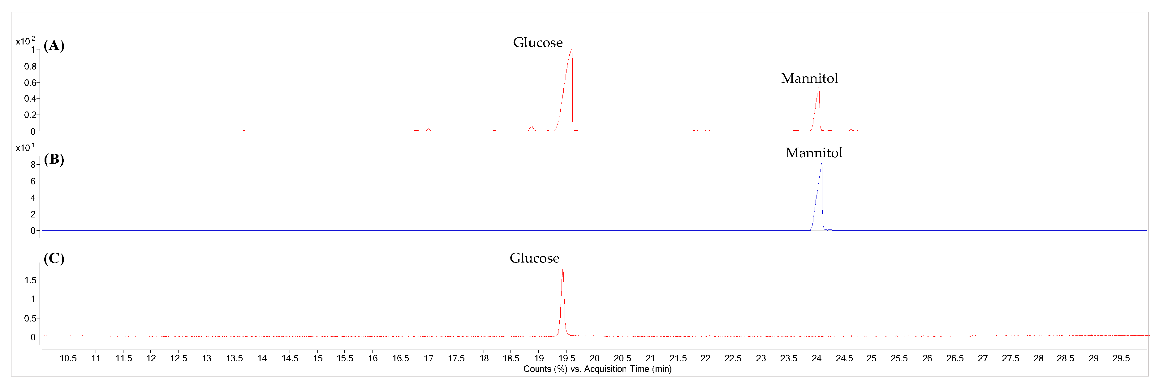
2.3.5. Sugar Analysis
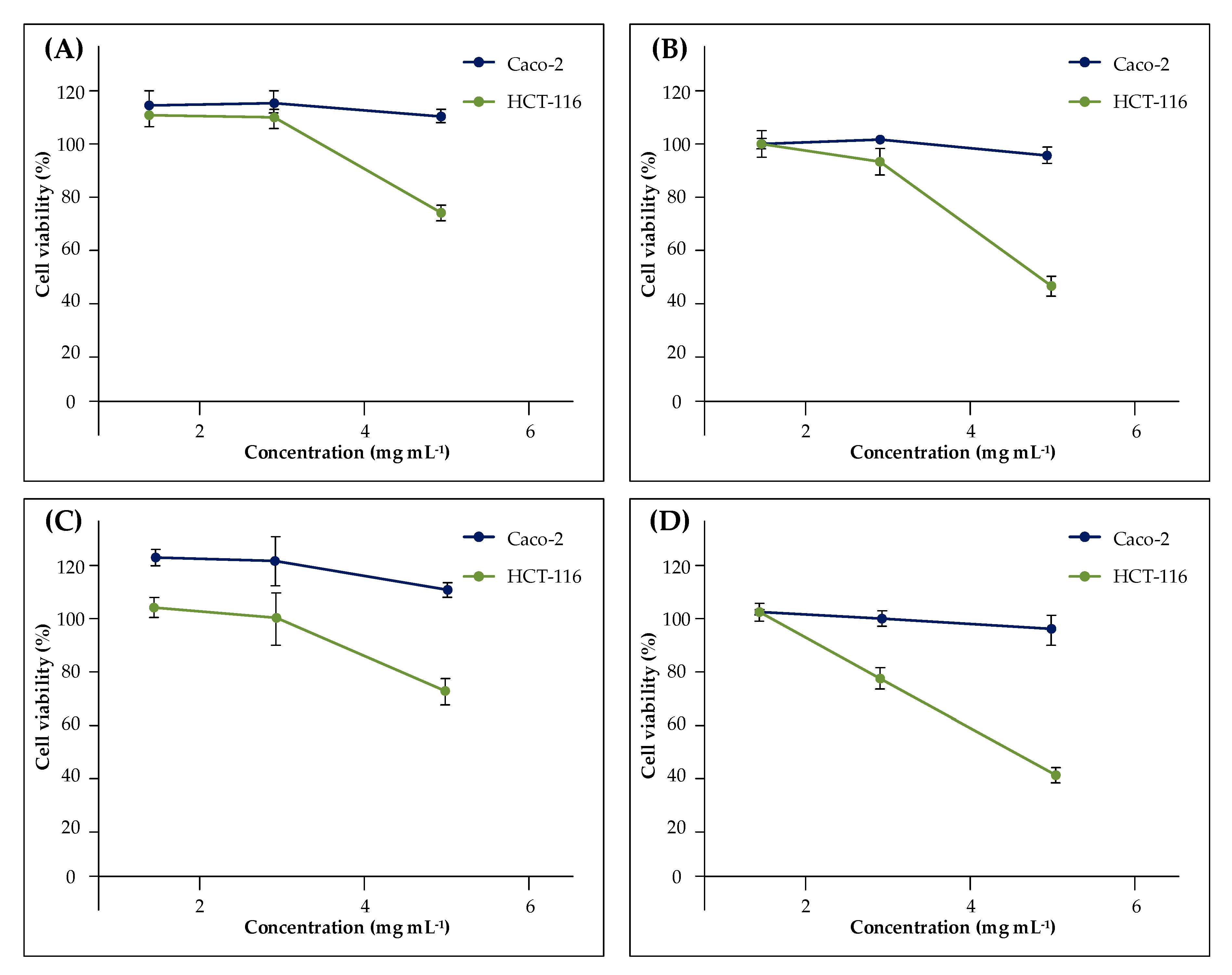
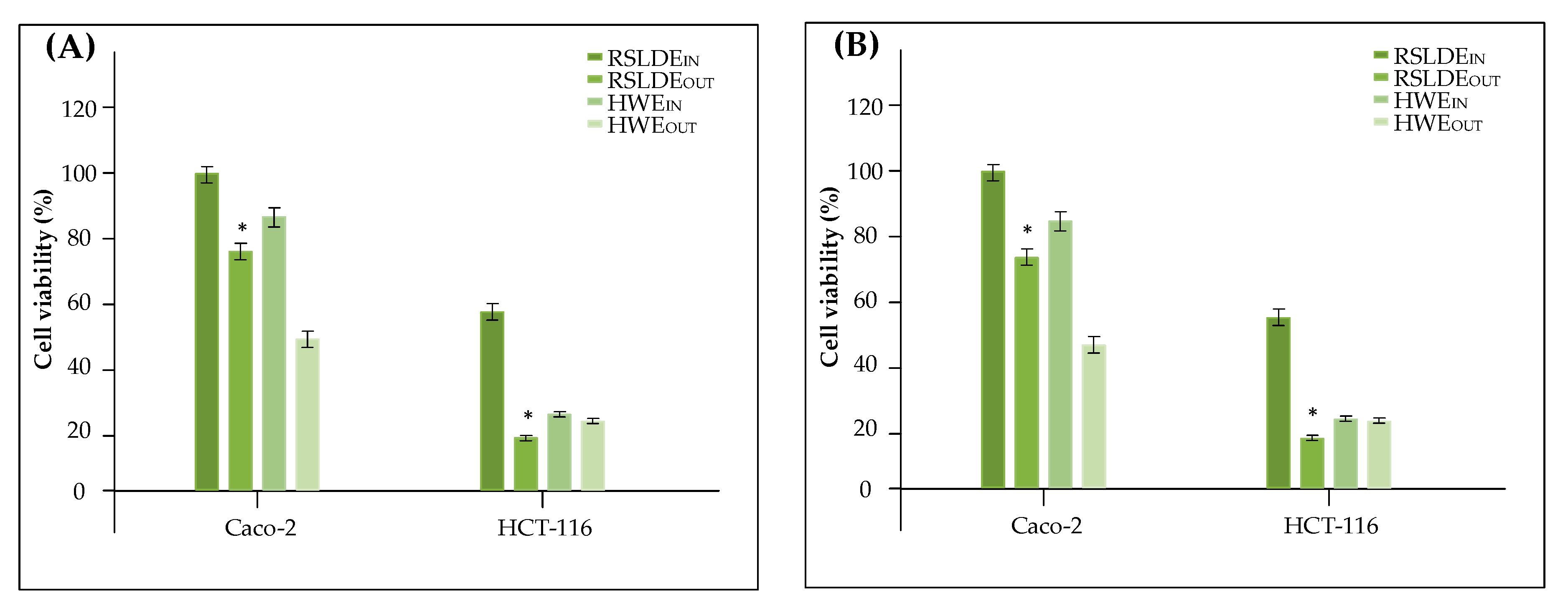
2.4. Assays of Anticancer Activity
2.5. Statistical Analysis
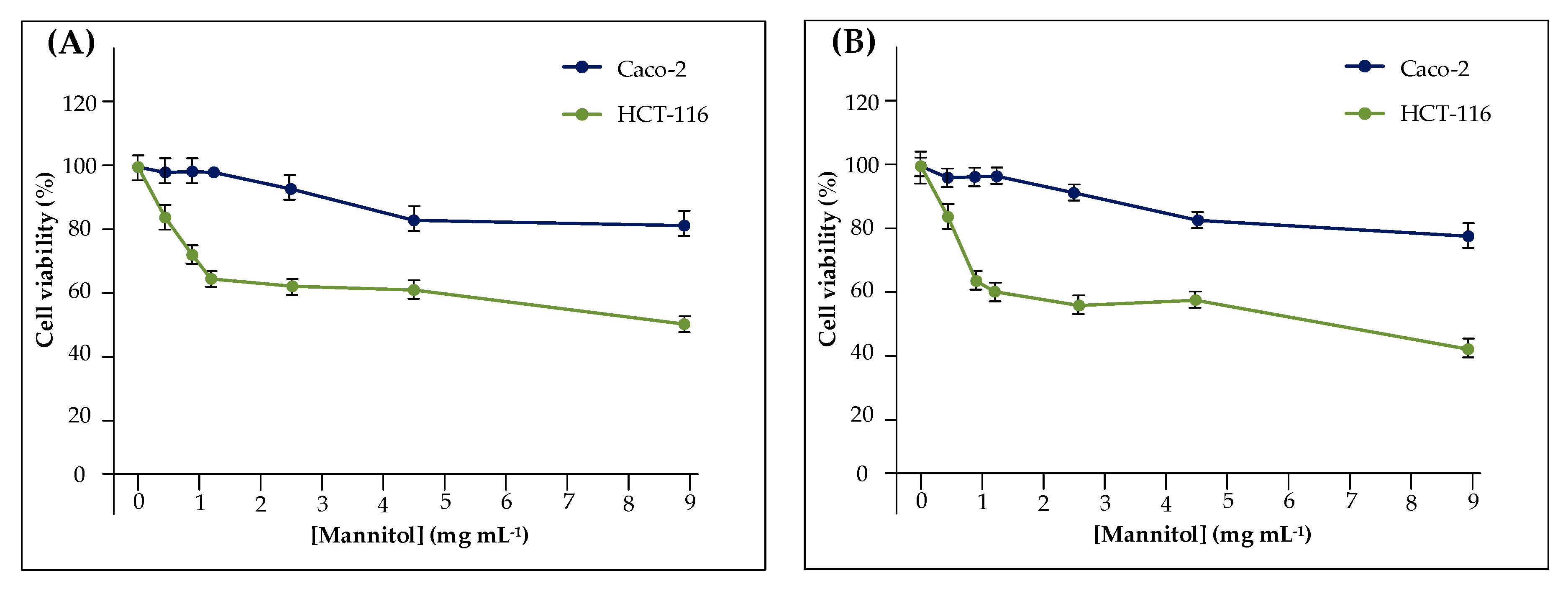
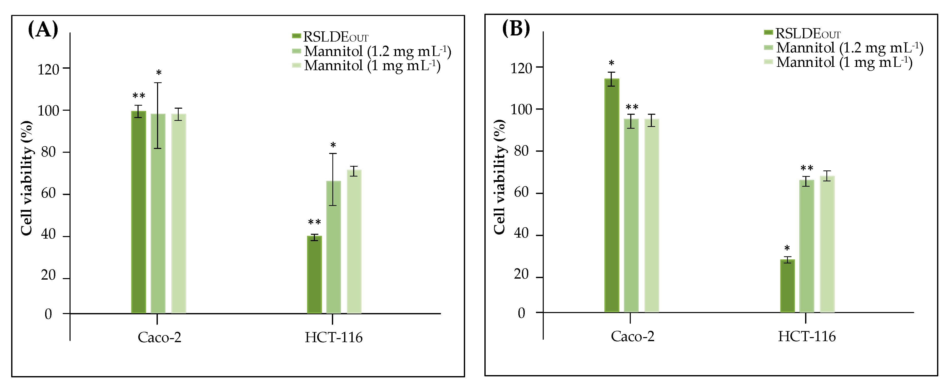
3. Results
4. Discussion
5. Conclusions
Supplementary Materials
Author Contributions
Funding
Acknowledgments
Conflicts of Interest
References
- Wasser, S.P. Medicinal mushrooms in human clinical studies. Part I. Anticancer, oncoimmunological, and immunomodulatory activities: A review. Int. J. Med. Mushrooms 2017, 19, 279–317. [Google Scholar] [CrossRef] [PubMed]
- Thu, Z.M.; Myo, K.K.; Aung, H.T.; Clericuzio, M.; Armijos, C.; Vidari, G. Bioactive phytochemical constituents of wild edible mushrooms from southeast Asia. Molecules 2020, 25, 1972. [Google Scholar] [CrossRef] [PubMed]
- Ivanova, T.S.; Krupodorova, T.A.; Barshteyn, V.Y.; Artamonova, A.B.; Shlyakhovenko, V.A. Anticancer substances of mushroom origin. Exp. Oncol. 2014, 36, 58–66. [Google Scholar] [PubMed]
- Badalyan, S.M.; Barkhudaryan, A.; Rapior, S. Recent progress in research on the pharmacological potential of mushrooms and prospects for their clinical application. In Medicinal Mushrooms; Springer: Singapore, 2019; pp. 1–70. ISBN 978-981-13-6381-8. [Google Scholar]
- Wang, P.Y.; Zhu, X.L.; Lin, Z.B. Antitumor and immunomodulatory effects of polysaccharides from broken-spore of Ganoderma lucidum. Front. Pharmacol. 2012, 3, 135. [Google Scholar] [CrossRef] [Green Version]
- Atay, S.; Ak, H.; Kalmis, E.; Kayalar, H.; Aydin, H.H. Diverse effects of the lingzhi or reishi medicinal mushroom, Ganoderma lucidum (Agaricomycetes), in combination with tamoxifen citrate and doxorubicin in MCF-7 breast cancer cells. Int. J. Med. Mushrooms 2016, 18. [Google Scholar] [CrossRef]
- Yang, Y.; Nirmagustina, D.E.; Kumrungsee, T.; Okazaki, Y.; Tomotake, H.; Kato, N. Feeding of the water extract from Ganoderma lingzhi to rats modulates secondary bile acids, intestinal microflora, mucins, and propionate important to colon cancer. Biosci. Biotechnol. Biochem. 2017, 81, 1796–1804. [Google Scholar] [CrossRef] [Green Version]
- Veena, R.K.; Ajith, T.A.; Janardhanan, K.K. Lingzhi or reishi medicinal mushroom, Ganoderma lucidum (Agaricomycetes), prevents doxorubicin-induced cardiotoxicity in rats. Int. J. Med. Mushrooms 2018, 20. [Google Scholar] [CrossRef]
- Sohretoglu, D.; Huang, S. Ganoderma lucidum polysaccharides as an anti-cancer agent. Anticancer Agents Med. Chem. 2018, 18, 667–674. [Google Scholar] [CrossRef]
- Min, B.S.; Nakamura, N.; Miyashiro, H.; Bae, K.W.; Hattori, M. Triterpenes from the spores of Ganoderma lucidum and their inhibitory activity against HIV-1 protease. Chem. Pharm. Bull. 1998, 46, 1607–1612. [Google Scholar] [CrossRef] [Green Version]
- Liu, X.; Yuan, J.P.; Chung, C.K.; Chen, X.J. Antitumor activity of the sporoderm-broken germinating spores of Ganoderma lucidum. Cancer Lett. 2002, 182, 155–161. [Google Scholar] [CrossRef]
- Liu, X.; Xu, S.P.; Wang, J.H.; Yuan, J.P.; Guo, L.X.; Li, X.; Huang, X.N. Characterization of ganoderma spore lipid by stable carbon isotope analysis: Implications for authentication. Anal. Bioanal. Chem. 2007, 388, 723–731. [Google Scholar] [CrossRef]
- Xu, J.; Li, P. Researches and Application of Ganoderma Spores Powder. In Ganoderma and Health; Springer: Singapore, 2019; pp. 157–186. ISBN 978-981-13-9866-7. [Google Scholar]
- Dubois, M.; Gilles, K.A.; Hamilton, J.K.; Rebers, P.T.; Smith, F. Colorimetric method for determination of sugars and related substances. Anal. Chem. 1956, 28, 350–356. [Google Scholar] [CrossRef]
- Restaino, O.F.; Finamore, R.; Diana, P.; Marseglia, M.; Vitiello, M.; Casillo, A.; Bedini, E.; Corsaro, M.M.; Parrilli, M.; Trifuoggi, M.; et al. A multi-analytical approach to better assess the keratan sulfate contamination in animal origin chondroitin sulfate. Anal. Chim. Acta 2017, 958, 59–70. [Google Scholar] [CrossRef]
- Salvatore, M.M.; Elvetico, A.; Gallo, M.; Salvatore, F.; DellaGreca, M.; Naviglio, D.; Andolfi, A. Fatty acids from Ganoderma lucidum spores: Extraction, identification and quantification. Appl. Sci. 2020, 10, 3907. [Google Scholar] [CrossRef]
- Bishop, K.S.; Kao, C.H.; Xu, Y.; Glucina, M.P.; Paterson, R.R.M.; Ferguson, L.R. From 2000 years of Ganoderma lucidum to recent developments in nutraceuticals. Phytochemistry 2015, 114, 56–65. [Google Scholar] [CrossRef] [Green Version]
- Boh, B. Ganoderma lucidum: A potential for biotechnological production of anti-cancer and immunomodulatory drugs. Recent Pat. Anticancer Drug Discov. 2013, 8, 255–287. [Google Scholar] [CrossRef]
- Shahveisi, K.; Mousavi, S.H.; Hosseini, M.; Rad, A.K.; Jalali, S.A.; Rajaei, Z.; Sadeghnia, H.R.; Hadjzadeh, M.A.R. The role of local renin-angiotensin system on high glucose-induced cell toxicity, apoptosis and reactive oxygen species production in PC12 cells. Iran. J. Basic Med. Sci. 2014, 17, 613. [Google Scholar]
- Baby, S.; Johnson, A.J.; Govindan, B. Secondary metabolites from Ganoderma. Phytochemistry 2015, 114, 66–101. [Google Scholar] [CrossRef]
- Ahmad, M.F. Ganoderma lucidum: Persuasive biologically active constituents and their health endorsement. Biomedicine 2018, 107, 507–519. [Google Scholar] [CrossRef]
- Gao, Y.; Gao, H.; Chan, E.; Tang, W.; Xu, A.; Yang, H.; Huang, M.; Lan, J.; Li, X.; Duan, W.; et al. Antitumor activity and underlying mechanisms of ganopoly, the refined polysaccharides extracted from Ganoderma lucidum, in mice. Immunol. Investig. 2005, 34, 171–198. [Google Scholar] [CrossRef]
- Wang, X.C.; Xi, R.J.; Li, Y.; Wang, D.M.; Yao, Y.J. The species identity of the widely cultivated Ganoderma, ‘G. lucidum’(Ling-zhi), in China. PLoS ONE 2012, 7, e40857. [Google Scholar] [CrossRef] [Green Version]
- Sharma, C.; Bhardwaj, N.; Sharma, A.; Tuli, H.S.; Katyal, P.; Beniwal, V.; Gupta, G.K.; Sharma, A.K. Bioactive metabolites of Ganoderma lucidum: Factors, mechanism and broad spectrum therapeutic potential. J. Herb. Med. 2019, 17–18, 100268. [Google Scholar] [CrossRef]
- Huang, S.Q.; Li, J.W.; Wang, Z.; Pan, H.X.; Chen, J.X.; Ning, Z.X. Optimization of alkaline extraction of polysaccharides from Ganoderma lucidum and their effect on immune function in mice. Molecules 2010, 15, 3694–3708. [Google Scholar] [CrossRef] [PubMed]
- Naviglio, D. Naviglio’s principle and presentation of an innovative solid–liquid extraction technology: Extractor Naviglio®. Anal. Lett. 2003, 36, 1647–1659. [Google Scholar] [CrossRef]
- Naviglio, D.; Scarano, P.; Ciaravolo, M.; Gallo, M. Rapid Solid-Liquid Dynamic Extraction (RSLDE): A powerful and greener alternative to the latest solid-liquid extraction techniques. Foods 2019, 8, 245. [Google Scholar] [CrossRef] [PubMed] [Green Version]
- De Vries, J.E.; Dinjens, W.N.M.; De Bruyne, G.K.; Verspaget, H.W.; Van Der Linden, E.P.M.; De Bruine, A.P.; Mareel, M.M.; Bosman, F.T.; Ten Kate, J. In vivo and in vitro invasion in relation to phenotypic characteristics of human colorectal carcinoma cells. Br. J. Cancer 1995, 71, 271–277. [Google Scholar] [CrossRef] [PubMed] [Green Version]
- Rajput, A.; San Martin, I.D.; Rose, R.; Beko, A.; LeVea, C.; Sharratt, E.; Mazurchuk, R.; Hoffman, R.M.; Brattain, M.G.; Wang, J. Characterization of HCT116 human colon cancer cells in an orthotopic model. J. Surg. Res. 2008, 147, 276–281. [Google Scholar] [CrossRef]
- Patel, T.K.; Williamson, J.D. Mannitol in plants, fungi, and plant–fungal interactions. Trends Plant Sci. 2016, 21, 486–497. [Google Scholar] [CrossRef]
- André, P.; Villain, F. Free radical scavenging properties of mannitol and its role as a constituent of hyaluronic acid fillers: A literature review. Int. J. Cosmet. Sci. 2017, 39, 355–360. [Google Scholar] [CrossRef] [Green Version]
- Naviglio, D.; Formato, A.; Vitulano, M.; Cozzolino, I.; Ferrara, L.; Zanoelo, E.F.; Gallo, M. Comparison between the kinetics of conventional maceration and a cyclic pressurization extraction process for the production of lemon liqueur using a numerical model. J. Food Process Eng. 2017, 40, e12350. [Google Scholar] [CrossRef] [Green Version]
- Gallo, M.; Vitulano, M.; Andolfi, A.; DellaGreca, M.; Naviglio, D. Rapid solid-liquid dynamic extraction (RSLDE), a new rapid and greener method to extract two steviol glycosides (stevioside and rebaudioside a) from stevia leaves. Plant Foods Hum. Nutr. 2017, 72, 141–148. [Google Scholar] [CrossRef] [PubMed]
- Salvatore, M.M.; Ciaravolo, M.; Cirino, P.; Toscano, A.; Salvatore, F.; Gallo, M.; Naviglio, D.; Andolfi, A. Fatty Acids from Paracentrotus lividus Sea Urchin Shells Obtained via Rapid Solid Liquid Dynamic Extraction (RSLDE). Separations 2019, 6, 50. [Google Scholar] [CrossRef] [Green Version]
- Scarano, P.; Naviglio, D.; Prigioniero, A.; Tartaglia, M.; Postiglione, A.; Sciarrillo, R.; Guarino, C. Sustainability: Obtaining natural dyes from waste matrices using the prickly pear peels of Opuntia ficus-indica (L.) Miller. Agronomy 2020, 10, 528. [Google Scholar] [CrossRef] [Green Version]


© 2020 by the authors. Licensee MDPI, Basel, Switzerland. This article is an open access article distributed under the terms and conditions of the Creative Commons Attribution (CC BY) license (http://creativecommons.org/licenses/by/4.0/).
Share and Cite
Salvatore, M.M.; De Gregorio, V.; Gallo, M.; Corsaro, M.M.; Casillo, A.; Vecchione, R.; Andolfi, A.; Naviglio, D.; Netti, P.A. Evaluation of Two Extraction Methods for the Analysis of Hydrophilic Low Molecular Weight Compounds from Ganoderma lucidum Spores and Antiproliferative Activity on Human Cell Lines. Appl. Sci. 2020, 10, 4033. https://doi.org/10.3390/app10114033
Salvatore MM, De Gregorio V, Gallo M, Corsaro MM, Casillo A, Vecchione R, Andolfi A, Naviglio D, Netti PA. Evaluation of Two Extraction Methods for the Analysis of Hydrophilic Low Molecular Weight Compounds from Ganoderma lucidum Spores and Antiproliferative Activity on Human Cell Lines. Applied Sciences. 2020; 10(11):4033. https://doi.org/10.3390/app10114033
Chicago/Turabian StyleSalvatore, Maria Michela, Vincenza De Gregorio, Monica Gallo, Maria Michela Corsaro, Angela Casillo, Raffaele Vecchione, Anna Andolfi, Daniele Naviglio, and Paolo Antonio Netti. 2020. "Evaluation of Two Extraction Methods for the Analysis of Hydrophilic Low Molecular Weight Compounds from Ganoderma lucidum Spores and Antiproliferative Activity on Human Cell Lines" Applied Sciences 10, no. 11: 4033. https://doi.org/10.3390/app10114033