Next Issue
Volume 7, June
Previous Issue
Volume 7, April


Med. Sci., Volume 7, Issue 5 (May 2019) – 2 articles

Cover Story (view full-size image): 
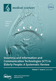
Diaphragmatic dysfunction is a common cause of slow weaning in mechanically ventilated patients. The aim of our study was to evaluate the motion of the diaphragm in patients who were ventilated for a protracted period in comparison with healthy controls by using magnetic resonance imaging (MRI). The advantage of MRI lies in its capability of directly acquiring coronal and sagittal images of the entire diaphragm, which can reveal focal, unilateral, or global diaphragmatic motion abnormalities. By using MRI dynamic sequences, we found that the vertical displacement of both right and left hemi-diaphragms at various anatomical locations had different values in both controls and patients. The ventilator induced diaphragm dysfunction in our patients was regional as well as global. View this paper.
  • Issues are regarded as officially published after their release is announced to the table of contents alert mailing list.
  • You may sign up for e-mail alerts to receive table of contents of newly released issues.
  • PDF is the official format for papers published in both, html and pdf forms. To view the papers in pdf format, click on the "PDF Full-text" link, and use the free Adobe Reader to open them.
Order results
Result details
Section
Select all
Export citation of selected articles as:
15 pages, 258 KiB  
Review
TRP Channels as Lower Urinary Tract Sensory Targets
by Karl-Erik Andersson
Med. Sci. 2019, 7(5), 67; https://doi.org/10.3390/medsci7050067 - 22 May 2019
Cited by 28 | Viewed by 4017
Abstract
Several members of the transient receptor potential (TRP) superfamily, including TRPV1, TRPV2, TRPV4, TRM4, TRPM8 and TRPA1, are expressed in the lower urinary tract (LUT), not only in neuronal fibers innervating the bladder and urethra, but also in the urothelial and muscular layers [...] Read more.
Several members of the transient receptor potential (TRP) superfamily, including TRPV1, TRPV2, TRPV4, TRM4, TRPM8 and TRPA1, are expressed in the lower urinary tract (LUT), not only in neuronal fibers innervating the bladder and urethra, but also in the urothelial and muscular layers of the bladder and urethral walls. In the LUT, TRP channels are mainly involved in nociception and mechanosensory transduction. Animal studies have suggested the therapeutic potential of several TRP channels for the treatment of both bladder over- and underactivity and bladder pain disorders,; however translation of this finding to clinical application has been slow and the involvement of these channels in normal human bladder function, and in various pathologic states have not been established. The development of selective TRP channel agonists and antagonists is ongoing and the use of such agents can be expected to offer new and important information concerning both normal physiological functions and possible therapeutic applications. Full article
(This article belongs to the Special Issue Targeting TRP Channels: from Drug Development to Clinical Trials)
8 pages, 634 KiB  
Article
MRI Assessment of Global and Regional Diaphragmatic Motion in Critically Ill Patients Following Prolonged Ventilator Weaning
by Khurram Saleem Khan, James Meaney, Ignacio Martin-Loeches and Daniel V. Collins
Med. Sci. 2019, 7(5), 66; https://doi.org/10.3390/medsci7050066 - 22 May 2019
Cited by 2 | Viewed by 2617
Abstract
Introduction: diaphragmatic dysfunction is a common cause of slow weaning in mechanically ventilated patients. Diaphragmatic dysfunction in ventilated patients can be global or regional. The aim of our study was to evaluate the motion of the entire diaphragm in patients who were ventilated [...] Read more.
Introduction: diaphragmatic dysfunction is a common cause of slow weaning in mechanically ventilated patients. Diaphragmatic dysfunction in ventilated patients can be global or regional. The aim of our study was to evaluate the motion of the entire diaphragm in patients who were ventilated for a protracted period in comparison with healthy controls by using Magnetic Resonance Imaging (MRI). Methods: Intensive care patients who had a prolonged ventilator wean and required tracheostomies were enrolled based on extensive exclusion criteria. MRI dynamic sequence and subtraction images were used to measure vertical displacement at five different points on each hemi-diaphragm during normal tidal breathing. Tidal displacement of each point on the right and left hemi-diaphragms of the patients were compared to the precise respective points on the right and left hemi-diaphragms of enrolled controls. Results: Eight intensive care patients and eight controls were enrolled. There were observed significant differences in the displacements of the left hemi-diaphragm between the two groups (median 6.4 mm [Interquartile range (IQR), 4.6–12.5]) vs. 11.6 mm [IQR, 9.5–14.5], p = 0.02). There were also observed significant differences in the displacements at five evaluated study points on the left hemi-diaphragms of the patients when compared to the precise respective points in controls, especially at the dome (median 6.7 mm [IQR, 5.0–11.4] vs. 13.5 mm [IQR 11.5-18], p value = 0.005) and the anterior zone of apposition (median 5.0 mm [IQR, 3.3–7.1] vs. 7.8mm [IQR, 7.1–10.5], p value = 0.01). The intensive care patients showed lower minimal and maximal values of displacement of right hemi-diaphragms compared to the controls, suggesting that the differences in the displacement of right hemi-diaphragm are possible; however, the differences in the mean values of displacement of right hemi-diaphragm between the intensive care patient group and the control group (median 9.8 mm [IQR (Interquartile range), 5.0–12.3] vs. 10.1 mm [IQR 8.3–18.5], p = 0.12) did not reach the level of significance. Conclusion: Although frequently global, diaphragm dysfunction in ventilated patients after prolonged ventilation can also be regional or focal when assessed by MRI dynamic sequence. The vertical displacement of both right and left hemi-diaphragms at various anatomical locations had different values in both controls, and patients. There were significant focal variations in the movement of diaphragm in patients with ventilator-induced diaphragmatic dysfunction. Full article
(This article belongs to the Section Pneumology and Respiratory Diseases)
Show Figures

Figure 1

Previous Issue
Next Issue
Back to TopTop