Article

Role of Maternal Antibodies in the Protection of Broiler Chicks against Campylobacter Colonization in the First Weeks of Life

1
Department of Pathobiology, Pharmacology and Zoological Medicine, Ghent University, B9820 Merelbeke, Belgium
2
Terrestrial Ecology Unit (TEREC), Ghent University, B9000 Ghent, Belgium
3
Technology & Food Sciences Unit, Flanders Research Institute for Agriculture, Fisheries and Food (ILVO), B9090 Melle, Belgium
*
Author to whom correspondence should be addressed.
Animals 2024, 14(9), 1291; https://doi.org/10.3390/ani14091291
Submission received: 28 February 2024 / Revised: 8 April 2024 / Accepted: 23 April 2024 / Published: 25 April 2024
(This article belongs to the Special Issue The Animal Microbiome in Health and Disease)

Abstract

:

Simple Summary

Campylobacter is the main cause of foodborne human gastroenteritis worldwide. Most of these cases originate from the consumption of poultry products. Broiler chickens are often colonized by this bacterium, with a high prevalence at slaughter age. However, in the first 2–3 weeks of life, chicks are resistant to Campylobacter infection, and this resistance has been attributed to the transfer of maternal antibodies from the hen’s serum to the yolk and then to the hatched chick’s bloodstream and intestines. In this study, the role of maternal antibodies in the protection of broilers against Campylobacter colonization was investigated. Field monitoring of broiler flocks from breeders with varying antibody levels showed a trend of lower Campylobacter prevalence in offspring from high-antibody-level breeders. In a series of trials, breeders were vaccinated to increase maternal antibody levels in offspring, with the aim of increasing the chicks’ resistance to infection. A minor reduction in Campylobacter prevalence was obtained only in the second week of life. Immunization of breeders thus showed limited efficacy in enhancing protection against Campylobacter infection in the offspring.

Abstract

Thermophilic Campylobacter species are the most common cause of bacterium-mediated diarrheal disease in humans globally. Poultry is considered the most important reservoir of human campylobacteriosis, but so far, no effective countermeasures are in place to prevent the bacterium from colonizing broiler flocks. This study investigated maternal antibodies’ potential to offer protection against Campylobacter in broiler chicks via a field trial and an immunization trial. In the field trial, breeder flocks with high and low anti-Campylobacter antibody levels in the yolk were selected based on serological screening. Offspring were subsequently monitored for maternal antibodies and Campylobacter prevalence during early life. Although maternal antibodies declined rapidly in the serum of broilers, offspring from flocks with lower anti-Campylobacter antibody levels seemed to be more susceptible to colonization. In the immunization trial, breeders from a seropositive breeder flock were vaccinated with an experimental bacterin or subunit vaccine. Immunization increased antibody levels in the yolk and consequently in the offspring. Elevated maternal antibody levels were significantly associated with reduced Campylobacter susceptibility in broilers at 2 weeks old but not at 1 and 3 weeks old. Overall, the protective effect of maternal immunity should be cautiously considered in the context of Campylobacter control in broilers. Immunization of breeders may enhance resistance but is not a comprehensive solution.

1. Introduction

Campylobacter, a genus of Gram-negative bacteria, is the leading cause of human gastroenteritis worldwide. The most common symptoms of campylobacteriosis include fever, diarrhea and abdominal cramps. Although most of these infections are self-limiting, they can lead to more serious pathologies such as extraintestinal diseases and chronic sequelae like reactive arthritis or Guillain-Barré syndrome [1]. In 2022, 137,107 confirmed cases of campylobacteriosis were reported in 27 EU countries, with an overall incidence rate of 43.1 cases per 100,000. The majority (87.6%) of these infections were caused by Campylobacter (C.) jejuni, followed by C. coli (10.7%) [2].
The transmission of the bacteria to humans typically occurs through direct or indirect contact with animal-derived products, particularly poultry products. In Belgium, 30 to 70% of broiler flocks are positive for Campylobacter at slaughter age [3,4,5,6]. Thermophilic Campylobacter species colonize the ceca of chickens to a high degree (106–109 colony forming units (cfu)/g) and are often regarded as a commensal. However, colonization of broiler chickens can be associated with intestinal inflammation, leading to diarrhea and subsequent associated pathology [7]. More likely, the interaction with the chicken host suggests that their relationship can better be described as “immunological tolerance”, as Campylobacter may escape or alter the inflammatory response via different mechanisms [8].
Currently, there are no effective counter measures in place to prevent the colonization of broilers at the farm level [9,10]. Campylobacter is typically introduced horizontally in the chicken flock through different environmental sources and various vectors such as rodents and insects [11]. Once present in the poultry house, the bacteria spread quickly, and one week after initial colonization, most chickens in the flock are already colonized [12]. Although it has been suggested [13] that fecal contamination of the eggshell can lead to the possible survival of Campylobacter in the eggshell membranes and transmission to the progeny, other authors claim that the transfer of Campylobacter from parent stock to the offspring is unlikely in commercial settings [14,15]. Moreover, Campylobacter is rarely detected in chicks under 2 to 3 weeks of age [16,17], the time before which is referred to as the lag period. This lag could be attributed to competition among microflora, which inhibits proliferation [18], as well as to the presence of maternal antibodies, which decline by about 2 weeks of age [19,20]. These antibodies contribute to the complement-mediated killing of C. jejuni in a strain-specific manner [20,21]. The immunization of specific-pathogen-free (SPF) broiler breeders with a bacterin and subunit vaccine led to the protection of their offspring against Campylobacter colonization during the first two to three weeks of their life due the transfer of maternal antibodies [22].
This study seeks to further investigate the potential protective role of maternal antibodies against Campylobacter in chicks. As there is a lack of field studies investigating the impact of anti-Campylobacter maternal antibodies, this research first aimed to examine the link between the presence of anti-Campylobacter antibodies in broiler breeders and the onset of Campylobacter colonization in their offspring. Then, we immunized seropositive breeders using two experimental vaccines to assess the extent to which resulting anti-Campylobacter antibody levels might improve protection of their progeny against Campylobacter colonization.

2. Materials and Methods

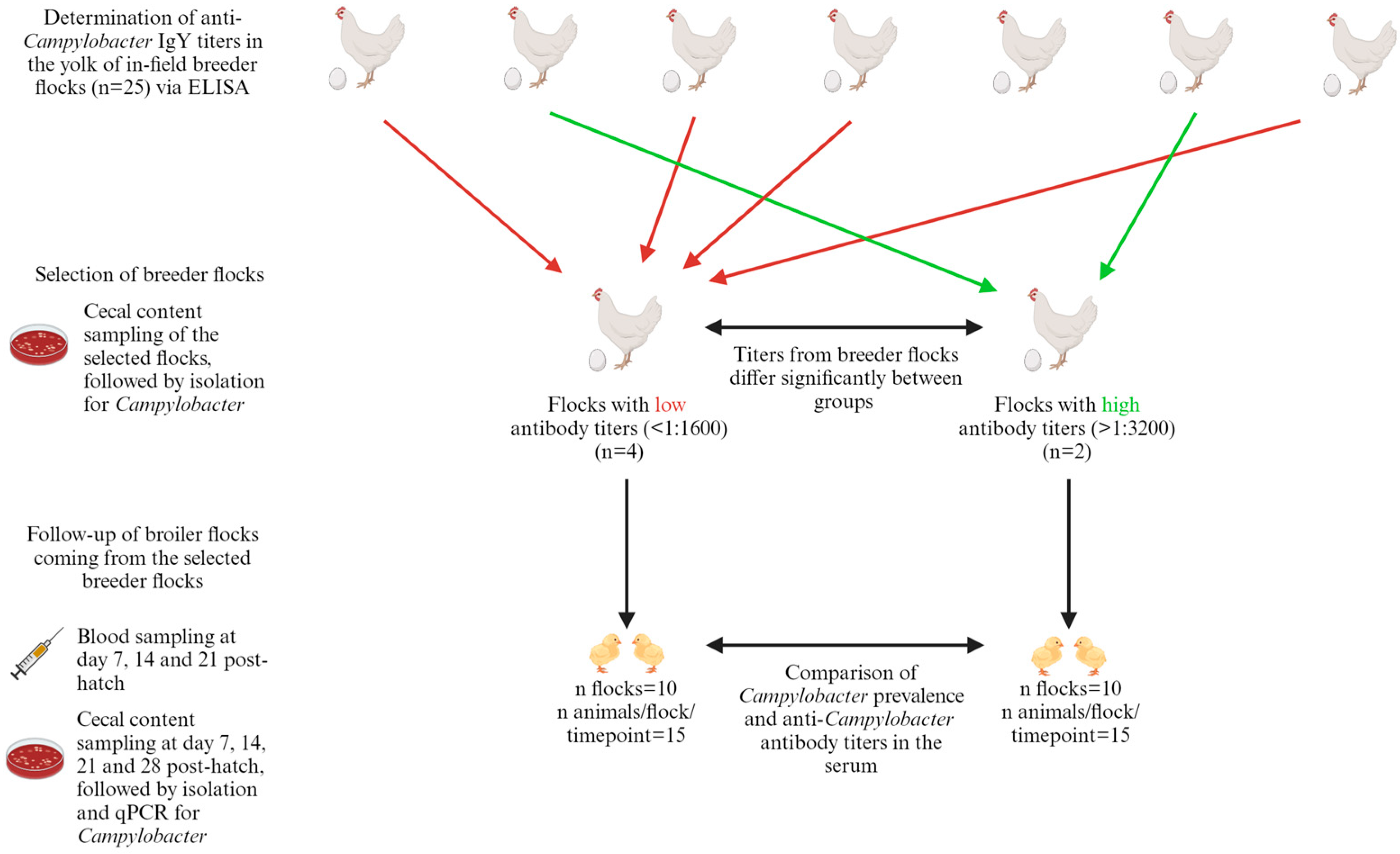
2.1. Field Study: Anti-Campylobacter IgY in Broiler Breeders’ Egg Yolks and Resulting IgY Titers and Probability of Colonization in Their Offspring

A total of 20 eggs per flock were obtained from 25 different broiler breeder flocks via a local hatchery (Belgabroed NV, Merksplas, Belgium), with varying ages ranging from 27 to 44 weeks. For quantification of the IgY titers, the egg yolks were analyzed via ELISA, as described below (Section 2.7). At first, the eggs were pooled by flock. Based on these results, flocks were assigned to one of two groups: breeder flocks with high anti-Campyloacter IgY titers and breeder flocks with low anti-Campylobacter IgY titers. The cut-off values for the group with low and high antibody titers were anti-Campylobacter IgY titers ≤ 1:1600 and ≥1:3200 respectively. Subsequently, all the eggs from the selected flocks were analyzed individually. Flocks from the group with low IgY titers that differed significantly in optical density 450 nm (OD450) from the flocks in the group with high IgY titers were selected for follow-up of the progeny flocks and vice versa. Pooled cecal samples from the selected flocks were collected for Campylobacter culture, as described below (Section 2.4).
Following selection, 20 broiler flocks descended from the selected breeder flocks were monitored. These flocks included 10 flocks from the group with low anti-Campylobacter IgY titers and 10 flocks from the group with high anti-Campylobacter IgY titers. Fifteen randomly selected broiler chicks per flock were monitored at weekly intervals, with monitoring starting at 7 days of age and ending at 28 days of age. A fixed volume of 1 mL of serum was collected from each chick, and the collected serum from all chicks on the same farm was pooled and mixed weekly. The pooled samples were subsequently analyzed for maternal antibodies via ELISA, following the procedure described below (Section 2.7). In anticipation of processing, the samples were stored at −20 °C. Next, the chicks were euthanized by injection of an overdose (100 mg/kg) of sodium pentobarbital (Kela, Elversele, Belgium) in the wing vein, and the cecal content was collected for detection of Campylobacter, as described below (Section 2.4). For a schematic overview, see Figure 1.

2.2. Vaccination Study: Selection and Immunization of in Field Seropositive Broiler Breeders and Determination of Maternal Antibodies after Vaccination

To select the breeder flock for the vaccination trial, eggs (n = 10/flock) from 4 different broiler breeder flocks with ages between 23 and 26 weeks were collected. These eggs were acquired from the same local hatchery as sampled previously (Belgabroed NV, Merksplas, Belgium). The anti-Campylobacter IgY titers in the yolk of individual eggs were analyzed via ELISA as described below (Section 2.7). The flock with the highest titer (1:2000 ± 400) and a low coefficient of variation (CV%) (63%) was selected for vaccination purposes. On the day of sampling, this flock was 26 weeks old. The other breeder flocks had titers of 1:180 ± 47, 1:920 ± 298 and 1:840 ± 139. From the selected flock, 45 commercial Ross 308 broiler breeder hens and 10 Ross 308 broiler breeder roosters were purchased. On arrival, pooled cecal droppings were collected for Campylobacter culture, as described below (Section 2.4). Flocks were subjected to Campylobacter screening to characterize the colonizing strains. This investigation aimed to determine whether the antibodies circulating in the flock, which originated from field infections, were homologous or heterologous to the challenge strain utilized for offspring inoculation. The goal was to assess the efficacy of the maternal antibodies in providing protection in the offspring of sham-immunized negative-control breeders. Hens were randomly assigned to three vaccine groups, and each group was separated into three subgroups: bacterin (n = 15), subunit (n = 15) and sham-vaccinated negative control (n = 15), in a design similar to that used in the preceding study [22]. Birds were housed in a minimum enclosure space of 0.21 m2 per bird. The birds were given restricted commercial food (Versele Laga, Deinze, Belgium) once per day in the morning. The feeding schedule and light schedule were adapted based on the Ross Broiler Breeder Management Guide and the conditions on the originating farm. Drinking water was provided ad libitum.
At 30 weeks of age, breeders were primo-vaccinated. This vaccination was followed by 3 boosters at two-week intervals, in a design similar to that used in the previous study [22]. The vaccine was administered by intramuscular injection in the pectoral muscle. The composition of the vaccines is described below (Section 2.6). Two weeks after each vaccination and 2 and 4 months after the final booster, yolk and blood derived from the wing vein was collected and pooled by group (control, bacterin and subunit). In anticipation of processing, the samples were stored at −20 °C. The anti-Campylobacter IgY titers were determined via ELISA, as described below (Section 2.7). Two weeks and two months after the last booster, the first fertilized eggs from these (sham-)vaccinated broiler breeders were collected and incubated. Post-hatch, the progeny were used to determine the probability of colonization with Campylobacter in threshold trials, as described below (Section 2.3). Husbandry, euthanasia methods, experimental procedures and biosafety precautions were approved by the Ethical Committee (EC2021_086) of the Faculty of Veterinary Medicine, Ghent University, Belgium.

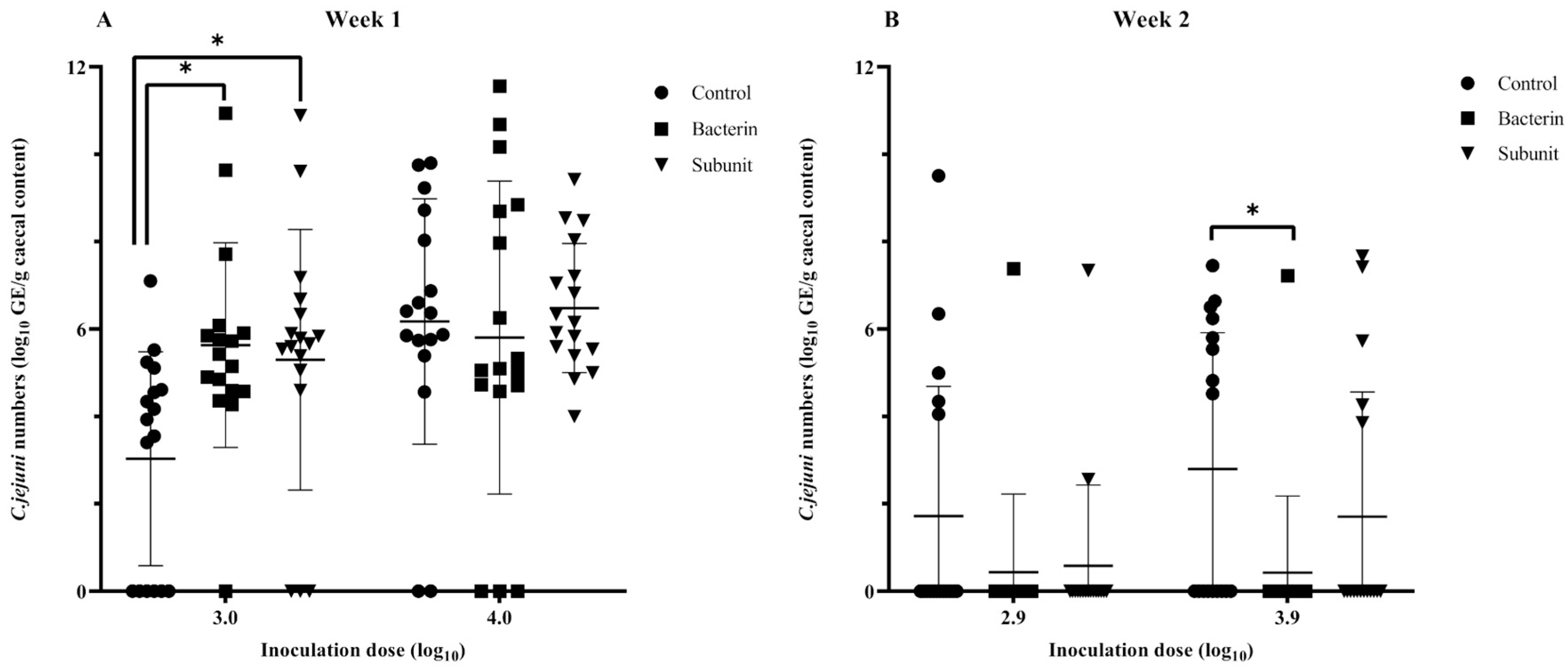
2.3. Vaccination Study: Protective Efficacy of Bacterin and Subunit Derived Maternal Antibodies on Cecal Colonization in Broilers Using a Threshold Model

The susceptibility of young chicks derived from immunized field breeders to initial colonization by Campylobacter was studied using a threshold model [22,23]. Chicks were housed individually to prevent the spread of Campylobacter to other chicks so that the individual probability of a chick becoming colonized by a fixed dose of Campylobacter could be assessed. Fertilized eggs from the three (sham-)vaccinated groups (bacterin, subunit and control) were hatched and housed in groups until the age of inoculation. Birds were housed according to the guidelines set by the ethical committee, and conditions were similar to those used in the preceding study [22]. This means that they were raised on a bedding of wood shavings and under a heat lamp. The minimum enclosure space depended on the weight of the birds (from 0.025 m2 per bird at week 1 to 0.09 m2 at week 3). From the day of hatching until the end of the experiment, chicks received ad libitum commercial starting feed (Farm 1 & 2, Versele Laga, Belgium) and drinking water. From different time-points onwards (7, 14 and 21 days of age), each chick was housed individually in a box on a bedding of wood shavings with an enclosure space of 0.16 m2 per chick and audiovisual contact with the other chicks. Before chicks were challenged, cecal samples were collected, pooled by group and tested for Campylobacter isolation. Chicks were orally inoculated in the crop with Campylobacter jejuni strain KC40 [22,23,24,25,26] using a syringe, administering 1 mL inoculum containing either a low dose (2.7–3.0 log10 cfu) or a high dose (3.6–4.0 log10 cfu). Two days after inoculation, the chicks were euthanized by injection of an overdose (100 mg/kg) of sodium pentobarbital (Kela, Hoogstraten, Belgium) in the wing vein and the cecal content was collected for qPCR analysis, as described below (Section 2.8). Before euthanization, blood was collected from the wing vein and processed for ELISA, as described below (Section 2.7). In anticipation of processing, the samples were stored at −20 °C. The number of chicks per trial varied depending on the number of hatched chicks, and the numbers are listed below in Table 1. As the number of chicks per hatching was limited, the in vivo trials were conducted over two time periods: in the first in vivo trials, the fertilized eggs were collected and incubated two weeks after the last booster; in the second in vivo trials, they were collected and incubated two months after the last booster. The bacterial challenge at two weeks of age was repeated to compare the results with those from the chicks incubated at two weeks after the final booster, a design choice that accounts for possible differences in antibody titers between the two time periods. Husbandry, euthanasia methods, experimental procedures and biosafety precautions were approved by the Ethical Committee (EC2023_015) of the Faculty of Veterinary Medicine, Ghent University, Belgium.

2.4. Bacterial Strains, Culture Conditions and Isolation

C. jejuni KC40 was used as challenge strain in the threshold trials. This strain was isolated at the Flanders Research Institute for Agriculture, Fisheries and Food (ILVO, Melle, Belgium) and has been used in preceding research and challenge trials, where it proved to be a high colonizer of broiler ceca [22,23,24,25,26]. The bacterin vaccine and corresponding coating of the whole-cell ELISA plates consisted of 13 Campylobacter strains, 11 C. jejuni strains and 2 C. coli strains, as listed in Table S1. All strains were isolated from chicken samples [25] and provided by Sciensano (Brussels, Belgium). For the selection of these strains, genetic heterogeneity, prevalence ratio in broilers and role in human campylobacteriosis cases were considered [25]. These strains were stored at −80 °C.
Campylobacter bacteria were cultured on modified charcoal cefoperazone deoxycholate agar (mCCDA, CM0739; Oxoid Ltd., Basingstoke, Hampshire, UK) followed by microaerobic incubation (5% O2, 5% CO2, 5% H2, 85% N2) at 42 °C for 24 h. Next, bacteria were grown in Nutrient Broth No. 2 (NB2, CM0067; Oxoid Ltd., Basingstoke, Hampshire, UK) enriched with Modified Preston Campylobacter-selective supplement (SR0204E; Oxoid Ltd., Basingstoke, Hampshire, UK) and Campylobacter-specific growth supplement (SR0232E; Oxoid Ltd., Basingstoke, Hampshire, UK) at 37 °C for 16 h under microaerobic conditions.
Before and after inoculation, the concentration of Campylobacter bacteria in the bacterial suspensions was determined by titration. Tenfold dilutions in Hank’s Balanced Salt Solution (HBSS; GIBCO-BRL, Invitrogen, Carlsbad, CA, USA) were plated on mCCDA supplemented with charcoal cefoperazone deoxycholate agar (CCDA) selective supplement (SR0155E; Oxoid Ltd., Basingstoke, Hampshire, UK) and Campylobacter-specific growth supplement (SR0232E; Oxoid Ltd., Basingstoke, Hampshire, UK). The plates were then subjected to microaerobic incubation at 37 °C for 48 h.
Via spectrophotometry (Genesys 10S UV-VIS, Thermo Scientific, Waltham, MA, USA) at 600 nm, the bacterial concentration was estimated in a suspension. For the preparation of the challenge-strain inoculum, the enriched broth containing C. jejuni KC40 was diluted in non-enriched NB2 to the desired concentration.
To isolate Campylobacter from the cecal content of breeders and broilers, samples were diluted 1:9 (w/v) in enriched NB2 and incubated at 42 °C for 24 h under microaerobic conditions. Subsequently, these samples were plated on mCCDA agar plates supplemented with CCDA selective supplement and Campylobacter-specific growth supplement under the same culturing conditions. After culture, Campylobacter colonies were identified with matrix-assisted laser desorption ionization time-of-flight mass spectrometry (MALDI-TOF MS; Autoflex Speed LRF, Bruker, Billerica, MA, USA) [27] using the direct-transfer method and using α-cyano-4-hydroxycinnamic acid (HCCA) as a matrix, according to the manufacturer’s guidelines. The spectra were obtained and analyzed with MBT Compass software version 4.1 (Bruker Daltonik, Billerica, MA, USA) and compared with a database of 6120 mean spectra projections. Identifications with a score value > 2.000 were considered reliable to the species level. The Campylobacter colonies were purified by plating them again on blood agar plates under the same culturing conditions. In anticipation of processing, the samples were stored at −80 °C.

2.5. Characterization of Campylobacter Isolates

Campylobacter isolates were identified and further characterized. The species was determined via MALDI-TOF MS (Autoflex Speed LRF, Bruker, Billerica, MA, USA) [27], as described above (Section 2.4). Hereafter, clonal complexes (CC) and sequence types (ST) of the strains were determined via multilocus sequence typing (MLST). Via the DNeasy Blood and Tissue kit (Qiagen, Venlo, Hilden, Germany), DNA was extracted from the isolates, and the DNA yield and quality were examined spectrophotometrically (NanoDrop, Thermo Scientific, Waltham, MA, USA). Finally, the samples were diluted to 25 ng/µL. Primers, PCR conditions, sequencing and MLST scheme were used as described by Jolley et al. [28]. The isolated strains were analyzed via MLST to determine whether the strains colonizing broiler flocks were homologous or heterologous strains compared to those colonizing the respective breeder flocks.

2.6. Bacterin and Subunit Vaccine Preparation

For the preparation of the bacterin and subunit vaccine/ELISA, the protocol was used the same as that in [25], with minor adaptions. To summarize, to prepare the bacterin, the 13 Campylobacter strains (Table S1) were cultured and enumerated separately in enriched NB2, as discussed above. Next, to kill the bacteria, suspensions were incubated overnight with 36% formaldehyde (0.005 mL/mL) (Sigma-Aldrich, St. Louis, MO, USA) at 37 °C. Bacterial suspensions were subsequently centrifuged for 30 min at 5718 relative centrifugal force (rcf) at room temperature. The pellets were resuspended in 5 mL of phosphate-buffered saline (PBS) with the same formaldehyde concentration (36% formaldehyde/L) mentioned above and incubated overnight at 37 °C. The next day, the successful killing of the bacteria was checked by culturing the suspensions on sheep blood agar plates under microaerobic conditions at 42 °C for 24 h. Finally, the bacterial cultures were stored at 4 °C for further use.
Six immunodominant antigens (AtpA, CheV, Ef-Tu, GroEL, LivJ and Tig) were integrated into the subunit vaccine (Table S2), based on preceding studies [22,24,25]. The inclusion of the recombinant proteins in the subunit vaccine was based on the reactivity of IgY from C. jejuni KC40 immunized layer hens [24], the association of these proteins with the bacterial cell membrane [24,29,30,31] and positive results in previous vaccination studies [22,25].
To generate the antigens sourced from the C. jejuni reference strain KC40, the Escherichia coli Expression System using Gateway® Technology (Invitrogen, Carlsbad, CA, USA) was used, following the outlined methodology of Vandeputte et al. [25] with minor adaptions. Changes included the addition of 10% glycerol (Sigma-Aldrich, St. Louis, MO, USA) to the elution buffer and a subsequent dialysis step in phosphate-buffered saline (PBS) and 10% glycerol (Sigma-Aldrich, St. Louis, MO, USA) overnight after elution. These adaptations were introduced to counter protein precipitation and conserve the biological activity of the proteins.
To make the bacterin vaccine, 7 log10 cfu of each Campylobacter strain was mixed. Thus, a bacterial suspension consisting of 8.1 log10 cfu inactivated Campylobacter was obtained [22,25]. For the subunit vaccine, 75 µg of protein (12.5 µg of each recombinant antigen as listed in Table S2) was used per dose [22,25]. The negative controls were vaccinated with a sham vaccine containing 150 µL of pure HBSS. HBSS was added to the antigen until a volume of 150 µL was reached, and the (sham) vaccine was combined with Montanide ISA 71 VG adjuvant (SEPPIC, Paris, France) at a dosage of 500 µL per chick at a 3:7 ratio.

2.7. Determination of IgY Titers in Egg Yolk and Serum

To determine the IgY titers in egg yolks and serum, the same protocol used by Hermans et al. [24] and Vandeputte et al. [25] was used, with some small adaptations. Prior to ELISA, the egg yolks were diluted 1/5 (v/v) in PBS and mixed. Samples were incubated overnight at 4 °C; eventually, the watery-soluble supernatant was collected for processing. After the collected blood was allowed to clot, the blood was centrifuged (1425 rcf, 15 min). Finally, the serum was collected for IgY quantification. In anticipation of processing, the samples were stored at −20 °C.
To quantify IgY titers against Campylobacter, both a whole-cell and a recombinant-protein ELISA were used. Overnight, 96-well flat-bottom plates (Nunc MaxiSorp, Nalge Nunc Int., Rochester, NY, USA) were coated (4 °C) with 13 different Campylobacter whole-cell strains (for the field screening of broiler breeders and broilers), the whole-cell Campylobacter KC40 strain (bacterin-vaccinated chicks’ serum collected during the threshold trial) or a mixture of 6 subunit antigens (for the subunit breeder group and subunit-vaccinated chicks). Whole-cell plates were coated with 106 cfu/well, and subunit plates were coated with a mixture of 6 µg/well recombinant proteins. Both coatings were diluted in 50 µL coating buffer. After coating, plates were washed (3× washing buffer: 0.1% Tween-20 in PBS) and the wells were blocked (90 min, 37 °C) with 100 µL blocking buffer (5% skim milk powder in washing buffer). A ½ dilution series of the samples was made in blocking buffer, and 100 µL was incubated in duplo for 60 min at room temperature. The washing procedure was performed as described earlier, and the samples were incubated with 100 µL 1/10,000 horseradish peroxidase (HRP)-labelled anti-chicken IgY (Sigma-Aldrich, St. Louis, MO, USA) in washing buffer for 90 min at room temperature. Chromogenic substrate 3,3′,5,5′-tetramethyl benzidine (TMB) (Sigma-Aldrich, St. Louis, MO, USA) was used, and the plates were incubated with 50 µL at room temperature in the dark. After 10 min, 50 µL 0.5 M H2SO4 was added as stopping solution to each well and the absorbance was measured at 450 nm (OD450) using an automated spectrophotometer (Pharmacia LKB Ultrospec III, Gemini BV, Apeldoorn, The Netherlands).
The cut-off in the dilution series, which was used to determine the IgY titer of a sample, was analyzed using the R package “changepoint” [22,32,33]. The changepoint in the dilution series of the negative control acted as cut-off value. The limit of detection was set to the dilution of the negative control at which this changepoint occurred. The IgY titer of the sample was determined based on the last dilution with an OD450 value greater than the cut-off value. When the dilution at which the changepoint took place was lower than the limit of detection, the sample was considered negative. To ensure reliability and repeatability between the different ELISA assays, the same positive-control dilution series was used, depending on the matrix (serum, yolk), the antigen (bacterin, subunit, KC40) and the age of the birds (broiler chicks, breeders).

2.8. Cecal Campylobacter Enumeration by qPCR

DNA was extracted using the QIAamp® Fast DNA Stool Mini Kit (Qiagen, Venlo, Hilden, Germany) according to the manufacturer’s instructions, with a small adaptation. To elute the DNA, 100 µL ATE buffer was used instead of 200 µL. To test for the presence of thermophilic Campylobacter spp. and quantify these bacteria in the ceca, qPCR (Model CFX 96, Biorad, Hercules, CA, USA) was used, as described by Lund et al. [34] and Botteldoorn et al. [35] and applied by Vandeputte et al. [25]. The quality and the concentration of the DNA were examined spectrophotometrically (NanoDrop, Thermo Scientific, Waltham, MA, USA). Samples underwent triplicate processing, and the mean of these measurements was computed. To ensure reliability and repeatability between the different qPCR assays, the same positive-control dilution series was used.

2.9. Statistical Analysis

Figure 1 was made in BioRender (www.biorender.com, accessed on 28 March 2024), and the remaining figures were made in GraphPad Prism 9. In the field trial, GraphPad Prism 9 served as the analytical tool. Significance testing aimed to ascertain whether the antibody titers of breeder flocks in the MAB− and MAB+ groups differed significantly (p < 0.05) from each other. This assessment utilized the OD450 values obtained from ELISA at dilution 1:400 and involved a comprehensive comparison with the OD450 values of breeder flocks in the other group at the same dilution. As not all flocks had parametric OD450 values, Kruskal−Wallis was used as a test to determine significance.
The Campylobacter prevalence data from the monitored broiler flocks in the field study were analyzed with R.4.2.0. A generalized (i.e., binomial) linear mixed-effects regression model was used, with Campylobacter presence or absence as the dependent variable and age and antibody titer of the respective breeder flocks as independent variables. A random intercept term was included for each separate farm to allow the intercept to vary across trials. The significance of the impact of the maternal antibody titer on the outcome was determined via the “summary” function.
For analysis of the in vivo challenge trials, R.4.2.0 was also used. C. jejuni counts were transformed to log10 counts. To assess how Campylobacter colonization probability (disease prevalence) varies with treatment (i.e., control versus subunit- and bacterin-vaccinated birds), inoculation dose and week, we fitted Bayesian generalized linear models (R function ‘bayesglm’, binomial error distribution) using Campylobacter infection presence or absence as the dependent variable. First, analyses were run for each week and inoculation dose separately. Secondly, an overall analysis was conducted specifying treatment, inoculation dose, week and all two-way interactions between them as independent variables. As AabTiter differed between trials, this factor was included in the overall analysis as an additional covariate. Analysis of CampyTiterCounts followed a similar framework, using a Gaussian error distribution where possible (R function ‘lm’) or a zero-inflated model where needed (R function glmmTMB). Model fit and assumptions were checked using Shapiro−Wilk W values of regression residuals (i.e., W > 0.95 for a Gaussian distribution) and using glmmTMB’s built-in significance test for zero-inflation. Model selection followed a stepwise backward-selection procedure, eliminating non-significant interactions and terms until a minimal model was obtained at p < 0.05. Differences between the control and vaccine treatments at different inoculation doses were obtained using the ‘emmeans’ R function, using the Tukey method to adjust p values for multiple pairwise comparisons.

3. Results

3.1. Field Study: Trend of Decreased Susceptibility to Campylobacter Colonization in Broilers Originating from Breeders with High Antibody Levels

All sampled breeder flocks showed anti-Campylobacter IgY antibodies in their yolks, with pooled titers ranging from 1:200 to 1:3200 (Figure 2). No correlation was observed between the age of the flocks and the antibody titers (Spearman, p > 0.05). From the sampled breeder flocks, six were selected for further monitoring. The selection was based primarily on the antibody titers but also involved logistical factors (e.g., the supply of Flemish broiler farms) and the willingness of the farmer to cooperate. Four breeder flocks (D, E, P and U) with low titers were selected. The titers of these flocks were 1:680 ± 72, 1120 ± 290, 1140 ± 197 and 1:200 ± 62, respectively. Additionally, two flocks (J and W) with high antibody levels were selected, with 1:3560 ± 816 and 1:4480 ± 610 as their respective titers. The titers of the selected breeder flocks with low antibodies (MAB−) differed (p < 0.05) from those of the selected flocks with high antibodies (MAB+). Flock J differed significantly from flock D (p < 0.0001), flock E (p < 0.0001), flock P (p = 0.003) and flock U (p < 0.0001). Flock W also differed significantly from flock D (p < 0.0001), flock E (p < 0.001), flock P (p = 0.049) and flock U (p < 0.0001). Four weeks after the initial sampling, flocks D, E, J and W were retested to ensure that the titers in the breeder flocks’ yolks remained stable. This finding was confirmed through pooled ELISA analysis of yolk samples collected from 10 eggs per stable per flock, as discussed above (Section 2.7). Flocks P and U were not retested, as the sampling of their progeny occurred within a relatively short timeframe (<4 weeks). All selected breeder flocks tested positive for Campylobacter after culture (Table 2). For breeder flock E, however, no results could be obtained, as no permission was granted to take cecal samples from the farm.
Already in the second week post-hatch, one MAB− broiler flock tested positive for Campylobacter. Among the MAB+ broiler flocks, two were positive at 3 weeks post-hatch. At 4 weeks post hatch, the endpoint of the trial, 40% of the monitored MAB− broiler flocks and 20% of the MAB+ broiler flocks had been colonized by Campylobacter. This pattern of colonization resulted in an overall trend in which there were more positive MAB+ broiler flocks than MAB− broiler flocks during the first 4 weeks of life (Figure 3). The overall Campylobacter prevalence did not differ significantly (p = 0.309) between MAB− and MAB+ broiler flocks. No difference was observed in Campylobacter load among the different positive broiler flocks. The sampling of cecal content was limited to 4 weeks of age, as almost all broiler flocks were negative for anti-Campylobacter maternal antibodies at week 1. Even if some undetectable levels were present, the effect of these antibodies would have worn off by 4 weeks of age.

3.2. Field Study: C. coli Dominant in Breeders, an Equal Prevalence of C. jejuni and C. coli in Broilers

After they had been identified to the species level via MALDI-TOF, the Campylobacter isolates were further characterized via MLST. C. coli was found in all breeder flocks (7/7). C. jejuni was encountered only in the breeder flock that had been selected for immunization (1/7). C. jejuni and C. coli were equally prevalent (both 3/6) in positive broiler flocks. In this study, no broiler flocks were colonized with the same ST as their respective breeder flock. The most frequently encountered clonal complex (CC) was CC828 (C. coli), which had colonized 6 out of 7 breeder flocks and 2 out of 6 broiler flocks. Although the same clonal complexes were found in different barns of parent flocks U and W, the same sequence type was not found across all barns. Two of the broiler flocks had been colonized by the same CC as their respective parent flock samples, with one being MAB+ and the other being MAB−. However, the STs were not the same, indicating colonization by a different strain (Table 2). The Campylobacter isolates derived from breeder flock J and P were not further typed, as none of their offspring was positive for Campylobacter.

3.3. Field Study: Low Maternal Antibody Titers in Broilers from Breeders with Varying Antibody Levels

Throughout the early stages of life, pooled serum samples from the progeny flocks were subjected to weekly analysis. Almost all pooled serum samples from the different broiler flocks proved to be negative for anti-Campylobacter antibodies at the different timepoints, with the exceptions of flock J1 at week 1 (1:400) and flock W4 (1:200) at week 2. It is noteworthy that these two broiler flocks both originated from MAB+ breeder flocks. As all samples were negative at three weeks of age, serum was not further examined after this timepoint.

3.4. Vaccination Study: Immunization of Seropositive Broiler Breeders Results in Temporarily Higher Anti-Campylobacter Titers in Broiler Breeders

As already mentioned above, the seropositive breeder flock selected for vaccination (flock Delta, average yolk titer 1:2000 ± 400, CV 63%) proved to be positive for both C. jejuni and C. coli. The C. jejuni CCs included 677 (ST-764) and 353 (ST-7571), and the C. coli CC included 828 (ST-860).
Immunization of Campylobacter-seropositive breeders led to an increase in the antibody titers in yolk in both subunit- and bacterin-vaccinated breeders. A peak of antibody titers was reached in both subunit- and bacterin-vaccinated breeders after the second booster, and titers started to decline two months after the third booster. After this point, the titers seemed to stabilize for the subunit vaccine but continued to decrease for the bacterin vaccine until the final sampling point (4 months after the final booster). At all timepoints after vaccination, titers in the yolks of both groups were higher than in the yolks from the seropositive breeders vaccinated with the sham vaccine. At peak titers, 64 times more antibodies were measured in the yolks from the bacterin-vaccinated breeders than in the yolks from the negative control. For the subunit-vaccinated animals, 640 times more antibodies were measured in the yolks (Figure 4, Table S3). It is imperative to note that two breeders of the bacterin-vaccinated group died 2 and 3 months after the last booster. Notably, neither case appeared to be linked to the immunization process; instead, both were attributed to fatty liver hemorrhagic syndrome. Moreover, no adverse effects, local or systemic, were observed in the months subsequent to immunization.

3.5. Vaccination Study: Protective Efficacy of Bacterin and Subunit Derived Maternal Antibodies on Cecal Colonization in Broilers Using a Threshold Model

In order to evaluate the susceptibility of broiler chicks to Campylobacter colonization during the early stages of life and to investigate the influence of maternal antibodies, a threshold model was employed following the immunization of seropositive broiler breeders. After the first incubation period (two weeks after the last booster),when the animals were challenged at one week of age with a low or a high Campylobacter inoculation dose, no differences were observed with regard to the presence of Campylobacter-positive animals in the different experimental groups (subunit, bacterin and control) (p > 0.05) (Figure 5). It is noteworthy that the majority of animals (>60%) were colonized and that the mean number of cecal genomic equivalents (GE) of the control group was lower than those of the bacterin (p = 0.015) and subunit (p = 0.038) groups following challenge with a low dose (Figure 6). Over the two incubation periods of the fertilized eggs (two weeks and two months after the last booster), the antibody titers decreased considerably in immunized breeders and their offspring. When looking at the results separately, only a reduction (p = 0.049) in Campylobacter prevalence could be noted in bacterin-vaccinated chicks compared to control chicks when fertilized eggs were incubated two weeks after the last booster and challenged with a high dose at week 2 (Figure 5). No other statistically significant differences were observed (p > 0.05). However, when we take into account the results from week 2 overall, including both the chicks inoculated at two weeks and those inoculated at two months after the last booster, and account for the differences in antibody titers, higher levels of antibody titers are significantly linked with a lower prevalence of Campylobacter. This is true both for bacterin versus control (p = 0.008) and subunit versus control (p = 0.003). On analysis of level of colonization, no such differences were observed (p > 0.05). In chicks challenged at three weeks, no differences in prevalence could be observed between the different groups (p > 0.05) (Figure 7). A higher cecal GE number was observed in the subunit broilers in comparison with the control (p = 0.031) and bacterin (p = 0.005) broilers when the animals were challenged with a high dose (Figure 8).
In the first in vivo trials, eggs were incubated two weeks after the final booster. The control chicks were positive for anti-KC40 antibodies (1:600) in the first week and for antibodies against both the KC40 (<1:400) and recombinant antigens (<1:1600) in the second week. In the first week, antibodies against the recombinant antigens could not be detected. The pooled anti-KC40 antibody titer in bacterin-vaccinated chicks proved to be twice as high in week 1 (1:16,000) in comparison with week 2 (1:8000). The pooled titers against the recombinant antigens in subunit-animals were 1:48,000 in week 1 and 1:19,200 in week 2 (Table 3).
For the second in vivo trials, eggs were incubated two months after the final booster. Antibodies against KC40 were observed only at week 2 in the control group. A sharp decrease in levels of both anti-KC40 and anti-subunit antibodies could be noted in the bacterin-vaccinated and subunit-vaccinated chicks, respectively, at 2 weeks of age in comparison with the chicks from the first in vivo trials. For the bacterin-vaccinated chicks, this decrease in the pooled titer was five-fold (1:1600), and for the subunit-vaccinated chicks, three times fewer (1:6400) antibodies were detected. At 3 weeks of age, antibodies were still found in bacterin- and subunit-vaccinated chicks. These pooled titers were quantified at 1:600 and 1:2400 respectively (Table 3).

4. Discussion

In this study, the potential role of maternal antibodies in the protection of chicks against Campylobacter in the first weeks of life was investigated. In a preceding study [22], the role of maternal antibodies was demonstrated by immunizing seronegative SPF breeders with a bacterin vaccine and a subunit vaccine and subsequently challenging their offspring with different doses of Campylobacter in a controlled environment. In that study, a protective effect of the maternal antibodies was observed during the first 1 (bacterin) and 2 (subunit) weeks of life. Our results confirm the (very) high prevalence of Campylobacter in breeder flocks in the field [36], which was reflected in a high seroprevalence. Thus questions remained: whether antibodies elicited in broiler breeders by natural colonization and passed on to their progeny confer protection against in-field colonization in broilers; and whether immunization of naturally colonized broiler breeders can increase the protection in their offspring against a controlled challenge with Campylobacter due to elevated maternal antibody titers.
Previous studies [19,20,22] were able to demonstrate the protective effect of maternal antibodies in broilers against Campylobacter colonization under experimental conditions. However, so far, no such studies have been conducted under commercial field conditions. To explore to what extent naturally elicited maternal antibodies might confer protection to broilers in the field, a field study was conducted in which broiler flocks from different breeder flocks with high or low anti-Campylobacter IgY levels were monitored. All sampled breeder flocks (n = 25) tested positive for Campylobacter antibodies, indicating widespread exposure to Campylobacter in breeder flocks. In all breeder flocks that were subsequently selected for further follow-up of their progeny and for which permission was granted to take faecal samples for further analysis, Campylobacter could be isolated. The high Campylobacter seroprevalence and colonization rates in broiler breeders observed in the current study are in line with the results of previous samplings [22,36]. Significant differences in titers made it possible to divide the breeder flocks into a group with high serum anti-Campylobacter IgY titers and a group with low titers. The presence of low antibody titers in combination with ongoing colonization could possibly be attributed to colonization by a less immunogenic strain or to an older colonization. In another study [20], a substantial anti-Campylobacter serum-antibody titer was observed one week after experimental oral inoculation of Ross breeders and layers. However, these titers seemed to have dropped a few weeks after initial colonization, although they increased upon rechallenge with Campylobacter.
According to the results of monitoring the offspring of MAB+ and MAB− breeder flocks, broiler flocks with lower anti-Campylobacter IgY titers tended to be more susceptible for Campylobacter, as a higher percentage of positive flocks was recorded early in the study (at 2 weeks of age) and at the endpoint of the study (at 4 weeks of age). These results were not statistically significant, possibly due to the limited sample size. The serological investigations demonstrated that almost all sampled broilers, including both those originating from both MAB− and MAB+ breeder flocks, were negative for anti-Campylobacter antibodies at the first sample point (one week of age). Antibodies might still have been present, but if so, they were undetectable by our ELISA method used. The changepoint observed in the dilution series of the negative control on ELISAs, occurring either at 1:200 or 1:400, suggests that antibody titers below this threshold were undetectable. In other words, the detection limit ranged between 1:200 and 1:400 [22,32,33]. Other studies investigating anti-Campylobacter antibodies in broilers’ serum after vaccination reported the same starting dilutions on ELISA (1:200), indicating detection limits similar to that observed in our study [37,38,39]. Although they were not significant, the results of the field study indicate possible early protection by maternal antibodies against Campylobacter colonization. However, as maternal antibody titers decreased rapidly and as Campylobacter-positive MAB+ flocks were observed, the protection thus obtained is most likely only partial. Numerous other factors likely contribute to the early onset of colonization, including biosecurity measures [16,40,41], flock size [42,43], seasonality and weather conditions [42,44], the presence of other livestock [45] and the current intestinal microflora [18,46,47].
Not many studies have investigated the prevalence and species prevalence of Campylobacter in broiler breeders. In our study, all sampled breeder field flocks tested positive for C. coli. Co-colonization with two C. jejuni strains was observed only in the breeder flock selected for immunization. In previous studies conducted in Belgium [36] and Spain [48], a 100% prevalence of Campylobacter in breeder flocks was also observed. However, in these studies, C. jejuni was observed as the predominant species, followed by C. coli. In contrast, our current findings indicate a noteworthy shift in Belgium, with C. coli currently assuming the role of the dominant species in breeder populations.
In the monitored progeny broiler flocks, C. jejuni and C. coli were equally prevalent. Just as in breeders, more C. coli strains were encountered than expected, as C. jejuni is generally considered the dominant Campylobacter species in broilers, both in Belgium [36,40] and in Europe [41,42,43]. Lately, there have also been reports of C. coli being more frequently recovered from poultry meat in Europe [44,45,46]. However, as pointed out by Andritsos et al. [47], multiple factors should be taken into account when interpreting these data, such as seasonality of the sampling, broiler age, sampling method and isolation procedure. In the study by Babacan et al. [48], it was found that birds slaughtered earlier were more frequently colonized by C. jejuni compared with birds slaughtered at a later timepoint, which were more frequently colonized by C. coli. El-Shibiny et al. [49] observed that a C. jejuni strain and a C. coli strain could both colonize and compete with each other equally until the birds were 35 days old. After this timepoint, C. jejuni numbers decreased, leaving the C. coli strain as the dominant isolate. It should be noted that these results could be strain-specific and that they do not explain the high prevalence of C. coli in broilers in this study, as all birds were sampled at a young age. However, the results could potentially explain the high prevalence of C. coli in the breeders. The shift from C. jejuni to C. coli holds significance, as it is hypothesized that C. coli harbors fewer virulence genes affecting humans [50]. Nonetheless, the actual impact of an elevated ratio on reducing cases of human campylobacteriosis remains a subject requiring further investigation. It would also be interesting to continue monitoring whether C. coli has indeed expanded its foothold in Belgium and, by extension, in Europe. If so, the reasons behind this change would be interesting to investigate. An interesting factor to explore is the direct effect of climate change and changing weather patterns. As the season-dependent patterns of C. jejuni and C. coli appear to differ from each other [42,51,52], this could be a contributing factor.
The analysis via MLST of the colonizing strains in breeders and their offspring was intended to further explore the correlation between maternal antibodies and their potential protective effect against Campylobacter colonization. For example, early colonization of maternal-antibody-negative (MAB−) broiler flocks with homologous strains and its absence in maternal-antibody-positive (MAB+) broiler flocks would support the hypothesis of a potential protective effect of maternal antibodies, whereas the opposite finding would challenge this hypothesis. However, as breeder flocks and their progeny were colonized by different strains, no further conclusions could be drawn regarding the role of maternal antibodies in the protection of broiler flocks against Campylobacter colonization. Despite the assumptions of Cox et al. [13] about vertical transmission, no identical Campylobacter STs were found in breeders and their offspring in the field study. Also, in the offspring of our (sham-)immunized breeder flock from the field, which were colonized by three different Campylobacter strains, no Campylobacter was found before the experimental challenge using culture techniques. The claim could be made that these chicks had maternal antibodies and the Campylobacter population was suppressed by these, which made the Campylobacter undetectable by conventional culture methods, but even at 3 weeks of age, when maternal antibodies had decreased significantly in the control group, no Campylobacter was found. It is possible that Campylobacter could survive on the eggshells (and maybe in the outer membranes), but the assumption that this could lead to the full-blown colonization of newly hatched chicks is too far-fetched. Based on the MLST analysis, six out of seven breeder flocks and two out of three broiler flocks were colonized by C. coli CC828 complex, which also colonized the breeder flock selected for the immunization trial. This was no surprise, as CC828 has been recognized worldwide as the predominant clonal complex, originating from different clinical and environmental sources [53,54,55,56,57,58]. This CC has successfully established itself in an agricultural niche and subsequently causes most human campylobacteriosis cases attributed to C. coli. In other Belgian studies on C. coli in broiler carcasses and diarrheal patients [59,60], most (>80%) of the isolates belonged to CC828. The most frequently observed STs were ST-854, ST-825, ST-860, ST-5163, ST-829, ST-832 and ST-872. In our study, ST-832 was present in two broiler flocks and ST-860 was present in the breeder flock selected for immunization, but the other STs were not encountered. ST-828 was the predominant ST in our study, with three identified breeder isolates. Regarding C. jejuni, other Belgian studies [59,61,62] have mentioned CC-21 as one of the most common STs. Clonal Complexes, both in human and poultry samples, although the C. jejuni population is much more diverse. Within this CC, ST-50 and ST-21 are considered the most dominant STs. Although the number of C. jejuni strains in our study was limited, CC-21 was encountered in two broiler flocks, with one strain belonging to ST-50. CC-677 (ST-764) and 353 (ST-7571), isolated from the breeder flock selected for the immunization trial, have not been described in previous Belgian studies. To conclude, most of our findings regarding MLST types in C. jejuni and C. coli are in line with the findings of previous studies.
In the immunization trial, the seropositive breeder flock selected for immunization tested positive for 3 strains of Campylobacter, namely C. jejuni CC-677 (ST-764) and CC-353 (ST-7571) and C. coli CC-828 (ST-860), which might explain the strong serological reaction on ELISA. One strain even belonged to the same CC as our C. jejuni challenge strain (KC40; CC-677 ST-794). Next, this seropositive breeder flock was immunized using a bacterin vaccine and a subunit vaccine. The selection of C. jejuni and C. coli strains incorporated into the bacterin vaccine was initially based on their prevalence in poultry and in human campylobacteriosis cases [25]. The bacterin contains strains belonging to the same clonal complexes encountered in the current study, except for CC-353, supporting their relevance. Immunization with both the bacterin vaccine and the subunit vaccine resulted in increased anti-Campylobacter titers in the yolks compared to the yolks from sham-immunized seropositive breeders. The maximum titers in both bacterin-vaccinated and subunit-vaccinated breeders were reached after the second booster, which suggests that the third booster might not be necessary. However, these titers proved unstable, as after the second and third booster, they started to decline. Despite this decline, they were still higher than the titers of the sham-immunized breeders.
Next, to assess the protective efficacy of the increased maternal antibodies against Campylobacter colonization, a threshold challenge model was conducted. No trends or differences in prevalence were observed between the different groups following challenge at weeks 1 and 3. It is possible that the challenge dose was too high in week 1, even for the lower challenge dose, as the lower challenge dose was slightly higher than that in the preceding study by Haems et al. [22] (3.0 log10 versus 2.5 log10), in which a protective effect of maternal antibodies elicited by immunization was observed. Additionally, more than 64% of each group was colonized, which also points towards a strong challenge. In week 3, maternal antibodies had possibly decreased too much to make a difference, although subunit-vaccinated animals had a higher Campylobacter load in their caeca than did the two other groups following challenge with a high dose. If we account for the differences in antibody levels and examine the overall results from week 2, a significant link between decreased prevalence in bacterin-vaccinated chicks and subunit-vaccinated chicks and their respective antibody levels can be observed. This finding confirms that in week 2, both bacterin- and subunit-elicited antibodies played a role in the protection of chicks against Campylobacter challenge. However, in a previous study in which SPF breeders were immunized [22], the protective efficacy of the maternal antibodies induced by the bacterin and subunit vaccines proved to be more significant [22]. This difference can be explained by the higher baseline levels of antibodies present in the breeders and their progeny, as the SPF breeders in the study of Haems et al. [22] were seronegative. The in-field breeders used in this study were colonized by a Campylobacter strain belonging to the same clonal complex as the challenge strain, which provided the sham-immunized breeders and their chicks with homologous humoral protection. Although it was proven in a previous study [22] that immunization of SPF breeders was able to increase the level of anti-Campylobacter IgY antibodies in the intestinal mucus, the enhanced maternal immunity induced by the vaccines in seropositive breeders had only a limited effect with regard to protection against Campylobacter colonization in week 2 in both subunit- and bacterin-vaccinated chicks.
Finally, an important factor to consider is the way birds were challenged. It was suggested by other studies [63] that the handling of the chicks may induce stress and that the experimental challenge with a pure Campylobacter culture at a high dose may disrupt the native microflora, potentially facilitating easier colonization by Campylobacter. However, a comparative study between different treatment groups requires that birds be inoculated with a standardized challenge dose, ensuring scientific rigor in drawing conclusions regarding treatment efficacy. To address this necessity, the lowest challenge dose at which C. jejuni KC40 strain maximally colonizes broiler chicks from SPF breeders was chosen as the low challenge dose (unpublished data [22]). However, the possibility that the experimental conditions themselves may have favored Campylobacter colonization, thereby potentially obscuring the true treatment effect, cannot be excluded. Despite this possibility, significant Campylobacter reductions were also obtained with this strain at much higher challenge doses (8 × 10³–105 cfu) when chicks were orally supplemented with yolk from hyperimmunized laying hens [24,25].

5. Conclusions

To conclude, seropositivity for Campylobacter in breeder flocks is widespread, indicating regular contact of these flocks with Campylobacter. Additionally, C. coli was the most commonly isolated species both in broiler and breeder flocks, indicating a potential shift from C. jejuni to C. coli as the dominant species on Belgian poultry farms. On farms, chicks with higher levels of maternal antibodies tend to be less susceptible to Campylobacter colonization in the first weeks of life. However, taking into account the rapid decline of titers in the serum of the chicks, maternal antibody transfer probably offers only partial protection. Using immunization, it was possible to increase anti-Campylobacter antibody titers in the serum of seropositive breeders and consequently also in the serum of the offspring. Indications for improved resistance against Campylobacter colonization in offspring from these immunized breeds are limited and inconsistent. All findings taken into account, the presumed protective effect of maternal immunity should be considered limited and should not be overestimated in the context of Campylobacter control in broilers.

Supplementary Materials

The following supporting information can be downloaded at: https://www.mdpi.com/article/10.3390/ani14091291/s1, Table S1: The Campylobacter strains used in the bacterin vaccine and coating.; Table S2: The recombinant proteins used in the subunit vaccine and coating.; Table S3: Pooled bacterin and subunit-vaccine induced egg yolk titers in broiler breeders as determined by ELISA at different time points. For comparison, titers of the seropositive breeders vaccinated with the sham vaccine against the respective bacterin- and subunit-coating are included.

Author Contributions

Conceptualization, K.H., A.M., F.P. and A.G.; methodology, K.H., N.V.R., G.R., A.M., F.P. and A.G.; software, D.S.; validation, K.H., A.M., F.P. and A.G.; formal analysis, K.H. and D.S.; investigation, K.H.; resources, G.R., A.M., F.P. and A.G.; data curation, K.H.; writing—original draft preparation, K.H.; writing—review and editing, D.S., G.R., A.M., F.P. and A.G.; visualization, K.H.; supervision, A.M., F.P. and A.G.; project administration, A.G.; funding acquisition, A.G. All authors have read and agreed to the published version of the manuscript.

Funding

This research was funded by Federal Public Service for Health, Food Chain Safety and Environment (FOD, Brussels, Belgium), grant FOD RF 19/6333 Campybroilerbreed.

Institutional Review Board Statement

The animal study protocol was approved by the Ethics Committee of Ghent University (EC2021_086, 28 September 2021).

Informed Consent Statement

Not applicable.

Data Availability Statement

The data presented in this study are openly available in Open Science Network at DOI 10.17605/OSF.IO/GHT7F, reference number ght7f.

Acknowledgments

The authors are grateful to Filip Boel and Belgabroed (Merksplas, Belgium), for providing us with breeder eggs and access to the different breeder and broiler farms of the field-trial and Ross 308 breeder chickens for the immunization trial. Also, the cooperation of Galluvet (Lummen, Belgium) and Dierenkliniek Venhei (Kasterlee, Belgium) was essential for the collection of cecal samples in the different breeder flocks. For the immunization, the Montanide adjuvant was generously provided free of charge by SEPPIC (Courbevoie, France).

Conflicts of Interest

The authors affirm that there are no conflicts of interest to disclose. While the study design underwent review by the funder, it’s important to note that they were not involved in data collection, analysis, interpretation, manuscript composition, or the decision to publish the findings.

References

  1. Nachamkin, I.; Allos, B.M.; Ho, T. Campylobacter Species and Guillain-Barré Syndrome. Clin. Microbiol. Rev. 1998, 11, 555–567. [Google Scholar] [CrossRef] [PubMed]
  2. European Food Safety Authority and European Centre for Disease Prevention and Control (EFSA and ECDC). The European Union One Health 2022 Zoonoses Report. EFSA J. 2023, 21, e8442. [Google Scholar] [CrossRef] [PubMed]
  3. Herman, L.; Heyndrickx, M.; Grijspeerdt, K.; Vandekerckhove, D.; Rollier, I.; De ZUTTER, L. Routes for Campylobacter Contamination of Poultry Meat: Epidemiological Study from Hatchery to Slaughterhouse. Epidemiol. Infect. 2003, 131, 1169–1180. [Google Scholar] [CrossRef] [PubMed]
  4. Rasschaert, G.; Houf, K.; Van Hende, J.; De Zutter, L. Campylobacter Contamination during Poultry Slaughter in Belgium. J. Food Prot. 2006, 69, 27–33. [Google Scholar] [CrossRef] [PubMed]
  5. Seliwiorstow, T.; Duarte, A.; Baré, J.; Botteldoorn, N.; Dierick, K.; Uyttendaele, M.; De Zutter, L. Comparison of Sample Types and Analytical Methods for the Detection of Highly Campylobacter-Colonized Broiler Flocks at Different Stages in the Poultry Meat Production Chain. Foodborne Pathog. Dis. 2015, 12, 399–405. [Google Scholar] [CrossRef] [PubMed]
  6. Hertogs, K.; Haegeman, A.; Schaumont, D.; Gelaude, P.; De Zutter, L.; Dewulf, J.; Heyndrickx, M.; Rasschaert, G. Contamination Sources and Transmission Routes for Campylobacter on (Mixed) Broiler Farms in Belgium, and Comparison of the Gut Microbiota of Flocks Colonized and Uncolonized with Campylobacter. Pathogens 2021, 10, 66. [Google Scholar] [CrossRef] [PubMed]
  7. Humphrey, S.; Chaloner, G.; Kemmett, K.; Davidson, N.; Williams, N.; Kipar, A.; Humphrey, T.; Wigley, P. Campylobacter Jejuni Is Not Merely a Commensal in Commercial Broiler Chickens and Affects Bird Welfare. mBio 2014, 5, e01364-14. [Google Scholar] [CrossRef]
  8. Pédron, T.; Sansonetti, P. Commensals, Bacterial Pathogens and Intestinal Inflammation: An Intriguing Ménage à Trois. Cell Host Microbe 2008, 3, 344–347. [Google Scholar] [CrossRef] [PubMed]
  9. Hermans, D.; Van Deun, K.; Messens, W.; Martel, A.; Van Immerseel, F.; Haesebrouck, F.; Rasschaert, G.; Heyndrickx, M.; Pasmans, F. Campylobacter Control in Poultry by Current Intervention Measures Ineffective: Urgent Need for Intensified Fundamental Research. Vet. Microbiol. 2011, 152, 219–228. [Google Scholar] [CrossRef]
  10. Abd El-Hack, M.E.; El-Saadony, M.T.; Shehata, A.M.; Arif, M.; Paswan, V.K.; Batiha, G.E.-S.; Khafaga, A.F.; Elbestawy, A.R. Approaches to Prevent and Control Campylobacter spp. Colonization in Broiler Chickens: A Review. Environ. Sci. Pollut. Res. 2021, 28, 4989–5004. [Google Scholar] [CrossRef]
  11. Sahin, O.; Morishita, T.Y.; Zhang, Q. Campylobacter Colonization in Poultry: Sources of Infection and Modes of Transmission. Anim. Health Res. Rev. 2002, 3, 95–105. [Google Scholar] [CrossRef] [PubMed]
  12. Newell, D.G.; Elvers, K.T.; Dopfer, D.; Hansson, I.; Jones, P.; James, S.; Gittins, J.; Stern, N.J.; Davies, R.; Connerton, I.; et al. Biosecurity-Based Interventions and Strategies to Reduce Campylobacter spp. on Poultry Farms. Appl. Environ. Microbiol. 2011, 77, 8605–8614. [Google Scholar] [CrossRef] [PubMed]
  13. Cox, N.A.; Richardson, L.J.; Maurer, J.J.; Berrang, M.E.; Fedorka-Cray, P.J.; Buhr, R.J.; Byrd, J.A.; Lee, M.D.; Hofacre, C.L.; O’Kane, P.M.; et al. Evidence for Horizontal and Vertical Transmission in Campylobacter Passage from Hen to Her Progeny. J. Food Prot. 2012, 75, 1896–1902. [Google Scholar] [CrossRef] [PubMed]
  14. Callicott, K.A.; Friðriksdóttir, V.; Reiersen, J.; Lowman, R.; Bisaillon, J.-R.; Gunnarsson, E.; Berndtson, E.; Hiett, K.L.; Needleman, D.S.; Stern, N.J. Lack of Evidence for Vertical Transmission of Campylobacter spp. in Chickens. Appl. Environ. Microbiol. 2006, 72, 5794–5798. [Google Scholar] [CrossRef] [PubMed]
  15. Shanker, S.; Lee, A.; Sorrell, T.C. Campylobacter jejuni in Broilers: The Role of Vertical Transmission. J. Hyg. 1986, 96, 153–159. [Google Scholar] [CrossRef] [PubMed]
  16. Evans, S.J.; Sayers, A.R. A Longitudinal Study of Campylobacter Infection of Broiler Flocks in Great Britain. Prev. Vet. Med. 2000, 46, 209–223. [Google Scholar] [CrossRef]
  17. Jacobs-Reitsma, W.F.; Van de Giessen, A.W.; Bolder, N.M.; Mulder, R.W.A.W. Epidemiology of Campylobacter spp. at Two Dutch Broiler Farms. Epidemiol. Infect. 1995, 114, 413–421. [Google Scholar] [CrossRef]
  18. Schoeni, J.L.; Doyle, M.P. Reduction of Campylobacter Jejuni Colonization of Chicks by Cecum-Colonizing Bacteria Producing Anti-C. Jejuni Metabolites. Appl. Environ. Microbiol. 1992, 58, 664–670. [Google Scholar] [CrossRef] [PubMed]
  19. Sahin, O.; Luo, N.; Huang, S.; Zhang, Q. Effect of Campylobacter-Specific Maternal Antibodies on Campylobacter jejuni Colonization in Young Chickens. Appl. Environ. Microbiol. 2003, 69, 5372–5379. [Google Scholar] [CrossRef]
  20. Cawthraw, S.A.; Newell, D.G. Investigation of the Presence and Protective Effects of Maternal Antibodies Against Campylobacter jejuni in Chickens. Avian Dis. 2010, 54, 86–93. [Google Scholar] [CrossRef]
  21. Sahin, O.; Zhang, Q.; Meitzler, J.C.; Harr, B.S.; Morishita, T.Y.; Mohan, R. Prevalence, Antigenic Specificity, and Bactericidal Activity of Poultry Anti- Campylobacter Maternal Antibodies. Appl. Environ. Microbiol. 2001, 67, 3951–3957. [Google Scholar] [CrossRef]
  22. Haems, K.; Van Rysselberghe, N.; Goossens, E.; Strubbe, D.; Rasschaert, G.; Martel, A.; Pasmans, F.; Garmyn, A. Reducing Campylobacter Colonization in Broilers by Active Immunization of Naive Broiler Breeders Using a Bacterin and Subunit Vaccine. Poult. Sci. 2023, 102, 103075. [Google Scholar] [CrossRef]
  23. Hermans, D.; Martel, A.; Garmyn, A.; Verlinden, M.; Heyndrickx, M.; Gantois, I.; Haesebrouck, F.; Pasmans, F. Application of Medium-Chain Fatty Acids in Drinking Water Increases Campylobacter jejuni Colonization Threshold in Broiler Chicks. Poult. Sci. 2012, 91, 1733–1738. [Google Scholar] [CrossRef] [PubMed]
  24. Hermans, D.; Van Steendam, K.; Verbrugghe, E.; Verlinden, M.; Martel, A.; Seliwiorstow, T.; Heyndrickx, M.; Haesebrouck, F.; De Zutter, L.; Deforce, D.; et al. Passive Immunization to Reduce Campylobacter jejuni Colonization and Transmission in Broiler Chickens. Vet. Res. 2014, 45, 27. [Google Scholar] [CrossRef]
  25. Vandeputte, J.; Martel, A.; Canessa, S.; Van Rysselberghe, N.; De Zutter, L.; Heyndrickx, M.; Haesebrouck, F.; Pasmans, F.; Garmyn, A. Reducing Campylobacter jejuni Colonization in Broiler Chickens by in-Feed Supplementation with Hyperimmune Egg Yolk Antibodies. Sci. Rep. 2019, 9, 8931. [Google Scholar] [CrossRef]
  26. Van Deun, K.; Pasmans, F.; Ducatelle, R.; Flahou, B.; Vissenberg, K.; Martel, A.; Van den Broeck, W.; Van Immerseel, F.; Haesebrouck, F. Colonization Strategy of Campylobacter jejuni Results in Persistent Infection of the Chicken Gut. Vet. Microbiol. 2008, 130, 285–297. [Google Scholar] [CrossRef] [PubMed]
  27. Yang, J.Y.; Phelan, V.V.; Simkovsky, R.; Watrous, J.D.; Trial, R.M.; Fleming, T.C.; Wenter, R.; Moore, B.S.; Golden, S.S.; Pogliano, K.; et al. Primer on Agar-Based Microbial Imaging Mass Spectrometry. J. Bacteriol. 2012, 194, 6023–6028. [Google Scholar] [CrossRef] [PubMed]
  28. Jolley, K.A.; Bray, J.E.; Maiden, M.C.J. Open-Access Bacterial Population Genomics: BIGSdb Software, the PubMLST.Org Website and Their Applications. Wellcome Open Res. 2018, 3, 124. [Google Scholar] [CrossRef]
  29. Nieves, W.; Heang, J.; Asakrah, S.; Höner zu Bentrup, K.; Roy, C.J.; Morici, L.A. Immunospecific Responses to Bacterial Elongation Factor Tu during Burkholderia Infection and Immunization. PLoS ONE 2010, 5, e14361. [Google Scholar] [CrossRef]
  30. Tsugawa, H.; Ito, H.; Ohshima, M.; Okawa, Y. Cell Adherence-Promoted Activity of Plesiomonas shigelloides GroEL. J. Med. Microbiol. 2007, 56, 23–29. [Google Scholar] [CrossRef]
  31. Zhang, M.; Meng, F.; Cao, F.; Qiao, B.; Liu, G.; Liu, H.; Zhou, Y.; Dong, H.; Gu, Y.; Xiao, D.; et al. Cloning, Expression, and Antigenicity of 14 Proteins from Campylobacter jejuni. Foodborne Pathog. Dis. 2012, 9, 706–712. [Google Scholar] [CrossRef] [PubMed]
  32. Lardeux, F.; Torrico, G.; Aliaga, C. Calculation of the ELISA’s Cut-off Based on the Change-Point Analysis Method for Detection of Trypanosoma cruzi Infection in Bolivian Dogs in the Absence of Controls. Memórias Inst. Oswaldo Cruz 2016, 111, 501–504. [Google Scholar] [CrossRef] [PubMed]
  33. Killick, R.; Eckley, I.A. Changepoint: An R Package for Changepoint Analysis. J. Stat. Softw. 2014, 58, 1–19. [Google Scholar] [CrossRef]
  34. Lund, M.; Nordentoft, S.; Pedersen, K.; Madsen, M. Detection of Campylobacter spp. in ChickenFecal Samples by Real-TimePCR. J. Clin. Microbiol. 2004, 42, 5125–5132. [Google Scholar] [CrossRef] [PubMed]
  35. Botteldoorn, N.; Van Coillie, E.; Piessens, V.; Rasschaert, G.; Debruyne, L.; Heyndrickx, M.; Herman, L.; Messens, W. Quantification of Campylobacter spp. in Chicken Carcass Rinse by Real-Time PCR. J. Appl. Microbiol. 2008, 105, 1909–1918. [Google Scholar] [CrossRef]
  36. Rasschaert, G.; Houf, K.; Van Hende, J.; De Zutter, L. Investigation of the Concurrent Colonization with Campylobacter and Salmonella in Poultry Flocks and Assessment of the Sampling Site for Status Determination at Slaughter. Vet. Microbiol. 2007, 123, 104–109. [Google Scholar] [CrossRef] [PubMed]
  37. Meunier, M.; Guyard-Nicodème, M.; Vigouroux, E.; Poezevara, T.; Béven, V.; Quesne, S.; Amelot, M.; Parra, A.; Chemaly, M.; Dory, D. A DNA Prime/Protein Boost Vaccine Protocol Developed against Campylobacter jejuni for Poultry. Vaccine 2018, 36, 2119–2125. [Google Scholar] [CrossRef] [PubMed]
  38. Mortada, M.; Cosby, D.E.; Akerele, G.; Ramadan, N.; Oxford, J.; Shanmugasundaram, R.; Ng, T.T.; Selvaraj, R.K. Characterizing the Immune Response of Chickens to Campylobacter jejuni (Strain A74C). PLoS ONE 2021, 16, e0247080. [Google Scholar] [CrossRef]
  39. Zhang, X.-Y.; Zhou, Q.; Tang, M.-J.; Pu, J.-H.; Fan, Y.-F.; Lu, J.-X.; Huang, J.-L.; Gao, Y.-S. Expression of the Campylobacter jejuni FliD Protein and Its Reaction to Chicken Sera. FEMS Microbiol. Lett. 2020, 367, fnaa115. [Google Scholar] [CrossRef]
  40. Vandeplas, S.; Dubois-Dauphin, R.; Palm, R.; Beckers, Y.; Thonart, P.; Théwis, A. Prevalence and Sources of Campylobacter spp. Contamination in Free-Range Broiler Production in the Southern Part of Belgium. Biotechnol. Agron. Soc. Environ. 2010, 14, 279–288. [Google Scholar]
  41. Schets, F.M.; Jacobs-Reitsma, W.F.; van der Plaats, R.Q.J.; Heer, L.K.-D.; van Hoek, A.H.A.M.; Hamidjaja, R.A.; de Roda Husman, A.M.; Blaak, H. Prevalence and Types of Campylobacter on Poultry Farms and in Their Direct Environment. J. Water Health 2017, 15, 849–862. [Google Scholar] [CrossRef]
  42. Iannetti, S.; Calistri, P.; Di Serafino, G.; Marotta, F.; Alessiani, A.; Antoci, S.; Neri, D.; Perilli, M.; Iannitto, G.; Iannetti, L.; et al. Campylobacter jejuni and Campylobacter coli: Prevalence, Contamination Levels, Genetic Diversity and Antibiotic Resistance in Italy. Vet. Ital. 2020, 56, 23–34. [Google Scholar] [CrossRef]
  43. García-Sánchez, L.; Melero, B.; Diez, A.M.; Jaime, I.; Canepa, A.; Rovira, J. Genotyping, Virulence Genes and Antimicrobial Resistance of Campylobacter spp. Isolated during Two Seasonal Periods in Spanish Poultry Farms. Prev. Vet. Med. 2020, 176, 104935. [Google Scholar] [CrossRef]
  44. Marinou, I.; Bersimis, S.; Ioannidis, A.; Nicolaou, C.; Mitroussia-Ziouva, A.; Legakis, N.J.; Chatzipanagiotou, S. Identification and Antimicrobial Resistance of Campylobacter Species Isolated from Animal Sources. Front. Microbiol. 2012, 3, 58. [Google Scholar] [CrossRef]
  45. Repérant, E.; Laisney, M.J.; Nagard, B.; Quesne, S.; Rouxel, S.; Le Gall, F.; Chemaly, M.; Denis, M. Influence of Enrichment and Isolation Media on the Detection of Campylobacter spp. in Naturally Contaminated Chicken Samples. J. Microbiol. Methods 2016, 128, 42–47. [Google Scholar] [CrossRef]
  46. Wieczorek, K.; Kania, I.; Osek, J. Prevalence and Antimicrobial Resistance of Campylobacter spp. Isolated from Poultry Carcasses in Poland. J. Food Prot. 2013, 76, 1451–1455. [Google Scholar] [CrossRef]
  47. Andritsos, N.D.; Tzimotoudis, N.; Mataragas, M. Prevalence and Distribution of Thermotolerant Campylobacter Species in Poultry: A Comprehensive Review with a Focus on the Factors Affecting the Detection and Enumeration of Campylobacter jejuni and Campylobacter coli in Chicken Meat. Appl. Sci. 2023, 13, 8079. [Google Scholar] [CrossRef]
  48. Babacan, O.; Harris, S.A.; Pinho, R.M.; Hedges, A.; Jørgensen, F.; Corry, J.E.L. Factors Affecting the Species of Campylobacter Colonizing Chickens Reared for Meat. J. Appl. Microbiol. 2020, 129, 1071–1078. [Google Scholar] [CrossRef]
  49. El-Shibiny, A.; Connerton, P.L.; Connerton, I.F. Campylobacter Succession in Broiler Chickens. Vet. Microbiol. 2007, 125, 323–332. [Google Scholar] [CrossRef] [PubMed]
  50. Lapierre, L.; Gatica, M.A.; Riquelme, V.; Vergara, C.; Yañez, J.M.; San Martín, B.; Sáenz, L.; Vidal, M.; Martínez, M.C.; Araya, P.; et al. Characterization of Antimicrobial Susceptibility and Its Association with Virulence Genes Related to Adherence, Invasion, and Cytotoxicity in Campylobacter jejuni and Campylobacter coli Isolates from Animals, Meat, and Humans. Microb. Drug Resist. 2016, 22, 432–444. [Google Scholar] [CrossRef] [PubMed]
  51. Nilsson, A.; Johansson, C.; Skarp, A.; Kaden, R.; Bertilsson, S.; Rautelin, H. Survival of Campylobacter jejuni and Campylobacter coli Water Isolates in Lake and Well Water. APMIS 2018, 126, 762–770. [Google Scholar] [CrossRef] [PubMed]
  52. Wieczorek, K.; Bocian, Ł.; Osek, J. Prevalence and Antimicrobial Resistance of Campylobacter Isolated from Carcasses of Chickens Slaughtered in Poland—A Retrospective Study. Food Control 2020, 112, 107159. [Google Scholar] [CrossRef]
  53. Dingle, K.E.; Colles, F.M.; Falush, D.; Maiden, M.C.J. Sequence Typing and Comparison of Population Biology of Campylobacter coli and Campylobacter jejuni. J. Clin. Microbiol. 2005, 43, 340–347. [Google Scholar] [CrossRef]
  54. Kittl, S.; Heckel, G.; Korczak, B.M.; Kuhnert, P. Source Attribution of Human Campylobacter Isolates by MLST and Fla-Typing and Association of Genotypes with Quinolone Resistance. PLoS ONE 2013, 8, e81796. [Google Scholar] [CrossRef]
  55. Miller, W.G.; Englen, M.D.; Kathariou, S.; Wesley, I.V.; Wang, G.; Pittenger-Alley, L.; Siletz, R.M.; Muraoka, W.; Fedorka-Cray, P.J.; Mandrell, R.E. Identification of Host-Associated Alleles by Multilocus Sequence Typing of Campylobacter coli Strains from Food Animals. Microbiology 2006, 152, 245–255. [Google Scholar] [CrossRef]
  56. Nohra, A.; Grinberg, A.; Midwinter, A.C.; Marshall, J.C.; Collins-Emerson, J.M.; French, N.P. Molecular Epidemiology of Campylobacter coli Strains Isolated from Different Sources in New Zealand between 2005 and 2014. Appl. Environ. Microbiol. 2016, 82, 4363–4370. [Google Scholar] [CrossRef]
  57. Sheppard, S.K.; Dallas, J.F.; Strachan, N.J.C.; MacRae, M.; McCarthy, N.D.; Wilson, D.J.; Gormley, F.J.; Falush, D.; Ogden, I.D.; Maiden, M.C.J.; et al. Campylobacter Genotyping to Determine the Source of Human Infection. Clin. Infect. Dis. 2009, 48, 1072–1078. [Google Scholar] [CrossRef]
  58. Thakur, S.; Morrow, W.E.M.; Funk, J.A.; Bahnson, P.B.; Gebreyes, W.A. Molecular Epidemiologic Investigation of Campylobacter coli in Swine Production Systems, Using Multilocus Sequence Typing. Appl. Environ. Microbiol. 2006, 72, 5666–5669. [Google Scholar] [CrossRef] [PubMed]
  59. Duarte, A.; Seliwiorstow, T.; Miller, W.G.; De Zutter, L.; Uyttendaele, M.; Dierick, K.; Botteldoorn, N. Discriminative Power of Campylobacter Phenotypic and Genotypic Typing Methods. J. Microbiol. Methods 2016, 125, 33–39. [Google Scholar] [CrossRef] [PubMed]
  60. Elhadidy, M.; Miller, W.G.; Arguello, H.; Álvarez-Ordóñez, A.; Dierick, K.; Botteldoorn, N. Molecular Epidemiology and Antimicrobial Resistance Mechanisms of Campylobacter coli from Diarrhoeal Patients and Broiler Carcasses in Belgium. Transbound. Emerg. Dis. 2019, 66, 463–475. [Google Scholar] [CrossRef] [PubMed]
  61. Habib, I.; Uyttendaele, M.; De Zutter, L. Survival of Poultry-Derived Campylobacter jejuni of Multilocus Sequence Type Clonal Complexes 21 and 45 under Freeze, Chill, Oxidative, Acid and Heat Stresses. Food Microbiol. 2010, 27, 829–834. [Google Scholar] [CrossRef]
  62. Elhadidy, M.; Arguello, H.; Álvarez-Ordóñez, A.; Miller, W.G.; Duarte, A.; Martiny, D.; Hallin, M.; Vandenberg, O.; Dierick, K.; Botteldoorn, N. Orthogonal Typing Methods Identify Genetic Diversity among Belgian Campylobacter jejuni Strains Isolated over a Decade from Poultry and Cases of Sporadic Human Illness. Int. J. Food Microbiol. 2018, 275, 66–75. [Google Scholar] [CrossRef]
  63. Bailey, R.A.; Kranis, A.; Psifidi, A.; Watson, K.A.; Rothwell, L.; Hocking, P.M.; Kaiser, P.; Stevens, M.P.; Avendano, S. Colonization of a Commercial Broiler Line by Campylobacter Is under Limited Genetic Control and Does Not Significantly Impair Performance or Intestinal Health. Poult. Sci. 2018, 97, 4167–4176. [Google Scholar] [CrossRef]
Figure 1. Schematic overview of the field study, where broiler flocks coming from breeder flocks with high or low anti-Campylobacter antibody titers in the yolk were sampled for the presence of Campylobacter and anti-Campylobacter IgY antibodies in the first 4 weeks post-hatch.
Animals 14 01291 g001
Figure 2. Anti-Campylobacter IgY titers in pooled yolks from 25 broiler breeder field flocks ranked by age. Flocks with statistically significant high titers (in green) and flocks with statistically significant low titers (in red) were selected for follow up of their progeny. Flock J differed significantly from flock D (p < 0.0001), flock E (p < 0.0001), flock P (p = 0.003) and flock U (p < 0.0001). Flock W also differed significantly from flock D (p < 0.0001), flock E (p < 0.001), flock P (p = 0.049) and flock U (p < 0.0001). Flocks that were not selected are represented in grey.
Animals 14 01291 g002
Figure 3. Percentage of Campylobacter positive broiler flocks at each sample point, as determined by isolation from pooled cecal content and confirmed by qPCR. The broiler flocks are progeny from breeders with low anti-Campylobacter IgY yolk antibodies (MAB−) or high anti-Campylobacter IgY yolk antibodies (MAB+).
Animals 14 01291 g003
Figure 4. Evolution of anti-Campylobacter IgY titers in the yolks of broiler breeders vaccinated with the bacterin (A), the subunit (B) and the negative control (A,B) vaccine as determined by ELISA on the plates coated with the bacterin antigen (A) and the antigen subunit (B). For comparison, titers of the seropositive breeders vaccinated with the sham vaccine against the bacterin antigen and subunit antigen are included.
Animals 14 01291 g004
Figure 5. Percentage Campylobacter-positive broilers after experimental inoculation with C. jejuni strain KC40 at the ages of 1 (A) and 2 (B) weeks using a threshold model. The fertilized eggs of these chicks were incubated two weeks after the last booster. The individually housed broilers were progeny from Campylobacter-positive broiler breeders immunized with a bacterin or subunit vaccine. The positive controls were progeny from Campylobacter-seropositive broiler breeders vaccinated with the sham vaccine. The statistical significance of differences between groups are summarized as * p ≤ 0.05. When no symbol representing statistical significance is displayed between groups, p ≥ 0.05.
Animals 14 01291 g005
Figure 6. Individual and mean numbers of cecal Campylobacter genomic equivalents (log10 GE/g) in broilers inoculated with a fixed dose of C. jejuni strain KC40 at the ages of 1 week (A) and 2 weeks (B) using a threshold model. The fertilized eggs of these chicks were incubated two weeks after the last booster. The individually housed broilers were progeny from broiler breeders immunized with a bacterin or subunit vaccine. The positive controls were progeny from sham-immunized broiler breeders. The statistical significance of differences between groups are summarized as * p ≤ 0.05. When no symbol representing statistical significance is displayed between groups, p ≥ 0.05.
Animals 14 01291 g006
Figure 7. Percentage Campylobacter-positive broilers after experimental inoculation with C. jejuni strain KC40 at the ages of 2 (A) and 3 (B) weeks using a threshold model. The fertilized eggs of these chicks were incubated two months after the last booster. The individually housed broilers are progeny from broiler breeders immunized with a bacterin or subunit vaccine. The positive controls are progeny from sham-immunized broiler breeders. When no symbol representing statistical significance is displayed between groups, p ≥ 0.05.
Animals 14 01291 g007
Figure 8. Individual and mean numbers of caecal Campylobacter genomic equivalents (log10 GE/g) in broilers inoculated with a fixed dose of C. jejuni strain KC40 at the age of 2 weeks (A) and 3 weeks (B) using a threshold model. The fertilized eggs of these chicks were incubated two months after the last booster. The individually housed broilers are progeny from broiler breeders immunized with a bacterin or subunit vaccine. The positive controls are progeny from sham-immunized broiler breeders. The statistical significance between groups is summarized as * p ≤ 0.05, ** p ≤ 0.01. When no statistical significance is displayed between groups, p ≥ 0.05.
Animals 14 01291 g008
Table 1. Summary of the threshold trials design.
Time of InoculationChallenge Dose in log10 cfuSample Size per Group (n)
Week 1 *3.0Bacterin, subunit, control (n = 17)
4.0Bacterin, subunit, control (n = 17)
Week 2 *2.9Bacterin, subunit, control (n = 17)
3.9Bacterin, subunit, control (n = 17)
Week 2 2.7Bacterin, subunit, control (n = 19)
3.6Bacterin, subunit, control (n = 14)
Week 3 2.8Bacterin, subunit, control (n = 19)
3.7Bacterin, subunit, control (n = 14)
* = the fertilized eggs were incubated 2 weeks after the last booster. ‡ = the fertilized eggs were incubated 2 months after the last booster.
Table 2. The clonal complex (CC) and sequence type (ST), as determined by MLST analysis of the Campylobacter strains isolated from the in field-monitored breeder and broiler flocks. These flocks included breeder flocks with high (≥1:3200) anti-Campylobacter IgY titers (MAB+), breeder flocks with low (≤1:1600) titers and their respective progeny flocks.
MAB−
Breeder FlockSpeciesCCSTBroiler FlockSpeciesCCST
D (Pooled)C. coliUA *6549D1C. jejuni2121
ENo permission grantedE1C. jejuniUA2274
E2C. coliUA10,044
E3Negative
E4Negative
P (Stable 1)C. coliNot analyzed *P1Negative
P (Stable 2)C. coliNot analyzedP2Negative
P3Negative
P4Negative
U (Stable 1)C. coli828UAU1C. coli828832
U (Stable 2)C. coli828828
MAB+
Breeder FlockSpeciesCCSTBroiler FlockSpeciesCCST
J (Pooled)C. coliNot analyzedJ1Negative
J2Negative
W (Stable 1)C. coli8281058W1C. coli828832
W (Stable 2)C. coli828828W2C. jejuni2150
W (Stable 3)C. coli828828W3Negative
W4Negative
W5Negative
W6Negative
W7Negative
W8Negative
* UA = Unassigned; the isolate could not be assigned to a specific CC or ST. * Not analyzed; as none of the offspring broiler flocks was positive and no link could be made between the strains colonizing the breeder and the broiler flock, the strain was not further characterized.
Table 3. Pooled serum IgY titers present in bacterin- and subunit-vaccinated chicks versus control chicks, challenged with C. jejuni strain KC40 in threshold trials and quantified via ELISA. Birds were sampled when they were euthanized, two days after inoculation. Bacterin-vaccinated, subunit-vaccinated, and control chicks were the offspring of bacterin-, subunit- and sham-vaccinated breeders, respectively. Control samples were analyzed both on a KC40 whole-cell coating and on a subunit coating.
GroupInoculation Dose (log10 cfu)KC40 Whole-Cell CoatingSubunit Coating
BacterinControlSubunitControl
Week 1 *3.01:16,0001:4001:32,000Negative
4.01:16,0001:8001:64,000Negative
Week 2 *2.91:8000Negative1:25,600Negative
3.91:8000Negative1:12,800Negative
Week 2 2.71:1600Negative1:6400Negative
3.61:16001:4001:6400Negative
Week 3 2.81:400Negative1:3200Negative
3.71:800Negative1:1600Negative
* = the fertilized eggs were incubated 2 weeks after the last booster. ‡ = the fertilized eggs were incubated 2 months after the last booster.
Disclaimer/Publisher’s Note: The statements, opinions and data contained in all publications are solely those of the individual author(s) and contributor(s) and not of MDPI and/or the editor(s). MDPI and/or the editor(s) disclaim responsibility for any injury to people or property resulting from any ideas, methods, instructions or products referred to in the content.

