
Description of Zoonotic Pseudocowpoxvirus Infection of Cattle in Russia


Abstract
:Simple Summary
Abstract
1. Introduction
2. Materials and Methods
2.1. Sample Collection
2.2. Molecular Diagnostics and Analysis
2.3. Virus Isolation
3. Results
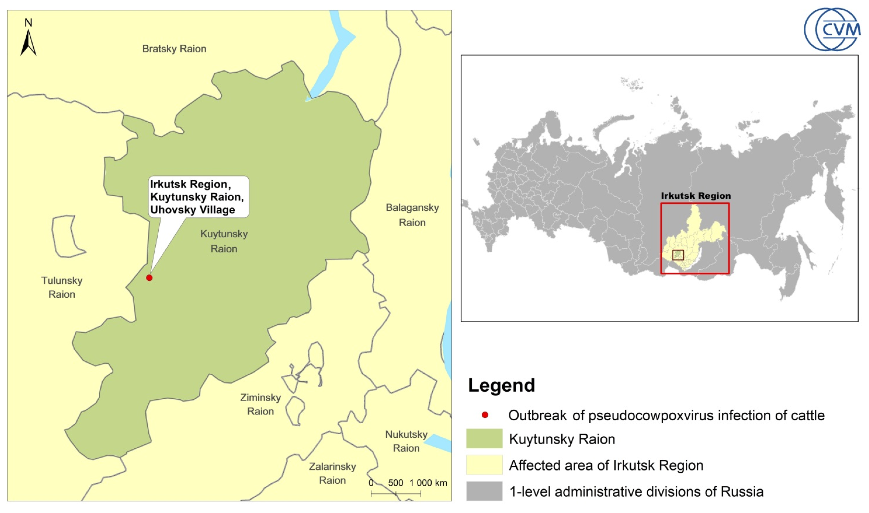
3.1. Epidemiology
3.2. Clinical Signs
3.3. Molecular Genetic Analysis
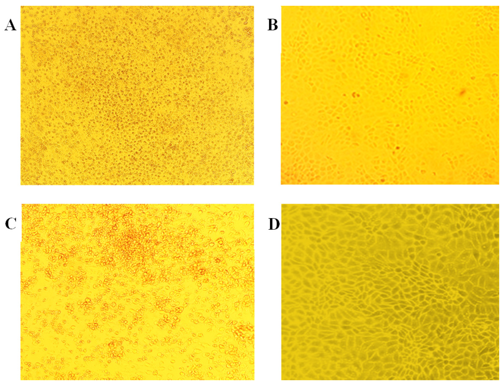
3.4. Virus Isolation
4. Discussion
5. Conclusions
Author Contributions
Funding
Institutional Review Board Statement
Informed Consent Statement
Data Availability Statement
Acknowledgments
Conflicts of Interest
References
- Friederichs, S.; Krebs, S.; Blum, H.; Wolf, E.; Lang, H.; Von Buttlar, H.; Büttner, M. Comparative and Retrospective Molecular Analysis of Parapoxvirus (PPV) Isolates. Virus Res. 2014, 181, 11–21. [Google Scholar] [CrossRef] [PubMed]
- de Oliveira Lopes, G.A.; Ferreira, L.R.; de Souza Trindade, G.; Fonseca, A.A.; dos Reis, J.K.P. qPCR Assay for the Detection of Pseudocowpox Virus. Arch. Virol. 2021, 166, 243–247. [Google Scholar] [CrossRef] [PubMed]
- Cargnelutti, J.F.; Flores, M.M.; Teixeira, F.R.M.M.; Weiblen, R.; Flores, E.F. An Outbreak of Pseudocowpox in Fattening Calves in Southern Brazil. J. Vet. Diagn. Investig. 2012, 24, 437–441. [Google Scholar] [CrossRef] [PubMed]
- Tikkanen, M.K.; McInnes, C.J.; Mercer, A.A.; Büttner, M.; Tuimala, J.; Hirvelä-Koski, V.; Neuvonen, E.; Huovilainen, A. Recent Isolates of Parapoxvirus of Finnish Reindeer (Rangifer tarandus Tarandus) Are Closely Related to Bovine Pseudocowpox Virus. J. Gen. Virol. 2004, 85, 1413–1418. [Google Scholar] [CrossRef] [PubMed]
- Vikøren, T.; Lillehaug, A.; Åkerstedt, J.; Bretten, T.; Haugum, M.; Tryland, M. A Severe Outbreak of Contagious Ecthyma (Orf) in a Free-Ranging Musk Ox (Ovibos moschatus) Population in Norway. Vet. Microbiol. 2008, 127, 10–20. [Google Scholar] [CrossRef]
- MacLachlan, N.J.; Dubovi, E.J. (Eds.) Chapter 7—Poxviridae. In Fenner’s Veterinary Virology, 5th ed.; Academic Press: Boston, FL, USA, 2017; pp. 157–174. ISBN 978-0-12-800946-8. [Google Scholar]
- Barker, I.K.; Van Dreumel, A.A.; Palmer, N. Chapter 1—The Alimentary System. In Pathology of Domestic Animals, 5th ed.; Jubb, K.V.F., Kennedy, P.C., Palmer, N., Eds.; Academic Press: San Diego, CA, USA, 1993; pp. 1–318. ISBN 978-0-12-391606-8. [Google Scholar]
- McKeever, D.J.; Jenkinson, D.M.; Hutchison, G.; Reid, H.W. Studies of the Pathogenesis of Orf Virus Infection in Sheep. J. Comp. Pathol. 1988, 99, 317–328. [Google Scholar] [CrossRef]
- Coetzer, J.A.W.; Tustin, R.C. Infectious Diseases of Livestock; Oxford University Press: Oxford, UK, 2004; ISBN 978-0-19-578202-8. [Google Scholar]
- CDC Human Orf Virus Infection from Household Exposures—United States, 2009–2011. Morb. Mortal. Wkly. Rep. 2012, 61, 245–248.
- Horner, G.W.; Robinson, A.J.; Hunter, R.; Cox, B.T.; Smith, R. Parapoxvirus Infections in New Zealand Farmed Red Deer (Cervus elaphus). N. Z. Vet. J. 1987, 35, 41–45. [Google Scholar] [CrossRef]
- Scagliarini, A.; Vaccari, F.; Turrini, F.; Bianchi, A.; Cordioli, P.; Lavazza, A. Parapoxvirus Infections of Red Deer, Italy. Emerg. Infect. Dis. 2011, 17, 684–687. [Google Scholar] [CrossRef] [PubMed]
- Moens, U.; Wold, I.; Mathiesen, S.D.; Jørgensen, T.; Sørensen, D.; Traavik, T. Parapoxvirus Papillomatosis in the Muskoxen (Ovibos moschatus): Genetical Differences between the Virus Causing New Outbreak in a Vaccinated Herd, the Vaccine Virus and a Local Orf Virus. Acta Vet. Scand. 1990, 31, 17–25. [Google Scholar] [CrossRef] [PubMed]
- Ali, O.A.; Kheir, S.A.; Abu Damir, H.; Barri, M.E. Camel (Camelus dromedarius) Contagious Ecthyma in the Sudan. A Case Report. Rev. Elev. Med. Vet. Pays Trop. 1991, 44, 143–145. [Google Scholar]
- Yeruham, I.; Nyska, A.; Abraham, A. Parapox Infection in a Gazelle Kid (Gazella gazella). J. Wildl. Dis. 1994, 30, 260–262. [Google Scholar] [CrossRef]
- Suzuki, Y.; Sugimura, M.; Atoji, Y.; Minamoto, N.; Kinjo, T. Widespread of Parapox Infection in Wild Japanese Serows, Capricornis Crispus. Nihon Juigaku Zasshi 1986, 48, 1279–1282. [Google Scholar] [CrossRef]
- Tompkins, D.M.; Sainsbury, A.W.; Nettleton, P.; Buxton, D.; Gurnell, J. Parapoxvirus Causes a Deleterious Disease in Red Squirrels Associated with UK Population Declines. Proc. Biol. Sci. 2002, 269, 529–533. [Google Scholar] [CrossRef] [PubMed]
- Müller, G.; Gröters, S.; Siebert, U.; Rosenberger, T.; Driver, J.; König, M.; Becher, P.; Hetzel, U.; Baumgärtner, W. Parapoxvirus Infection in Harbor Seals (Phoca vitulina) from the German North Sea. Vet. Pathol. 2003, 40, 445–454. [Google Scholar] [CrossRef]
- Rietschel, W. Parapox in a pigmy chimpanzee. Tierarztl. Prax. 1992, 20, 99–101. [Google Scholar]
- Gelaye, E.; Mach, L.; Kolodziejek, J.; Grabherr, R.; Loitsch, A.; Achenbach, J.E.; Nowotny, N.; Diallo, A.; Lamien, C.E. A Novel HRM Assay for the Simultaneous Detection and Differentiation of Eight Poxviruses of Medical and Veterinary Importance. Sci. Rep. 2017, 7, 42892. [Google Scholar] [CrossRef] [PubMed]
- Cargnelutti, J.F.; Weiblen, R.; Flores, E.F. A Multiplex PCR for Viruses Associated with Exanthematic and Vesicular Disease in Cattle. J. Virol. Methods 2017, 239, 38–41. [Google Scholar] [CrossRef] [PubMed]
- Hall, T.A. BioEdit: A User-Friendly Biological Sequence Alignment Editor and Analysis Program for Windows 95/98/NT. Nucleic Acids Symp. Ser. 1999, 41, 95–98. [Google Scholar]
- Kumar, S.; Stecher, G.; Li, M.; Knyaz, C.; Tamura, K. MEGA X: Molecular Evolutionary Genetics Analysis across Computing Platforms. Mol. Biol. Evol. 2018, 35, 1547–1549. [Google Scholar] [CrossRef]
- Inoshima, Y.; Morooka, A.; Sentsui, H. Detection and Diagnosis of Parapoxvirus by the Polymerase Chain Reaction. J. Virol. Methods 2000, 84, 201–208. [Google Scholar] [CrossRef]
- Ohtani, A.; Yokoyama, A.; Narushige, H.; Inoshima, Y. First Isolation and Genetic Characterization of Pseudocowpox Virus from Cattle in Japan. Virol. J. 2017, 14, 172. [Google Scholar] [CrossRef]
- Ziba, M.W.; Chitala, C.; Settypalli, T.B.K.; Mumba, M.; Cattoli, G.; Fandamu, P.; Lamien, C.E. First Detection and Molecular Characterisation of Pseudocowpox Virus in a Cattle Herd in Zambia. Virol. J. 2020, 17, 152. [Google Scholar] [CrossRef] [PubMed]
- Lederman, E.; Khan, S.U.; Luby, S.; Zhao, H.; Braden, Z.; Gao, J.X.; Karem, K.; Damon, I.; Reynolds, M.; Li, Y. Zoonotic Parapoxviruses Detected in Symptomatic Cattle in Bangladesh. BMC Res. Notes 2014, 7, 816. [Google Scholar] [CrossRef] [PubMed]
- Yaegashi, G.; Sasaki, I.; Chiba, S.; Murakami, K. Molecular Analysis of Parapoxvirus Detected in Eight Calves in Japan. J. Vet. Med. Sci. 2013, 75, 1399–1403. [Google Scholar] [CrossRef] [PubMed]
- Buettner, M.; Rziha, H.-J. Parapoxviruses: From the Lesion to the Viral Genome. J. Vet. Med. 2002, 49, 7–16. [Google Scholar] [CrossRef] [PubMed]
- Dixit, D.; Tang, J.W.; Grolla, A. Pseudocowpox Lesions in an Agricultural Student. CMAJ Can. Med. Assoc. J. 2023, 195, E305. [Google Scholar] [CrossRef]
- Bayindir, Y.; Bayraktar, M.; Karadag, N.; Ozcan, H.; Kayabas, U.; Otlu, B.; Durmaz, R.; Doganay, M. Investigation and Analysis of a Human Orf Outbreak among People Living on the Same Farm. New Microbiol. 2011, 34, 37–43. [Google Scholar] [PubMed]
- Lederman, E.R.; Austin, C.; Trevino, I.; Reynolds, M.G.; Swanson, H.; Cherry, B.; Ragsdale, J.; Dunn, J.; Meidl, S.; Zhao, H.; et al. Orf Virus Infection in Children: Clinical Characteristics, Transmission, Diagnostic Methods, and Future Therapeutics. Pediatr. Infect. Dis. J. 2007, 26, 740–744. [Google Scholar] [CrossRef]
- Uzel, M.; Sasmaz, S.; Bakaris, S.; Cetinus, E.; Bilgic, E.; Karaoguz, A.; Ozkul, A.; Arican, O. A Viral Infection of the Hand Commonly Seen after the Feast of Sacrifice: Human Orf (Orf of the Hand). Epidemiol. Infect. 2005, 133, 653–657. [Google Scholar] [CrossRef]
- Nougairede, A.; Fossati, C.; Salez, N.; Cohen-Bacrie, S.; Ninove, L.; Michel, F.; Aboukais, S.; Buttner, M.; Zandotti, C.; de Lamballerie, X.; et al. Sheep-to-Human Transmission of Orf Virus during Eid al-Adha Religious Practices, France. Emerg. Infect. Dis. 2013, 19, 102–105. [Google Scholar] [CrossRef] [PubMed]
- Shivanna, V.; Cino-Ozuna, A.G.; Heskett, C.; Marthaler, D.G.; Ganta, C. Pseudocowpox Virus Infection in an American Bison (Bison bison). BMC Vet. Res. 2020, 16, 241. [Google Scholar] [CrossRef] [PubMed]
- Fairley, R.A.; Mercer, A.A.; Copland, C.I.; Craig, S.M.; Heslip, P.A. Persistent Pseudocowpox Virus Infection of the Skin of a Foot in a Cat. N. Z. Vet. J. 2013, 61, 242–243. [Google Scholar] [CrossRef] [PubMed]
- Hautaniemi, M.; Ueda, N.; Tuimala, J.; Mercer, A.A.; Lahdenpera, J.; McInnes, C.J. The Genome of Pseudocowpoxvirus: Comparison of a Reindeer Isolate and a Reference Strain. J. Gen. Virol. 2010, 91, 1560–1576. [Google Scholar] [CrossRef]
- Oğuzoğlu, T.Ç.; Koç, B.T.; Kirdeci, A.; Tan, M.T. Evidence of Zoonotic Pseudocowpox Virus Infection from a Cattle in Turkey. Virusdisease 2014, 25, 381–384. [Google Scholar] [CrossRef]


Disclaimer/Publisher’s Note: The statements, opinions and data contained in all publications are solely those of the individual author(s) and contributor(s) and not of MDPI and/or the editor(s). MDPI and/or the editor(s) disclaim responsibility for any injury to people or property resulting from any ideas, methods, instructions or products referred to in the content. |
© 2024 by the authors. Licensee MDPI, Basel, Switzerland. This article is an open access article distributed under the terms and conditions of the Creative Commons Attribution (CC BY) license (https://creativecommons.org/licenses/by/4.0/).
Share and Cite
Sindryakova, I.; Blokhin, A.; Lyska, V.; Titov, I. Description of Zoonotic Pseudocowpoxvirus Infection of Cattle in Russia. Animals 2024, 14, 969. https://doi.org/10.3390/ani14060969
Sindryakova I, Blokhin A, Lyska V, Titov I. Description of Zoonotic Pseudocowpoxvirus Infection of Cattle in Russia. Animals. 2024; 14(6):969. https://doi.org/10.3390/ani14060969
Chicago/Turabian StyleSindryakova, Irina, Andrey Blokhin, Valentina Lyska, and Ilya Titov. 2024. "Description of Zoonotic Pseudocowpoxvirus Infection of Cattle in Russia" Animals 14, no. 6: 969. https://doi.org/10.3390/ani14060969