Article

Molecular Characterization, Expression Pattern, DNA Methylation and Gene Disruption of Figla in Blotched Snakehead (Channa maculata)

1
School of Life Sciences, Hunan University of Science and Technology, Xiangtan 411201, China
2
Key Laboratory of Tropical and Subtropical Fishery Resources Application and Cultivation, Ministry of Agriculture and Rural Affairs, Pearl River Fisheries Research Institute, Chinese Academy of Fishery Sciences, Guangzhou 510380, China
3
Chongqing Fisheries Technical Extension Center, Chongqing 404100, China
*
Authors to whom correspondence should be addressed.
These authors contributed equally to this work.
Animals 2024, 14(3), 491; https://doi.org/10.3390/ani14030491
Submission received: 6 January 2024 / Revised: 24 January 2024 / Accepted: 30 January 2024 / Published: 1 February 2024

Abstract

:

Simple Summary

There is a significant sexual dimorphism in the blotched snakehead (Channa maculata). However, studies on sex differentiation and gonadal development of C. maculata are limited. Factor in the germ line alpha (Figla) is an oocyte-specific basic helix–loop–helix (bHLH) transcription factor, involved in ovarian development. In the present research, the expression patterns and cellular location of Figla, as well as the CpG methylation of its promoter, demonstrated that Figla participated in ovarian differentiation and development in C. maculata. Meanwhile, the gene disruption of Figla was realized by the CRISPR/Cas9 system for further functional research.

Abstract

Figla is one of the earliest expressed genes in the oocyte during ovarian development. In this study, Figla was characterized in C. maculata, one of the main aquaculture species in China, and designated as CmFigla. The length of CmFigla cDNA was 1303 bp, encoding 197 amino acids that contained a conserved bHLH domain. CmFigla revealed a female-biased expression patterns in the gonads of adult fish, and CmFigla expression was far higher in ovaries than that in testes at all gonadal development stages, especially at 60~180 days post-fertilization (dpf). Furthermore, a noteworthy inverse relationship was observed between CmFigla expression and the methylation of its promoter in the adult gonads. Gonads at 90 dpf were used for in situ hybridization (ISH), and CmFigla transcripts were mainly concentrated in oogonia and the primary oocytes in ovaries, but undetectable in the testes. These results indicated that Figla would play vital roles in the ovarian development in C. maculata. Additionally, the frame-shift mutations of CmFigla were successfully constructed through the CRISPR/Cas9 system, which established a positive foundation for further investigation on the role of Figla in the ovarian development of C. maculata. Our study provides valuable clues for exploring the regulatory mechanism of Figla in the fish ovarian development and maintenance, which would be useful for the sex control and reproduction of fish in aquaculture.

1. Introduction

The bHLH superfamily contains numerous transcription factors that are essential for gene regulation in most eukaryotes [1], such as sex determination [2], cell proliferation, and differentiation [3]. Figla is an oocyte-specific bHLH transcription factor, which has been reported to play crucial roles in oocyte growth and development [4]. In mice (Mus musculus), Figla transcripts can be first detected at embryonic stage 13.5, which is restricted to oocytes [5]. Figla can promote the formation of primordial follicles and activate the expression of oocyte-related genes. After knockout of Figla in M. musculus, oocyte apoptosis was observed and all primordial follicles were lost right after birth [5], and genes related to germ cell differentiation and oogenesis were significantly reduced, such as Lhx8, Sohlh1, and Gdf9 [6]. Additionally, Figla was thought to be one of the genes that caused premature ovarian failure in women [7].
Figla was also identified in several teleosts, and displayed the sexually dimorphic expression pattern in the gonads, which was dominantly expressed in the ovaries. In Nile tilapia (Oreochromis niloticus), Figla mRNA was remarkably expressed during ovarian development, especially in the early primary oocytes, while no specific signals could be detected in testes [8]. In Japanese flounder (Paralichthys olivaceus), Figla showed a higher expression in ovaries than in testes, and Figla was specifically expressed in germ cells, in which Figla signals were largely detected in oocytes and weakly in spermatogonia and primary spermatocytes [9]. Figla expression was also changed during sex transformation. In the protogynous wrasse (Halichoeres poecilopterus), Figla expression levels dwindled during the female-to-male sex change, accompanied by oocytes degeneration [10]. In protandrous black porgy (Acanthopagrus schlegelii), Figla was even presumed to induce the transformation of Sertoli cells into follicle-like cells during the male-to-female sex change [11]. Similar to the study in M. musculus, the gonads were masculinized after the disruption of Figla in zebrafish (Danio rerio), resulting in an all-male phenotype in the mutants [12]. Furthermore, the over-expression of Figla in XY O. niloticus led to defective spermatogenesis, accompanied by the attrition of meiotic spermatocytes as well as spermatids [8]. These results indicated that Figla also played a key role in the ovarian differentiation and development of fish.
Epigenetic regulation is thought to play an important role in gonadal differentiation and development, in which DNA methylation has been considered as a vital actor [13]. Studies have indicated that the relationship between Figla expression and the methylation of its promoter plays a vital role in gonadal development. In half-smooth tongue sole (Cynoglossus semilaevis), Figla was less expressed and its promoter was highly methylated in male individuals, whereas in the pseudomales and females, the expression levels of Figla were significantly higher than that in males, and the methylation status was lower than that in males [13]. In channel catfish (Ictalurus punctatus), the expression of Figla was low, with high methylation levels in males; however, it became progressively demethylated after 17β-oestradiol exposure. Meanwhile, Figla expression was also gradually increased [14]. Therefore, it is necessary to investigate whether DNA methylation affects gonadal development by regulating the expression of Figla in fish.
Blotched snakehead (Channa maculata) is well received by consumers for its delicious flavor, high protein content, few intramuscular spines, and medicinal properties. Males grow much faster than females; therefore, an all-male population is greatly beneficial for aquaculture. In our previous study, XY sex-reversal females (XY-F) and YY super-males (YY-M) were produced by the combination of 50 mg/kg 17β-estradiol induction and a sex-specific molecular marker [15]. However, the mechanisms of sex differentiation and gonadal development in C. maculata was unclear. The gonadal histology and transcriptome analysis of 6-month-old C. maculata indicated that the ovarian development in C. maculata was asynchronous, where oocytes of all stages of development were present. In addition, Figla was shown to be a female-biased gene in C. maculata, whose expression was dominantly higher in the ovaries of normal XX females (XX-F) and XY-F than in the testes of normal XY males (XY-M) and YY-M [16]. Therefore, we would like to know if Figla also plays a role in the ovarian development in C. maculata, similar to studies in other fish. In this study, the Figla gene from C. maculata was cloned and analyzed, and the expression patterns of Figla were expounded in different adult tissues and gonads at different developmental stages. Furthermore, the CpG methylation levels of Figla promoter were also illuminated in gonads. In order to further deepen the functional research of Figla in the future, the CRISPR/Cas9 system was used to construct Figla mosaic F0 generation. This study explores the role of Figla in the gonadal differentiation and development, and is expected to provide a reference for the study of the sex differentiation and gonadal development mechanism of blotched snakehead.

2. Materials and Methods

2.1. Experimental Fish and Sampling

Experimental fish were reared in the fish laboratory of the Model Animal Research Center, Pearl River Fisheries Research Institute (Guangzhou City, Guangdong Province, China). Adult XY-M, XX-F, XY-F, and YY-M individuals (n = 3) were determined by sex-specific molecular marker and gonadal histology [15]. After anesthesia with MS222 anesthetic (Sigma-Aldrich, St. Louis, MO, USA), at a dose of 1.25 g/mL, the multitudinous tissues, such as liver (L), gills (G), middle kidney (MK), spleen (S), head kidney (HK), intestines (I), muscle (M), heart (H), gonads (ovaries or testes (O/T)), brain (B), hypothalamus (Hy), and pituitary (P) were collected from one-year-old XX-F, XY-F, XY-M, and YY-M individuals (n = 3), respectively. These tissue samples were used for gene cloning, expression, and DNA methylation analysis.
The gonads from XX-F and XY-M individuals (n = 3) at different developmental stages (30, 60, 90, 120, 150, 180, and 365 dpf) were obtained for measuring the expression levels of Figla during the developmental stages of gonadal differentiation, and the genetic sex was identified as mentioned above. All samples were quickly frozen in liquid nitrogen and stored at −80 °C. Additionally, the gonads from 90 dpf XX-F and XY-M individuals were fixed in Bouin’s solution (ABI, Foster City, CA, USA) for ISH. All fish operations complied with the regulations of the National Institutes of Health guide for the care and use of laboratory animals.

2.2. RNA Extraction and cDNA Synthesis

Total RNAs of the above samples were isolated by TRIzol (Invitrogen, Carlsbad, CA, USA). The RNA quality and quantity were determined by spectrophotometer (Thermo Fisher, Waltham, MA, USA). Then, 1 μg RNA was electrophoresed with a 1.0% agarose gel to analyze the RNA integrity. The remaining RNA sample was frozen at −80 °C. The 5′-RACE-Ready and 3′-RACE-Ready cDNA were obtained by SMART RACE cDNA Amplification Kit (TaKaRa, Otsu, Japan). cDNA was synthesized by the ReverTra Ace qPCR RT Kit (Toyobo, Osaka, Japan) [17]. All operations were performed according to the manufacturer’s instructions.

2.3. Full-Length cDNA Cloning and Sequence Analysis

The predicted cDNA sequence of Figla was obtained from the genome of blotched snakehead (SRA Accession No. PRJNA730430). Figla-F1/R1 primer pair was designed by Primer 5.0 to identify the predicted cDNA sequence of Figla. Then, 5′-RACE and 3′-RACE primers were designed for PCR amplification with 5′-RACE-Ready and 3′-RACE-Ready cDNA as templates to amplify 5′ and 3′untranslated regions (UTRs), respectively. Thereafter, the full-length cDNA sequence of Figla was amplified by Figla-F2/R2 primer pair with 5′ and 3′UTRs. The primers are showed in Table 1. PCR product containing the target fragment was purified by Gel Extraction Kit (Omega Bio-Tek, Norcross, GA, USA), purified product was ligated into pMD19-T vectors (TaKaRa, Japan), and the ligated product was transformed into Escherichia coli DH5α competent cells (TransGen Biotech, Beijing, China). Positive colonies were selected and sequenced by a commercial company (Tianyi huiyuan, Guangzhou, China) [17].
Homologous Figla protein sequences from other species were gained from NCBI (https://www.ncbi.nlm.nih.gov/, accessed on 23 October 2023), including C. argus, C. striata, Lates calcarifer, L. japonicus, C. semilaevis, Oryzias latipes, D. rerio, Xenopus tropicalis, Microcaecilia unicolor, Pelodiscus sinensis, Gopherus evgoodei, Gallus gallus, Strigops habroptila, Bos taurus, M. musculus, and Homo sapiens. The deduced amino acid sequences of Figla in C. maculata and its homologous counterparts from other species were aligned using DNAMAN. Then, the neighbor-joining method was used to construct the phylogenetic tree of Figla proteins from different species using MEGA 11.0, and bootstrap values were calculated with 1000 replications.

2.4. Genome DNA Sequence Cloning and Structure Analysis

Genomic DNA was extracted from the tail using the Universal Genomic DNA Kit (CWBIO, Beijing, China). The predicted genome DNA sequence of Figla was obtained from the blotched snakehead genome (SRA Accession No. PRJNA730430). Specific primer pairs were designed to verify the predicted genome DNA sequence of Figla (Table 2). PCR products were recovered, ligated, transformed, and sequenced as mentioned above. The genomic structure of Figla was determined by comparing the genome DNA sequence with the cDNA sequence in conformity with the GT/AG principle. Softberry (http://www.softberry.com/berry.phtml, accessed on 24 October 2023) was applied to predict the transcription start site (TSS). Animal TFDB (http://bioinfo.life.hust.edu.cn/AnimalTFDB/, accessed on 24 October 2023) was used to predict the potential transcription factor binding sites (TFBS) [17].

2.5. Quantitative Real-Time PCR (qRT-PCR)

The primers for qRT-PCR are revealed in Table 1. The housekeeping genes β-actin and EF1α were utilized as internal controls for cDNA normalization [17]. qRT-PCR was implemented on the StepOnePlus™ Real-Time PCR System (ABI, USA) by SYBR® Green Master Mix (Toyobo, Japan) according to the manufacturer´s instruction. The reaction system was (20 μL): 10 μL SYBR® Green Master Mix, 0.8 μL for both forward and reverse primers, 7.4 μL ddH2O, and 1 μL cDNA. The qRT-PCR program was as follows: 95 °C for 2 min, 39 cycles of 95 °C for 15 s, 50 °C for 15 s, and 72 °C for 30 s. The default melting curve acquisition procedure of the instrument was used, 60 °C for 5 s and 95 °C for 5 s. qRT-PCR was performed for each sample in triplicates. Expression levels of Figla gene in different tissues and gonads at different developmental stage were calculated using the 2−△△Ct method. For tissue expression, the expression level of the pituitary (P) in XX-F individuals was used as the baseline (1.0), and the expression levels of the different tissues in each type of fish were relative to that of the baseline. For gonad expression at different developmental stages, the expression level of the gonad in 30 dpf XX-F individuals was used as the baseline (1.0), and the expression levels of the gonads at different developmental stages were relative to that of the baseline.

2.6. In Situ Hybridisation (ISH)

The gonads of 90 dpf XX-F and XY-M were used for ISH analysis. Probe synthesis was performed as described by Feng et al. (2011) with minor modification [18]. The primers used for the amplification are displayed in Table 1. DIG RNA Labeling Mix (Roche, Mannheim, Germany) was employed for probe labeling, according to the manufacturer’s instructions. Additionally, the steps of ISH were conducted according to Gao et al. (2013) with slight emendation [19]. Briefly, gonad samples were dehydrated in a series gradient of ethanol and xylene before embedding in paraffin wax. The sections (7 μm thick) were fixed on amino slide and dried on a dryer (Kedee, Jinhua, Zhejiang, China) overnight at 40 °C. The next day, after the sections were dewaxed, each sample section was fixed with 4% paraformaldehyde (PFA) for 15 min and incubated with 10 μg/mL protease K at 37 °C for 10–15 min. The hybridization was carried out at 65 °C for 19 h (3 h for the pre-hybridization process and 16 h for the hybridization process). After hybridization, all sample sections were washed twice in mixed solution (5 mL 20 × SSC + 25 mL formamide deionized + 50 μL tween + distilled water, total 50 mL) at 65 °C for 30 min, 2 × SSC for 15 min, 0.2 × SSC washed twice for 30 min each time, and 1 × PBST three times for 5 min each time at room temperature. After samples were blocked in 2% blocking reagent for 3 h, they were incubated with blocking buffer mixed with anti-Digoxigenin-AP Fab fragments (Roche, Mannheim, Germany) for 16 h at 4 °C. Subsequently, all sample sections were washed six times in 1 × PBST for 10 min. After 5 min of NTMT equilibrium, they were then developed with NTMT mixed with DIG Nuleic Acid Delection Kit (Roche, Mannheim, Germany) for 3–5 h at room temperature. When the ideal chroma was reached, the sections were washed three times in 1 × PBST for 10 min each time to terminate the dyeing. The sections were covered with 50% glycerin, observed, and photographed using Nikon Eclipse Ti-U microscope (Nikon, Tokyo, Japan).

2.7. Bisulfite Sequencing PCR (BSP)

Gonads from the adult XX-F, XY-F, XY-M, and YY-M individuals (n = 3) were used for genomic DNA extraction and BSP analysis. BSP analysis was conducted as previously described [17]. In brief, genomic DNA from the gonads of each type of fish was extracted by the Universal Genomic DNA Kit (CWBIO, China) and mixed separately, and the EZ DNA Methylation-Gold Kit (Zymo Research, Irvine, CA, USA) was used to obtain the methylated DNA according to the manufacturer’s instructions. The DNA methylated CpG islands of Figla were predicted by MethPrimer 2.0 online software (http://www.urogene.org/cgi-bin/methprimer/methprimer.cgi, accessed on 25 October 2023), and the suitable CpG islands regions were selected as target fragments. Then, Figla-BSP-F/R primer pair was designed for PCR amplification (Table 1). The amplified DNA fragments were cloned into pMD19-T vectors and transformed into E. coli DH5α competent cells, and positive clones of each type of fish (n = 15) were randomly chosen and sequenced. CpG methylation percentage was calculated as (number of methylated sites)/(total number of CpG sites).

2.8. Knockout of Figla Gene in C. maculata

CRISPR/Cas9 system was used to knockout Figla in C. maculata. The target sites were designed using the CRISPRscan online tool (https://www.crisprscan.org/, accessed on 25 October 2023), and two guide RNAs (gRNAs) were designed to target Figla gene (Table 3). These gRNAs were synthesized using MAXIscript T7 In Vitro Transcription Kit (Invitrogen USA). Subsequently, gRNAs were purified by LiCl/ethanol precipitation method. gRNAs were severally mixed with TrueCut Cas9 Protein v2 (Invitrogen, USA) before microinjection, then 200 pg gRNA and 300 pg Cas9 protein was co-injected into 1–4 cell stage embryos of C. maculata. Five microinjected fry were collected after 48 h post-fertilization (hpf) and used for genomic DNA extraction. The primer pair Figla-JC-F/R was used for mutation detection (Table 1). The artificial insemination, microinjection, mutagenesis analysis, and fish rearing were based on our previous studies [20].

2.9. Statistical Analysis

The experimental data was exhibited as mean ± standard deviation (SD). All results were subjected to one-way ANOVA, followed by Dunnett’s test using SPSS Statistics 20.0. p < 0.05 was considered to be statistically significant. The correlation coefficient of CpG methylation percentage of Figla promoter and the mRNA expression of Figla was calculated using Excel 2021.

3. Results

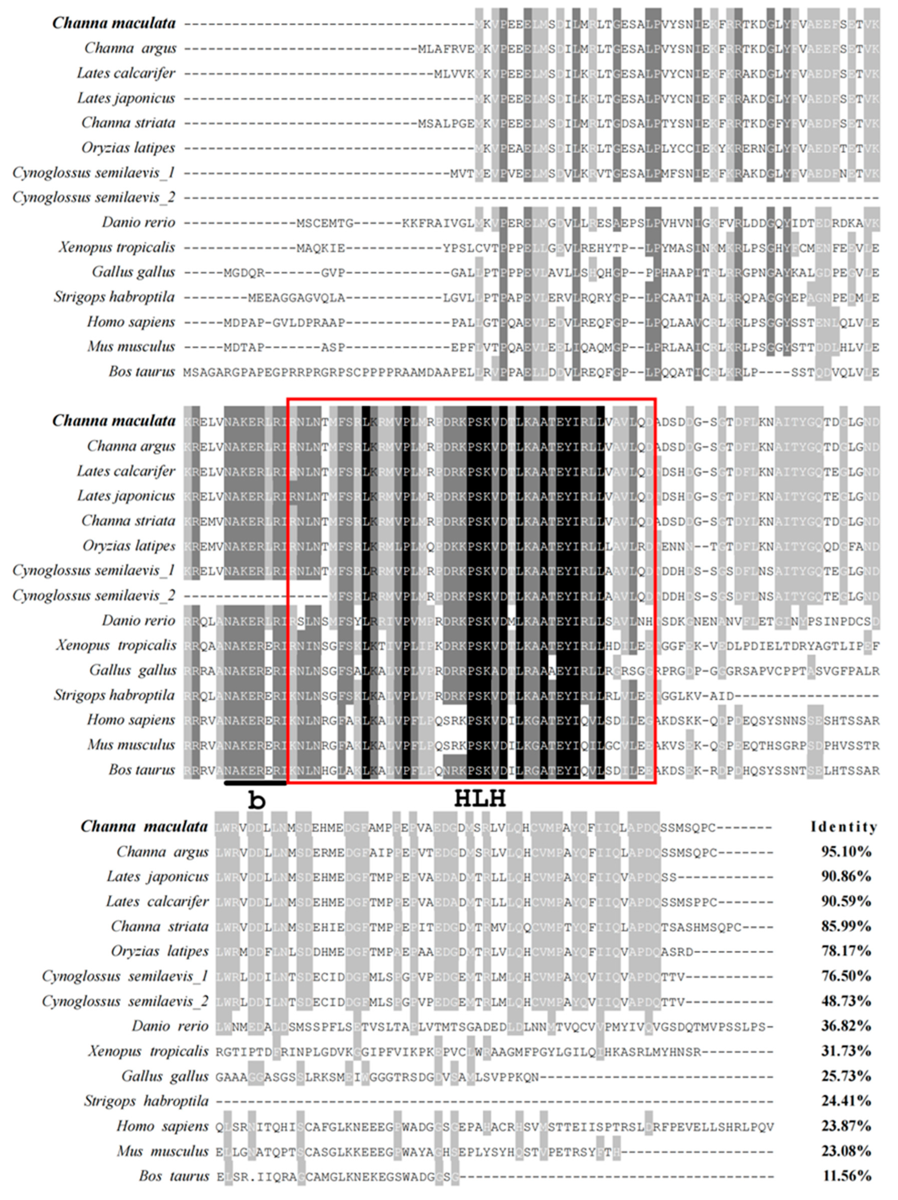
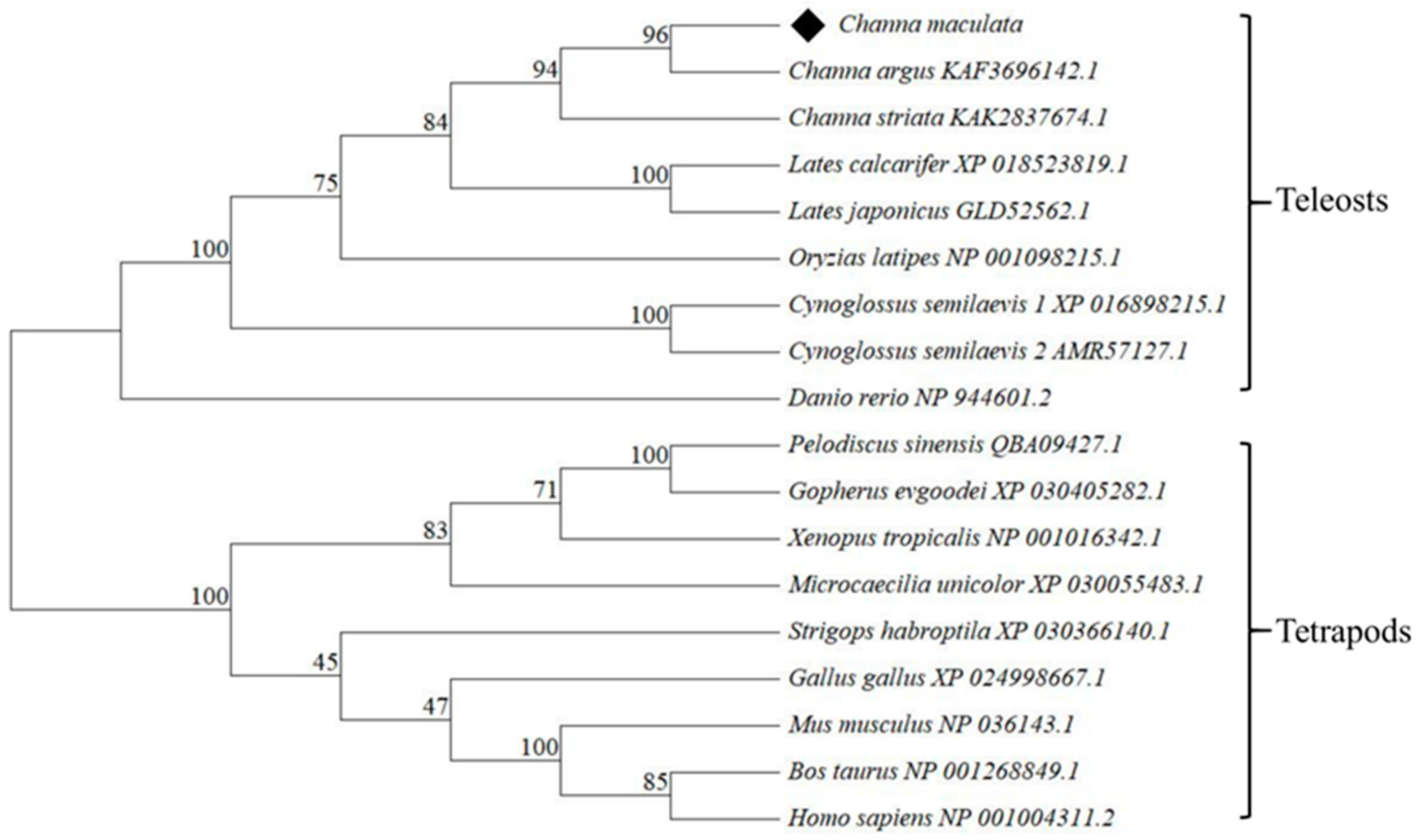
3.1. Sequence Analysis of Figla in C. maculata

One Figla transcript was identified in C. maculata, named as CmFigla. The cDNA sequence of CmFigla was 1303 bp in long (GenBank accession no. PP067023), which included 431 bp 5′-UTR, 594 bp opening reading frame (ORF), and 278 bp 3′-UTR (Figure S1a). CmFigla encoded a putative protein of 197 amino acids (aa), and a highly conserved bHLH domain was predicted in 56-108 aa (Figure S1a).
The multiple sequence alignments were conducted by DNAMAN, and the result revealed that Figla protein in vertebrates contained a conservative bHLH domain (Figure 1). CmFigla had high similarities to Figla in Perciformes, which was 95.10% identical to C. argus, 90.86% to L. japonicus, 90.59% to L. calcarifer, and 85.99% to C. striata. Nevertheless, the similarities between CmFigla and MmFigla (Figla in M. musculus) and BtFigla (Figla in B. taurus) were very low, only 23.08% and 11.56%, respectively. The phylogenetic tree showed Figla in C. maculata first clustered with Figla from the same genus species, C. argus and C. striata, then clustered with Figla from the same order, L. calcarifer and L. japonicus. Thereafter, they clustered with other teleosts, for example O. latipes, C. semilaevis, and D. rerio. Figla proteins from tetrapods clustered another clade, which was distant from the teleosts group, including X. tropicalis, M. unicolor, P. sinensis, G. evgoodei, G. gallus, S. habroptila, B. taurus, M. musculus, and H. sapiens (Figure 2).
By amplification, splicing, and alignment, the genome DNA sequence of CmFigla was verified with 8033 bp in long, which contained 929 bp 5′-flanking region, 6090 bp coding region and 1014 bp 3′-flanking region. The coding region covered five exons and four introns (Figure 3). A great deal of putative TFBS were predicted in CmFigla, including Estrogen Receptor 2 (Esr2), Sp1 Transcription Factor (Sp1), Sex-Determining Region Y Protein (SRY), and SRY-Box Transcription Factor 3 (SOX3), which were essential for the regulation of gene promoter activities. The CpG island of the CmFigla promoter was also predicted to be present (−445~+314) (Figure S1b).

3.2. Expression Patterns of CmFigla

The expression levels of CmFigla in different tissues of XY-M, XX-F, XY-F, and YY-M individuals were detected by qRT-PCR. CmFigla transcripts were specifically expressed in gonads, especially in ovaries (O), whose expression was 215.33 ± 8.69-fold changes in the ovaries of XX-F, and 108.30 ± 4.21-fold changes in the ovaries of XY-F. In contrast, CmFigla mRNA was extremely low in the testes, with only 2.13 ± 0.14-fold changes in the testes of XY-M and 1.62 ± 0.12-fold changes in the testes of YY-M. Interestingly, there was also a small amount of CmFigla mRNA in the hypothalamus (Hy) (Figure 4a).
Figure 4b showed that the mRNA expression of CmFigla in the gonads existed an obvious sexual dimorphism, which was far higher in female ovaries than that in male testes at all developmental stages. In the ovaries, CmFigla transcripts sharply increased from 30 dpf (1.00 ± 0.17-fold changes), and peaked at 180 dpf (158.14 ± 11.43-fold changes), then decreased at 365 dpf (23.54 ± 1.18-fold changes). However, CmFigla always maintained very low expression levels at different testicular developmental stages, whose values ranged from 0.20 ± 0.03-fold changes (30 dpf) to 9.28 ± 0.03-fold changes (120 dpf).

3.3. CpG Methylation Levels of CmFigla Promoter

Differential CpG methylation levels of CmFigla promoter in the gonads from four types of fish were evaluated by BSP, and twenty-two CpG methylation sites were identified (Figure 5). The DNA methylation levels of CmFigla promoter were much lower in the ovaries of XX-F and XY-F individuals compared to those in the testes of XY-M and YY-M individuals. In the ovaries of XX-F and XY-F, the CpG methylation levels of CmFigla promoter were 20.6 ± 5.1% and 32.1 ± 6.5%, respectively. The CpG sites of CmFigla promoter maintained the hypermethylated status in the testes of XY-M and YY-M, with 86.1 ± 5.3% in XY-M and 89.7 ± 3.2% in YY-M. Meanwhile, most CpG sites of CmFigla promoter were not methylated at all in the ovaries of XX-F and XY-F. In XX-F ovaries, only locus 16 was fully methylated, while seven loci (11, 12, 14, 15, 17, 18, and 22) were completely unmethylated (Figure 5a). In XY-F ovaries, three loci (13, 15, and 16) had 100% methylation status, while eleven loci (1, 2, 5, 6, 7, 8, 9, 11, 12, 14, and 17) had 100% non-methylation status (Figure 5b). Conversely, the hypermethylation of CmFigla promoter resided in testes. Eight loci (3, 10, 12, 13, 14, 15, 16, and 21) in XY-M testes (Figure 5c) and fourteen loci (1, 2, 3, 4, 5, 11, 13, 14, 15, 16, 18, 19, and 21) in YY-F testes (Figure 5d) were wholly methylated, and no loci were not methylated.
Meanwhile, the correlation analysis indicated that the CpG methylation level of the CmFigla promoter was violently negatively correlated to CmFigla expression in the gonads of C. maculata, and the correlation coefficient (R2) was 0.9053 (Figure S2).

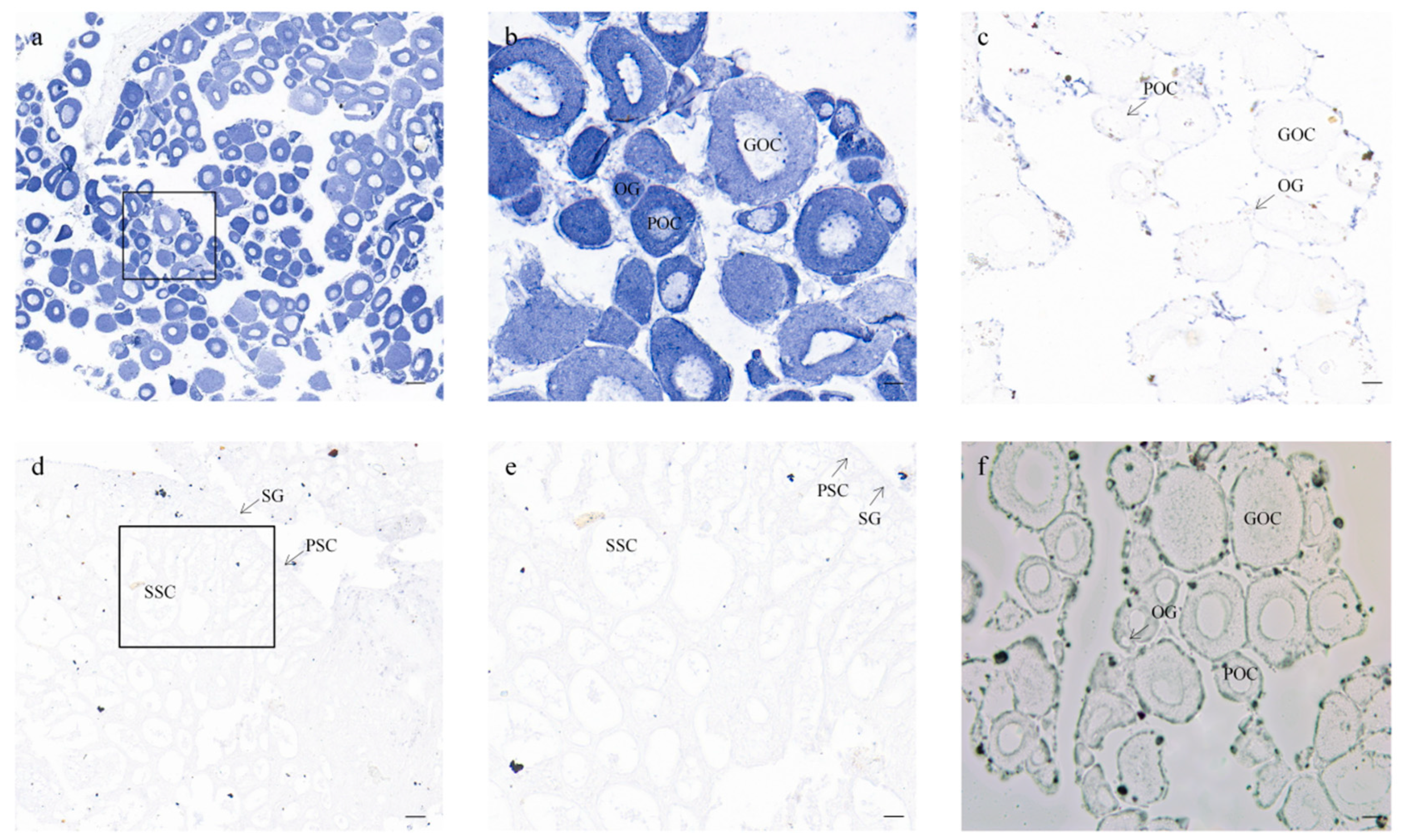
3.4. Cellular Location of CmFigla in Gonads

To confirm the cellular localization of the CmFigla transcripts, ISH analysis was performed in the gonads. In the ovaries of 90 dpf XX-F individuals, CmFigla signals were dominantly expressed in oocytes, and the intensity of the positive signals in oogonia and primary oocytes seemed to be stronger than that in growing oocytes (Figure 6a,b). There were no obvious signals observed in the germ cells of the XY-M testes (Figure 6d,e), in accordance with the results of qRT-PCR (Figure 4b). There were no signals to be detected in the negative controls with sense probe hybridization (Figure 6c).

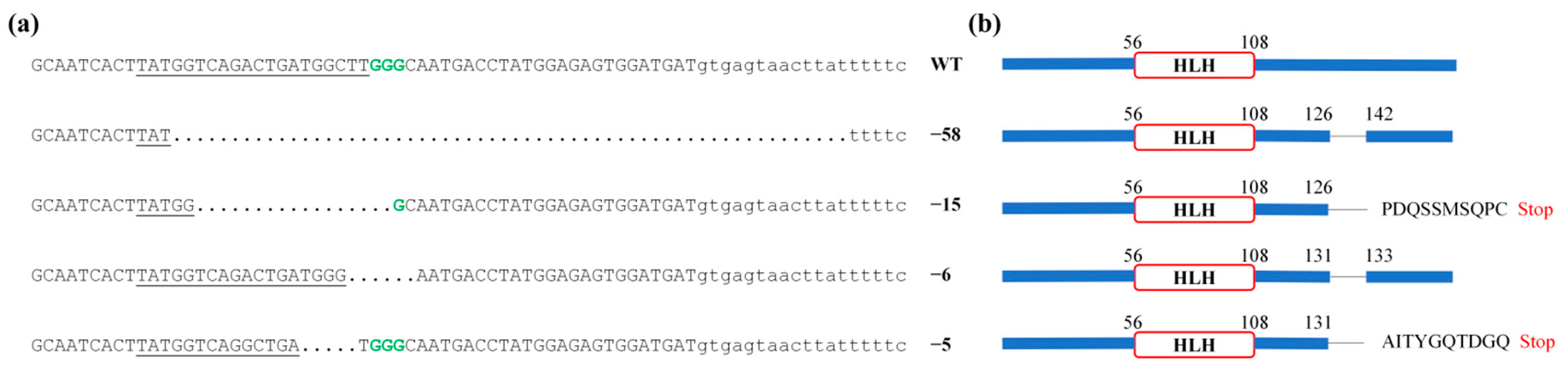
3.5. Generation of the CmFigla Mutantion Using the CRISPR/Cas9 System

To induce CmFigla disruption, two CRISPR/Cas9 target sites were designed within the mature mRNA sequence of CmFigla located in Exon 2 and Exon 3 (Figure 3). The mutation analysis of the target sites in CRISPR/Cas9 system were performed by PCR amplification, sub-cloning, and sequencing in 48 hpf fry. By aligning mutated DNA sequences with wild-type sequences, it was found that gRNA1 located in Exon 2 did not cause mutation, but the gRNA2 located in Exon3 generated four forms of deletion: −58, −17, −6, and −5 bp (Figure 7a), and the mutagenesis frequency was 45.0% (9 mutated clones/20 total sequenced clones). Missing 6 bp and 58 bp resulted in the deletion of two and fifteen amino acids, respectively. Meanwhile, the other two forms of deletion (−5, −17 bp) led to the premature termination of transcription due to frame-shift mutations (Figure 7b). These results demonstrated that Figla-gRNA had the ability to edit the Figla gene in C. maculata, and mutation types and frequency indicated that most forms of deletion brought about frame shifts, leading to the disruption of gene structure.

4. Discussion

As an oocyte-specific transcription factor, Figla plays an essential role in the formation of primordial follicles [6], the transition from primordial to primary follicle [4], and gonadal development and differentiation [21]. In the present study, one homology Figla containing the conservative bHLH domain was illustrated in C. maculata, and named CmFigla. In most fish, such as P. olivaceus [22] or turbot (Scophthalmus maximus) [23], only one Figla transcript exists. However, two transcriptional variants (Figla_tv1 and Figla_tv2) had been identified in C. semilaevis [24], and the difference between the two transcriptional variants was that Figla_tv2 lacked the bHLH region. In order to know whether multiple transcripts existed in other fish, Figla sequences in most teleosts were obtained from NCBI (https://www.ncbi.nlm.nih.gov/, accessed on 23 December 2023). It was found that there was more than one transcript in some fish, such as two forms in climbing bass (Anabas testudineus) (GenBank accession no. XM_026343723.1, XM_026343724.1), four forms in I. punctatus (GenBank accession no. XM_047149866.2, XM_017452432.3, XM_047149867.2, XM_047149868.2), three forms in Siamese fighting fish (Betta splendens) (GenBank accession no. XM_055511459.1, XM_029164147.3, XM_029164148.3), and so on. Similar to C. semilaevis, there were two different Figla protein in large yellow croaker (Larimichthys crocea) (GenBank accession no. XP_019121559.1, XP_027132394.1), river trout (Salmo trutta) (GenBank accession no. XP_029617814.1, XP_029617813.1), lake whitefish (Coregonus clupeaformis) (GenBank accession no. XP_041691711.1, XP_041691710.1), flier cichlid (Archocentrus centrarchus) (GenBank accession no. XP_030593618.1, XP_030593617.1, XP_030593616.1), sockeye salmon (Oncorhynchus nerka) (GenBank accession no. XP_029534804.1, XP_029534805.1), and Atlantic salmon (Salmo salar) (GenBank accession no. XP_014017212.1, XP_045560154.1); one Figla protein was common form, and the other lacked the conserved bHLH region. Perhaps different Figla transcripts were discovered due to the depth or coverage of sequencing, or the teleost-specific third round of whole-genome duplication [25]. Of course, more studies are needed to verify the possibility in the future. The protein sequence alignments indicated that the similarities of Figla between C. maculata and other fish species were high, and those between teleosts and tetrapods were low; similarly, the phylogenetic relationship of Figla proteins was in accordance with the evolutionary distance among the organisms.
Figla has shown an obvious sexual dimorphic expression pattern in the gonads of most teleosts. In O. niloticus, Figla expression was absolutely dominant in the early primary oocytes in ovaries, but almost undetectable in testes [8]. In S. salar, Figla showed significantly higher expression in the ovaries compared to the testes by qRT-PCR, and ISH indicated that Figla specific signals were only present within the oocytes [26]. Extensive transcriptome analysis of gonads also confirmed that Figla was female-biased, such as spotted scat (Scatophagus argus) [27] and tiger grouper (Epinephelus fuscoguttatus) [28]. Consistently, CmFigla expression in the ovaries was significantly higher than that in the testes, the intensity of the positive signals of CmFigla in oogonia and primary oocytes seemed to be stronger than that in growing oocytes, and negligible expression could be detected in the testes (Figure 4a and Figure 6). Furthermore, CmFigla expression in XY individuals could have been changed after 17β-estradiol induction; CmFigla showed high expression levels in the gonads of XY-F that was similar to that in the gonads of XX-F, which was different from the gonads of XY-M. Similar results have been observed in Japanese eel (Anguilla japonica) [29], A. schlegelii [11], and I. punctatus [14], in which Figla expression was up-regulated during ovarian differentiation after estrogen induction. These results revealed the conservative role of Figla in the ovarian development of C. maculata. Two transcripts (Figla_tv1 and Figla_tv2) were identified in C. semilaevis; Figla_tv1 was mainly expressed in the ovaries, skin and liver, but Figla_tv2 was detected in all examined tissues, especially in male muscle [24]. In addition, there was considerable expression in the female brain of P. olivaceus [22], indicating that Figla may also play a role in other tissues. In our study, CmFigla was also detected in the male hypothalamus (Hy), indicating a role for CmFigla in hormone production through the hypothalamic–pituitary–gonadal (HPG) axis, which requires more study.
Many studies illustrated that Figla was essential during ovarian differentiation and development. In O. niloticus, Figla expression was significantly up-regulated within 30 days after hatching (dah) ovaries, then peaked at 90 dah and descended from 120 dah [8]. In C. semilaevis, Figla_tv1 mRNA was first examined at 120 dah in ovaries and reached the maximum at 1 year after hatching (yah) before sharply declining at 2 yah [24]. In P. olivaceus, Figla expression in ovaries ascended from 8-month-old stage to 1-year-old and 1.5-year-old stage [9]. In sablefish (Anoplopoma fimbria), Figla expression was significantly increased when XX females reach 120 mm, which coincided with the emergence of CN oocytes, and with the development of PN oocytes [30]. Similarly, the expression of CmFigla could be detected at 30 dpf ovaries (the onset of ovarian differentiation), then gradually raised from 60 dpf and reached to the peak at 180 dpf (the critical stage of ovarian development). Subsequently, CmFigla expression descended to a similar level of 60 dpf at 365 dpf (ovulation stage) (Figure 4b). These finding indicated that Figla is closely related to differentiation and development of ovary in C. maculata.
DNA methylation has been considered as a vital actor in sex determination and gonadal development, which also regulates the expression of numerous genes [31]. The expression of Figla was low in male testes of I. punctatus with high methylation levels of Figla promoter; however, it became progressively demethylated after E2 exposure [14]. In C. semilaevis, the expression levels of Figla were significantly higher in pseudomales and females than that in males, and the methylation status of Figla promoter was lower in pseudomales and females than that in males [13]. Analogous findings were observed in our study, wherein it was found that the CpG methylation levels of CmFigla promoter in XY-M and YY-M testes were high, whereas XY-F and XX-F ovaries showed abundant CmFigla expression, and the CpG methylation levels of CmFigla promoter were significantly reduced compared to those in the testes (Figure 5). There was also a violently negative correlation between the CpG methylation level of CmFigla promoter and CmFigla expression in the gonads (Figure S2); similar results have been reported in M. musculus [32] and D. rerio [33]. It was suspected that DNA methylation might regulate the expression of Figla and participate in ovarian development and maintenance in C. maculata.
The deletion of the Figla gene in vertebrates usually leads to ovarian dysfunction. In O.latipes, two TALENs targets led to premature truncations and the bHLH region was destroyed, thus XX Figla−/− mutants did not form follicles and the expression of female-specific genes (Gdf9 and Bmp15) was reduced [34]. However, XX Figlα−/− medaka obtained by CRISPR/Cas9 developed ovarian lamellae and ovarian cavities, but the Figla deficient oocytes were blocked at or around pachytene [35]. The destruction of Figla in D. rerio blocked the transition from cystic CN oocytes to individual follicular perinucleolar oocytes, resulting in an all-male phenotype in the homozygous mutant [12]. In this study, we introduced CRISPR/Cas9 system to construct CmFigla mutants. The designed gRNA effectively worked at the target sites, and four forms of deletion were obtained. The majority of deletions were frame-shift mutations that generated a premature termination codon, underwent early termination in translation, and disrupted the molecular functions of the CmFigla protein. Successful knockout of CmFigla established a research basis for further investigating the role of CmFigla in the ovarian development of C. maculata. Due to the young age of the current gene knockout fish, two consecutive generations were required to obtain CmFigla−/− homozygous mutants; thus, there were no results on whether the deletion of Figla will affect the gonadal development and oogenesis related genes expression in C. maculata, and we will continue to conduct the relevant studies in the future.

5. Conclusions

In conclusion, we characterized and analyzed the CmFigla gene in this study. CmFigla has a conserved bHLH domain, and its sequence characteristics and gene expression patterns are similar to those reported in vertebrates. CmFigla revealed consumingly female-biased expression patterns in the gonads; furthermore, a noteworthy inverse relationship was observed between CmFigla expression and the CpG methylation status of its promoter in the gonads. CmFigla expression also indicated obvious sexual dimorphism at all gonadal development stages, which was far higher in female ovaries than in male testes. ISH proved again that CmFigla transcript was only detected in oocytes. According to these results, it was concluded that Figla plays a vital role in the ovarian differentiation and development in C. maculata. Additionally, we successfully constructed frame-shift CmFigla mutations through the CRISPR/Cas9 system, which established a positive foundation for further investigation on the role of CmFigla in the ovarian development of C. maculata. Our study provides valuable clues for exploring the potential function of Figla in fish ovarian development and maintenance, which may be useful for the sex control and reproduction of fish in aquaculture. Simultaneously, our study demonstrates that the CRISPR/Cas9 system is an effective tool for genetics studies in C. maculata, promoting genetic engineering breeding and gene function research in C. maculata.

Supplementary Materials

The following supporting information can be downloaded at: https://www.mdpi.com/article/10.3390/ani14030491/s1, Figure S1. The sequence analysis of CmFigla. (a) Nucleotide, deduced amino acid sequences of CmFigla. The nucleotide and amino acid sequences are numbered on the left. The start codon (ATG) and stop codon (TGA) are in bold red. The motif associated with mRNA instability (ATTTA) is shown as wavy line, and the poly-adenylation signal sequence (AATAA) is labeled with underline. In the deduced amino acid sequence, the conserved bHLH domain is highlighted in yellow background. (b) Prediction of transcription factor binding sites. The putative TSS is marked in bold (designated as +1), the putative TFBS are highlighted with different symbols, the putative CpG islands are blue-shaded, and the CpG sites are in the box; Figure S2. The relationship between the CpG methylation levels and the expression levels of the CmFigla gene in the gonads.

Author Contributions

Conceptualization, Y.Z. and Y.L.; methodology, F.X., X.Z. and Y.W.; software, Q.L. and H.L.; validation, S.F., X.C. and Y.S.; investigation, X.Z., Y.W., J.Z. and K.C.; data curation, M.O.; writing—original draft preparation, Y.Z., Y.L., F.X. and S.F.; writing—review and editing, J.Z., Q.L., H.L. and X.C.; supervision, K.C., J.Z., Y.S. and M.O.; project administration, M.O.; funding acquisition, M.O. and J.Z. All authors have read and agreed to the published version of the manuscript.

Funding

This research was funded by the National Natural Science Foundation of China (32373127), the China Agriculture Research System of MOF and MARA (CARS-46), and the Central Public-interest Scientific Institution Basal Research Fund, CAFS (2023XT0202, 2023TD37).

Institutional Review Board Statement

All fish experiments in the present study were approved by the Pearl River Fisheries Research Institute and the Chinese Academy of Fishery Sciences under contract LAEC-PRFRI-2023-01-01 (approved on 10 January 2023), and the experimental process complied with protocols of international guidelines for the ethical use of animals in research.

Informed Consent Statement

Not applicable.

Data Availability Statement

Data are contained within the article and Supplementary Materials.

Conflicts of Interest

The authors declare no conflicts of interest.

References

  1. Tutukova, S.; Tarabykin, V.; Hernandez-Miranda, L.R. The role of neurod genes in brain development, function, and disease. Front. Mol. Neurosci. 2021, 14, 662774. [Google Scholar] [CrossRef] [PubMed]
  2. Li, A.-M.; He, W.-Z.; Wei, J.-L.; Chen, Z.-L.; Liao, F.; Qin, C.-X.; Pan, Y.-Q.; Shang, X.-K.; Lakshmanan, P.; Wang, M.; et al. Transcriptome profiling reveals genes related to sex determination and differentiation in Sugarcane borer (Chilo sacchariphagus Bojer). Insects 2022, 13, 500. [Google Scholar] [CrossRef]
  3. Mor, E.; Pernisová, M.; Minne, M.; Cerutti, G.; Ripper, D.; Nolf, J.; Andres, J.; Ragni, L.; Zurbriggen, M.D.; De Rybel, B.; et al. bHLH heterodimer complex variations regulate cell proliferation activity in the meristems of Arabidopsis thaliana. iScience 2022, 25, 105364. [Google Scholar] [CrossRef]
  4. Hamazaki, N.; Kyogoku, H.; Araki, H.; Miura, F.; Horikawa, C.; Hamada, N.; Shimamoto, S.; Hikabe, O.; Nakashima, K.; Kitajima, T.S.; et al. Reconstitution of the oocyte transcriptional network with transcription factors. Nature 2021, 589, 264–269. [Google Scholar] [CrossRef]
  5. Soyal, S.M.; Amleh, A.; Dean, J. FIGα, a germ cell-specific transcription factor required for ovarian follicle formation. Development 2000, 127, 4645–4654. [Google Scholar] [CrossRef] [PubMed]
  6. Wang, Z.; Liu, C.-Y.; Zhao, Y.; Dean, J. FIGLA, LHX8 and SOHLH1 transcription factor networks regulate mouse oocyte growth and differentiation. Nucleic Acids Res. 2020, 48, 3525–3541. [Google Scholar] [CrossRef]
  7. Chen, M.; Jiang, H.; Zhang, C. Selected genetic factors associated with primary ovarian insufficiency. Int. J. Mol. Sci. 2023, 24, 4423. [Google Scholar] [CrossRef]
  8. Qiu, Y.; Sun, S.; Charkraborty, T.; Wu, L.; Sun, L.; Wei, J.; Nagahama, Y.; Wang, D.; Zhou, L. Figla favors ovarian differentiation by antagonizing spermatogenesis in a teleosts, Nile tilapia (Oreochromis niloticus). PLoS ONE 2015, 10, e0123900. [Google Scholar] [CrossRef]
  9. Qu, J.; Li, R.; Liu, Y.; Sun, M.; Yan, W.; Liu, J.; Wang, X.; Zhang, Q. Molecular characterization, expression pattern and transcriptional regulation of Figla during gonad development in Japanese founder (Paralichthys olivaceus). J. Ocean Univ. China 2022, 21, 1037–1050. [Google Scholar] [CrossRef]
  10. Miyake, Y.; Sakai, Y.; Kuniyoshi, H. Molecular cloning and expression profile of sex-specific genes, Figla and Dmrt1, in the protogynous hermaphroditic fish, Halichoeres poecilopterus. Zool. Sci. 2012, 29, 690. [Google Scholar] [CrossRef] [PubMed]
  11. Wu, G.-C.; Chang, C.-F. Oocytes survive in the testis by altering the soma fate from male to female in the protandrous black porgy, Acanthopagrus schlegeli. Biol. Reprod. 2013, 88, 19. [Google Scholar] [CrossRef]
  12. Qin, M.; Zhang, Z.; Song, W.; Wong, Q.W.-L.; Chen, W.; Shirgaonkar, N.; Ge, W. Roles of Figla/figla in juvenile ovary development and follicle formation during zebrafish gonadogenesis. Endocrinology 2018, 159, 3699–3722. [Google Scholar] [CrossRef]
  13. Shao, C.; Li, Q.; Chen, S.; Zhang, P.; Lian, J.; Hu, Q.; Sun, B.; Jin, L.; Liu, S.; Wang, Z.; et al. Epigenetic modification and inheritance in sexual reversal of fish. Genome Res. 2014, 24, 604–615. [Google Scholar] [CrossRef]
  14. Wang, W.; Tan, S.; Yang, Y.; Zhou, T.; Xing, D.; Su, B.; Wang, J.; Li, S.; Shang, M.; Gao, D.; et al. Feminization of channel catfish with 17β-oestradiol involves methylation and expression of a specific set of genes independent of the sex determination region. Epigenetics 2022, 17, 1820–1837. [Google Scholar] [CrossRef]
  15. Zhao, J.; Ou, M.; Wang, Y.; Liu, H.; Luo, Q.; Zhu, X.; Chen, B.; Chen, K. Breeding of YY super-male of blotched snakehead (Channa maculata) and production of all-male hybrid (Channa argus ♀ × C. maculata ♂). Aquaculture 2021, 538, 736450. [Google Scholar] [CrossRef]
  16. Ou, M.; Chen, K.; Gao, D.; Wu, Y.; Chen, Z.; Luo, Q.; Liu, H.; Zhao, J. Comparative transcriptome analysis on four types of gonadal tissues of blotched snakehead (Channa maculata). Comp. Biochem. Physiol. Part D Genom. Proteom. 2020, 35, 100708. [Google Scholar] [CrossRef]
  17. Huang, S.; Wu, Y.; Chen, K.; Zhang, X.; Zhao, J.; Luo, Q.; Liu, H.; Wang, F.; Li, K.; Fei, S.; et al. Gene expression and epigenetic modification of aromatase during sex reversal and gonadal development in blotched snakehead (Channa maculata). Fishes 2023, 8, 129. [Google Scholar] [CrossRef]
  18. Feng, Z.-F.; Zhang, Z.-F.; Shao, M.-Y.; Zhu, W. Developmental expression pattern of the Fc-vasa-like gene, gonadogenesis and development of germ cell in Chinese shrimp, Fenneropenaeus chinensis. Aquaculture 2011, 314, 202–209. [Google Scholar] [CrossRef]
  19. Gao, J.; Wang, J.; Jiang, J.; Fan, L.; Wang, W.; Liu, J.; Zhang, Q.; Wang, X. Identification and characterization of a nanog homolog in Japanese flounder (Paralichthys olivaceus). Gene 2013, 531, 411–421. [Google Scholar] [CrossRef] [PubMed]
  20. Ou, M.; Wang, F.; Li, K.; Wu, Y.; Huang, S.; Luo, Q.; Liu, H.; Zhang, X.; Fei, S.; Chen, K.; et al. Generation of myostatin gene-edited blotched snakehead (Channa maculata) using CRISPR/Cas9 system. Aquaculture 2023, 563, 738988. [Google Scholar] [CrossRef]
  21. Wu, K.; Zhai, Y.; Qin, M.; Zhao, C.; Ai, N.; He, J.; Ge, W. Genetic evidence for differential functions of Figla and Nobox in zebrafish ovarian differentiation and folliculogenesis. Commun. Biol. 2023, 6, 1185. [Google Scholar] [CrossRef]
  22. Liang, S.; Wang, W.; Wang, L.; Wu, Z.; Zou, Y.; Tan, X.; Liu, Y.; Peng, Z.; You, F. Figla gene roles in the proliferation of oocytes in the olive flounder Paralichthys olivaceus. Aquaculture 2020, 528, 735493. [Google Scholar] [CrossRef]
  23. Zhou, Q.; Zhu, Y.; Chen, Z.; Wang, L.; Li, W.; Lu, S.; Chen, S. Identification and expression analysis of Figla gene in the turbot, Scophthalmus maximus. Aquac. Res. 2018, 49, 3483–3490. [Google Scholar] [CrossRef]
  24. Li, H.; Xu, W.; Zhang, N.; Shao, C.; Zhu, Y.; Dong, Z.; Wang, N.; Jia, X.; Xu, H.; Chen, S. Two Figla homologues have disparate functions during sex differentiation in half-smooth tongue sole (Cynoglossus semilaevis). Sci. Rep. 2016, 6, 28219. [Google Scholar] [CrossRef] [PubMed]
  25. Lin, C.-J.; Maugars, G.; Lafont, A.-G.; Jeng, S.-R.; Wu, G.-C.; Dufour, S.; Chang, C.-F. Basal teleosts provide new insights into the evolutionary history of teleost-duplicated aromatase. Gen. Comp. Endocrinol. 2020, 291, 113395. [Google Scholar] [CrossRef] [PubMed]
  26. Kleppe, L.; Edvardsen, R.B.; Furmanek, T.; Andersson, E.; Juanchich, A.; Wargelius, A. Bmp15l, Figla, Smc1bl, and Larp6l are preferentially expressed in germ cells in Atlantic salmon (Salmo salar L.): Germ cell factors in salmon. Mol. Reprod. Dev. 2017, 84, 76–87. [Google Scholar] [CrossRef] [PubMed]
  27. He, F.-X.; Jiang, D.-N.; Huang, Y.-Q.; Mustapha, U.F.; Yang, W.; Cui, X.-F.; Tian, C.-X.; Chen, H.-P.; Shi, H.-J.; Deng, S.-P.; et al. Comparative transcriptome analysis of male and female gonads reveals sex-biased genes in spotted scat (Scatophagus argus). Fish Physiol. Biochem. 2019, 45, 1963–1980. [Google Scholar] [CrossRef] [PubMed]
  28. Palma, P.; Nocillado, J.; Superio, J.; Ayson, E.G.D.J.; Ayson, F.; Bar, I.; Elizur, A. Gonadal response of juvenile protogynous grouper (Epinephelus fuscoguttatus) to long-term recombinant follicle-stimulating hormone administration. Biol. Reprod. 2019, 100, 798–809. [Google Scholar] [CrossRef]
  29. Jeng, S.-R.; Wu, G.-C.; Yueh, W.-S.; Kuo, S.-F.; Dufour, S.; Chang, C.-F. Gonadal development and expression of sex-specific genes during sex differentiation in the Japanese eel. Gen. Comp. Endocrinol. 2018, 257, 74–85. [Google Scholar] [CrossRef]
  30. Hayman, E.S.; Fairgrieve, W.T.; Luckenbach, J.A. Molecular and morphological sex differentiation in sablefish (Anoplopoma fimbria), a marine teleost with XX/XY sex determination. Gene 2021, 764, 145093. [Google Scholar] [CrossRef]
  31. Matsumoto, Y.; Hannigan, B.; Crews, D. Temperature shift alters DNA methylation and histone modification patterns in gonadal aromatase (Cyp19a1) gene in species with temperature-dependent sex determination. PLoS ONE 2016, 11, e0167362. [Google Scholar] [CrossRef]
  32. Pan, B.; Chao, H.; Chen, B.; Zhang, L.; Li, L.; Sun, X.; Shen, W. DNA methylation of germ-cell-specific basic helix-loop-helix (HLH) transcription factors, Sohlh2 and Figlα during gametogenesis. Mol. Hum. Reprod. 2011, 17, 550–561. [Google Scholar] [CrossRef] [PubMed]
  33. Ribas, L.; Vanezis, K.; Imués, M.A.; Piferrer, F. Treatment with a DNA methyltransferase inhibitor feminizes zebrafish and induces long-term expression changes in the gonads. Epigenet. Chromatin 2017, 10, 59. [Google Scholar] [CrossRef] [PubMed]
  34. Nishimura, T.; Yamada, K.; Fujimori, C.; Kikuchi, M.; Kawasaki, T.; Siegfried, K.R.; Sakai, N.; Tanaka, M. Germ cells in the teleost fish medaka have an inherent feminizing effect. PLoS Genet. 2018, 14, e1007259. [Google Scholar] [CrossRef] [PubMed]
  35. Kamimura, K.; Kinoshita, M.; Kanamori, A. Medaka factor in the germline alpha (Figα) is indispensable for oogenesis but not for gonadal sex differentiation. Aquac. Fish. 2023; in press. [Google Scholar] [CrossRef]
Figure 1. Multiple sequence alignments of Figla between C. maculata and other species. The presumed basic region (b) is underlined with black line and the conservative HLH domain is boxed in red.
Animals 14 00491 g001
Figure 2. Phylogenetic analysis among the Figla amino acid sequences from different species. The bootstrap values were calculated with 1000 replications, and the bar indicates the distance. Figla of C. maculata was marked with a rhombus.
Animals 14 00491 g002
Figure 3. Genomic structure of CmFigla gene. Box indicates exons, and introns are between exons. The numbers above and below represent the lengths of exons and introns, respectively. CRISPR/Cas9 target sites are in Exon 2 and Exon 3, the gRNA target sites are shown as underscore followed by PAM in bold green.
Animals 14 00491 g003
Figure 4. Gene expression analysis of CmFigla in different tissues of four types of C. maculata (n = 3) (a) and in the gonads of XX-F and XY-M individuals at different developmental stages (n = 3) (b). Values are expressed as mean ± SD. ** means extremely significant difference (p < 0.01).
Animals 14 00491 g004
Figure 5. The CpG methylation profiles of CmFigla in gonads of adult XX-F (a), XY-F (b), XY-M (c), and YY-M (d) individuals (n = 3). Values are expressed as mean ± SD. The hollow circles mean the unmethylated CpG loci, and the solid circles mean the methylated CpG loci.
Animals 14 00491 g005
Figure 6. Cellular localization of CmFigla mRNA in gonads visualized by ISH. Sections of ovary (ac,f), sections of testis (d,e). (a,b) and (d,e) were labeled with CmFigla antisense probe. Positive signals with the antisense probe were indicated in blue (a,b), panels (b) and (e) are the magnifications of black box in panels (a) and (d), respectively. The negative control with sense probe hybridization is (c). The arrows indicated the location of cell. The blank control is (f). OG: oogonia, POC: primary oocytes, GOC: growing oocytes, SG: spermatogonia, PSC: primary spermatocyte, SSC: secondary spermatocyte. Scale bar = 50 μm (a), scale bar = 10 μm (b,c,e,f); scale bar = 25 μm (d).
Animals 14 00491 g006
Figure 7. The schematic diagram of CmFigla in blotched snakehead. (a) CRISPR/Cas9 system-mediated modifications of CmFigla in C. maculata. The wild-type (WT) CmFigla gene sequences are shown on the top. The target sites of gRNA sequences are shown as underscore followed by PAM sequence (in bold green). Numbers in bracket indicate the number of nucleotides deleted (−) after editing. (b) Schematic diagrams show the predicted truncated proteins that would be produced from the mutated blotched snakehead. The numbers show the positions of amino acid residues. The loss of amino acid is displayed with single black lines in the domains.
Animals 14 00491 g007
Table 1. Primers for full-length cDNA cloning, qRT-PCR, BSP analysis, and ISH.
Primer NameSequence (5′→3′)Application
Figla-F1CAGTCTGTCAGGTTGGGTATPartial sequence obtaining
Figla-R1TTGGTACGCAGGCATCACA
Figla-5′RoutTACAAGCCGTCCTTAGTCCGTCTGAA5′-Race PCR amplification
Figla-5′RinCCGTTAAACGCATCAAAATGTCACTC
Figla-3′FoutAGACTGATGGCTTGGGCAATGACCT3′-Race PCR amplification
Figla-3′F inCGGAGGATGGAGATATGAGCAGGC
Figla-F2GAGTGGAAAACACACTGCORF qualifying
Figla-R2AGAGCAGATGGCTTCTAC
Figla-BSP-FTTTCGGTTTATTTCGTGAGTTTDNA methylation analysis
Figla-BSP-RACCCTCTACCCACGACTACCT
Figla-qFCAACGCCAAGGAACGACTGQuantitative real-time PCR
Figla-qRCTCTCCATAAGTCATTGCCCA
β-actin-qFGCAAGCAGGAGTATGATGAG
β-actin-qRTTGGGATTGTTTCAGTCAGT
EF1α-qFGGGACACCCACAATAACATCC
EF1α-qRCCAGGCATACTTGAAGGAGC
Figla-JC-FACCGCAAACCCAGTAAAGTCKnockout detection
Figla-JC-RTACATTCATAAAGAGTATTTCCACA
Figla-ISH-FCGGACTAAGGACGGCTTGTIn situ hybridization
Figla-ISH-RAATTGGTACGCAGGCATCAC
Figla-ISH-T7-zy-FTAATACGACTCACTATAGGGCGGACTAAGGACGGCTTGT
Figla-ISH-T7-zy-RAATTGGTACGCAGGCATCAC
Figla-ISH-T7-fy-FCGGACTAAGGACGGCTTGT
Figla-ISH-T7-fy-RTAATACGACTCACTATAGGGAATTGGTACGCAGGCATCAC
Table 2. Primers for genome DNA sequences cloning.
Primer NameSequence (5′→3′)Length (bp)
Figla-gDNA-F1GAGTGTAAAGGGGTTGGTAC1969
Figla-gDNA-R1ATTCGCTTTTGAGATTTATGTA
Figla-gDNA-F2GTAGAAGTACCTCAAAAACCGT1116
Figla-gDNA-R2CACTTACGCTTGGCTTCTT
Figla-gDNA-F3GTCTAAGAACAGACTATGAGCTTGA1841
Figla-gDNA-R3TGTACGGCAAAGCCCAAT
Figla-gDNA-F4AGGATGTACCATGCCATAGTAG1879
Figla-gDNA-R4GATAGGGGTCCATGTAACAGTAC
Figla-gDNA-F5AACAAAGACGGCAGAGGTGAAT1773
Figla-gDNA-R5AACAGTCCATCTCCCTCCTCG
Table 3. The sequences of guide RNAs and the common primer used to target CmFigla.
Primer NameSequence (5′→3′)Locus
Figla-gRNA1TAATACGACTCACTATAGTGTCGACTTTACTGGGTTTGGTTTTAGAGCTAGAAATAGCExon 2
Figla-gRNA2TAATACGACTCACTATAgTATGGTCAGACTGATGGCTTGTTTTAGAGCTAGAAATAGCExon 3
Common reverseAAAAAAAGCACCGACTCGGT/
Note: The underline sequences are the target sites.
Disclaimer/Publisher’s Note: The statements, opinions and data contained in all publications are solely those of the individual author(s) and contributor(s) and not of MDPI and/or the editor(s). MDPI and/or the editor(s) disclaim responsibility for any injury to people or property resulting from any ideas, methods, instructions or products referred to in the content.

