Article

Modeling Co-Infection by Streptococcus suis and Haemophilus parasuis Reveals Influences on Biofilm Formation and Host Response

1
College of Animal Science and Technology, Henan University of Science and Technology, Luoyang 471000, China
2
Henan Provincial Engineering Research Center for Detection and Prevention and Control of Emerging Infectious Diseases in Livestock and Poultry, Luoyang 471000, China
3
College of Life Science, Luoyang Normal University, Luoyang 471934, China
*
Authors to whom correspondence should be addressed.
Animals 2023, 13(9), 1511; https://doi.org/10.3390/ani13091511
Submission received: 13 March 2023 / Revised: 22 April 2023 / Accepted: 28 April 2023 / Published: 29 April 2023
(This article belongs to the Special Issue Bacterial Diseases in Livestock and Poultry)

Abstract

:

Simple Summary

Clinically, Streptococcus suis and Haemophilus parasuis often co-occur or mix with each other, causing great harm to the pig industry. Thus, we established a mixed infection model in vitro and a co-infected mice model. We found that the co-existence of S. suis and H. parasuis can interfere with each other. There was competition between S. suis and H. parasuis in co-culture. Compared to single cultures, co-cultures showed enhanced biofilm formation, changes in virulence genes, and increased resistance to drugs. The number of bacteria in the co-infected mice increased and the inflammatory response changed. Ultimately, the study elucidated the interaction between S. suis and H. parasuis. This provides new ideas for the prevention and treatment of porcine respiratory disease syndrome caused by bacteria.

Abstract

Streptococcus suis (S. suis) and Haemophilus parasuis (H. parasuis) are two primary pathogens currently affecting the porcine industry. They often cause encephalitis and arthritis. They also frequently co-infect in clinical settings. In the current study, we identified significant correlations between S. suis and H. parasuis. The results from CI versus RIR suggested that S. suis and H. parasuis were competitive in general. Compared to mono-species biofilm, the biomass, bio-volume, and thickness of mixed-species biofilms were significantly higher, which was confirmed using crystal violet staining, confocal laser scanning microscopy, and scanning electron microscopy. Compared to mono-species biofilm, the viable bacteria in the mixed-species biofilms were significantly lower, which was confirmed using the enumeration of colony-forming units (CFU cm−2). The susceptibility of antibiotics in the co-culture decreased in the planktonic state. In contrast, biofilm state bacteria are significantly more difficult to eradicate with antibiotics than in a planktonic state. Whether in planktonic or biofilm state, the expression of virulence genes of S. suis and H. parasuis in mixed culture was very different from that in single culture. Subsequently, by establishing a mixed infection model in mice, we found that the colonization of the two pathogens in organs increased after mixed infection, and altered the host’s inflammatory response. In summary, our results indicate that S. suis and H. parasuis compete when co-cultured in vitro. Surprisingly, S. suis and H. parasuis synergistically increased colonization capacity after co-infection in vivo. This study elucidated the interaction between S. suis and H. parasuis during single infections and co-infections. Future studies on bacterial disease control and antibiotic treatment should consider the interaction of mixed species.

1. Introduction

Streptococcus suis (S. suis) is a commensal bacterium of the upper respiratory tract of pigs and an important zoonotic pathogen that can cause a variety of diseases such as sepsis, arthritis, meningitis, and endocarditis, resulting in huge economic losses [1]. Serotypes have been identified by capsular polysaccharide, and S. suis serotype 2 (S. suis 2) is the most virulent and most prevalent strain [2]. Studies from our laboratory have demonstrated that S. suis 2 has a strong biofilm formation ability, and the biofilm formation ability of the S. suis 2 virulent strain is stronger than that of the S. suis 2 avirulent strain [3]. Meanwhile, Meng et al., demonstrated that S. suis biofilm formation can reduce sensitivity to many antibiotics [4]. Lee, K.W. et al., demonstrated that mixed-species biofilms are more resistant to adverse environments than single-species biofilms [5].
Haemophilus parasuis (H. parasuis) is a symbiotic Gram-negative bacterial pathogen in the upper respiratory tract of pigs [6]. It is the causative agent of Glaser’s disease, a systemic disease characterized by polyarthritis, fibrinous polyserositis and meningitis [7]. In total, 15 serovars of H. parasuis have been identified, with serovars 4 and 5 being the most prevalent worldwide. H. parasuis plays an important role during infection after forming a biofilm [8]. Under normal circumstances, H. parasuis does not cause disease, but in the case of stress factors or disease infections, it can invade the body and cause serious systemic diseases [9]. H. parasuis is an opportunistic pathogen that often co-infected the host with other pathogens. For example, pigs infected with Porcine Reproductive and Respiratory Syndrome Virus (PRRSV) are prone to secondary infections with H. parasuis, which induces a strong inflammatory response [10].
Since both S. suis and H. parasuis are present in the upper respiratory tract and both are the main pathogens of the Porcine Respiratory Disease Complex (PRDC), thus they interact when infecting the host [11]. Mathieu-Denoncourt et al., showed no significant interaction between S. suis and H. parasuis, but they believe their interaction may be related to biofilm [12]. Bacterial biofilm is a bacterial adherent aggregate encapsulated by extracellular polymer (EPS) [13]. More than 90% of bacteria in nature can form biofilms [14]. Most studies focus on biofilms of a single species, whereas mixed biofilms can more closely mimic the natural disease. The interaction between bacteria can be roughly divided into competition and association, and it will affect the growth and spatial distribution of the population within the biofilm [15,16]. Manyu Jin et al., developed a TaqMan real-time PCR assay that detected S. suis and H. parasuis, which exhibited efficient identification in mixed biofilms [17]. Reddinger, R.M. et al., showed that Streptococcus pneumoniae inhibits Staphylococcus aureus biofilm dispersion after S. aureus and S. pneumoniae form a bi-species biofilm [18]. Pompilio, A. et al., showed that there is mutual interference between Stenotrophomonas maltophilia and Pseudomonas aeruginosa in cystic fibrosis lung [19]. Cope, E.K. et al., showed that the interaction between nontypeable Haemophilus influenzae and Streptococcus pneumoniae affects the physical and chemical mechanisms of virulence gene expression in a mixture of biofilm communities [20]. In fact, interspecies interactions are unavoidable and may affect the dynamics of biofilm formation in each species.
Therefore, we investigated the complex interactions between S. suis and H. parasuis during in vitro biofilm formation and virulence gene expression. We examined the impact of co-infection on bacterial physiology and pathogenesis by observing the entire infection process in mice models with chronic infection. Specifically, we worked with S. suis and H. parasuis bacterial strains to better understand how these pathogens interact during biofilm formation and infection. This provides new ideas for the prevention and treatment of porcine respiratory disease syndrome caused by bacteria.

2. Materials and Methods

2.1. Bacterial Strains and Growth Conditions

H. parasuis and S. suis from clinically ill pigs were isolated by standard methods. The identified causative bacteria were H. parasuis type 5 and S. suis type 2. S. suis was grown in Tryptic Soy Broth (TSB) or Tryptic Soy Agar (TSA) medium at 37 °C. H. parasuis was grown in TSB or TSA supplemented with 10 μg/mL of nicotinamide adenine dinucleotide (NAD) and 5% (v/v) inactivated bovine serum (T/V/S) at 37 °C. Amoxicillin, gentamicin and enrofloxacin were purchased from the China Veterinary Drug Research Institute (Beijing, China). The stock solution (1280 mg/L) of antibiotics was stored at −20 °C.

2.2. Mono-Culture and Co-Culture Planktonic Growth Assays In Vitro

Co-culture (3 mL) were inoculated by pure cultures of S. suis or H. parasuis grown to exponential phase (OD600, 0.5–0.6), diluted to OD600 = 0.05, and mix in a 1:1 ratio in TSB with 10 mg/mL NAD and 5% bovine serum. At different growth stages (0 h, 2 h, 4 h, 6 h, 8 h, 10 h, 12 h and 24 h), samples were serially diluted in sterile phosphate-buffered saline (PBS), plated on TSA and TSA supplemented with 10 mg/mL NAD and 5% bovine serum. In TSA without NAD, H. parasuis does not grow. After the plate was incubated at 37 °C for 24 h, the CFU number was recorded. All growth cultures were performed in triplicate.

2.3. Establishment of Single and Mixed Biofilms In Vitro

Briefly, pure cultures of S. suis or H. parasuis that grew to the exponential phase (OD600, 0.5–0.6) were inoculated individually or in a ratio of 1:1 into fresh TSB with 5% bovine serum and 10 mg/mL NAD. Subsequently, the inoculated samples were then individually transferred to 96-well polystyrene, flat bottom, tissue culture-treated microtiter. Add 200 μL of each diluted sample to the sample wells. It was then incubated at 37 °C.

2.4. Measurement of Biofilm Biomass

The biomass of the biofilm was determined according to the method previously described [21]. To mimic chronic infection, the medium was changed daily during biofilm formation. The biofilm in each well was washed 3 times with sterile phosphate-buffered saline (PBS, pH 7.0) at each time point (12 h, 24 h, 48 h, 72 h). After drying, it was fixed with 95% methanol for 20 min. Then it was rinsed one time with sterile saline. After drying, it was stained with 0.1% (w/v) crystal violet for 10 min. It was then rinsed with distilled water and dried. It was then dissolved in 95% ethanol for 5 min. The absorbance was measured at 590 nm using a microplate reader (Infinite 200, Tecan, Switzerland).
After incubation for 24 h to form a biofilm, each well was washed three times with PBS. 100 μL of PBS was added to each well and then sonicated for 5 min using an ultrasonic bath. Serial dilutions were made and plated onto TSA plates with 5% bovine serum and 10 mg/mL NAD to determine the CFU present in the biofilm. The bacteria were counted on the plates as described above.

2.5. Observation of Biofilms by Confocal Laser Scanning Microscopy

Mono-culture of S. suis or H. parasuis and co-culture of a mixture (1:1 ratio) grown in TSB supplemented with 5% bovine serum and 10 mg/mL NAD, were inoculated into 24-well polystyrene tissue culture-treated microtiter plates. The plates were then incubated at 37 °C for 24 h. After washing the plates thrice with PBS, the planktonic cells were removed. After drying at room temperature, the biofilm was labelled with SYTO 9 by the instruction manual from LIVE/DEAD BIOFILM (ABI L10316, Invitrogen, Waltham, MA, USA). Confocal Laser Scanning Microscope (CLSM), Carl Zeiss LSM800, Jena, Germany, was used to analyze the samples.

2.6. Observation of Biofilms by Scanning Electron Microscope

Bacterial suspensions of S. suis, H. parasuis and a combination of S. suis and H. parasuis in a ratio of 1:1 grown in TSB supplemented with 5% bovine serum and 10 mg/mL NAD, were inoculated into 24-well polystyrene, flat bottom, tissue culture-treated microtiter, then placed in sterile coverslips of appropriate size and incubated at 37 °C for 24 h. Coverslips were washed three times with PBS and fixed with 2.5% glutaraldehyde for 24 h. Then washed three times with PBS for 10 min each. The samples were dehydrated in series of 30%, 50%, 70%, 80%, 90%, and 100% (twice) ethanol for 15 min each. The dehydrated samples were freeze-dried and platinum-coated with an IB-5 ion coater before being examined using a JSM-7800F ultra-high-resolution thermal field emission scanning electron microscope (Japan Electronics, Tokyo, Japan). All SEM images were magnified 5000 times. The images were acquired for three independent copies.

2.7. Analysis of Gene Expression in Mixed Planktonic and Biofilm Cells

The virulence gene expression of S. suis was measured in combination with H. parasuis using relation PCR(RT-PCR) Different combinations of bacterial suspensions (S. suis, H. parasuis and 1:1 combination) were grown to logarithmic growth phase, according to the method described previously. Following the abovementioned culturing biofilm, it was incubated at 37 °C for 24 h. Then the biofilm was washed with PBS two to three times and sonicated for ten minutes. RNA was extracted using TRIzon (CoWin Biosciences Co., Ltd., Beijing, China) according to the manufacturer’s instructions. Purity was checked by a NanoDrop-2000 spectrophotometer (Thermo Scientific Italia, Milan, Italy). RNA samples were reverse transcribed into cDNA using Prime Script RT-PCR kit (Takara). Gene expression was assessed using SYBR green (Applied Biosystems, Waltham, MA, USA) RT-PCR assay. The gene expression level (arbitrary units) was normalized using 16sRNA as an internal reference. The primer sequences are listed in Table A1, and gene quantification analysis was performed using the 2−ΔΔCt method.

2.8. Antibiotic Susceptibility of Planktonic and Biofilm Cultures

The MIC and MBC of the antibiotic were evaluated using a Micro broth dilution method from the Clinical and Laboratory Standards Association standard [22]. Briefly, serial dilutions of antibiotics were prepared in TSB with 5% bovine serum and 10 mg/mL NAD and 100 µL of dilution was added to 96-well microplates (Corning/Costar, NY, USA). Overnight cultures of S. suis and A. pleuropneumoniae were diluted at 1:100 with TSB contained in 5% bovine serum and 10 mg/mL NAD, and 100 µL of S. suis, H. parasuis, or combination in 1:1 was added to 96-well plates and incubated at 37◦C for 24 h. The microplates were statically incubated at 37 °C for 24 h. The minimum inhibitory concentration (MIC) values were determined by reading the visual observation of the turbidity. MIC is defined as the lowest concentration of test reagent that completely inhibits visible growth in TSB with 5% bovine serum and 10 mg/mL NAD. MBC were determined by inoculating 2 µL from each well with no visible bacterial growth in TSA plate with 5% bovine serum and 10 mg/mL NAD. The plates were then incubated at 37 °C for 24 h. MBC is defined as the lowest concentration of test reagent that kills 99.9% of the test bacteria after inoculation onto the TSA plate with 5% bovine serum and 10 mg/mL NAD.
Biofilm MIC and MBC measurements were performed as previously described [23]. Different combinations of bacterial suspensions were added to 96-well plates to culture biofilms as described above. After incubation for 24 h at 37 °C, the wells were washed 2–3 times with PBS. Serial dilutions of antibiotics were prepared in TSB with 5% bovine serum and 10 mg/mL NAD, then added to the washed wells and incubated for an additional 24 h. The MIC value was determined by visual observation of the turbidity gradient. Bacteria were counted by CFU count to establish a minimum bactericidal concentration (MBC).

2.9. Establishment of Mixed Infection Mice Model

To evaluate the influence of S. suis and H. parasuis virulence in co-infection, a model of mixed infection with S. suis and H. parasuis was established. A mixed infection mice model was performed according to a protocol previously published by our group [24].

2.10. Determination of Live Bacteria in Organs

To determine the distribution of S. suis and H. parasuis in different tissues of mice with single infection or co-infection, we divided the experiment into the S. suis infection, H. parasuis infection, and co-infection (1:1) groups. S. suis and H. parasuis were cultured overnight at 37 °C under the speed of 190 rpm and diluted serially with sterile PBS to a concentration of 5 × 10⁶ CFU/mL. The diluted bacterial suspension along with PBS were intraperitoneally injected into female BALB/c mice having specific pathogen-free (SPF) into 3 groups (5 mice per group, 4 weeks old). Five mice in each group were euthanized 24 h post-infection. The heart, liver, spleen, lung, brain and trachea were removed and homogenized in 1 mL PBS with 1 mm-diameter zirconia/silica beads, serially diluted in PBS and plated as described above to determine the number of bacterial colonies. Colonies were counted and expressed in colony-forming units CFU/mL.

2.11. Inflammatory Factor Expression Assay

The expression of inflammatory factors in the spleen of mice when S. suis and H. parasuis coexisted were determined. The experiment was divided into the S. suis infection, H. parasuis infection, and co-infection (1:1) groups. S. suis and H. parasuis were cultured overnight at 37 °C and then diluted with sterile PBS to a concentration of 5 × 10⁶ CFU/mL. The diluted bacterial suspension and PBS were injected into the peritoneum of specific pathogen-free (SPF) female BALB/c mice (4 weeks old, 3 mice per group). Three mice in each group were euthanized 24 h after infection. The total RNA of each lung sample was extracted using TRIzon (CoWin Biosciences Co., Ltd., Beijing, China) according to the manufacturer’s instructions. The purity was checked by a NanoDrop-2000 spectrophotometer (Thermo Scientific Italia, Milan, Italy). RNA samples were reverse transcribed into cDNA using Prime Script RT-PCR kit (Takara, Shiga, Japan). Gene expression was assessed using a SYBR green (Applied Biosystems) RT-PCR assay.

2.12. Statistical Analysis and Interpretative Criteria

GraphPad Prism v7 software is used to analyze the data. One-way analysis of variance (ANOVA) was used to analyze planktonic growth and biofilm formation, and RT-PCR results were used for two-way ANOVA. For in vivo infection experiments, log-rank tests were used to analyze survival data. Values of p < 0.05 are considered significant, and all data are expressed as mean ± SD. ** p < 0.01; * p < 0.05.
In co-culture, the Competitive Index (CI) was defined as the S. suis/H. parasuis ratio within the output sample divided by the corresponding ratio in the inoculum (input): CI = (S. suis/H. parasuis) output/(S. suis/H. parasuis) input, where output and input samples were assessed after plating onto selective media serial dilutions of the sample taken at fixed times or the inoculum (t = 0), respectively [25]. For statistical analyses, CI values were first subjected to a Log transformation for normal distribution, then interpreted as follows: a CI value equal to 0 indicates equal competition of the two species; a positive CI value indicates a competitive advantage for S. suis; a negative CI value indicates a competitive advantage for H. parasuis. Similar to CI, the Relative Increase Ratio (RIR) was calculated based on the growth results obtained from monocultures of each strain [25]. RIR was calculated as the CFU ratio between S. suis and H. parasuis after growth within individual infections, divided by the CFU ratio between the strains in their respective initial inocula, and was represented side by side with the corresponding CI for comparison. Comparison of CI and RIR for a given experiment using unpaired Student’s t-test, significant differences suggest meaningful competition between species.

3. Results

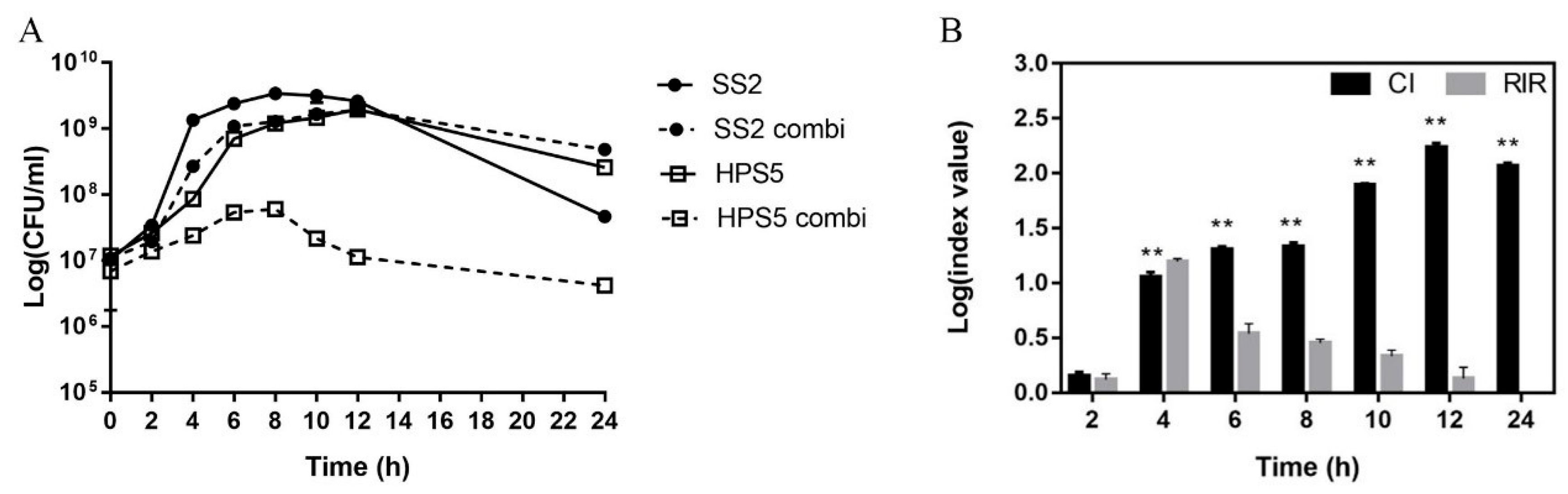
3.1. Competition between S. suis and H. parasuis during Co-Culture In Vitro

The growth kinetics of S. suis and H. parasuis in the single or mixed cultures were evaluated by a 24-h colony count (Figure 1A). Both S. suis and H. parasuis in the co-cultures during both log and stationary phases were inhibited compared to the single culture.
To further evaluate the meaning of the differences observed in the single culture and the co-culture, CI and RIR were calculated and compared (Figure 1B). The CI of S. suis and H. parasuis was significantly different from the corresponding RIR values between 4 h- and 24 h-incubation and was greater than 0 after Log, indicating that S. suis is the dominant bacteria.

3.2. Formation of Mixed Biofilms of S. suis and H. parasuis In Vitro

3.2.1. Single or Mixed Biofilms were Visualized by CLSM

The biofilm formed by S. suis is lumpy (Figure 2A). The biofilm formed by H. parasuis is thinner and more dispersed (Figure 2B). Mixed biofilms formed by S. suis and H. parasuis are dense and flake (Figure 2C). Furthermore, the viable counts of S. suis and H. parasuis were more significantly reduced in the co-culture than in the mono-culture.

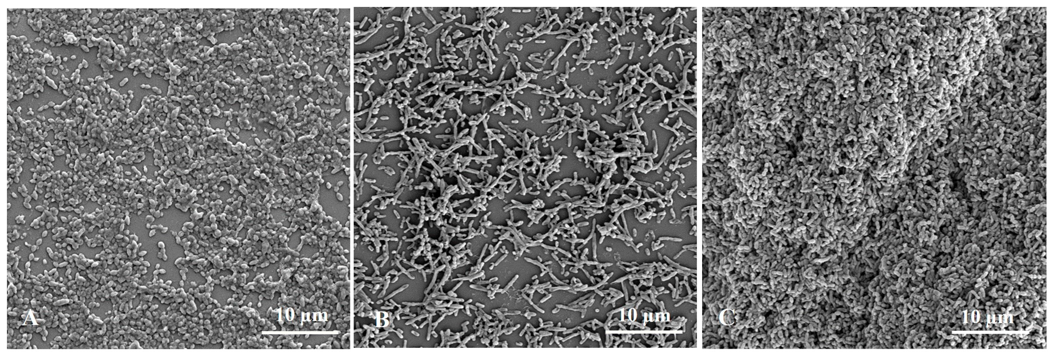
3.2.2. 3D images of Single and Mixed Biofilms Visualized by Scanning Electron Microscopy

Single or mixed biofilms were visualized by Scanning Electron Microscope (SEM). The biofilm formed by S. suis is lumpy (Figure 3A). The biofilm formed by H. parasuis is thinner (Figure 3B). The mixed biofilms formed by S. suis and H. parasuis are dense and flake, S. suis and H. parasuis are intertwined and closely linked (Figure 3C).

3.2.3. S. suis and H. parasuis Were Inhibited When Cultured in Mixed Biofilms

Kinetics of biofilm formation assessed by crystal violet staining (Figure 4A). From 24 h of culture, the biomass of the mixed biofilm is significantly increased compared with the single species biofilm formed by S. suis and H. parasuis. From 72 h of culture, the biomass of the mixed biofilm is significantly reduced compared with the single-species biofilm formed by S. suis and H. parasuis.
The interaction of the two bacteria in the biofilm cultured for 24 h was evaluated by viable count (Figure 4B). The number of viable bacteria in S. suis and H. parasuis biofilms was similar in a single culture. Compared with the single-species biofilms, the viable counts of S. suis and H. parasuis in the mixed biofilms were significantly reduced and their viable counts were similar.

3.3. Mixed Culture of S. suis and H. parasuis Decrease Antibiotic Susceptibility

As shown in Table 1, in the planktonic state, for amoxicillin, MIC increased to 5 μg/mL when co-cultured with S. suis and H. parasuis. For gentamicin, MIC increased to 20 μg/mL when co-cultured with S. suis and H. parasuis. In the biofilm state, for gentamicin, the MIC of the mixed biofilms formed by S. suis and H. parasuis increased to 20 μg/mL. MBC values are higher than 320 μg/mL for both single and mixed biofilms.

3.4. H. parasuis Has a Significant Effect on the Virulence of S. suis in In-Vitro Co-Culture

The expressions of virulence-related genes of S. suis and H. parasuis were determined by RT-PCR (Table A1). In the planktonic state (Figure 5A,C), the virulence-related genes (cps2, ef, mrp, pdh, luxs) of S. suis were down-regulated (p < 0.05), and the virulence-related genes (capD, clpX, Group1, luxs, OmpP2) of H. parasuis were down-regulated (p < 0.05). In the mixed biofilms (Figure 5B,D), the cps2, ef, mrp expression of S. suis was up-regulated (p < 0.05), the expression of pdh was down-regulated (p < 0.05), and the expression of luxs was not significantly different (p > 0.05). The virulence-related genes (capD, clpX, Group1, luxs, OmpP2) of H. parasuis were up-regulated (p > 0.05). cps2, ef, mrp, pdh, luxs codifying for capsular polysaccharide, extracellular factor, muramidase-release proteins, Pyruvate dehydrogenase, and S-Ribosylhomocysteinase, respectively. capD, clpX, Group1, luxs, OmpP2 codifying for capsular polysaccharide, casein hydrolytic protease, trimer autotransporter, s-ribohomocysteinase and outer membrane protein respectively.

3.5. Coexistence of S. suis and H. parasuis Increase the Bacterial Load in Mice

In the mixed infection, the bacterial load of S. suis and H. parasuis in each organ increased. After collecting the heart, liver, spleen, lung, brain, and trachea of the mice, the distribution of S. suis or H. parasuis in various organs of the mice was analyzed. As shown in (Figure 6A,B), there was a significant difference in the content of S. suis and H. parasuis in mice after single infection and mixed infection. The bacteria count of S. suis and H. parasuis was significantly increased in mice after mixed infection with S. suis and H. parasuis.

3.6. Single and Mixed Infections Affect Inflammatory Factor Expression

As shown in Figure 6C, compared with S. suis-infected mice, the mRNA expressions of IFN-γ, IL-12 and IL-β were down-regulated, while mRNA expression of MCP-1, TNF-α and IL-6 were up-regulated in the co-infected mice. (p < 0.05); As shown in Figure 6D, compared with H. parasuis-infected mice, the mRNA expressions of IFN-γ, IL-12 and TNF-α were down-regulated, while mRNA expression of MCP-1, IL-β and IL-6 were up-regulated in the co-infected mice. (p < 0.05).

4. Discussion

Although the role of biofilms in porcine respiratory infections has garnered increased attention, previous research has primarily focused on single-species biofilms. The research findings highlighted that S. suis and H. parasuis were the major respiratory tract pathogens responsible for respiratory disease syndrome in pigs. Accordingly, we established mixed biofilm to explore their interactions. Firstly, we used CLSM and SEM to image the biofilm formation and confirm that S. suis and H. parasuis can form mixed biofilms. It can be seen from CLSM images that the mixed biofilms are denser compared to a single-species biofilm. It can be seen from the CLSM images that the spatial distribution of the mixed biofilms belongs to the “Coaggregation” mode [25]. It cannot be completely determined by imaging that the mixed biofilm becomes stronger or weaker. Therefore, we measured the biomass of the biofilm and the number of viable bacteria under the biofilm by crystal violet staining and colony-forming units (CFUs) respectively. The results showed that the mixed biofilm formed by S. suis and H. parasuis increased, but the proportion of dead bacteria increased. It is worth noting that the competitiveness of H. parasuis in the biofilm state is enhanced compared to the planktonic state. Competitive interactions have been shown to exist primarily between phylogenetically and metabolically similar species [26]. Previous work has shown that the mixed biofilm formed by S. aureus and E. coli was more complex and changed over time [27]. Studies by Kreth et al., have shown that oral Streptococcus, haemorrhagic Streptococci and Gordon’s Streptococcus inhibit the growth of other oral bacteria, including Streptococcus mutants, by producing hydrogen peroxide (H2O2) in a mixed biofilm [28]. An et al., suggested that P. aeruginosa uses strong athletic ability to “cover” Agrobacterium tumefaciens to change the spatial distribution in mixed biofilms, thereby leading the competition in hybrid biofilms [29]. It has not been confirmed that S. suis has antibacterial activity against H. parasuis, and vice versa. Additionally, the imaging results did not show an “overlay” phenomenon. Consequently, the main factor that reduces the number of viable bacteria in the mixed biofilm formed by S. suis and H. parasuis is the competition for nutrients. The competitive ability of H. parasuis is enhanced in the biofilm state in comparison to the planktonic state. This suggests that the balance of the competition between S. suis and H. parasuis in a mixed biofilm community has changed. Currently, it has been confirmed that S. suis and H. parasuis have LuxS/AI-2 quorum sensing systems [30,31]. Recent research indicates that AI-2 plays a significant role in regulating virulence-related genes, pathogenicity, and biofilm formation, the extent of which varies depending on the type of bacterium. “Coaggregation” mode of direct contact between microorganisms in the mixed biofilms can facilitate communication more effectively [32]. It can be inferred that the interaction between S. suis and H. parasuis in the mixed biofilm is related to the bacterial communication.
It is well known that bacteria in a biofilm state are more difficult to eradicate than bacteria in a planktonic state [33,34]. Compared to biofilms formed by a single species, mixed biofilms can more effectively evade host immune defenses and display reduced antibiotic sensitivity [35]. The study showed that co-cultures of S. suis and H. parasuis in planktonic states reduced sensitivity to antibiotics. Although the coexistence of S. suis and H. parasuis caused a reduction in the number of bacteria, the sensitivity to the drug did not increase. In the biofilm state, compared with the single biofilms, the mixed biofilms formed by S. suis and H. parasuis had no significant difference in antibiotic sensitivity, and only the sensitivity to gentamicin was found to be weakened. It is worth noting that none of the drugs utilized in this study entirely eradicated the formed biofilm. In short, whether in the planktonic state or in the biofilm state, the coexistence of the two substances will change the sensitivity of certain antibiotics.
The interactions between microorganisms are complex and play an important role in the infection process [36]. There is competition and coexistence between them. This study indicates that S. suis and H. parasuis compete, and the CI and RIR values indicate that S. suis has a competitive advantage. Antagonistic mechanisms occur in various forms, including one bacterium’s antibacterial effect on another bacterium, chemical signaling that disrupts behavior or physiology, and competition for nutrition and living space. [37]. From the results of a number of viable bacteria under biofilm, S. suis has no antibacterial activity against H. parasuis, and vice versa. To date, there have been no reports of S. suis or H. parasuis producing chemical signals that interfere with another bacterium.
Research has demonstrated that virulence and gene expression of bacterial pathogens can be modulated by the presence of other bacterial species [38]. Therefore, we have compared the transcription levels of virulence factors of S. suis and H. parasuis during the mixed infection in both the planktonic and the biofilm states. Quantitative PCR analysis revealed that the presence of H. parasuis reduced the virulence of S. suis in the planktonic state and changed the virulence of S. suis in the biofilm state. The mixed biofilm significantly increased the expression of S. suis capsular polysaccharide (CPS2) and muramidase-release proteins (MRP). The presence of S. suis reduced the virulence of H. parasuis in the planktonic state and changed the virulence of H. parasuis in the biofilm state. The gene expression caused by the interaction between S. suis and H. parasuis in a biofilm state is more complicated.
Mice models of S. suis infection are often established through intraperitoneal [39], intranasal [40], intravenous [41], and intracisternal [42] routes of infection. Mice models of H. parasuis infection are often established through intraperitoneal routes of infection [43]. However, intraperitoneal injection models are often used in virulence studies [44,45,46,47]. To further evaluate whether the virulence of co-infection changes, we established a mixed infection mice model of S. suis and H. parasuis through intraperitoneal routes of infection. Through organ infection experiments and the determination of inflammatory factor transcription levels, we confirmed that the virulence of S. suis or H. parasuis was enhanced in the mixed infection. The presence of both S. suis and H. parasuis in mice was found to enhance the colonization of these bacteria in the heart, liver, spleen, lungs and trachea. Co-infection of mice with S. suis and H. parasuis was found to alter the inflammatory response. In summary, our research shows that the colonization of mice organs by S. suis and H. parasuis is increased in the presence of co-infection.

5. Conclusions

The study confirmed a competitive and co-existing relationship between S. suis and H. parasuis in vitro. S. suis is more competitive than H. parasuis in planktonic co-cultures (Figure 7A). Additionally, planktonic co-culture enhances drug resistance. A coaggregation pattern developed when S. suis and H. parasuis were co-cultured in the biofilm state. S. suis and H. parasuis have similar competition, and the expression of virulence genes is changed (Figure 7B). Compared to bacteria in the planktonic state, bacteria in the biofilm state are significantly more resistant to antibiotics and hinder the eradication of these bacteria. At the same time, we established a mixed infection model of S. suis and H. parasuis in mice, and we found that the colonization of S. suis and H. parasuis in organs increased after mixed infection (Figure 7C). The co-culture of S. suis and H. parasuis caused an alteration in the host inflammatory response. Interactions between different bacterial species could be accountable for the escalation in pathogenicity. Our research establishes a basis for future research on the co-infection of S. suis and H. parasuis.

Author Contributions

Conceptualization, Y.W. (Yang Wang) and L.Y.; methodology, M.G.; software, Y.W. (Yuxin Wang); formal analysis, J.Z. and S.G.; investigation, M.G.; data curation, Y.S. and S.Y.; writing—original draft preparation, M.G. and J.Z.; writing—review and editing, Y.W. (Yang Wang) and L.Y.; funding acquisition, Y.W. (Yang Wang) and L.Y. All authors have read and agreed to the published version of the manuscript.

Funding

This research was financially supported by the National Natural Science Foundation of China (32172852, 31902309), the Excellent Youth Foundation of Henan Scientific Committee (222300420005) and the Henan Provincial Science and Technology Research Project (232102110095).

Institutional Review Board Statement

The animal study protocol was approved by the Animal Care and Use Committee of Henan University of Science and Technology (approval number: SKKUIACUC-21-04-14-3, date of approval: 14 April 2021).

Informed Consent Statement

Not applicable.

Data Availability Statement

All the data generated or analyzed during this study are available from the corresponding author upon reasonable request.

Conflicts of Interest

The authors declare no conflict of interest.

Appendix A

Table A1. Primers used in this study.
PrimersPrimers Sequence (5′–3′)Target Gene
CPS2-FATTGGTAGGCACTGTCGTTGGTCcps2
CPS2-RAGAACTTAGCATTGTTGCGGTGGcps2
EF-FTCCAATCACAGATCCAGATAGCGef
EF-RCTGACCCATTTGGACCATCTAAGef
MRP-FCAAGGAAAGTGAACAGAACGAGCmrp
MRP-ATAGTCGTCCAAACCTGAGTAGCGmrp
PDH-FCGCGAATTCATGCAACAAATCCGTGATpdh
PDH-RCCCTCGAGGCTAGTCTACAAACACATCpdh
LUXS-FATGAAAAAAGAAGTCACTluxs
LUXS-RTTAGATTGGTTTTCTTTCluxs
16S RNA-FGTTGCGAACGGGTGAGTAA16sRNA
16S RNA-RTCTCAGGTCGGCTATGTATCG16sRNA
CAPD-FATGTTAATGCCATTAATTTATTCATTGCapD
CAPD-RTCGAACCGATAGAACCAGCAGCACCAGTCCapD
CLPX-FAGAGTGAGGGCGTTGAGTclpX
CLPX-RTTCTTGTTTCGGGTGTTTclpX
GROUP 1-FTTTAGGTAAAGATAAGCAAGGAAATCCGroup 1
GROUP 1-RCCACACAAAACCTACCCCTCCTCCGroup 1
LUXS-FGCATCAGCAAGAGAATGTTCCT luxs
LUXS-RATGTCGTTAATTGGTTCACCTTCAluxs
OMPP2-FATGAAAAAAACACTAGTAGCAompP2
OMPP2-RTTACCATAATACACGTAAACCompP2
16S RNA-FGGCTTCGTCACCCTCTGT16sRNA
16S RNA-RGTGATGAGGAAGGGTGGTGT16sRNA
IL-6-FCTGCAAGAGACTTCCATCCAGIL-6
IL-6-RAGTGGTATAGACAGGTCTGTTGGIL-6
IL-12-FGTCCTCAGAAGCTAACCATCTCCIL-12
IL-12-RCCAGAGCCTATGACTCCATGTCIL-12
IL-1β-FGAAATGCCACCTTTTGACAGTG IL-1β
IL-1β-RTGGATGCTCTCATCAGGACAGIL-1β
MCP-FGCATCCACGTGTTGGCTCA MCP-1
MCP-RCTCCAGCCTACTCATTGGGATCMCP-1
TNF-α-FCAGGCGGTGCCTATGTCTCTNF-α
TNF-α-RCGATCACCCCGAAGTTCAGTAGTNF-α
IFN-γ-FACAGCAAGGCGAAAAAGGATGIFN-γ
IFN-γ-RTGGTGGACCACTCGGATGAIFN-γ
GAPDH-FAGGTCGGTGTGAACGGATTTGgapdh
GAPDH-RGGGGTCGTTGATGGCAACAgapdh

References

  1. Dutkiewicz, J.; Zajac, V.; Sroka, J.; Wasinski, B.; Cisak, E.; Sawczyn, A.; Kloc, A.; Wojcik-Fatla, A. Streptococcus suis: A re-emerging pathogen associated with occupational exposure to pigs or pork products. Part II—Pathogenesis. Ann. Agric. Environ. Med. 2018, 25, 186–203. [Google Scholar] [CrossRef]
  2. Dutkiewicz, J.; Sroka, J.; Zajac, V.; Wasinski, B.; Cisak, E.; Sawczyn, A.; Kloc, A.; Wojcik-Fatla, A. Streptococcus suis: A re-emerging pathogen associated with occupational exposure to pigs or pork products. Part I—Epidemiology. Ann. Agric. Environ. Med. 2017, 24, 683–695. [Google Scholar] [CrossRef] [PubMed]
  3. Wang, Y.; Zhang, W.; Wu, Z.; Lu, C. Reduced virulence is an important characteristic of biofilm infection of Streptococcus suis. FEMS Microbiol. Lett. 2011, 316, 36–43. [Google Scholar] [CrossRef] [PubMed]
  4. Meng, X.; Shi, Y.; Ji, W.; Meng, X.; Zhang, J.; Wang, H.; Lu, C.; Sun, J.; Yan, Y. Application of a bacteriophage lysin to disrupt biofilms formed by the animal pathogen Streptococcus suis. Appl. Environ. Microbiol. 2011, 77, 8272–8279. [Google Scholar] [CrossRef]
  5. Lee, K.W.K.; Periasamy, S.; Mukherjee, M.; Xie, C.; Kjelleberg, S.; Rice, S.A. Biofilm development and enhanced stress resistance of a model, mixed-species community biofilm. ISME J. 2014, 8, 894–907. [Google Scholar] [CrossRef] [PubMed]
  6. Zhang, B.; Tang, C.; Liao, M.; Yue, H. Update on the pathogenesis of Haemophilus parasuis infection and virulence factors. Vet. Microbiol. 2014, 168, 1–7. [Google Scholar] [CrossRef]
  7. Macedo, N.; Rovira, A.; Torremorell, M. Haemophilus parasuis: Infection, immunity and enrofloxacin. Vet. Res. 2015, 46, 128. [Google Scholar] [CrossRef] [PubMed]
  8. Bello-Orti, B.; Costa-Hurtado, M.; Martinez-Moliner, V.; Segales, J.; Aragon, V. Time course Haemophilus parasuis infection reveals pathological differences between virulent and non-virulent strains in the respiratory tract. Vet. Microbiol. 2014, 170, 430–437. [Google Scholar] [CrossRef] [PubMed]
  9. Oliveira, S.; Pijoan, C. Haemophilus parasuis: New trends on diagnosis, epidemiology and control. Vet. Microbiol. 2004, 99, 1–12. [Google Scholar] [CrossRef]
  10. Li, J.; Wang, S.; Li, C.; Wang, C.; Liu, Y.; Wang, G.; He, X.; Hu, L.; Liu, Y.; Cui, M.; et al. Secondary Haemophilus parasuis infection enhances highly pathogenic porcine reproductive and respiratory syndrome virus (HP-PRRSV) infection-mediated inflammatory responses. Vet. Microbiol. 2017, 204, 35–42. [Google Scholar] [CrossRef] [PubMed]
  11. Cheong, Y.; Oh, C.; Lee, K.; Cho, K.H. Survey of porcine respiratory disease complex-associated pathogens among commercial pig farms in Korea via oral fluid method. J. Vet. Sci. 2017, 18, 283–289. [Google Scholar] [CrossRef] [PubMed]
  12. Mathieu-Denoncourt, A.; Letendre, C.; Auger, J.-P.; Segura, M.; Aragon, V.; Lacouture, S.; Gottschalk, M. Limited Interactions between Streptococcus Suis and Haemophilus Parasuis in In Vitro Co-Infection Studies. Pathogens 2018, 7, 7. [Google Scholar] [CrossRef]
  13. Flemming, H.C.; Wingender, J. The biofilm matrix. Nat. Rev. Microbiol. 2010, 8, 623–633. [Google Scholar] [CrossRef]
  14. Costerton, W.J. Bacterial Biofilms: A Common Cause of Persistent Infections. Science 1999, 284, 1318–1322. [Google Scholar] [CrossRef] [PubMed]
  15. Yuan, L.; Hansen, M.F.; Roder, H.L.; Wang, N.; Burmolle, M.; He, G. Mixed-species biofilms in the food industry: Current knowledge and novel control strategies. Crit. Rev. Food Sci. Nutr. 2019, 60, 1–17. [Google Scholar] [CrossRef] [PubMed]
  16. Wuertz, S.; Okabe, S.; Hausner, M. Microbial communities and their interactions in biofilm systems: An overview. Water Sci. Technol. 2004, 49, 327. [Google Scholar] [CrossRef]
  17. Elias, S.; Banin, E. Multi-species biofilms: Living with friendly neighbors. FEMS Microbiol. Rev. 2012, 36, 990–1004. [Google Scholar] [CrossRef]
  18. Reddinger, R.M.; Luke-Marshall, N.R.; Sauberan, S.L.; Hakansson, A.P.; Campagnari, A.A. Streptococcus pneumoniae Modulates Staphylococcus aureus Biofilm Dispersion and the Transition from Colonization to Invasive Disease. MBio 2018, 9, e02089-17. [Google Scholar] [CrossRef]
  19. Pompilio, A.; Crocetta, V.; De Nicola, S.; Verginelli, F.; Fiscarelli, E.; Di Bonaventura, G. Cooperative pathogenicity in cystic fibrosis: Stenotrophomonas maltophilia modulates Pseudomonas aeruginosa virulence in mixed biofilm. Front. Microbiol. 2015, 6, 951. [Google Scholar] [CrossRef] [PubMed]
  20. Cope, E.K.; Goldstein-Daruech, N.; Kofonow, J.M.; Christensen, L.; McDermott, B.; Monroy, F.; Palmer, J.N.; Chiu, A.G.; Shirtliff, M.E.; Cohen, N.A.; et al. Regulation of virulence gene expression resulting from Streptococcus pneumoniae and nontypeable Haemophilus influenzae interactions in chronic disease. PLoS ONE 2011, 6, e28523. [Google Scholar] [CrossRef] [PubMed]
  21. Grenier, D.; Grignon, L.; Gottschalk, M. Characterisation of biofilm formation by a Streptococcus suis meningitis isolate. Vet. J. 2009, 179, 292–295. [Google Scholar] [CrossRef] [PubMed]
  22. M100-S25; Performance Standards for Antimicrobial Susceptibility Testing; Twenty-Fifth Informational Supplement. Clinical and Laboratory Standards Institute (CLSI): Wayne, PA, USA, 2015.
  23. Wiegand, I.; Hilpert, K.; Hancock, R.E. Agar and broth dilution methods to determine the minimal inhibitory concentration (MIC) of antimicrobial substances. Nat. Protoc. 2008, 3, 163–175. [Google Scholar] [CrossRef] [PubMed]
  24. Wang, Y.; Wang, Y.; Liu, B.; Wang, S.; Li, J.; Gong, S.; Sun, L.; Li, Y. Pdh modulate virulence through reducing stress tolerance and biofilm formation of serotype 2. Virulence 2019, 10, 588–599. [Google Scholar] [CrossRef] [PubMed]
  25. Macho, A.P.; Zumaquero, A.; Ortiz-Martin, I.; Beuzon, C.R. Competitive index in mixed infections: A sensitive and accurate assay for the genetic analysis of Pseudomonas syringae-plant interactions. Mol. Plant. Pathol. 2007, 8, 437–450. [Google Scholar] [CrossRef]
  26. Russel, J.; Roder, H.L.; Madsen, J.S.; Burmolle, M.; Sorensen, S.J. Antagonism correlates with metabolic similarity in diverse bacteria. Proc. Natl. Acad. Sci. USA 2017, 114, 10684–10688. [Google Scholar] [CrossRef]
  27. Reece, E.; Doyle, S.; Greally, P.; Renwick, J.; McClean, S. Aspergillus fumigatus Inhibits Pseudomonas aeruginosa in Co-culture: Implications of a Mutually Antagonistic Relationship on Virulence and Inflammation in the CF Airway. Front. Microbiol. 2018, 9, 1205. [Google Scholar] [CrossRef]
  28. Kreth, J.; Merritt, J.; Shi, W.; Qi, F. Competition and Coexistence between Streptococcus mutans and Streptococcus sanguinis in the Dental Biofilm. J. Bacteriol. 2005, 187, 7193–7203. [Google Scholar] [CrossRef]
  29. An, D.; Danhorn, T.; Fuqua, C.; Parsek, M.R. Quorum sensing and motility mediate interactions between Pseudomonas aeruginosa and Agrobacterium tumefaciens in biofilm cocultures. Proc. Natl. Acad. Sci. USA 2006, 103, 3828–3833. [Google Scholar] [CrossRef]
  30. Wang, Y.; Wang, Y.; Sun, L.; Grenier, D.; Yi, L. The LuxS/AI-2 system of Streptococcus suis. Appl. Microbiol. Biotechnol. 2018, 102, 7231–7238. [Google Scholar] [CrossRef]
  31. Zhang, B.; Ku, X.; Zhang, X.; Zhang, Y.; Chen, G.; Chen, F.; Zeng, W.; Li, J.; Zhu, L.; He, Q. The AI-2/luxS Quorum Sensing System Affects the Growth Characteristics, Biofilm Formation, and Virulence of Haemophilus parasuis. Front. Cell Infect. Microbiol. 2019, 9, 62. [Google Scholar] [CrossRef]
  32. Egland, P.G.; Palmer, R.J., Jr.; Kolenbrander, P.E. Interspecies communication in Streptococcus gordonii-Veillonella atypica biofilms: Signaling in flow conditions requires juxtaposition. Proc. Natl. Acad. Sci. USA 2004, 101, 16917–16922. [Google Scholar] [CrossRef] [PubMed]
  33. Wang, Y.; Wang, Y.; Sun, L.; Grenier, D.; Yi, L. Streptococcus suis biofilm: Regulation, drug-resistance mechanisms, and disinfection strategies. Appl. Microbiol. Biotechnol. 2018, 102, 9121–9129. [Google Scholar] [CrossRef]
  34. Hoiby, N.; Bjarnsholt, T.; Givskov, M.; Molin, S.; Ciofu, O. Antibiotic resistance of bacterial biofilms. Int. J. Antimicrob. Agents. 2010, 35, 322–332. [Google Scholar] [CrossRef] [PubMed]
  35. Armbruster, C.E.; Hong, W.; Pang, B.; Weimer, K.E.; Juneau, R.A.; Turner, J.; Swords, W.E. Indirect pathogenicity of Haemophilus influenzae and Moraxella catarrhalis in polymicrobial otitis media occurs via interspecies quorum signaling. mBio 2010, 1, e00102-10. [Google Scholar] [CrossRef]
  36. Gabrilska, R.A.; Rumbaugh, K.P. Biofilm models of polymicrobial infection. Future Microbiol. 2015, 10, 1997–2015. [Google Scholar] [CrossRef] [PubMed]
  37. García-Bayona, L.; Comstock, L.E. Bacterial antagonism in host-associated microbial communities. Science 2018, 361, eaat2456. [Google Scholar] [CrossRef]
  38. Duan, K.; Dammel, C.; Stein, J.; Rabin, H.; Surette, M.G. Modulation of Pseudomonas aeruginosa gene expression by host microflora through interspecies communication. Mol. Microbiol. 2003, 50, 1477–1491. [Google Scholar] [CrossRef]
  39. Domínguez-Punaro, M.C.; Segura, M.; Plante, M.M.; Lacouture, S.; Rivest, S.; Gottschalk, M. Streptococcus suis serotype 2, an important swine and human pathogen, induces strong systemic and cerebral inflammatory responses in a mouse model of infection. J. Immunol. 2007, 179, 1842–1854. [Google Scholar] [CrossRef]
  40. Seitz, M.; Beineke, A.; Seele, J.; Fulde, M.; Valentin-Weigand, P.; Baums, C.G. A novel intranasal mouse model for mucosal colonization by Streptococcus suis serotype 2. J. Med. Microbiol. 2012, 61, 1311–1318. [Google Scholar] [CrossRef]
  41. Williams, A.E.; Blakemore, W.F.; Alexander, T.J. A murine model of Streptococcus suis type 2 meningitis in the pig. Res. Vet. Sci. 1988, 45, 394–399. [Google Scholar] [CrossRef]
  42. Domínguez-Punaro, M.C.; Koedel, U.; Hoegen, T.; Demel, C.; Klein, M.; Gottschalk, M. Severe cochlear inflammation and vestibular syndrome in an experimental model of Streptococcus suis infection in mice. Eur. J. Clin. Microbiol. Infect. Dis. 2012, 31, 2391–2400. [Google Scholar] [CrossRef] [PubMed]
  43. Zhang, L.; Li, Y.; Wen, Y.; Lau, G.W.; Huang, X.; Wu, R.; Yan, Q.; Huang, Y.; Zhao, Q.; Cao, S. HtrA Is Important for Stress Resistance and Virulence in Haemophilus parasuis. Infect. Immun. 2016, 84, 2209–2219. [Google Scholar] [CrossRef]
  44. Niu, K.; Meng, Y.; Liu, M.; Ma, Z.; Lin, H.; Zhou, H.; Fan, H. Phosphorylation of GntR reduces Streptococcus suis oxidative stress resistance and virulence by inhibiting NADH oxidase transcription. PLoS Pathog. 2023, 19, e1011227. [Google Scholar] [CrossRef]
  45. Gao, T.; Tan, M.; Liu, W.; Zhang, C.; Zhang, T.; Zheng, L.; Zhu, J.; Li, L.; Zhou, R. GidA, a tRNA Modification Enzyme, Contributes to the Growth, and Virulence of Streptococcus suis Serotype 2. Front. Cell. Infect. Microbiol. 2016, 6, 44. [Google Scholar] [CrossRef]
  46. Jiang, X.; Yang, Y.; Zhou, J.; Zhu, L.; Gu, Y.; Zhang, X.; Li, X.; Feng, W. Roles of the Putative Type IV-like Secretion System Key Component VirD4 and PrsA in Pathogenesis of Streptococcus suis Type 2. Front. Cell. Infect. Microbiol. 2016, 6, 172. [Google Scholar] [CrossRef] [PubMed]
  47. Dai, K.; Yang, Z.; Ma, X.; Chang, Y.F.; Cao, S.; Zhao, Q.; Huang, X.; Wu, R.; Huang, Y.; Wen, Y.; et al. Deletion of Polyamine Transport Protein PotD Exacerbates Virulence in Glaesserella (Haemophilus) parasuis in the Form of Non-biofilm-generated Bacteria in a Murine Acute Infection Model. Virulence 2021, 12, 520–546. [Google Scholar] [CrossRef] [PubMed]
Figure 1. Growth curves and competition indices of pure cultures and co-cultures of S. suis and H. parasuis. (A) Growth curves of S. suis and H. parasuis strains in pure culture (SS2, HPS5) and in co-culture (SS2 combi, HPS5 combi). (B) Competitive index (CI; black bars) and Relative Increase Ratio (RIR; gray bars) were calculated from single and dual planktonic cultures of S. suis and H. parasuis. The results are shown as mean ± SD (n = 3). ** p < 0.01, CI vs. RIR, unpaired t-test.
Animals 13 01511 g001
Figure 2. The figure shows the orthogonal views of CLSM images. (A) Biofilm formed by S. suis; (B) Biofilm formed by H. parasuis; (C) Mixed biofilms formed by S. suis and H. parasuis. CLSM uses a Carl Zeiss LSM800 confocal scanning system 40× objective lens. LIVE/DEAD BIOFILM staining was used: live cells (with intact cell membranes) stain green and dead or dying cells (with compromised cell membranes) stain red.
Animals 13 01511 g002
Figure 3. SEM images of S. suis and H. parasuis single or Mixed biofilms. (A) Representative SEM image of S. suis single species biofilms. (B) Representative SEM images of H. parasuis single species biofilms. (C) Representative SEM images of mixed biofilms of S. suis and H. parasuis.
Animals 13 01511 g003
Figure 4. Crystal violet assay and Viable count assay of S. suis and H. parasuis single or mixed biofilms. (A) Crystal violet assay. S. suis and H. parasuis single (SS2, HPS5) or mixed biofilms (SS2 + HPS5) formation after 12 h, 24 h, 48 h, and 72 h incubation were tested. (B) Viable count assay. S. suis and H. parasuis strains were tested in single (SS2, HPS5)or mixed biofilms (SS2 combi, HPS5 combi). The results are shown as mean ± SD (n = 3). * p < 0.05, *** p < 0.001.
Animals 13 01511 g004
Figure 5. Expression of virulence and quorum sensing-related genes in single and mixed cultures (A,C) Planktonic state. (B,D) Biofilm state. The 16SRNA gene was used as a reference. When only S. suis or H. parasuis is present, the level of S. suis or H. parasuis gene expression is 100% (SS2 or HPS5, black bars). When S. suis and H. parasuis coexisted, the level of S. suis or H. parasuis gene expression was relative to the level of S. suis or H. parasuis gene in the presence of only S. suis or H. parasuis (SS2 combi or HPS5 combi, gray bars). Data from three independent assays are expressed as mean ± SD.
Animals 13 01511 g005
Figure 6. (A,B) Bacterial counts in different organs. Number of S. suis or H. parasuis in heart, liver, spleen, lung, brain, and trachea after single infection with S. suis or H. parasuis (open square, SS2 or HPS5). Number of S. suis or H. parasuis in heart, liver, spleen, lung, brain and trachea after mixed infection with S. suis and H. parasuis in mice (closed circles, SS2 or HPS5 combi). The black horizontal line indicates the average value. (C,D) Expression of inflammatory factor-related genes in mice. The level of gene expression in mice infected with S. suis or H. parasuis was 100% (SS2 or HPS5, black bars). The level of gene expression in mice infected with S. suis and H. parasuis was relative to the level of gene expression in mice infected with S. suis or H. parasuis(SS2 + HPS5, gray bars). Data from three independent determinations are expressed as mean ± SD.
Animals 13 01511 g006
Figure 7. (A) Planktonic mixed culture. (B) Mixed biofilm model. (C) Mice mixed infection model.
Animals 13 01511 g007
Table 1. The MIC and MBC of mono-cultures and co-cultures of S. suis and H. parasuis in planktonic and biofilm states.
Bacterial CombinationsMIC (μg/mL)
(Planktonic)
MBC (μg/mL)
(Planktonic)
MIC (μg/mL)
(Biofilm)
MBC (μg/mL)
(Biofilm)
Amoxicillin
SS20.31252.55>320
HPS50.16250.62510>320
SS2 + HPS551010>320
Gentamicin
SS210405>320
HPS551010>320
SS2 + HPS5204020>320
Enrofloxacin
SS20.31250.6251.25>320
HPS50.16250. 6250.625>320
SS2 + HPS50.625201.25>320
Disclaimer/Publisher’s Note: The statements, opinions and data contained in all publications are solely those of the individual author(s) and contributor(s) and not of MDPI and/or the editor(s). MDPI and/or the editor(s) disclaim responsibility for any injury to people or property resulting from any ideas, methods, instructions or products referred to in the content.

