Communication

Characterization of Bacillus pumilus Strains Isolated from Bovine Uteri

1
Functional Microbiology Unit, Institute for Microbiology, Department of Pathobiology, University of Veterinary Medicine, 1210 Vienna, Austria
2
Clinical Unit for Herd Health Management in Ruminants, Department for Farm Animals and Veterinary Public Health, University of Veterinary Medicine, 1210 Vienna, Austria
3
Department of Veterinary Medicine, Freie Universität Berlin, Institute of Veterinary Biochemistry, 14163 Berlin, Germany
*
Author to whom correspondence should be addressed.
Animals 2023, 13(8), 1297; https://doi.org/10.3390/ani13081297
Submission received: 8 February 2023 / Revised: 6 April 2023 / Accepted: 8 April 2023 / Published: 10 April 2023
(This article belongs to the Collection Endometritis and Fibrosis: An Evolving Story)

Abstract

:

Simple Summary

Bound with proper productivity, reproductive performance in dairy cows is of utmost importance. One of the most important conditions hampering the reproductive performance of a dairy cow is bovine endometritis. In this paper, we aim to report the effect of B. pumilus strains, isolated from cows with or without endometritis, with regard to their pathogenicity to the bovine uterus by using a cell-culture-based approach. Our study revealed that B. pumilus strains possess specific genes related to enzyme production and harm the endometrial bovine cells in a dose-dependent pattern.

Abstract

Uterine infections are a major source of economic losses to dairy farmers. The uterine microbiota as well as opportunistic uterine contaminants can contribute to the development of endometritis in dairy cows during the postpartum period. Therefore, it is important to characterize potential pathogens and to further elucidate their role in the disease. In this study, we aimed to characterize Bacillus pumilus field isolates to obtain more details regarding their effect on uterine cells by using an in vitro endometrial epithelial primary cells model. We found that B. pumilus isolates possessed the keratinase genes ker1 and ker2 and therefore may produce keratinases. When primary endometrial epithelial cells were infected with 4 different B. pumilus strains, an effect on cellular viability was observed over the course of 72 h. The effect was dose-dependent and time-dependent. Nevertheless, significant differences between the strains were not observed. All tested strains reduced the viability of the primary cells after 72 h of incubation, indicating that B. pumilus potentially has a pathogenic effect on endometrial epithelial cells.

1. Introduction

Recent opinions pinpoint that the development of postpartum uterine diseases in cows may have three major components: avoidance, tolerance and resistance [1]. Therefore, the development of uterine diseases, such as endometritis, is affected by multiple factors. One of the most important of those factors is the uterine microbiota and the postpartum contamination of the uterus with bacteria [2]. Generally, the bovine uterus was considered sterile outside of the postpartum period, but newer studies debate that opinion [3,4]. Over the last decade more information regarding pathogenic bacteria and the postpartum uterine microflora in dairy cows has surfaced, as the use of both culture-dependent and culture-independent techniques provided a wealth of information regarding the postpartum uterine microbial community [5,6,7,8,9,10,11,12]. Under the current opinions, the bacteria involved in bovine endometritis can be categorized into three groups: defined pathogens, potential pathogens and opportunistic colonizers of the uterus [13]. Defined uterine pathogens, such as Trueperella pyogenes and Escherichia coli, have been thoroughly studied in the past, providing evidence regarding their role in the disease [14,15,16,17]. On the contrary, bacteria such as Streptococcus uberis, which are considered as potential pathogens, were only rarely studied regarding their role in endometritis [8,18], and only recently have new studies elucidated their potentially harmful effect on uterine cells [19]. Bacterial species considered as opportunistic contaminants of the uterus, such as Bacillus pumilus, were just recently brought to the attention of researchers regarding their potential role in the development of endometritis and their effect on uterine cells [20], even though many of them possess pathogenic molecular mechanisms, such as the production of potentially cell-harmful enzymes or toxins [21]. Nevertheless, the abovementioned categorization is not absolute, and studies which will clearly define and characterize the pathogenicity of field-isolated bacterial isolates are needed.
This scope of this study is to characterize case-isolated B. pumilus strains isolated from bovine uteri during the postpartum period, with a focus on strains isolated from animals with endometritis, and to evaluate the potential effect of these strains on the viability of endometrial cells.

2. Materials and Methods

2.1. Bacterial Strains Culture and Growth Kinetics

The strains were collected in a previous study [18]. A total of 14 strains from 40 Holstein-Friesian cows sampled and monitored throughout the first days of the postpartum period were selected. The samples were collected from animals showing clear symptoms of endometritis (vaginal discharge score 3; 19/40 cows) and animals without endometritis, by using the cytobrush technique [22]. Bacteria were revived from glycerol stocks at −80 °C and were recovered by streaking onto Columbia III agar supplemented with 5% sheep blood (Becton Dickinson, Heidelberg, Germany). Bacteria were incubated at 37 °C for 24 h under aerobic conditions. Bacteria were grown routinely for the analyses in Columbia III agar supplemented with 5% sheep blood at 37 °C for 24 h.
For co-culturing, endometrial epithelial cells and bacteria were prepared as before [20] with slight modifications. Bacteria were primarily cultured in brain heart infusion (BHI) broth (Sigma-Aldrich, Taufkirchen, Germany) at 37 °C for 24 h, under aerobic conditions. The bacterial suspension was centrifuged at 2000× g for 10 min, and the bacteria were collected from the solution. The bacterial pellet was washed once with Dulbecco’s phosphate-buffered saline (PBS, Biochrom, Berlin, Germany), re-suspended in BHI broth with 20% glycerol v/v and stored at −80 °C. Plate counting on BHI agar (Sigma-Aldrich) was performed to determine the number of colony-forming units (CFU) per ml. Bacteria were thawed briefly before the start of the experiments and serially diluted in epithelial cell culture medium without antibiotics (DMEM/Ham’s F-12 +10% fetal bovine serum (FBS), all from Biochrom) to obtain the required bacterial concentrations for each assay. For the growth kinetics, bacteria were cultivated from a starting inoculum of 103 bacterial cells in DMEM/Ham’s F-12, supplemented with 10% FBS for 48 h at 37 °C, 5% CO2. Optical density was measured every 4 h for the first 24 h, and every 8 h from 24 to 48 h. Optical density (OD600) was measured in a Jenway 6305 spectrophotometer (Stone, Staffordshire, UK).

2.2. Keratinase Gene PCR

Evaluation of the presence of the keratinase genes ker1 and ker2 was performed as described before by [21], with slight modifications. For the PCR reaction, a mixture consisting of 1 μM of the respective oligonucleotide primers, 0.4 μΜ dNTPs, 2.5 mM MgCl2, 1 × GoTaq Reaction Buffer and 0.75 U goTaq polymerase (Promega, Mannheim, Germany) was used, along with 1 μL template DNA.
The PCR protocol started with an initial denaturation step for 2 min at 95 °C, followed by 30 cycles of: denaturation at 95 °C for 35 s, annealing for 30 s at appropriate temperature and extension at 72 °C, with the time depending on the length of the amplicon. A final extension step of 4 min at 72 °C was performed before the products were cooled at 4 °C. The presence of the specific bands was checked by gel electrophoresis.

2.3. Primary Endometrial Epithelial Cell Culture

Primary endometrial epithelial cell culturing was carried out as described before [18,23]. Briefly, bovine uteri were collected from a local slaughterhouse, and small tissue pieces (approximately 0.1 cm2) from the area between the caruncles were collected. The collected tissue was minced with scalpels and then digested for 2 h at 37 °C in a solution containing 150 U/mL of collagenase, 150 U/mL of hyaluronidase, 200 U/mL of penicillin and 20 μg/mL of streptomycin (Sigma-Aldrich) in Hank’s balanced salt solution (Biochrom). Cells were then centrifuged, and the cell pellet was washed with cell culture medium (DMEM/Ham’s F-12 and 10% FBS, gentamicin and amphotericin B; all from Biochrom). The cells were seeded in 25 cm2 flasks at 37 °C + 5% CO2 for 18 h. During this time, fibroblast cells had already attached, while the medium containing non-attached cells was transferred to a new 25 cm2 flask in order to obtain a pure (>99%) epithelial cell culture. Cells were transferred to a 75 cm2 flask when a confluence level of >80% was achieved. Cells were allowed to grow and finally passaged into a 24-well plate at a final density of 2 × 10⁵ cells per well.

2.4. Viability Assay

Primary endometrial endothelial cells were infected with different multiplicity of infection (MOI) concentrations of B. pumilus and then incubated at 37 °C, 5% CO2 for 72 h. Evaluation of the cellular viability was carried out by using 0.5% trypan blue (ScienCell, CA, USA) staining as described before [20]. Cellular viability was determined at 24, 48 and 72 h post-inoculation. For calculating the viability, multiple optical fields were evaluated, and the numbers of unstained cells (viable) and stained cells (non-viable) were calculated as percentages of the total number of cells.

2.5. Statistical Analysis

For the analysis of data and for data visualization, GraphPad Prism version 7.00 for Windows, by GraphPad Software (La Jolla, San Diego, CA, USA), was used. For the growth curve data, a Shapiro–Wilk test was used in order to test the normality of the data as it is more fitted to smaller sample sizes. Comparisons were performed by using the Welch’s test. For the cell viability assay data, a Wilcoxon signed rank test was performed, as we were comparing cellular viability scores. A p-value of <0.05 was considered as significant, while p-values from 0.05 to 0.1 were considered as a trend.

3. Results

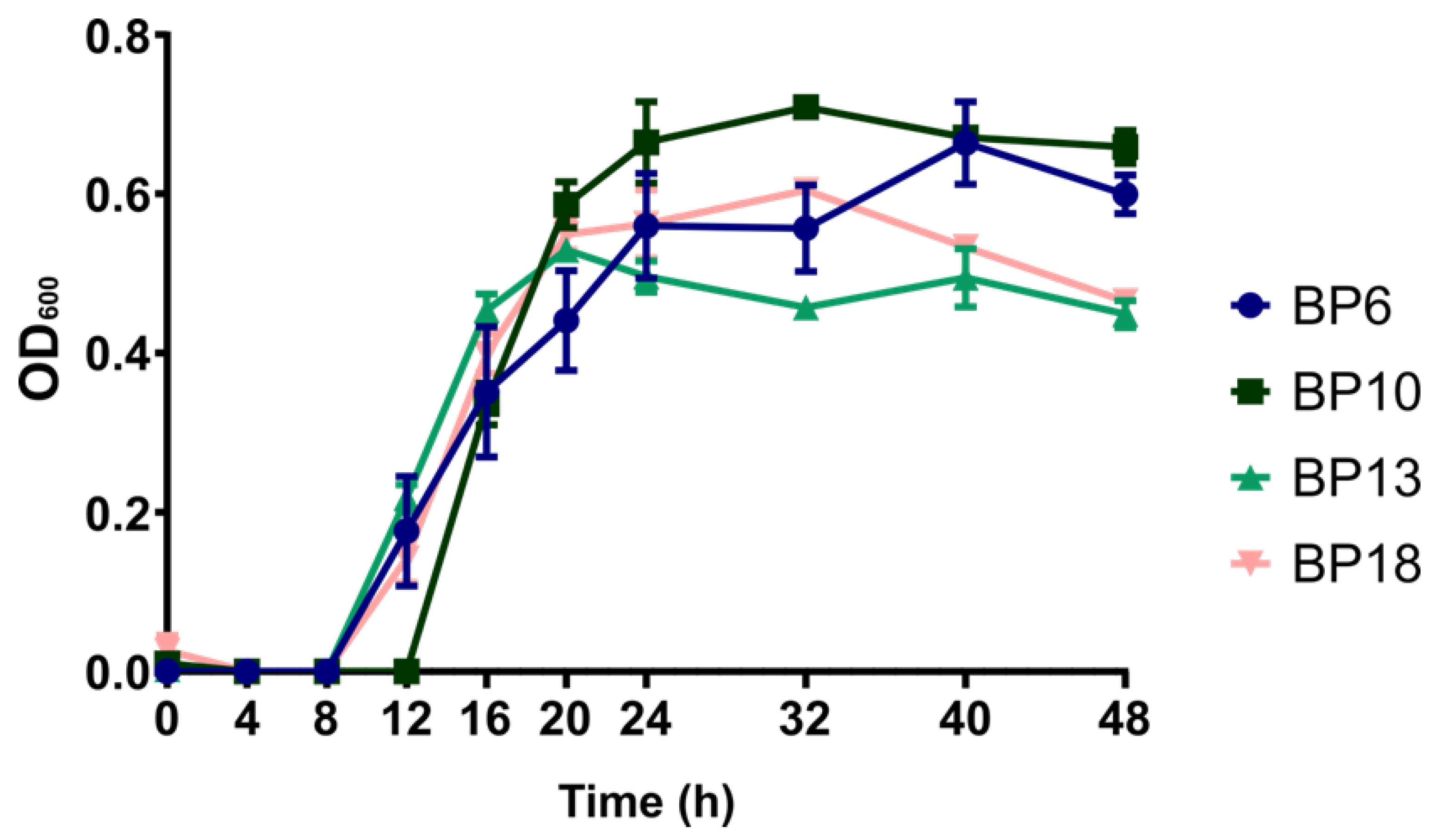
3.1. Keratinase Gene Screening, and Growth Kinetics

The keratinase genes screening was performed as described above. In the pool of tested strains, all strains were found positive for the ker1 gene, while only 1 out of 14 strains was positive for the ker2 gene. Because no specific pattern of gene presence between strains originating from healthy and diseased animals was found, B. pumilus strains originating from animals diagnosed with endometritis at day 21 postpartum were selected for further study. First, the growth of the bacteria in DMEM/Ham’s F-12 supplemented with 10% FBS was monitored. The four strains BP6, BP10, BP13 and BP18 showed generally similar growth patterns in the cell culture medium, as depicted in Figure 1.
Nevertheless, strain BP10 exhibited higher growth after 24 h compared to the strains BP6, BP13 and BP18, and after 32 h there was a significantly higher growth of strain BP10 compared to the strains BP13 and BP18 (p = 0.002 and p = 0.018, respectively), but also a delayed onset of the exponential growth phase.

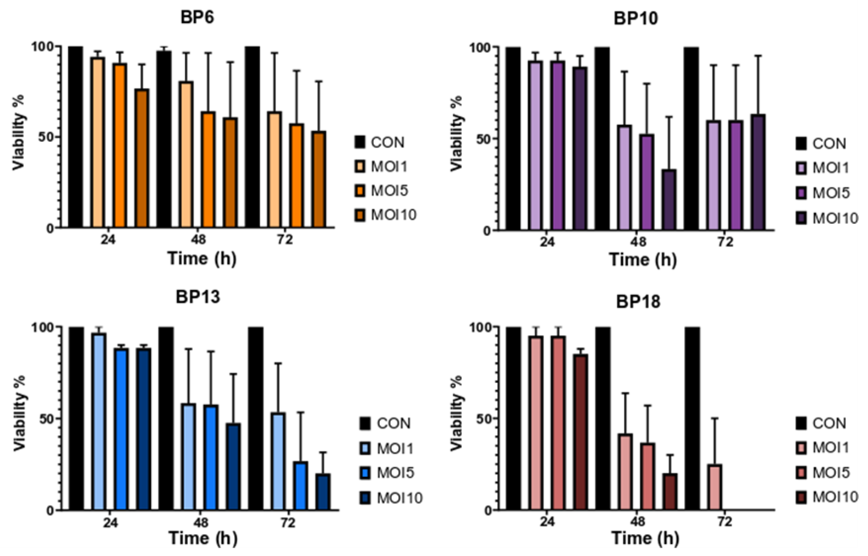
3.2. Effect of B. pumilus Isolates on Uterine Cells

B. pumilus strains prepared as indicated before were used to infect the primary endometrial epithelial cells. The viability of the cells was evaluated at 24, 48 and 72 h after inoculation. After 24 h of incubation, minimal impairment of cellular viability was observed, with a cellular viability of approximately 95% in MOI 1 (Figure 2).
Higher MOI had a stronger effect on reducing cellular viability. Cellular deterioration gradually increased over time, with cellular viability being reduced to approximately 80% for strain BP6, 60% for the strains BP10 and BP13 and 40% for BP18 in MOI 1 after 48 h (Figure 2). Cellular viability decreased further at 72 h post-inoculation, with the exception of cells infected with the strain BP10, in which the viability did not reduce further. In addition, no strain-specific differences in the killing capacity of the isolates were observed. Additionally, the color of the medium was monitored throughout the 72 h period. No changes were observed, indicating that there were no secondary metabolites or medium changes (such as pH drop). Therefore, it is assumed that the observed cytotoxic effect can be attributed to the presence of the bacterium itself, rather than to other factors related to medium changes or medium nutrient depletion. Furthermore, during the time window it was observed that the tested B. pumilus strains caused detachment of the uterine cells, as depicted in Figure 3.

4. Discussion

Information regarding the role of the uterine microbiota and invasive bacterial species in bovine endometritis has largely expanded over the last several years. In the past, the uterus was considered as a sterile environment before parturition, but recent studies have indicated that it harbors its own unique microbiome with a broad variety of bacterial species [3]. The microbiome and opportunistic pathogens of the uterus have a dynamic relationship which may contribute to the development of the disease. The bacteria which can be related to endometritis in dairy cows can be categorized as pathogens, potential pathogens and opportunistic contaminants [9]. Nevertheless, it is important to characterize the role of each bacterial species with robust in vivo and in vitro models. To this end, we sought to characterize B. pumilus field isolates collected during previous studies [18]. The selected field isolates were subjected to screening for the keratinase genes Ker1 and Ker2. Until now, no other study has considered a screening of B. pumilus isolates from a farm environment for the presence of the aforementioned genes, even though Bacillus spp. have the potential for production of keratinases. All strains tested positive for the Ker1 gene, but only one was positive for Ker2. Therefore, it is assumed that the strains possess the ability to produce at least one type of keratinase. B. pumilus keratinases are considered to be subtilisin-like serine proteases [21] and have been considered as a potential virulence factor in various fungi species, especially dermatophytes such as Microsporum canis [24]. Furthermore, recent studies in mice models have indicated that certain keratin types are present in the endometrium and could potentially be related to reproductive performance and decreased conception rates [25]. Therefore, it is tempting to speculate that the presence of bacterial keratinases could have an effect on the tissue keratin, thus reducing tissue resistance.
Additionally, previous studies have indicated that B. pumilus can increase the mRNA expression of certain pro-inflammatory factors in primary endometrial cells [20]. In our study, an effect on cellular viability was observed for all the B. pumilus strains tested. Although specific differences between strains were not found, it was observed that all the tested strains had the ability to reduce the viability of endometrial primary cells. This effect is in line with previous studies indicating that B. pumilus has a destructive effect on endometrial cells [20]. By measuring the growth of the strains in the cell culture medium under the same conditions used for the cell viability assay, it was ensured that the effect of B. pumilus on cellular viability could be attributed to the presence of the bacterium and not to secondary changes of the medium, such as pH change or the production of bacterial metabolites. Nevertheless, more studies will be needed to observe more precisely what happens at the bacterial-cellular level regarding the metabolites and the cytotoxic effect.

5. Conclusions

In summary, our study provides initial results on B. pumilus field isolates’ cytotoxicity towards endometrial cells, indicating a potential contribution of B. pumilus to bovine endometritis. Certain isolates were able to strongly reduce the cellular viability of uterine cells when tested in a cell-culture model, but extreme differences in the pathogenicity were not observed. The pathogenic effect shown is possibly attributed to the bacterial cell itself rather than to secondary metabolic products of the bacteria; however, further studies will be necessary to decipher the factors involved in the observed cytotoxic effects of B. pumilus on endometrial cells.

Author Contributions

Conceptualization, M.D. and M.E.-S.; methodology, P.B.; formal analysis, K.W. and P.B.; investigation, P.B. and K.W.; resources, M.D. and M.E.-S.; data curation, P.B. and K.W.; writing—original draft preparation, P.B.; writing—review and editing, K.W., M.D., C.G. and M.E.-S.; visualization, P.B.; supervision, C.G., M.D. and M.E.-S.; project administration, M.D. and M.E.-S.; funding acquisition, M.D. and M.E.-S. All authors have read and agreed to the published version of the manuscript.

Funding

This research was funded by the Vetmeduni Vienna Tandem PhD program. Open Access Funding by the University of Veterinary Medicine Vienna.

Institutional Review Board Statement

This study has been approved by the institutional ethics committee and the national authority according to § 8 of the Law for Animal Experiments (Tierversuchsgesetz-TVG BMWF-68.205/246-II/3b/2010). The ethic approval code is 08/01/97/2011 and the approval date is 1 February 2011.

Informed Consent Statement

Not applicable.

Data Availability Statement

Not applicable.

Acknowledgments

We acknowledge the support of Harald Pothmann for taking samples.

Conflicts of Interest

The authors declare no conflict of interest.

References

  1. Sheldon, I.M.; Molinari, P.C.C.; Ormsby, T.J.R.; Bromfield, J.J. Preventing Postpartum Uterine Disease in Dairy Cattle Depends on Avoiding, Tolerating and Resisting Pathogenic Bacteria. Theriogenology 2020, 150, 158–165. [Google Scholar] [CrossRef]
  2. Sheldon, I.M.; Noakes, D.E.; Rycroft, A.N.; Pfeiffer, D.U.; Dobson, H. Influence of Uterine Bacterial Contamination after Parturition on Ovarian Dominant Follicle Selection and Follicle Growth and Function in Cattle. Reproduction 2002, 123, 837–845. [Google Scholar] [CrossRef]
  3. Moore, S.G.; Ericsson, A.C.; Poock, S.E.; Melendez, P.; Lucy, M.C. Hot Topic: 16S rRNA Gene Sequencing Reveals the Microbiome of the Virgin and Pregnant Bovine Uterus. J. Dairy Sci. 2017, 100, 4953–4960. [Google Scholar] [CrossRef] [Green Version]
  4. Ballas, P.; Reinländer, U.; Schlegl, R.; Ehling-Schulz, M.; Drillich, M.; Wagener, K. Characterization of Intrauterine Cultivable Aerobic Microbiota at the Time of Insemination in Dairy Cows with and without Mild Endometritis. Theriogenology 2021, 159, 28–34. [Google Scholar] [CrossRef]
  5. Santos, T.M.A.; Gilbert, R.O.; Bicalho, R.C. Metagenomic Analysis of the Uterine Bacterial Microbiota in Healthy and Metritic Postpartum Dairy Cows. J. Dairy Sci. 2011, 94, 291–302. [Google Scholar] [CrossRef]
  6. Prunner, I.; Pothmann, H.; Wagener, K.; Giuliodori, M.; Huber, J.; Ehling-Schulz, M.; Drillich, M. Dynamics of Bacteriologic and Cytologic Changes in the Uterus of Postpartum Dairy Cows. Theriogenology 2014, 82, 1316–1322. [Google Scholar] [CrossRef]
  7. Jeon, S.J.; Vieira-Neto, A.; Gobikrushanth, M.; Daetz, R.; Mingoti, R.D.; Parize, A.C.B.; de Freitas, S.L.; da Costa, A.N.L.; Bicalho, R.C.; Lima, S.; et al. Uterine Microbiota Progression from Calving until Establishment of Metritis in Dairy Cows. Appl. Environ. Microbiol. 2015, 81, 6324–6332. [Google Scholar] [CrossRef] [Green Version]
  8. Wagener, K.; Prunner, I.; Pothmann, H.; Drillich, M.; Ehling-Schulz, M. Diversity and Health Status Specific Fluctuations of Intrauterine Microbial Communities in Postpartum Dairy Cows. Vet. Microbiol. 2015, 175, 286–293. [Google Scholar] [CrossRef]
  9. Bicalho, M.L.S.; Lima, S.; Higgins, C.H.; Machado, V.S.; Lima, F.S.; Bicalho, R.C. Genetic and Functional Analysis of the Bovine Uterine Microbiota. Part II: Purulent Vaginal Discharge versus Healthy Cows. J. Dairy Sci. 2017, 100, 3863–3874. [Google Scholar] [CrossRef] [Green Version]
  10. Paiano, R.B.; Moreno, L.Z.; Gomes, V.T.M.; Parra, B.M.; Barbosa, M.R.; Sato, M.I.Z.; Bonilla, J.; Pugliesi, G.; Baruselli, P.S.; Moreno, A.M. Assessment of the Main Pathogens Associated with Clinical and Subclinical Endometritis in Cows by Culture and MALDI-TOF Mass Spectrometry Identification. J. Dairy Sci. 2022, 105, 3367–3376. [Google Scholar] [CrossRef]
  11. Wang, M.L.; Liu, M.C.; Xu, J.; An, L.G.; Wang, J.F.; Zhu, Y.H. Uterine Microbiota of Dairy Cows with Clinical and Subclinical Endometritis. Front. Microbiol. 2018, 9, 2691. [Google Scholar] [CrossRef] [PubMed]
  12. Pascottini, O.B.; Van Schyndel, S.J.; Spricigo, J.F.W.; Rousseau, J.; Weese, J.S.; LeBlanc, S.J. Dynamics of Uterine Microbiota in Postpartum Dairy Cows with Clinical or Subclinical Endometritis. Sci. Rep. 2020, 10, 12353. [Google Scholar] [CrossRef] [PubMed]
  13. Carneiro, L.C.; Cronin, J.G.; Sheldon, I.M. Mechanisms Linking Bacterial Infections of the Bovine Endometrium to Disease and Infertility. Reprod. Biol. 2016, 16, 1–7. [Google Scholar] [CrossRef] [PubMed] [Green Version]
  14. Bicalho, R.C.; Machado, V.S.; Bicalho, M.L.S.; Gilbert, R.O.; Teixeira, A.G.V.; Caixeta, L.S.; Pereira, R.V.V. Molecular and Epidemiological Characterization of Bovine Intrauterine Escherichia Coli. J. Dairy Sci. 2010, 93, 5818–5830. [Google Scholar] [CrossRef] [PubMed]
  15. Sheldon, I.M.; Rycroft, A.N.; Dogan, B.; Craven, M.; Bromfield, J.J.; Chandler, A.; Roberts, M.H.; Price, S.B.; Gilbert, R.O.; Simpson, K.W. Specific Strains of Escherichia Coli Are Pathogenic for the Endometrium of Cattle and Cause Pelvic Inflammatory Disease in Cattle and Mice. PLoS ONE 2010, 5, e9192. [Google Scholar] [CrossRef] [PubMed] [Green Version]
  16. Ibrahim, M.; Peter, S.; Wagener, K.; Drillich, M.; Ehling-Schulz, M.; Einspanier, R.; Gabler, C. Bovine Endometrial Epithelial Cells Scale Their pro-Inflammatory Response in Vitro to Pathogenic Trueperella Pyogenes Isolated from the Bovine Uterus in a Strain-Specific Manner. Front. Cell. Infect. Microbiol. 2017, 7, 264. [Google Scholar] [CrossRef] [PubMed] [Green Version]
  17. Paiano, R.B.; Bonilla, J.; Moreno, A.M.; Baruselli, P.S. Clinical Endometritis with Trueperella Pyogenes Reduces Reproductive Performance and Milk Production in Dairy Cows. Reprod. Domest. Anim. 2021, 56, 1536–1542. [Google Scholar] [CrossRef]
  18. Wagener, K.; Grunert, T.; Prunner, I.; Ehling-Schulz, M.; Drillich, M. Dynamics of Uterine Infections with Escherichia coli, Streptococcus uberis and Trueperella pyogenes in Post-Partum Dairy Cows and Their Association with Clinical Endometritis. Vet. J. 2014, 202, 527–532. [Google Scholar] [CrossRef]
  19. Ballas, P.; Gabler, C.; Wagener, K.; Drillich, M.; Ehling-Schulz, M. Streptococcus Uberis Strains Originating from Bovine Uteri Provoke Upregulation of pro-Inflammatory Factors mRNA Expression of Endometrial Epithelial Cells in Vitro. Vet. Microbiol. 2020, 245, 108710. [Google Scholar] [CrossRef]
  20. Gärtner, M.A.; Peter, S.; Jung, M.; Drillich, M.; Einspanier, R.; Gabler, C. Increased mRNA Expression of Selected pro-Inflammatory Factors in Inflamed Bovine Endometrium in Vivo as Well as in Endometrial Epithelial Cells Exposed to Bacillus pumilus in Vitro. Reprod. Fertil. Dev. 2016, 28, 982–994. [Google Scholar] [CrossRef]
  21. Fellahi, S.; Chibani, A.; Feuk-Lagerstedt, E.; Taherzadeh, M.J. Identification of Two New Keratinolytic Proteases from a Bacillus Pumilus Strain Using Protein Analysis and Gene Sequencing. AMB Express 2016, 6, 42–50. [Google Scholar] [CrossRef] [PubMed] [Green Version]
  22. Kasimanickam, R.; Duffield, T.F.; Foster, R.A.; Gartley, C.J.; Leslie, K.E.; Walton, J.S.; Johnson, W.H. A Comparison of the Cytobrush and Uterine Lavage Techniques to Evaluate Endometrial Cytology in Clinically Normal Postpartum Dairy Cows. Can. Vet. J. 2005, 46, 255–259. [Google Scholar] [PubMed]
  23. Betts, J.G.; Hansen, P.J. Regulation of Prostaglandin Secretion from Epithelial and Stromal Cells of the Bovine Endometrium by Interleukin-1β, Interleukin-2, Granulocyte-Macrophage Colony Stimulating Factor and Tumor Necrosis Factor-α. Life Sci. 1992, 51, 1171–1176. [Google Scholar] [CrossRef]
  24. Viani, F.C.; Dos Santos, J.I.; Paula, C.R.; Larsony, C.E.; Gambale, W. Production of Extracellular Enzymes by Microsporum Canis and Their Role in Its Virulence. Med. Mycol. 2001, 39, 463–468. [Google Scholar] [CrossRef] [PubMed] [Green Version]
  25. Zhang, H.; Zhao, H.; Wang, X.; Cui, X.; Jin, L. Keratin 86 Is up-Regulated in the Uterus during Implantation, Induced by Oestradiol. BMC Dev. Biol. 2020, 20, 3. [Google Scholar] [CrossRef] [PubMed] [Green Version]
Figure 1. Growth profiles of the four selected B. pumilus strains. OD600 = optical density measured at 600 nm. Bars indicate standard error of mean.
Animals 13 01297 g001
Figure 2. Viability of primary endometrial epithelial cells after being infected with B. pumilus endometritis isolates. Bars represent viability, and error bars represent the standard error of mean.
Animals 13 01297 g002
Figure 3. Staining of endometrial epithelial cells with trypan blue after co-culturing with B. pumilus strain BP13 at MOI 5: (a) at 24 h; (b) at 48 h; (c) at 72 h post-infection. Cellular detachment is evident after 48 h.
Animals 13 01297 g003
Disclaimer/Publisher’s Note: The statements, opinions and data contained in all publications are solely those of the individual author(s) and contributor(s) and not of MDPI and/or the editor(s). MDPI and/or the editor(s) disclaim responsibility for any injury to people or property resulting from any ideas, methods, instructions or products referred to in the content.

