
Are Currently Selected Laboratory Animals Useful in the Research of How Female Hormones Influence Orthodontic Biomechanics?


Abstract
:Simple Summary
Abstract
1. The Use of Laboratory Animals in Orthodontics
2. Selected Studies
3. Cross-Species Comparison with Humans
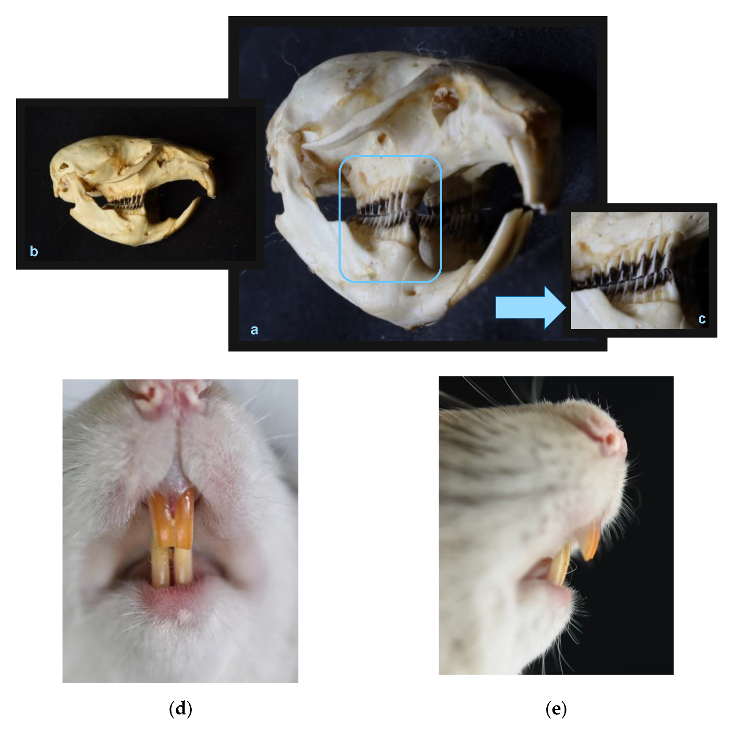
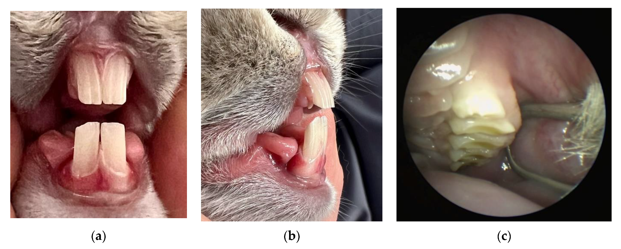
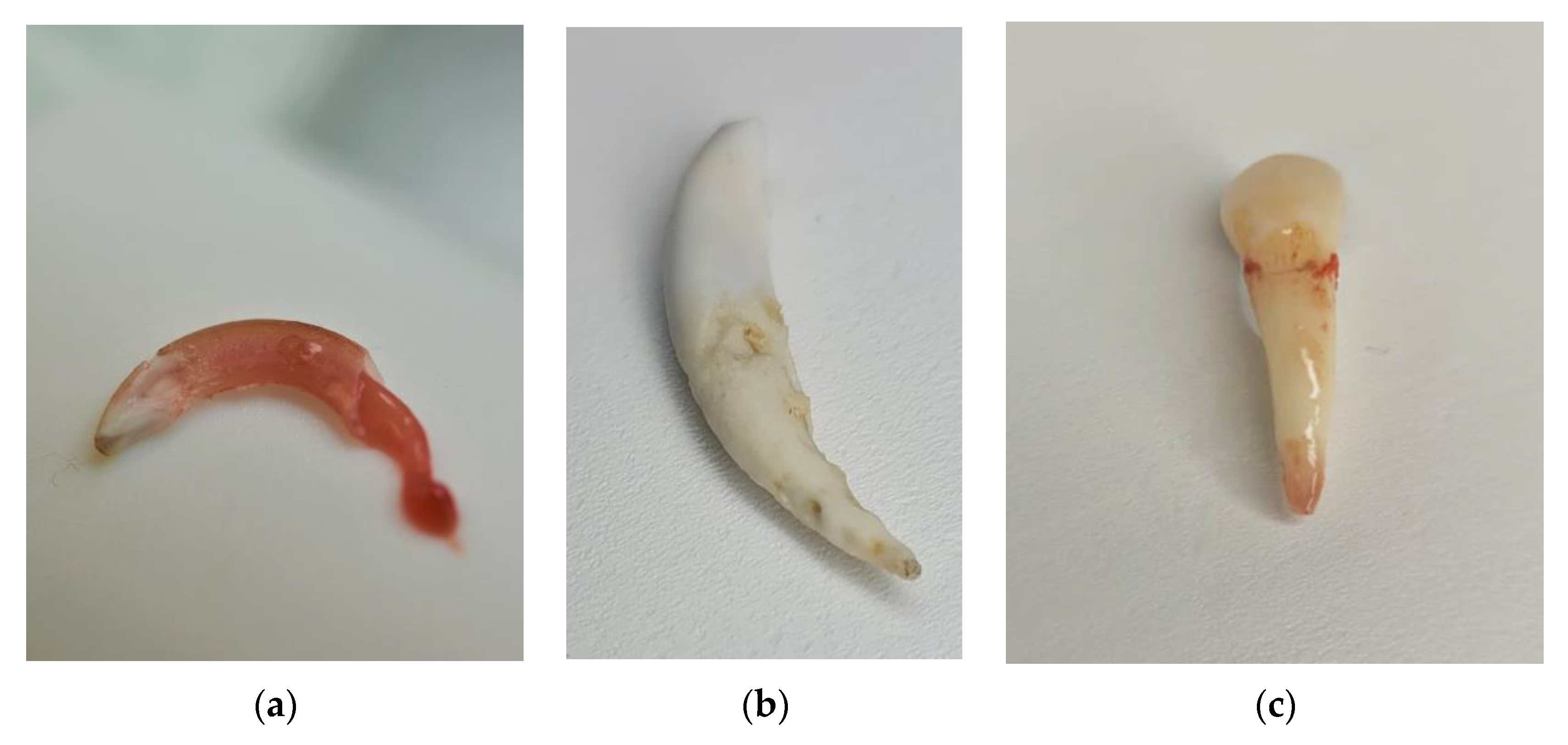
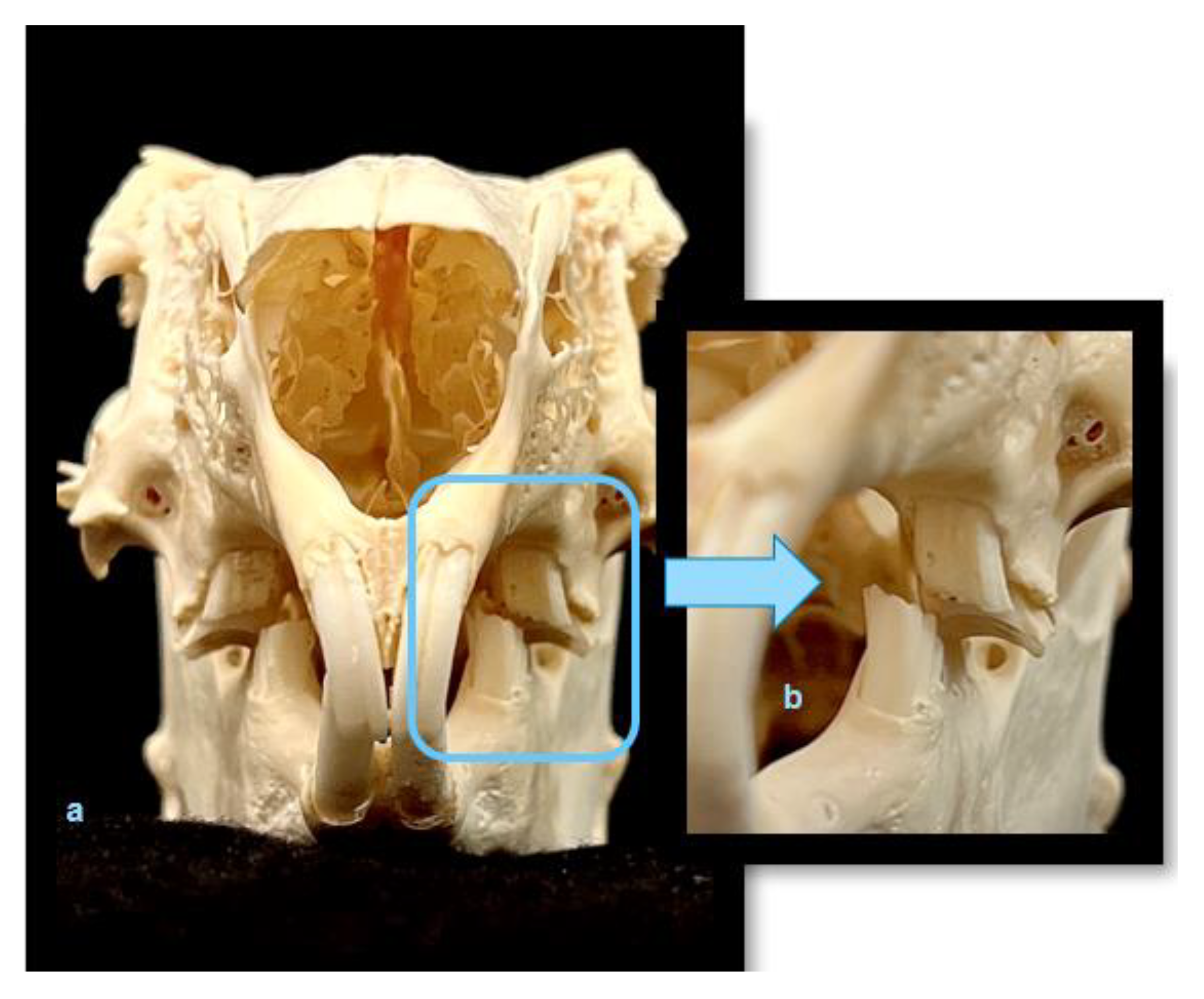
4. Rats
5. Rabbits
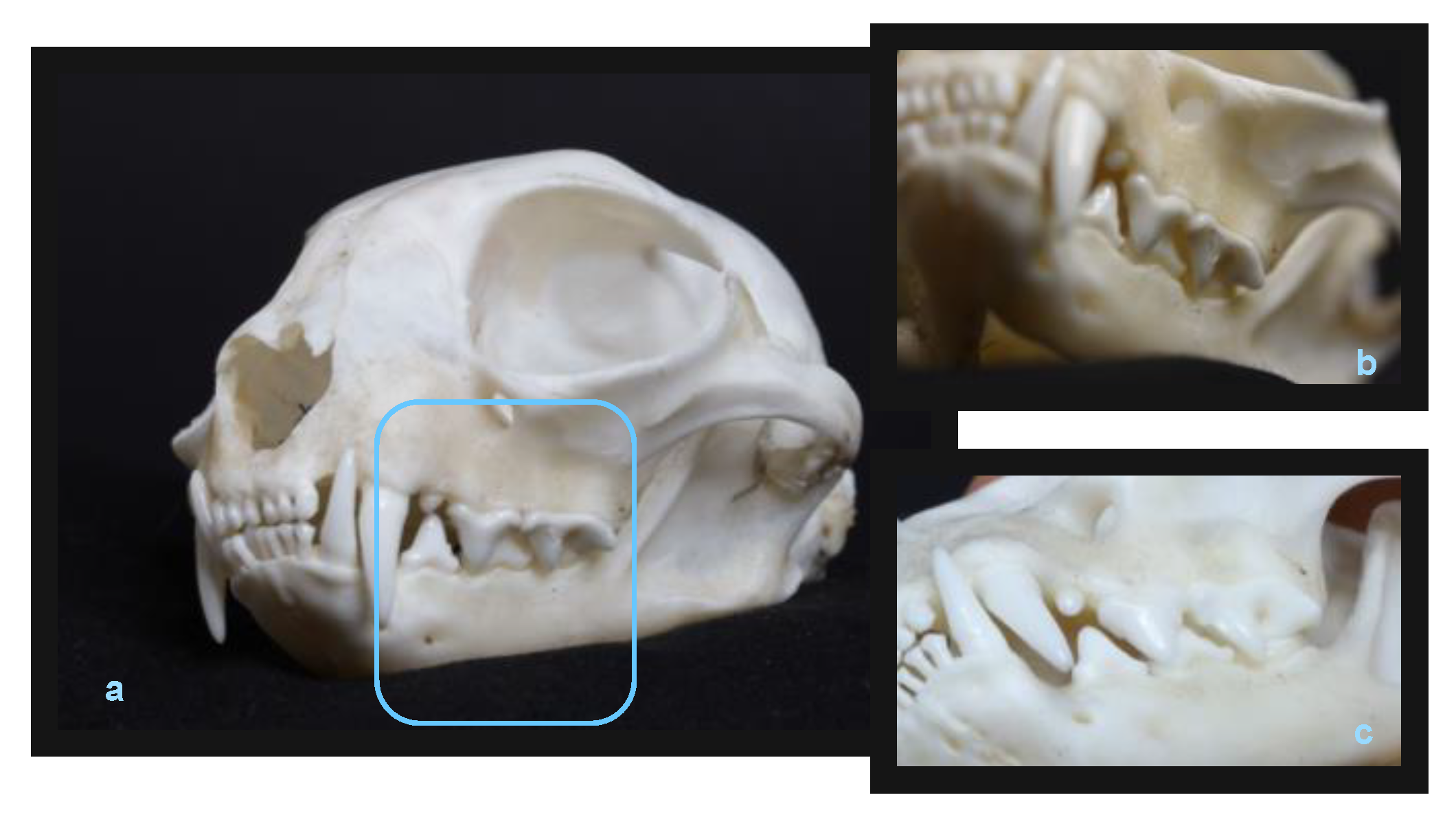
6. Cats
7. Hormone Cycle
8. Orthodontic Materials
9. Conclusions
Author Contributions
Funding
Institutional Review Board Statement
Informed Consent Statement
Data Availability Statement
Acknowledgments
Conflicts of Interest
References
- Midgley, M. Animals and Why They Matter; University of Georgia Press: Georgia, GA, USA, 1998; p. 28. [Google Scholar]
- Directive 2010/63/EU of the European Parliament and of the Council of 22 September 2010 on the Protection of Animals Used for Scientific Purposes. Available online: https://eur-lex.europa.eu/legal-content/EN/TXT/PDF/?uri=CELEX:32010L0063&from=pl (accessed on 9 February 2023).
- Ustawa z Dnia 15 Stycznia 2015 r. o Ochronie Zwierząt Wykorzystywanych do Celów Naukowych lub Edukacyjnych. Available online: https://isap.sejm.gov.pl/isap.nsf/DocDetails.xsp?id=WDU20150000266 (accessed on 9 February 2023).
- Goñi-Balentziaga, O.; Ortega-Saez, I.; Vila, S.; Azkona, G. A survey on the use of mice, pigs, dogs and monkeys as animal models in biomedical research in Spain. Lab. Anim. Res. 2022, 38, 14. [Google Scholar] [CrossRef]
- Ober, R.A.; Ho, J.W.; Kemp, M.T.; Keeney-Bonthrone, T.P.; Geist, G.E.; Alam, H.B. Culture and collaboration between the clinician-scientist and veterinary specialist: An essential interprofessional partnership in the translational sciences. Lab. Anim. 2022, 51, 95–97. [Google Scholar] [CrossRef]
- Azkona, G.; Sanchez-Pernaute, R. Mice in translational neuroscience: What R we doing? Prog. Neurobiol. 2022, 217, 102330. [Google Scholar] [CrossRef] [PubMed]
- Peruga, M.; Antoszewska-Smith, J. Orthodontic tooth movement and changes in hormone levels during the menstrual cycle. Clin. Orthod. 2018, 4, 23–27. [Google Scholar]
- Magowan, B.A.; Owen, P.; Thomson, A. Clinical Obstetrics and Gynaecology; Elsevier: Amsterdam, The Netherlands, 2022. [Google Scholar]
- Tal, R.; Taylor, H.S. Endocrinology of Pregnancy. In Endotext [Internet]; Feingold, K.R., Anawalt, B., Boyce, A., Chrousos, G., de Herder, W.W., Dhatariya, K., Dungan, K., Hershman, J.M., Hofland, J., Kalra, S., et al., Eds.; MDText.com, Inc.: South Dartmouth, MA, USA, 2000. [Google Scholar]
- Speroff, L.; Fritz, M.; Fritz, M.A. Clinical Gynecologic Endocrinology and Infertility; Lippincott Williams & Wilkins: Philadelphia, PA, USA, 2005. [Google Scholar]
- Sperling, M. Pediatric Endocrinology; Elsevier: Amsterdam, The Netherlands, 2014. [Google Scholar]
- Calina, D.; Docea, A.O.; Golokhvast, K.S.; Sifakis, S.; Tsatsakis, A.; Makrigiannakis, A. Management of Endocrinopathies in Pregnancy: A Review of Current Evidence. Int. J. Environ. Res. Public Health 2019, 16, 781. [Google Scholar] [CrossRef] [PubMed]
- Hamed, N.A.; Mirza, K.B.; AL-Rubaie, M.S. Effects of Oral Contraceptives Intake on the Gingiva. Iraqi Postgrad. Med. J. 2010, 9, 335–341. [Google Scholar]
- Brunet, L.; Miranda, J.; Farre, M. Gingival Enlargement induced by drugs. Drug Saf. 1996, 15, 219–231. [Google Scholar] [CrossRef]
- Olyaee, P.; Mirzakouchaki, B.; Ghajar, K.; Seyyedi, S.A.; Shalchi, M.; Garjani, A.; Dadgar, E. The effect of oral contraceptives on orthodontic tooth movement in rat. Med. Oral Patol. Oral Cir. Bucal 2013, 18, e146–e150. [Google Scholar] [CrossRef]
- WMA. The Declaration of Helsinki; WMA: Helsinki, Finland, 1964. [Google Scholar]
- Available online: https://ec.europa.eu/environment/chemicals/lab_animals/pdf/SWD_%20part_A_and_B.pdf (accessed on 9 February 2023).
- Available online: https://eur-lex.europa.eu/legal-content/EN/TXT/PDF/?uri=CELEX:52020DC0015&from=EN (accessed on 9 February 2023).
- Guo, J.; Zhao, Q.; Chen, Y.X.; Zeng, X.L. Effects of orthodontic tooth movements on serum and local estrogen expression. Shanghai Kou Qiang Yi Xue 2007, 16, 618–622. [Google Scholar]
- Guo, J.; Zhao, Q.; Chen, Y.X. A bio-mechanism study of differential orthodontic tooth moving speed during the estrous cycle. Hua Xi Kou Qiang Yi Xue Za Zhi 2008, 26, 327–330. [Google Scholar]
- Zhao, Q.; Tan, Z.; Guo, J.; Chen, Y.X. Influences of applying force during the different stages of estrous cycle on orthodontic tooth movement of rats. Hua Xi Kou Qiang Yi Xue Za Zhi 2005, 23, 480–482. [Google Scholar] [PubMed]
- Guo, J.; Che, X.X.; Zhao, Q.; Chen, Y.X. An experimental study on the relationship between the orthodontic tooth movements and menstrual cycle. Shanghai Kou Qiang Yi Xue 2007, 16, 187–191. [Google Scholar]
- Zhao, Q.; Tan, Z.; Guo, J.; Chen, Y.X. Influences of orthodontic tooth movement on estrous cycle and estrogen in rats. Chung Hua Kou Chiang Hsueh Tsa Chih 2006, 41, 90–91. [Google Scholar] [PubMed]
- Haruyama, N.; Igarashi, K.; Saeki, S.; Otsuka-Isoya, M.; Shinoda, H.; Mitani, H. Estrous-cycle-dependent variation in orthodontic tooth movement. J. Dent. Res. 2002, 81, 406–410. [Google Scholar] [CrossRef]
- Tan, Z.; Zhao, Q.; Chen, Y. The mutual effects between orthodontic tooth movement and estrous cycle or estrogen. Biol. Rhythm. Res. 2010, 41, 75–81. [Google Scholar] [CrossRef]
- Celebi, A.A.; Demirer, S.; Catalbas, B.; Arikan, S. Effect of ovarian activity on orthodontic tooth movement and gingival crevicular fluid levels of interleukin-1β and prostaglandin E(2) in cats. Angle Orthod. 2013, 83, 70–75. [Google Scholar]
- Sirisoontorn, I.; Hotokezaka, H.; Hashimoto, M.; Gonzales, C.; Luppanapornlarp, S.; Darendeliler, M.A.; Yoshida, N. Tooth movement and root resorption; The effect of ovariectomy on orthodontic force application in rats. Angle Orthod. 2011, 81, 570–577. [Google Scholar] [CrossRef] [PubMed]
- Mackie, M.A.; Momeni Danaei, S.H.; Habibagahi, S.; Tanide, N.; Montazeri, M. Effect of Ovariectomy on Orthodontic Tooth Movement in Rats. J. Iran. Dent. Assoc. 2016, 28, 86–91. [Google Scholar] [CrossRef]
- Poosti, M.; Basafa, M.; Eslami, N. Progesterone effects on experimental tooth movement in rabbits. J. Calif. Dent. Assoc. 2009, 37, 483–486. [Google Scholar]
- Kobryń, H.; Kobryńczuk, F.; Krysiak, K. Anatomia Zwierząt Tom 1–3 (Animal Anatomy Vol. 1–3); PWN: Warsaw, Poland, 2011. [Google Scholar]
- Bennett, J.C.; McLaughlin, R.P. Fundamentals of Orthodontic Treatment Mechanics; Legrander Publishing: London, UK, 2014. [Google Scholar]
- Mapara, M.; Thomas, B.S.; Bhat, K.M. Rabbit as an animal model for experimental research. Dent. Res. J. 2012, 9, 111–118. [Google Scholar]
- Mizoguchi, Y.; Matsuoka, T.; Asano, Y. Changes in blood parameters in New Zealand White rabbits during pregnancy. Lab. Anim. 2010, 44, 33–39. [Google Scholar] [CrossRef] [PubMed]
- Hanke, H.; Hanke, S.; Finking, G.; Muhic-Lohrer, A.; Muck, A.O. Different Effects of Estrogen and Progesterone on Experimental Atherosclerosis inFemale Versus Male Rabbits. Circulation 1996, 2, 175–181. [Google Scholar] [CrossRef]
- Ren, Y.; Maltha, J.C.; Kuijpers-Jagtman, A.N. The rat as a model for orthodontic tooth movement—A critical review and a proposed solution. Eur. J. Orthod. 2004, 26, 483–490. [Google Scholar] [CrossRef] [PubMed]
- Hellsing, E.; Hammarström, L. The effects of pregnancy and fluoride on orthodontic tooth movements in rats. Eur. J. Orthod. 1991, 13, 223–230. [Google Scholar] [CrossRef] [PubMed]
- He, Z.; Chen, Y.; Luo, S. Effects of pregnancy on orthodontic tooth movements: Effects of progesterone on orthodontic tooth movements in pregnant rats. Hua Xi Kou Qiang Yi Xue Za Zhi 1998, 16, 124–126. [Google Scholar]
- Bartzela, T.; Türp, J.C.; Motschall, E.; Maltha, J.C. Medication effects on the rate of orthodontic tooth movement: A systematic literature review. Am. J. Orthod. Dentofac. Orthop. 2009, 135, 16–26. [Google Scholar] [CrossRef]
- Dschietzig, T.; Bartsch, C.; Baumann, G.; Stangl, K. Relaxin—A pleiotropic hormone and its emerging role for experimental and clinical therapeutics. Pharmacol. Ther. 2006, 112, 38–56. [Google Scholar] [CrossRef] [PubMed]
- Liu, Z.J.; King, G.J.; Gu, G.M.; Shin, J.Y.; Stewart, D.R. Does human relaxin accelerate orthodontic tooth movement in rats? Ann. N. Y. Acad. Sci. 2005, 1041, 388–394. [Google Scholar] [CrossRef]
- Stewart, D.R.; Sherick, P.; Kramer, S.; Breining, P. Use of relaxin in orthodontics. Ann. N. Y. Acad. Sci. 2005, 1041, 379–387. [Google Scholar] [CrossRef] [PubMed]
- Nicozisis, J.L.; Nah-Cederquist, H.D.; Tuncay, O.C. Relaxin affects the dentofacial sutural tissues. Clin. Orthod. Res. 2000, 3, 192–201. [Google Scholar] [CrossRef] [PubMed]
- Tyrovola, J.B.; Spyropoulos, M.N. Effects of drugs and systemic factors on orthodontic treatment. Quintessence Int. 2001, 32, 365–371. [Google Scholar] [PubMed]
- Yamashiro, T.; Takano-Yamamoto, T. Influences of ovariectomy on experimental tooth movement in the rat. J. Dent. Res. 2001, 80, 1858–1861. [Google Scholar] [CrossRef] [PubMed]
- Arslan, S.G.; Arslan, H.; Ketani, A.; Hamamci, O. Effects of estrogen deficiency on tooth movement after force application: An experimental study in ovariectomized rats. Acta Odontol. Scand. 2007, 65, 319–323. [Google Scholar] [CrossRef] [PubMed]
- Brantley, W.; Eliades, T. Orthodontic Materials: Scientic and Clinical Aspects; Thieme: Stuttgart, Germany, 2001. [Google Scholar]


| Authors | Animals and Their Division | Tooth and Type of Tooth Shift | Material Used | Method Used to Test the Hormone Level | Results |
|---|---|---|---|---|---|
| Olyaee et al. [15] | Wistar rats Age—3 months N = 48 m = 250 ± 25 g females | Central incisors tipped distally | SS spring with a diameter of 0.35 mm | Administration of ethinyl estradiol/norgestrel | Ethinyl estradiol/norgestrel (oral contraceptives) can decrease the amount of tooth movement |
| Guo, Zhao, Chen [19] | Wistar rats Age—not given N = 120 m = not given females | Not given | Not given | Estradiol level in serum and periodontium tissue using radioimmune and immune-cytochemical methods | Estrogen affects teeth movement |
| Zhao, Than, Guo, Chen [20] | Wistar rats Age—not given N = not given m = not given females | Not given | Not given | Estradiol level in serum and periodontium tissue using radioimmune and immune-cytochemical methods | Estrogen affects teeth movement |
| Guo, Zhao, Chen [21] | Wistar rats Age—3 months N = 80 m = not given females | Left upper incisor and left upper molar Tipped | Not given | Not given | Teeth movement dependent on cycle |
| Zhao, Than, Guo, Chen [22] | Wistar rats Age—not given N = not given m = not given females | Not given | Not given | Not given | Estrogen affects teeth movement |
| Guo, Zhao, Chen [23] | Rats, strain not given Age—not given Not given N = 200 m = not given females | Not given | Not given | Estradiol level in serum | Estrogen affects teeth movement |
| Haruyama et al. [24] | Wistar rats Age—10 weeks N = 85 m = 136 g females | right and left, first upper molar Tipped | NiTi spring with a diameter of 0.012 inch | Acc. to estrous cycle with vaginal smear | Estrus can increase tooth movement |
| Tan et al. [25] | Wistar rats Age—3 months N = 200 m = 300 g females | Left distal incisor and first left molar Tipped | NiTi spring with a diameter of 0.012 inch | Monitoring estrus cycle and vaginal smear | Estrus can increase tooth movement |
| Celebi [26] | Domestic cat Age—2–4 years N = 18 m = not given females | Jaw canine and mini implant Tipped | NiTi spring with a diameter of 0.2 inch | According to estrus cycle | Teeth movement speed was higher in sterilized specimens |
| Sirisoontorn [27] | Wistar rats Age—10 weeks N = 10 m = 170–190 g females | Left distal incisor and and first left molar Tipped | NiTi spring with a diameter of 0.012 inch | Monitoring estrus cycle and vaginal smear | Teeth movement speed was higher in sterilized specimens |
| Mackie et al. [28] | Sprague Dawley rats Age—6 weeks N = 55 m = 160 ± 20 g females | Right distal incisor and first right molar Tipped | NiTi spring with a diameter of 0.03 × 0.01 inch | Not given | Estrus can increase tooth movement |
| Poosti et al. [29] | Rabbit Age—8 weeks N = 24 m = 1850 g females | Central incisors tipped distally | SS spring with a diameter of 0.014 inch | Administration of progesterone | Progesterone affects tooth movement |
| Species | Size | Dental Formula | Teeth | Periodontium | Hormones | Body Temperature |
|---|---|---|---|---|---|---|
| Human (Homo sapiens) | Target species | Diphyodont Heterodont Bunodont | Thecodont Brachydont | Cycle 24–33 days Ovulation 24 h | 36.6 °C | |
| Laboratory rat (Rattus) | Acceptable size | Monophyodont heterodont | Thecodont Monophyodont Incisors—hypsodont, elodont Molars—brachydont | Cycle 4–5 days Spontaneous ovulation Estrus 10–20 h | 37.5–39 °C | |
| Rabbit (Oryctolagus cuniculus) | Acceptable size | Monophyodont Heterodont | Thecodont Hypsodont Elodont | Cycle 16 days Induced ovulation | 38.5–40 °C | |
| Domestic cat (Felis catus) | Acceptable size | Diphiodont Heterodont Secodont | Thecodont Brachydont | 14–21 days | 38–39 °C |
Disclaimer/Publisher’s Note: The statements, opinions and data contained in all publications are solely those of the individual author(s) and contributor(s) and not of MDPI and/or the editor(s). MDPI and/or the editor(s) disclaim responsibility for any injury to people or property resulting from any ideas, methods, instructions or products referred to in the content. |
© 2023 by the authors. Licensee MDPI, Basel, Switzerland. This article is an open access article distributed under the terms and conditions of the Creative Commons Attribution (CC BY) license (https://creativecommons.org/licenses/by/4.0/).
Share and Cite
Peruga, M.; Kawala, B.; Sarul, M.; Kotowicz, J.; Lis, J. Are Currently Selected Laboratory Animals Useful in the Research of How Female Hormones Influence Orthodontic Biomechanics? Animals 2023, 13, 629. https://doi.org/10.3390/ani13040629
Peruga M, Kawala B, Sarul M, Kotowicz J, Lis J. Are Currently Selected Laboratory Animals Useful in the Research of How Female Hormones Influence Orthodontic Biomechanics? Animals. 2023; 13(4):629. https://doi.org/10.3390/ani13040629
Chicago/Turabian StylePeruga, Małgorzata, Beata Kawala, Michał Sarul, Jakub Kotowicz, and Joanna Lis. 2023. "Are Currently Selected Laboratory Animals Useful in the Research of How Female Hormones Influence Orthodontic Biomechanics?" Animals 13, no. 4: 629. https://doi.org/10.3390/ani13040629