Communication

Comparative Transcriptome Analysis Reveals Sexually Dimorphic Gene Expression in the Gonads of Brachymystax tsinlingensis Li

Key Laboratory of Freshwater Biodiversity Conservation, Ministry of Agriculture and Rural Affairs, Yangtze River Fisheries Research Institute, Chinese Academy of Fishery Sciences, Wuhan 430223, China
*
Authors to whom correspondence should be addressed.
These authors contributed equally to this work.
Animals 2023, 13(23), 3690; https://doi.org/10.3390/ani13233690
Submission received: 27 September 2023 / Revised: 12 November 2023 / Accepted: 24 November 2023 / Published: 29 November 2023

Abstract

:

Simple Summary

Brachymystax tsinlingensis Li has become an endangered species due to human activities and habitat degradation. However, little is known about the genes regulating its gonadal development. This study presents a high-quality gonadal transcriptome of B. tsinlingensis Li. A total of 22,864 differentially expressed genes between male and female were identified. Numerous sex-related differentially expressed genes, such as ccnb1, zp3, bmp15, dmrt1, and psmc3ip, were identified. Some signaling pathways involved in gonadal development, such as genes involved in base excision repair, the notch signaling pathway, neuroactive ligand-receptor interaction, the VEGF signaling pathway, and the estrogen signaling pathway, were found to be enriched through analysis using the Kyoto Encyclopedia of Genes and Genomes. Our results provide molecular data to support a better understanding of sexual development in B. tsinlingensis Li.

Abstract

Brachymystax tsinlingensis Li is an endangered cold-water salmonid fish native to China. This study aimed to identify sex-related genes and biological pathways via gonadal transcriptome sequencing of B. tsinlingensis Li. A total of 167,904 unigenes were identified with an average length of 836 bp and an N50 of 1452 bp, of which 84,977 (50.61%) unigenes were successfully annotated in six major databases. Comparative transcriptome analysis identified 22,864 differentially expressed genes (DEGs), of which 17,231 were up-regulated (male-biased genes, mDEGs) and 5633 were down-regulated (female-biased genes, fDEGs). Several DEGs associated with gonadal development were found through Gene Ontology enrichment analysis, such as ccnb1, zp3, bmp15, dmrt1, and psmc3ip. Signaling pathways related to gonadal development were found to be enriched through analysis using the Kyoto Encyclopedia of Genes and Genomes Pathway database, such as genes involves in base excision repair, the notch signaling pathway, neuroactive ligand-receptor interaction, the VEGF signaling pathway, and the estrogen signaling pathway. In addition, mRNA expression levels of 19 DEGs were determined to validate the reliability of the transcriptomic data by quantitative real-time polymerase chain reaction. These results revealed genes and signaling pathways potentially involved in gonadal development in B. tsinlingensis Li and provided basic molecular data for future research on reproductive regulation and breeding of B. tsinlingensis Li.

1. Introduction

Brachymystax tsinlingensis Li, which belongs to the Salmonidae family, is widely distributed in southern China. It originated from Siberia during the fourth glacial period and migrated to the cold-water streams of the Hei River, Shitou River, Xushui River, and Taibai River in the Tsinling Mountains as the glacier advanced [1]. However, due to natural and human factors, such as global warming, environmental destruction, and overfishing, the wild population of this species has declined severely and only 10% of its historical population currently remains. This species was listed as a second-class nationally protected animal in China in 1988 [2,3]. Artificial propagation and release is one effective method by which to restore the wild population of B. tsinlingensis Li. However, large-scale artificial propagation of this species has not been successful, partly because of our limited knowledge of the molecular mechanisms governing reproduction in B. tsinlingensis Li.
Previous studies in B. tsinlingensis Li mainly focused on behaviours [4], molecular genetic markers [5], feed additives [6], and morphology and the ultrastructure of the sperm [7]. However, research on reproductive development in B. tsinlingensis Li is still lacking, although such research is essential for artificial propagation and further species conservation. Transcriptome sequencing technology has been widely used to study gene-expression profiles and molecular mechanisms with high efficiency and low-cost [8]. Comparative gonadal transcriptome analysis has been used to reveal molecular pathways and genes involved in gonadal development and reproduction in aquatic animals. In Hong Kong catfish (Clarias fuscus), many differentially expressed genes (DEGs) were found to be enriched using sex-related gene ontology (GO) terms and analysis through Kyoto Encyclopedia of Genes and Genomes (KEGG) pathways, such as oocyte maturation, androgen secretion, gonadal development and steroid biosynthesis [9]. The dmrt1 gene has been revealed to play a dual role during testicular development in largemouth bass (Micropterus salmoides) [10]. In sea urchin (Strongylocentrotus intermedius), five DEGs related to gonadal quality have been identified [11]. Thus, the exploration of sex-related genes and biological pathways is an effective strategy by which to reveal sexual regulatory mechanisms.
This study presents a high-quality gonadal transcriptome of B. tsinlingensis Li. The gene-expression profiles of the testis and ovary were obtained, and candidate genes and biological pathways potentially related to gonadal development were further identified. Our study provides valuable insights for further research on the reproductive regulation and breeding of B. tsinlingensis Li.

2. Materials and Methods

2.1. Total RNA Extraction, cDNA Library Construction and Illumine Sequencing

A total of ten artificially cultured juvenile B. tsinlingensis Li (5 males and 5 females) collected from the Zhangjiachuan Malu Brachymystax tsinlingensis Li Artificial Breeding Farm in Gansu Province, China, were used for gonadal transcriptome analysis. The average length and weight of the male fish were 149.4 ± 11.52 mm and 58.4 ± 9.99 g, respectively, while those of the female fish were 143.2 ± 12.24 mm and 43.5 ± 11.03 g. After anesthesia, the gonads were sampled and stored in RNA Later (Sartorius, Gttingen, Germany) for RNA extraction; meanwhile, partial gonads were fixed in Bouin’s fixative solution (Scientific Phygene, Fuzhou, China), then subjected to gradient dehydration with ethanol, transparent tissue with xylene, paraffin dip, sectioning, and hematoxylin and eosin (HE) staining for tissue-structure evaluation. An RNeasy Plus Mini Kit (Qiagen, Dusseldorf, Germany) was used for extraction of total RNA. The total RNA was finally obtained by homogenizing gonadal tissue, removing the gDNA and purifying the RNA. The RNA quality was checked on a Nanodrop 2000 (Thermo Scientific, Waltham, MA, USA) and electrophoresis. High-quality RNA samples were sent to Meiji Biopharmaceutical Co., Ltd. (Shanghai, China) for construction of cDNA libraries and sequenced on the Illumina NovaSeq 6000 sequencing platform. All experimental procedures were carried out in accordance with the guiding principles for the care and use of laboratory animals set by the Yangtze River Fisheries Research Institute, Chinese Academy of Fishery Sciences, China.

2.2. De Novo Assembly and Functional Annotation

Fastp (version 0.19.5) was used to perform quality control for the original data. The numbers of raw reads, clean reads, raw bases and clean bases, as well as the error rate, Q30, and GC content were calculated to visualize the quality of library construction and sequencing. Generally, the error rate was below 0.1% and Q30 was above 80%. The clean reads have been submitted to the NCBI SRA database under the accession numbers SRR25744158, SRR25744157, SRR25744156, SRR25744155, SRR25744154, SRR25744153, SRR25744152, SRR25744151, SRR25744150, and SRR25744149.
The clean reads were then assembled into transcripts using Trinity (version v2.8.5), and the longest transcript for each gene was selected as the unigene. All unigenes were homology-annotated to the NCBI non-redundant protein database (NR), Swiss-Prot database, protein families database (Pfam), Evolutionary Genealogy of Genes: Non-supervised Orthologous Groups (eggNOG), GO, and KEGG, and the results were counted to obtain comprehensive functional information for the unigenes.

2.3. Identification of DEGs and Functional Enrichment Analysis

Gene-expression levels from each sample were estimated by RSEM (Version 1.3.1). The TPM (transcripts per million) algorithms were used to normalize mRNA expression levels. Sample correlation analysis and sample principal component analysis (PCA) were performed to validate the reliability of data and the rationality of the experimental design.
Based on the results for normalized reads, DESeq2 (Version 1.24.0) software was used to perform differential expression analysis of the genes. The p-values were corrected using the Benjamini–Hochberg (BH) method and represented as p-adjust. Unigenes with p-adjust < 0.001 & |log2FC| ≥ 4, which means that unigenes showing at least sixteen-fold expression difference between male and female were considered as DEGs. Compared with ovaries, DEGs with log2FC ≥ 4 were up-regulated in testes (male-biased genes, mDEGs), and those with log2FC ≤ −4 were down-regulated in testes (female-biased genes, fDEGs). Functional classification of DEGs was further analyzed by GO and KEGG annotation.

2.4. Quantitative Real-Time Polymerase Chain Reaction (qPCR) Verification

The accuracy of the RNA-Seq data was validated by qPCR. Gonad RNAs from five males and females used for transcriptomic sequencing were selected for gene-expression profiling. A total of 1 μg RNA was reverse-transcribed with the PrimeScript RT Reagent Kit With gDNA Eraser (Takara, Kyoto, Japan), and cDNA was diluted five-fold and stored at −20 °C until use. Quantitative real-time PCR was performed using a QuantStudio6 Flex PCR System (Applied Biosystems, CA, America) with PowerUpTM SYBRTM Green Master Mix. The sequence and amplification efficiency of the primers are listed in Table S1. β-actin was used as an internal reference. All samples were analyzed in triplicate, and relative gene expression was calculated using the 2−ΔΔCt method.

2.5. Statistics Analysis

Statistical analysis was performed using Student’s t-test, with final data expressed as mean ± standard error. Differences were considered as significant when * p < 0.05, ** p < 0.01, and *** p < 0.001. All statistical data were plotted using GraphPad Prism 8.0.

3. Results

3.1. Sex Identification and Histological Characteristics of Juvenile B. tsinlingensis Li Gonads

The histological characteristics of B. tsinlingensis Li gonads were determined by HE staining (Figure 1). The ovary had numerous primary oocytes and few oogonia, and the testes contained different types of germ cells, including spermatogonia, primary spermatocytes, secondary spermatocytes, spermatids, and spermatozoa.

3.2. Transcriptome Sequencing and Assembly

A total of 121.76 Gb of clean data was obtained. The Q30 values of the clean data were above 94.13%. The GC content was approximately 49.5%, with sequencing error rates no greater than 0.0251. A total of 167,904 unigenes were assembled. The minimum and maximum lengths of unigenes were 201 and 26,461 bp, respectively, with an N50 length of 1452 bp. The unigenes with sequence lengths exceeding 2000 bp and 3000 bp accounted for 10% and 5%, respectively. Moreover, the percentage of clean reads that could be mapped to the assembled transcripts ranged from 77.23% to 79.41%. Detailed information about transcriptome sequencing and assembly is shown in Table 1 and Table 2.

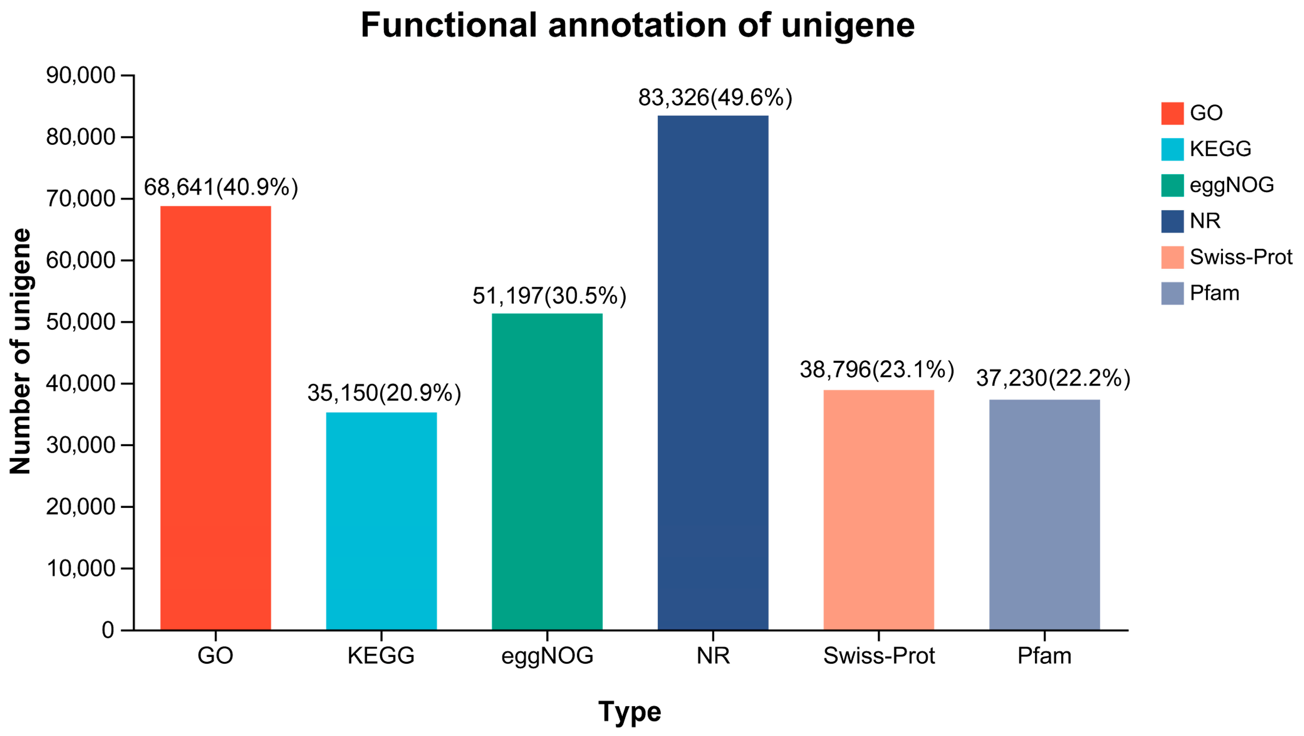
3.3. Functional Annotation

A total of 84,977 (50.61%) unigenes were annotated through six databases (NR, Swiss-Prot, Pfam, eggNOG, GO, and KEGG) (Figure 2). A large number of unigenes were matched to the NR and GO databases, accounting for 83,326 (49.6%) and 68,641 (40.9%), respectively. Based on the annotation in the NR database, 83,326 unigenes in B. tsinlingensis Li were matched to 165 species, of which 82,883 (99.5%) unigenes belonged to Actinopteri (Table S2). The greatest number of unigenes were homologous to genes of Oncorhynchus mykiss (38,174, 45.81%), followed by Salmo salar (19,293, 23.15%), Salvelinus alpinus (7064, 8.48%), etc. (Figure 3A). The number of unigenes with similarity above 80% was 58,464 (70.16%), and the number of unigenes with similarity of 60–80% was 19,981 (23.98%) (Figure 3B). Results with smaller E-values have higher confidence, and 47.62% of the E-values were distributed between 0 and 1 × 10⁻³⁰ (Figure 3C), suggesting high accuracy and reliability of the RNA-Seq sequencing.

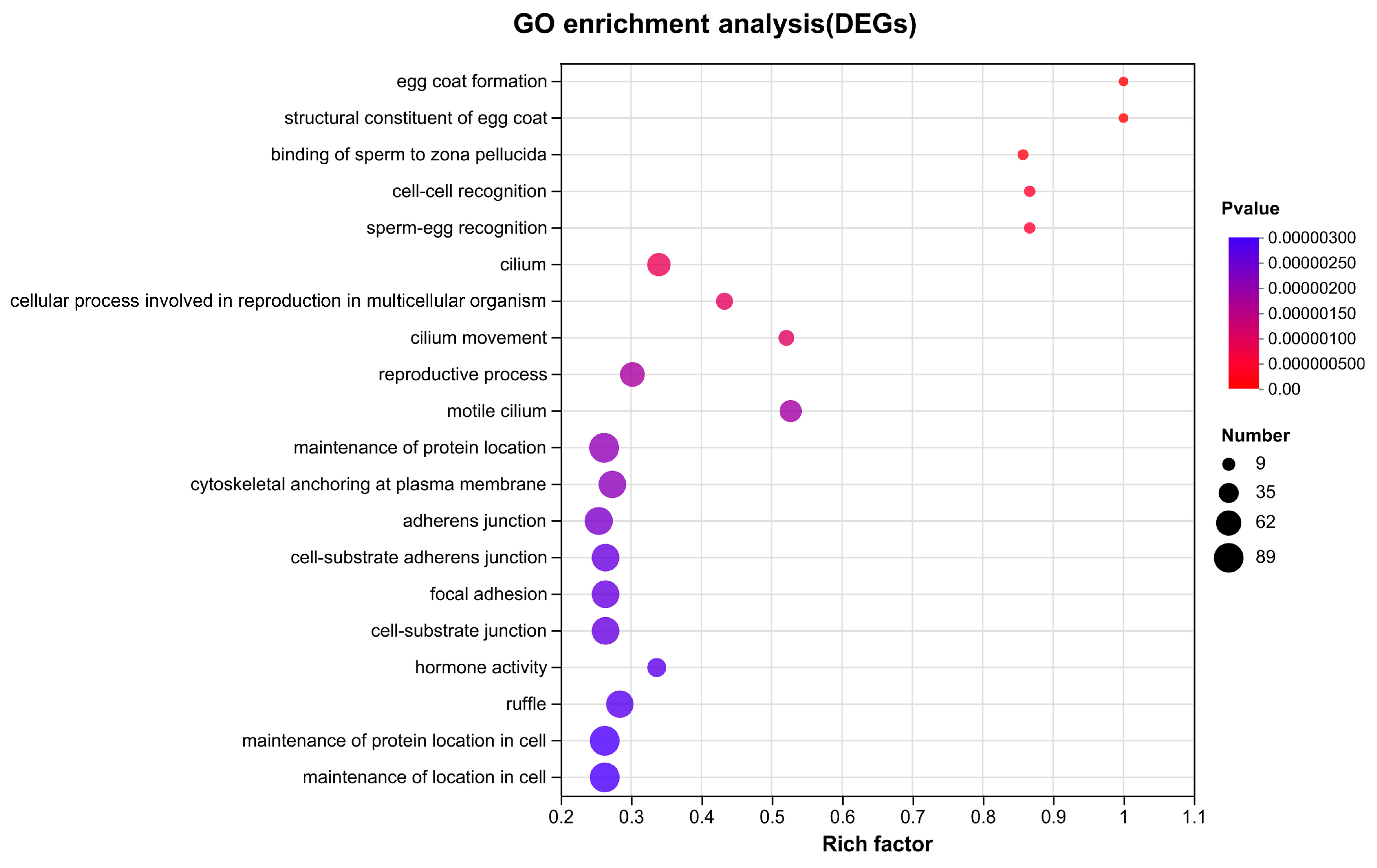
3.4. DEGs Identification and Enrichment Analysis

To analyze the global similarities and differences between the testis and the ovary, correlation analysis and PCA were performed. The results showed that samples in the same group displayed similar expression patterns, while samples in different groups were clearly distinguishable (Figure S1), indicating the reliability and repeatability of the data. Comparative analysis were performed to identify sex-biased genes. A total of 22,864 DEGs, including 17,231 male-biased genes and 5633 female-biased genes, were identified (Figure 4).
DEGs were further classified by GO and KEGG annotation to investigate their potential functions and metabolic pathways (Figure S2). The top 20 GO enrichment terms are shown in Figure 5. Sex-related terms, such as egg coat formation (BP, GO:0035803), structural constituent of egg coat (MF, GO:0035804), binding of sperm to zona pellucida (BP, GO:0007339), sperm-egg recognition (BP, GO:0035036), and reproductive process (BP, GO:0022414) were significantly enriched, with numerous reproductive-related genes included, such as zp3, bmp15, ccnb1, psmc3ip, hsd17b3, piwi, sycp1, pvrl3, zent9, syce1, syce2, syce3, catsper2, cep4, igf1r, diaph2, plcz, etc. A scatterplot of the top 20 enriched KEGG pathways of DEGs is presented in Figure 6. Several KEGG pathways related to reproduction were further identified, such as pathways related to base excision repair, the notch signaling pathway, cardiac muscle contraction, protein digestion and absorption, and ribosome biogenesis in eukaryotes in the testis (Figure 6A) and neuroactive ligand-receptor interaction, glycosphingolipid biosynthesis–globo and isoglobo series, cytokine-cytokine receptor interaction, the VEGF signaling pathway, and the estrogen signaling pathway in the ovary (Figure 6B).

3.5. Validation of DEGs by qPCR

To validate the reliability of the RNA-seq results, the expression levels of 19 DEGs (8 fDEGs and 11 mDEGs) were determined by qPCR (Figure 7). The transcript levels of fDEGs, such as cyp19a, zp1, trim25, parp7s, ccnb1, zar1, zp3, and bmp15, were significantly higher than those in the testis, while the transcript levels of mDEGs, such as nanos2, fshr, dmrt1, tcte1, theg, strbp, cep4, pamc3ip, pvrl3, sycp1, and znt9, were significantly higher than those in the ovary. Overall, the qPCR results strongly confirmed the accuracy and reliability of transcriptome expression analysis conducted for B. tsinlingensis Li.

4. Discussion

The population of B. tsinlingensis Li has decreased significantly due to human activities and natural environmental changes, resulting in its being assigned endangered status. Artificial propagation and release is considered an important strategy through which to conserve and restore this species. However, little is known about the genes and regulatory mechanisms of reproductive development in B. tsinlingensis Li. This study was the first report of gonadal transcriptome analysis in B. tsinlingensis Li and revealed sex-related DEGs and regulatory pathways. To some extent, these results provide new insights into sexual development in B. tsinlingensis Li.
Various sex-biased genes, including both female-biased genes (ccnb1, zp3, bmp15) and male-biased genes (psmc3ip, dmrt1), were identified by analyzing the gene-expression profiles in the gonads of B. tsinlingensis Li. It has been reported that ccnb1 has positive significance in the meiotic division of oocytes and in maintaining fertility [12,13]. Zp3, a critical component of the zona pellucida, plays a vital role in oocyte meiosis and maturation, and knockout of zp3 leads to complete infertility [14]. Previous studies demonstrated that bmp15 was necessary for the development and differentiation of oocytes [15,16]. In this study, ccnb1, zp3, and bmp15 were highly expressed in the ovary (Figure 7F,H,I), implying that they have important functions during oocyte development and maintenance of the female phenotype in B. tsinlingensis Li.
Some testicular genes of B tsinlingensis Li were also found. The dmrt1 gene exists on the Y chromosome in the males of some fishes [17,18] and regulates the differentiation and proliferation of male germ cells and germline stem cells [19,20,21,22]. Moreover, several studies have found that dmrt1 inhibits the sexual switch from male to female [23,24,25]. The psmc3ip gene is essential for homologous recombination during meiotic division, and deficiency of psmc3ip results in azoospermia [26,27]. Our results showed high transcription levels of dmrt1 and psmc3ip in the testis of B. tsinlingensis Li (Figure 7L,Q), suggesting their crucial roles as regulators of testicular development and spermatogenesis.
In this study, significantly different KEGG enrichment results for DEGs were acquired from a comparative analysis of the testis and ovary of B. tsinlingensis Li, indicating their different regulatory mechanisms for gonadal development and gametogenesis. The results showed that mDEGs were most enriched in base excision repair (BER) and the notch signaling pathway (Figure 6A). BER is the main pathway for repairing DNA base damage and lesions [28,29]. Our results showed that BER was the most enriched pathway in the testis, consistent with the detection of high levels of BER-related gene-transcription products in male gonads [30,31,32,33]. Additionally, BER was an important pathway for maintaining genomic integrity in male germ cells [34]. Notch signaling components can always be detected in the Sertoli and germ cells of male gonads, and research has shown that the notch signaling pathway is crucial for regulating testicular early development, spermatogonial differentiation, and spermatogenesis [35,36,37]. Our results emphasized the importance of BER and notch signaling in the testicular reproductive regulation of B. tsinlingensis Li.
The fDEGs were significantly enriched in the neuroactive ligand-receptor interaction pathway, the VEGF signaling pathway, and the estrogen signaling pathway. The neuroactive ligand-receptor interaction pathway can impact the synthesis of steroid hormones in the gonads via the HPG axis and plays an important regulatory role in ovulation [38,39]. Moreover, similar significant enrichment results were found in gonadal transcriptomic studies of chicken, sheep and goose gonads [40,41,42]. VEGF has been recognized as one of the most relevant angiogenetic factors in orchestrating folliculogenesis, and VEGF inhibition prevents follicle ovulation [43,44,45]. Moreover, estrogen has a significant regulatory role in oogenesis and ovulation [46]. Thus, our results revealed that these pathways may play vital roles in ovary development and gametogenesis in B. tsinlingensis Li. Additionally, many other biological pathways are also significantly enriched (Figure 6), and their functions require further investigation.

5. Conclusions

In this study, the first comparative transcriptome analysis between the testis and ovary of B. tsinlingensis Li was reported. A total of 22,864 DEGs were identified, including 17,231 male-biased DEGs and 5633 female-biased DEGs. Numerous genes related to reproductive development were explored, including ccnb1, zp3, bmp15, dmrt1, and psmc3ip. Biological pathways potentially involved in reproductive regulation were also identified. These pathways included base excision repair, the notch signaling pathway, neuroactive ligand-receptor interaction, the VEGF signaling pathway, and the estrogen signaling pathway. The results provide new insights into the reproductive development of B. tsinlingensis Li and will be beneficial for the further artificial breeding of this species.

Supplementary Materials

The following supporting information can be downloaded at: https://www.mdpi.com/article/10.3390/ani13233690/s1, Figure S1: correlation analysis (A) and principal component analysis (B) for samples; Figure S2: GO classification (A) and KEGG pathway analysis (B) of DEGs; Table S1: list of primers used for quantitative RT-PCR validation; Table S2: species distribution of NR protein database annotations.

Author Contributions

L.H. and H.Y. (Huan Ye), conceptualization, investigation, data curation, writing-original draft preparation; H.Y. (Huamei Yue), methodology, investigation, writing-original draft preparation; X.L., R.R. and H.D., methodology, resources; C.L. and J.W., conceptualization, supervision, project administration, writing—review & editing. All authors have read and agreed to the published version of the manuscript.

Funding

This research was funded by the National Key R&D Program of China (grant numbers 2021YFD1200304) and National Natural Science Foundation of China (grant number 31602165).

Institutional Review Board Statement

The experiments were approved by the Animal Experimental Ethical Inspection of Laboratory Animal Centre, Yangtze River Fisheries Research Institute, Chinese Academy of Fishery Sciences. The Ethics Approval Code is YFIYH01.

Informed Consent Statement

Not applicable.

Data Availability Statement

The data sets supporting the results of this article have been submitted to GenBank and accession numbers are shown in the article.

Conflicts of Interest

The authors declare that they have no known competing financial interests or personal relationships that could have appeared to influence the work reported in this paper.

References

  1. Xiong, D.; Meng, Y.; Zhang, X.; Wang, J.; Feng, G.; Shao, J.; Wang, L.; Meng, Y.; Zhang, X.; Wang, J.; et al. The validity of species of Brachymystax tsinlingensis Li based on mitochondria control region and microsatellite. Acta Hydrobiol. Sin. 2023, 47, 809–818, (In Chinese with English abstract). [Google Scholar]
  2. Xia, J.; Ma, Y.; Fu, C.; Fu, S.; Cooke, S.J. Effects of temperature acclimation on the critical thermal limits and swimming performance of Brachymystax lenok tsinlingensis: A threatened fish in Qinling Mountain region of China. Ecol. Res. 2017, 32, 61–70. [Google Scholar] [CrossRef]
  3. Zhao, Y.; Zhang, C. Threatened fishes of the world: Brachymystax lenok tsinlingensis Li, 1966 (Salmonidae). Environ. Biol. Fishes. 2009, 86, 11–12. [Google Scholar] [CrossRef]
  4. Li, P.; Liu, Q.; Li, J.; Wang, F.; Wen, S.; Li, N. Transcriptomic responses to heat stress in gill and liver of endangered Brachymystax lenok tsinlingensis. Comp. Biochem. Physiol. Part. D Genom. Proteom. 2021, 38, 100791. [Google Scholar] [CrossRef] [PubMed]
  5. Wen, S.; Li, P.; Wang, F.; Li, J.; Liu, H.; Li, N. De novo assembly and microsatellite marker development of the transcriptome of the endangered Brachymystax lenok tsinlingensis. Genes. Genom. 2020, 42, 727–734. [Google Scholar] [CrossRef]
  6. Ma, F.; Ma, R.; Zou, Y.; Zhao, L. Effect of astaxanthin on the antioxidant capacity and intestinal microbiota of tsinling lenok trout (Brachymystax lenok tsinlingensis). Mar. Biotechnol. 2022, 24, 1125–1137. [Google Scholar] [CrossRef]
  7. Guo, W.; Shao, J.; Li, P.; Wu, J.; Wei, Q. Morphology and ultrastructure of Brachymystax lenok tsinlingensis spermatozoa by scanning and transmission electron microscopy. Tissue Cell. 2016, 48, 321–327. [Google Scholar] [CrossRef]
  8. Wang, Z.; Sun, L.; Guan, W.; Zhou, C.; Tang, B.; Cheng, Y.; Huang, J.; Xuan, F. De novo transcriptome sequencing and analysis of male and female swimming crab (Portunus trituberculatus) reproductive systems during mating embrace (stage II). BMC Genet. 2018, 19, 3. [Google Scholar] [CrossRef]
  9. Lin, X.; Zhou, D.; Zhang, X.; Li, G.; Zhang, Y.; Huang, C.; Zhang, Z.; Tian, C. A first insight into the gonad transcriptome of hong kong catfish (Clarias fuscus). Animals 2021, 11, 1131. [Google Scholar] [CrossRef]
  10. Guan, W.Z.; Jiang, K.; Lai, X.L.; Dong, Y.T.; Qiu, G.F. Comprehensive transcriptome analysis of gonadal and somatic tissues for identification of sex-related genes in the largemouth bass Micropterus salmoides. Mar. Biotechnol. 2022, 24, 588–598. [Google Scholar] [CrossRef]
  11. Gou, P.; Wang, Z.; Yang, J.; Wang, X.; Qiu, X. Comparative transcriptome analysis of differentially expressed genes in the testis and ovary of sea urchin (Strongylocentrotus intermedius). Fishes 2022, 7, 152. [Google Scholar] [CrossRef]
  12. Du, C.; Davis, J.S.; Chen, C.; Li, Z.; Cao, Y.; Sun, H.; Shao, B.S.; Lin, Y.X.; Wang, Y.S.; Yang, L.G.; et al. FGF2/FGRG signaling promotes cumulus-oocyte complex maturation in vitro. Reproduction 2021, 161, 205–214. [Google Scholar] [CrossRef]
  13. Cheng, J.; Liu, Y. Knockout of cyclin B1 in granulosa cells causes female subfertility. Cell Cycle 2022, 21, 1867–1878. [Google Scholar] [CrossRef] [PubMed]
  14. Gao, L.L.; Zhou, C.X.; Zhang, X.L.; Liu, P.; Jin, Z.; Zhu, G.Y.; Ma, Y.; Li, J.; Yang, Z.X.; Zhang, D. ZP3 is required for germinal vesicle breakdown in mouse oocyte meiosis. Sci. Rep. 2017, 7, 41272. [Google Scholar] [CrossRef]
  15. Su, Y.Q.; Sugiura, K.; Wigglesworth, K.; O’Brien, M.J.; Affourtit, J.P.; Pangas, S.A.; Matzuk, M.M.; Eppig, J.J. Oocyte regulation of metabolic cooperativity between mouse cumulus cells and oocytes: BMP15 and GDF9 control cholesterol biosynthesis in cumulus cells. Development 2008, 135, 111–121. [Google Scholar] [CrossRef] [PubMed]
  16. Dranow, D.B.; Hu, K.; Bird, A.M.; Lawry, S.T.; Adams, M.T.; Sanchez, A.; Amatruda, J.F.; Draper, B.W. Bmp15 is an oocyte-produced signal required for maintenance of the adult female sexual phenotype in zebrafish. PLoS Genet. 2016, 12, e1006323. [Google Scholar] [CrossRef]
  17. Huang, Y.; Jiang, D.; Li, M.; Mustapha, U.F.; Tian, C.; Chen, H.; Huang, Y.; Deng, S.; Wu, T.; Zhu, C.; et al. Genome survey of male and female spotted scat (Scatophagus argus). Animals 2019, 9, 1117. [Google Scholar] [CrossRef]
  18. Nanda, I.; Kondo, M.; Hornung, U.; Asakawa, S.; Winkler, C.; Shimizu, A.; Shan, Z.; Haaf, T.; Shimizu, N.; Shima, A.; et al. A duplicated copy of DMRT1 in the sex-determining region of the Y chromosome of the medaka, Oryzias latipes. Proc. Natl. Acad. Sci. USA 2002, 99, 11778–11783. [Google Scholar] [CrossRef]
  19. Lei, N.; Hornbaker, K.I.; Rice, D.A.; Karpova, T.; Agbor, V.A.; Heckert, L.L. Sex-specific differences in mouse DMRT1 expression are both cell type- and stage-dependent during gonad development. Biol. Reprod. 2007, 77, 466–475. [Google Scholar] [CrossRef]
  20. Raymond, C.S.; Kettlewell, J.R.; Hirsch, B.; Bardwell, V.J.; Zarkower, D. Expression of Dmrt1 in the genital ridge of mouse and chicken embryos suggests a role in vertebrate sexual development. Dev. Biol. 1999, 215, 208–220. [Google Scholar] [CrossRef]
  21. Huang, S.; Ye, L.; Chen, H. Sex determination and maintenance: The role of DMRT1 and FOXL2. Asian J. Androl. 2017, 19, 619–624. [Google Scholar] [PubMed]
  22. Wei, Y.D.; Du, X.M.; Yang, D.H.; Ma, F.L.; Yu, X.W.; Zhang, M.F.; Li, N.; Peng, S.; Liao, M.Z.; Li, G.P.; et al. Dmrt1 regulates the immune response by repressing the TLR4 signaling pathway in goat male germline stem cells. Zool. Res. 2021, 42, 14–27. [Google Scholar] [CrossRef] [PubMed]
  23. Kobayashi, T.; Kajiura-Kobayashi, H.; Guan, G.; Nagahama, Y. Sexual dimorphic expression of DMRT1 and Sox9a during gonadal differentiation and hormone-induced sex reversal in the teleost fish nile tilapia (Oreochromis niloticus). Dev. Dyn. 2008, 237, 297–306. [Google Scholar] [CrossRef] [PubMed]
  24. Masuyama, H.; Yamada, M.; Kamei, Y.; Fujiwara-Ishikawa, T.; Todo, T.; Nagahama, Y.; Matsuda, M. Dmrt1 mutation causes a male-to-female sex reversal after the sex determination by Dmy in the medaka. Chromosom. Res. 2012, 20, 163–176. [Google Scholar] [CrossRef]
  25. Webster, K.A.; Schach, U.; Ordaz, A.; Steinfeld, J.S.; Draper, B.W.; Siegfried, K.R. Dmrt1 is necessary for male sexual development in zebrafish. Dev. Biol. 2017, 422, 33–46. [Google Scholar] [CrossRef] [PubMed]
  26. Miyamoto, T.; Koh, E.; Sakugawa, N.; Sato, H.; Hayashi, H.; Namiki, M.; Sengoku, K. Two single nucleotide polymorphisms in PRDM9 (MEISETZ) gene may be a genetic risk factor for japanese patients with azoospermia by meiotic arrest. J. Assist. Reprod. Genet. 2008, 25, 553–557. [Google Scholar] [CrossRef]
  27. Zangen, D.; Kaufman, Y.; Zeligson, S.; Perlberg, S.; Fridman, H.; Kanaan, M.; Abdulhadi-Atwan, M.; Abu Libdeh, A.; Gussow, A.; Kisslov, I.; et al. XX ovarian dysgenesis is caused by a PSMC3IP/HOP2 mutation that abolishes coactivation of estrogen-driven transcription. Am. J. Hum. Genet. 2011, 89, 572–579. [Google Scholar] [CrossRef]
  28. Karahalil, B.; Hogue, B.A.; de Souza-Pinto, N.C.; Bohr, V.A. Base excision repair capacity in mitochondria and nuclei: Tissue-specific variations. FASEB J. 2002, 16, 1895–1902. [Google Scholar] [CrossRef]
  29. Lindahl, T.; Karran, P.; Wood, R.D. DNA excision repair pathways. Curr. Opin. Genet. Dev. 1997, 7, 158–169. [Google Scholar] [CrossRef]
  30. Walter, C.A.; Trolian, D.A.; McFarland, M.B.; Street, K.A.; Gurram, G.R.; McCarrey, J.R. Xrcc-1 expression during male meiosis in the mouse. Biol. Reprod. 1996, 55, 630–635. [Google Scholar] [CrossRef]
  31. Walter, C.A.; Lu, J.; Bhakta, M.; Zhou, Z.Q.; Thompson, L.H.; McCarrey, J.R. Testis and somatic Xrcc-1 DNA repair gene expression. Somat. Cell Mol. Genet. 1994, 20, 451–461. [Google Scholar] [CrossRef] [PubMed]
  32. Mackey, Z.B.; Ramos, W.; Levin, D.S.; Walter, C.A.; McCarrey, J.R.; Tomkinson, A.E. An alternative splicing event which occurs in mouse pachytene spermatocytes generates a form of DNA ligase III with distinct biochemical properties that may function in meiotic recombination. Mol. Cell. Biol. 1997, 17, 989–998. [Google Scholar] [CrossRef] [PubMed]
  33. Alcivar, A.A.; Hake, L.E.; Hecht, N.B. DNA polymerase-beta and poly (ADP) ribose polymerase mRNAs are differentially expressed during the development of male germinal cells. Biol. Reprod. 1992, 46, 201–207. [Google Scholar] [CrossRef] [PubMed]
  34. Intano, G.W.; McMahan, C.A.; McCarrey, J.R.; Walter, R.B.; McKenna, A.E.; Matsumoto, Y.; MacInnes, M.A.; Chen, D.J.; Walter, C.A. Base excision repair is limited by different proteins in male germ cell nuclear extracts prepared from young and old mice. Mol. Cell. Biol. 2002, 22, 2410–2418. [Google Scholar] [CrossRef] [PubMed]
  35. Garcia, T.X.; DeFalco, T.; Capel, B.; Hofmann, M.C. Constitutive activation of NOTCH1 signaling in Sertoli cells causes gonocyte exit from quiescence. Dev. Biol. 2013, 377, 188–201. [Google Scholar] [CrossRef]
  36. Garcia, T.X.; Parekh, P.; Gandhi, P.; Sinha, K.; Hofmann, M.C. The NOTCH ligand JAG1 regulates GDNF expression in Sertoli cells. Stem Cells Dev. 2017, 26, 585–598. [Google Scholar] [CrossRef]
  37. Kamińska, A.; Marek, S.; Pardyak, L.; Brzoskwinia, M.; Bilinska, B.; Hejmej, A. Crosstalk between androgen-ZIP9 signaling and notch pathway in rodent sertoli cells. Int. J. Mol. Sci. 2020, 21, 8275. [Google Scholar] [CrossRef]
  38. Wang, X.; Guo, X.; He, X.; Di, R.; Zhang, X.; Zhang, J.; Chu, M. Integrated proteotranscriptomics of the hypothalamus reveals altered regulation associated with the FecB mutation in the BMPR1B gene that affects prolificacy in small tail han sheep. Biology 2022, 12, 72. [Google Scholar] [CrossRef]
  39. Chi, M.L.; Ni, M.; Li, J.F.; He, F.; Qian, K.; Zhang, P.; Chai, S.H.; Wen, H.S. Molecular cloning and characterization of gonadotropin subunits (GTHα, FSHβ and LHβ) and their regulation by hCG and GnRHa in Japanese sea bass (Lateolabrax japonicas) in vivo. Fish Physiol. Biochem. 2015, 41, 587–601. [Google Scholar] [CrossRef]
  40. Mu, R.; Yu, Y.Y.; Gegen, T.; Wen, D.; Wang, F.; Chen, Z.; Xu, W.B. Transcriptome analysis of ovary tissues from low- and high-yielding Changshun green-shell laying hens. BMC Genom. 2021, 22, 349. [Google Scholar] [CrossRef]
  41. Wei, S.; Kang, X.; Yang, C.; Wang, F.; Dai, T.; Guo, X.; Ma, Z.; Li, C.; Zhao, H.; Dan, X. Analysis of reproduction-related transcriptomes on pineal-hypothalamic-pituitary-ovarian tissues during estrus and anestrus in Tan sheep. Front. Vet. Sci. 2022, 9, 1068882. [Google Scholar] [CrossRef] [PubMed]
  42. Ouyang, Q.; Hu, S.; Wang, G.; Hu, J.; Zhang, J.; Li, L.; Hu, B.; He, H.; Liu, H.; Xia, L.; et al. Comparative transcriptome analysis suggests key roles for 5-hydroxytryptamlne receptors in control of goose egg production. Genes 2020, 11, 455. [Google Scholar] [CrossRef] [PubMed]
  43. Fraser, H.M. Regulation of the ovarian follicular vasculature. Reprod. Biol. Endocrinol. 2006, 4, 18. [Google Scholar] [CrossRef] [PubMed]
  44. Fraser, H.M.; Duncan, W.C. SRB reproduction, fertility and development award lecture 2008. Regulation and manipulation of angiogenesis in the ovary and endometrium. Reprod. Fertil. Dev. 2009, 21, 377–392. [Google Scholar] [CrossRef]
  45. Baerwald, A.R.; Adams, G.P.; Pierson, R.A. Ovarian antral folliculogenesis during the human menstrual cycle: A review. Hum. Reprod. Update 2012, 18, 73–91. [Google Scholar] [CrossRef]
  46. Asiamah, C.A.; Liu, Y.; Ye, R.; Pan, Y.; Lu, L.L.; Zou, K.; Zhao, Z.; Jiang, P.; Su, Y. Polymorphism analysis and expression profile of the estrogen receptor 2 gene in leizhou black duck. Poult. Sci. 2022, 101, 101630. [Google Scholar] [CrossRef]
Figure 1. Histological sections of juvenile Brachymystax tsinlingensis Li gonads stained with hematoxylin and eosin. (A) Section of B. tsinlingensis Li ovary stained with HE; (B) Section of B. tsinlingensis Li testis stained with HE. OG, oogonia; OC, oocyte; SG, spermatogonia; PSC, primary spermatocytes; SSC, secondary spermatocytes; ST, spermatids; SZ, spermatozoa. Scale bar: 100 μm.
Animals 13 03690 g001
Figure 2. Numbers and percentages of annotated unigenes in different databases.
Animals 13 03690 g002
Figure 3. Summary of annotations from the NR protein database. (A) Species distribution; (B) Similarity distribution; (C) E-value distribution.
Animals 13 03690 g003
Figure 4. Volcano plot of DEGs in testis versus ovary.
Animals 13 03690 g004
Figure 5. GO enrichment analysis of DEGs of Brachymystax tsinlingensis Li.
Animals 13 03690 g005
Figure 6. KEGG enrichment analysis of DEGs. (A) KEGG enrichment analysis of mDEGs; (B) KEGG enrichment analysis of fDEGs.
Animals 13 03690 g006
Figure 7. Quantitative real-time PCR validation of 19 DEGs between males and females. (AI) qPCR validation of fDEGs; (JT) qPCR validation of mDEGs. The data were expressed as the mean ± standard error of three independent experiments. * p < 0.05, ** p < 0.01, *** p < 0.001.
Animals 13 03690 g007
Table 1. Summary statistics for the gonadal transcriptome.
SampleRaw BasesClean BasesRaw ReadsClean ReadsMapped Reads(Ratio)Error Rate (%)Q30(%)GC Content (%)
F_113,429,748,53212,982,936,21288,938,73287,804,56069,352,106 (78.98%)0.02594.249.59
F_211,051,384,07410,653,775,48873,187,97472,275,08257,395,498 (79.41%)0.02594.2649.87
F_314,737,855,49214,309,137,43697,601,69296,375,31476,066,826 (78.93%)0.025194.1849.47
F_414,257,279,26813,826,078,96894,419,06893,274,92073,624,950 (78.93%)0.024994.349.54
F_511,127,009,40410,761,812,68173,688,80472,655,43257,117,684 (78.61%)0.025194.1349.58
M_112,286,990,49811,868,538,92981,370,79880,223,79863,205,096 (78.79%)0.024794.4449.32
M_212,421,889,06611,962,639,83182,264,16681,048,35062,965,986 (77.69%)0.02594.1750.2
M_311,909,431,91011,467,120,99578,870,41077,846,10261,366,132 (78.83%)0.024594.6950.05
M_412,657,506,14412,189,596,28483,824,54482,737,53064,825,426 (78.35%)0.024594.6849.86
M_512,159,281,64411,741,956,02980,525,04479,461,73461,365,664 (77.23%)0.024794.5350.09
Table 2. Evaluation of optimized results.
TypeUnigeneTranscript
200~500 (bp)95,750 (57%)123,592 (51%)
501~1000 (bp)38,003 (23%)53,103 (22%)
1001~2000 (bp)18,088 (11%)31,700 (13%)
2001~3000 (bp)7510 (5%)15,145 (7%)
3001~4000 (bp)4002 (2%)8236 (3%)
>4001 (bp)4551 (3%)9338 (4%)
Total number167,904241,114
Total base140,382,062240,137,939
Largest length (bp)26,46126,461
Smallest length (bp)201201
Average length (bp)836.09995.95
N50 length (bp)14521943
E90N50 length (bp)32273100
Fragment mapped percent (%)60.85677.793
GC percent (%)45.7946.68
TransRate score0.276960.35206
BUSCO scoreC:93.6% [S:78.8%; D:14.8%]C:93.6% [S:78.8%; D:14.8%]
Disclaimer/Publisher’s Note: The statements, opinions and data contained in all publications are solely those of the individual author(s) and contributor(s) and not of MDPI and/or the editor(s). MDPI and/or the editor(s) disclaim responsibility for any injury to people or property resulting from any ideas, methods, instructions or products referred to in the content.

