Article

First Molecular-Based Confirmation of Dermacentor marginatus and Associated Rickettsia raoultii and Anaplasma marginale in the Hindu Kush Mountain Range

1
Department of Zoology, Abdul Wali Khan University Mardan, Mardan 23200, Pakistan
2
King Abdulaziz City for Science and Technology, Riyadh 12354, Saudi Arabia
3
Department of Pharmacology and Toxicology, College of Pharmacy, King Saud University, Riyadh 11451, Saudi Arabia
4
Laboratory of Infectious Diseases, Joint Faculty of Veterinary Medicine, Kagoshima University, Kagoshima 890-0065, Japan
5
Department of Emergency Medicine, Ditmanson Medical Foundation Chia-Yi Christian Hospital, Chiayi 60002, Taiwan
6
Department of Pathology, Ditmanson Medical Foundation Chia-Yi Christian Hospital, Chiayi 60002, Taiwan
7
Department of Cosmetic Science, Chia Nan University of Pharmacy and Science, Tainan 717, Taiwan
8
Ph.D. Program in Translational Medicine, Rong Hsing Research Center for Translational Medicine, National Chung Hsing University, Taichung 402, Taiwan
9
Department of Biotechnology and Bioindustry Sciences, College of Bioscience and Biotechnology, National Cheng Kung University, Tainan 701, Taiwan
*
Authors to whom correspondence should be addressed.
Animals 2023, 13(23), 3686; https://doi.org/10.3390/ani13233686
Submission received: 7 October 2023 / Revised: 10 November 2023 / Accepted: 22 November 2023 / Published: 28 November 2023

Abstract

:

Simple Summary

Dermacentor ticks have a wide geographic range with an uneven distribution in the globe. They are not scientifically well known because they survive in hard topographic and harsh climatic regions along with elevated mountains. Many mammals serve as a primary host for Dermacentor ticks, like many other tick species. The present study aimed to provide the first morphological and molecular confirmation of Dermacentor marginatus and its related pathogens like Anaplasma marginale and Rickettsia raoultii in Pakistan. In this study, a total of 26 specimens (19 males and 7 females) were collected from goats and morphologically identified. A subset of 18 specimens were subjected for the molecular characterization of ticks and associated pathogen detection. In the BLAST and phylogenetic analyses, D. marginatus and their associated pathogen sequences showed close resemblance with their corresponding species. In the present study, we reported the first genetic characterization of D. marginatus and associated A. marginale and R. raoultii in Pakistan. Due to the difficult access and harsh climate, it is important to investigate the ticks and related pathogens in the northern parts of Pakistan due to their zoonotic threats.

Abstract

Ticks of the genus Dermacentor Koch, 1844 (Acari: Ixodidae) are poorly known systematically due to their habitation in harsh topographic environments and high mountains. Dermacentor ticks are diversely distributed in the Palearctic, Nearctic, and Oriental regions. There is no available information on the occurrence of Dermacentor marginatus in Pakistan; thus, the current investigation aimed the first morphological and molecular confirmation of this species and associated Anaplasma marginale and Rickettsia raoultii. Ticks were collected from goats (Capra hircus) and morphologically identified. Genomic DNA was extracted from 18/26 (69.23%) tick specimens, including 11 males and 7 females (1 unfed and 6 fed females). Extracted DNA was subjected to PCR for the amplification of genetic markers like 16S rDNA and cox1 for ticks, 16S rDNA for Anaplasma spp., and gltA and ompB for Rickettsia spp. A total of 26 D. marginatus ticks composed of 19 males (73.07%) and 7 females (26.9%) [1 (3.84%) unfed and 6 (23.07%) fed females] were collected from goats. According to amplicons via BLAST analysis, the 16S rDNA sequence showed 97.28–98.85% identity and the cox1 sequence showed 95.82–98.03% identity with D. marginatus. Additionally, the 16S rDNA sequence for Anaplasma sp. was detected in D. marginatus that showed 100% identity with Anaplasma marginale. Rickettsial gltA and ompB sequences for Rickettsia sp. showed 100% identity with Rickettsia raoultii. In phylogenetic analysis, ticks’ 16S rDNA and cox1 sequences clustered with the same species. In phylogenetic analysis, A. marginale based on 16 rDNA clustered with A. marginale, while gltA and ompB sequences clustered with R. raoultii. This is the first study on the genetic characterization of D. marginatus and associated A. marginale and R. raoultii in Pakistan. The northern areas of Pakistan, which need to be explored in terms of ticks and associated pathogens due to their zoonotic threats, have been neglected due to the inaccessible climatic conditions.

1. Introduction

Dermacentor Koch, 1844 (Acari: Ixodidae) ticks have a wide geographical range with an uneven distribution, and not a single species is reported from poles and remote islands. Like many other tick species, mammals act as a common host for Dermacentor ticks [1,2]. The adult ticks mainly feed on medium- to large-sized mammals, while the larvae feed on small mammals [3]. Dermacentor species are important from veterinary and public health perspectives, parasitizing artiodactyl, carnivores, rodents, and insectivores [4] and transmitting different protozoans and bacteria like Rickettsia spp. and Anaplasma spp. [1,5]. The greatest diversity of these ticks is confined to the Palearctic, Nearctic, and Oriental regions, while only a single species Dermacentor steini has been reported from the Australasian zoogeographical region [6].
Difficulties persist in the morphological separation of Dermacentor ticks in species complex [7,8]. Recently, Dermacentor atrosignatus has been reinstated as Dermacentor tricuspis, and D. atrosignatus has been considered as a synonym of D. tricuspis [9,10]. A single species of the genus Dermacentor, i.e., Dermacentor raskemensis, has been confirmed and reported from Pakistan based on the collection of immature and adult stages [11,12,13].
Dermacentor marginatus was named by German vulgars as ‘Schafzecke’ (sheep tick) due to its infestation with domestic sheep, but it has been also recorded from domestic and wild hosts including cattle, goat, dog, horse, hedgehog, hare, deer, wild boar, and hare [14,15]. This tick generally inhabits steppes, alpine steppes, forest steppes, and semi-desert steppes, particularly open sheep meadows [16]. D. marginatus is an ornate tick, geographically distributed from southern Europe to China [15]. The life cycle takes 1–2 years, and its developmental stages are interrupted by low temperatures or by snow cover in winter [17]. Previously, Pakistan was considered out of the adaptation range for D. marginatus [6].
These species are important vectors of microorganisms that cause diseases in domestic and wild animals and humans [3]. D. marginatus ticks act as potential vectors for the rickettsial group causing zoonotic diseases, which has an important role in the eco-epidemiology of rickettsiosis [18]. The Rickettsia species are divided on the basis of genotype and phenotypic characteristics into four major groups, namely spotted fever, typus, bellii, and limoniae groups [19]. Rickettsia species have been reported from D. marginatus, which include Rickettsia felis, Rickettsia monacensis, Rickettsia raoultii, Rickettsia slovaca, and “Candidatus Rickettsia rioja” [20,21,22].
There are insufficient data available on Dermacentor ticks and their associated pathogens in Pakistan. To the best of our knowledge, this is the first study to report D. marginatus and their associated pathogens by using genetic markers to spot the occurrence of this tick in the Hindu Kush Mountain Range in the northern areas of Khyber Pakhtunkhwa (KP), Pakistan.

2. Materials and Methods

2.1. Ethical Statement
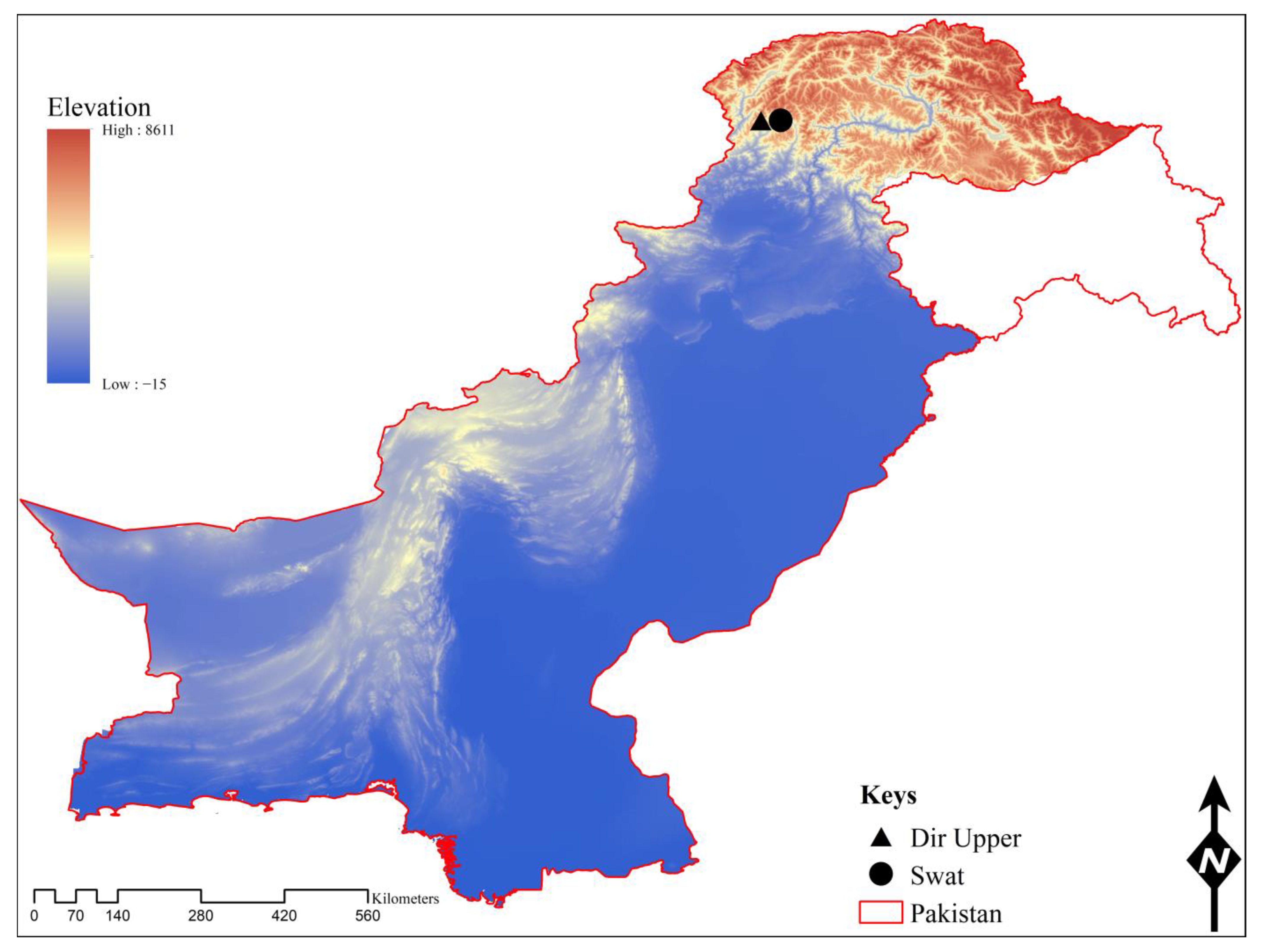
For the current study, ethical approval was obtained from the Advanced Studies and Research Board of the Faculty of Chemical and Life Sciences, with notification number Dir/A&R/AWKUM/2022/9674, Abdul Wali Khan University Mardan, KP, Pakistan. Oral permissions were taken from the animal owners.

2.2. Study Area

This study was conducted in the border region of the following districts: Dir Upper (35°30′00.1″ N, 72°20′43.0″ E) and Swat (35°34′27.3″ N 72°22′07.7″ E)—Badgoi pass of the Hindu Kush Mountain Range, the northern part of the KP province of Pakistan. The Badgoi pass is the link between districts Dir Upper and Swat, with an altitude of nearly 3520 m above sea level. The physiography of the area is composed of gently steep, rocky mountains covered with snow in winter and turned into green pastures in summer. The summer season is from moderate to warm, and temperatures rapidly fall in November to February. The average temperature ranges from −7 °C to 22 °C (climate-data.org). The geographical coordinates of the collection sites were obtained by using a Global Positioning System (GPS), and the map was designed via ArcGIS V. 10.3.1 (Figure 1).

2.3. Ticks Collection and Morphological Identification

Tick specimens were collected in June 2022 from goats (Capra hircus) in the study area. Ticks were washed with distilled water followed by 70% ethanol to remove the contaminants and extra tissues from the body surface. Ticks were morphologically identified under a stereo-microscope (StereoBlue-euromex SB.1302-1, Arnhem, The Netherlands), and their morphological characters were compared with the standard identification keys [23,24].

2.4. DNA Extraction and PCR

A total of 18/26 (69.23%) Dermacentor specimens comprising 9 specimens from each locations: Dir Upper (6 males and 1 unfed and 2 fed females) and Swat (5 males and 4 fed females) were subjected to genomic DNA extraction. The specimens were washed and crushed in a sterilized 1.5 mL Eppendorf tube. The genomic DNA was extracted by the phenol-chloroform method [25]. The DNA pellet was hydrated by adding 30 µL “nuclease-free” PCR water and quantified with NanoDrop (Optizen, Daejeon, South Korea).
A conventional PCR (GE-96G, BIOER, Hangzhou, China) was performed to amplify the16S rRNA and cox1 genes for tick identification (Table 1). All genomic DNA samples were utilized to test for associated pathogens through the amplification of the 16S rRNA partial gene for Anaplasma species and the gltA and ompB partial genes for Rickettsia species (Table 1).
Each PCR mixture was prepared in 25 µL, composed of 2 µL genomic DNA (100 ng/µL), 1 µL each (forward and reverse) primer (10 µM), 8.5 µL PCR water “nuclease-free”, and 12.5 µL DreamTaq green MasterMix (2×) (Thermo Fisher Scientific, Inc., Waltham, MA, USA). Each of the PCRs contained a positive control (DNA of Rickettsia massiliae and Anaplasma marginale for pathogens, and DNA of Rhipicephalus microplus for ticks) as well as a negative control (PCR water that was “nuclease-free” rather than DNA). The PCR products were run on a 2% agarose gel electrophoresis and observed via Gel documentation system (BioDoc-It™ Imaging Systems, UVP, LLC., Upland, CA, USA).

2.5. DNA Sequencing and Phylogenetic Analysis

The amplicons were purified by using a Gene Clean II Kit (Qbiogene, Il-lkirch, France) as per the manufacturer’s protocol. The PCR-amplified products were sent for bidirectional sequencing to Macrogen, Inc. (Seoul, Republic of Korea). The obtained sequences were trimmed to remove the contaminated and poor reading regions via SeqMan v. 5 (DNASTAR, Inc., Madison, WI, USA). The trimmed sequences were subjected to the Basic Local Alignment Search Tool (BLAST) in the National Center for Biotechnology Information (NCBI). The high-percentage identity sequences were downloaded in FASTA format from the NCBI for phylogenetic analyses. The downloaded sequences were aligned with the obtained sequences and an outgroup using ClustalW multiple alignments [31] in BioEdit Sequence Alignment Editor v. 7.0.5 [32]. The coding sequences were aligned by using MUSCLE statistical algorithms [33]. The phylogenetic trees were constructed through the maximum likelihood method and the Tamura–Nei model with a 1000 bootstrapping value in MEGA-X [34].

3. Results

3.1. Tick Record

Goat herds were observed for tick collection in 11 different localities in Badgoi pass Dir Upper and Swat. Among these goat herds, 11 goats (5 males and 6 females) were infested by D. marginatus ticks, counting 26 (19 males and 1 unfed and 6 fed females) ticks. Among these, 16 ticks (13 males and 1 unfed and 2 fed females) were from Dir Upper and 10 ticks (6 males and 4 fed females) from Swat. The Dermacentor-infested goats were of three age groups, namely 4 adults, 4 young, and 3 kids. Besides D. marginatus ticks, these goats were co-infested by Haemaphysalis montgomeryi. A total of 45 H. montgomeryi ticks including 23 males and 22 females (6 unfed and 16 fed females) were collected. Environmental and topographic conditions as well as the host records are mentioned in Table 2.

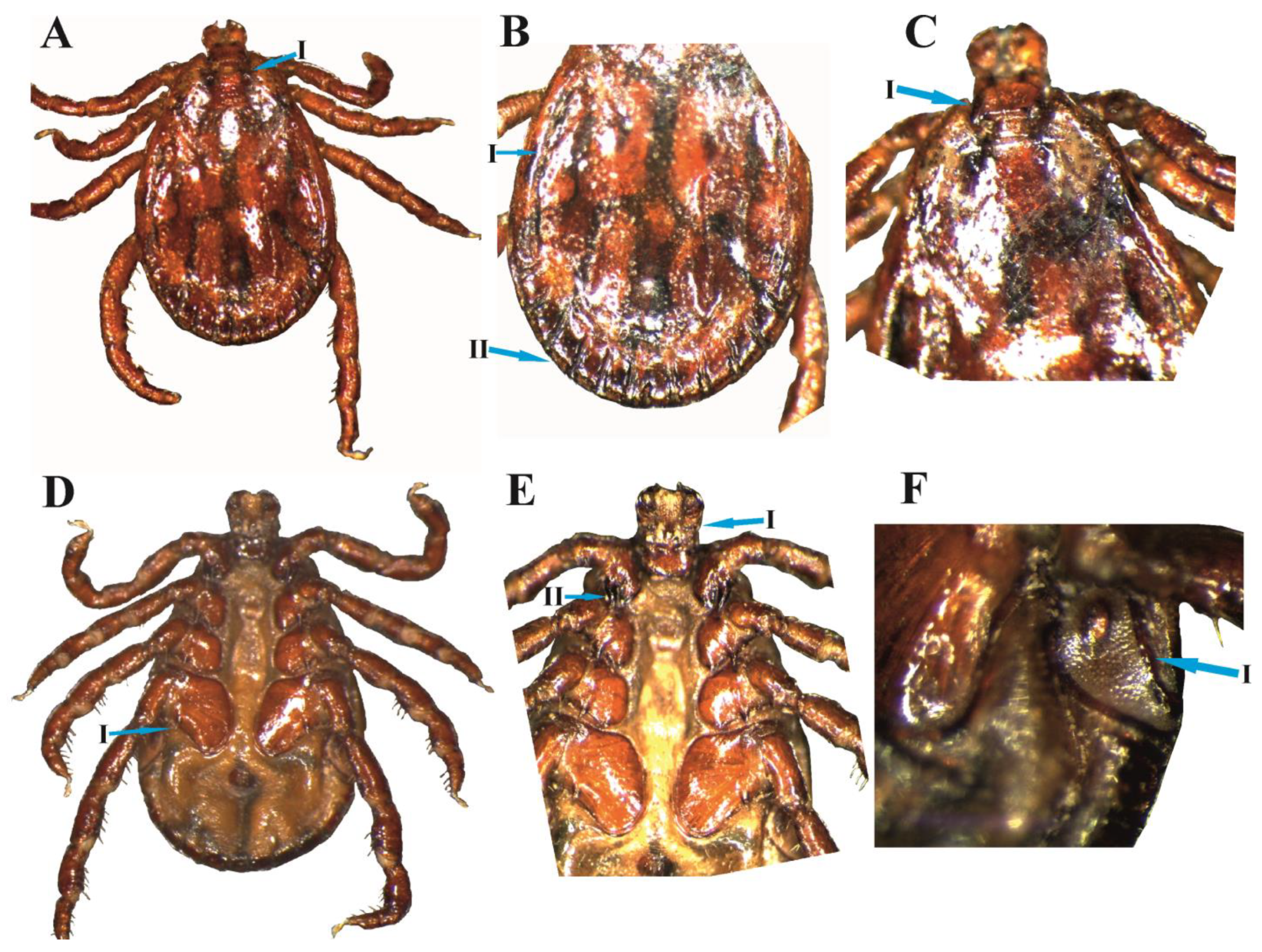
3.2. D. marginatus Male

Idiosoma: The body is medium-sized with an elongated oval shape. Conscutum is inornate, dark reddish with irregular black patches. Cervical grooves are short, deep, and comma-shaped converging anteriorly and posteriorly. Punctuations are deep, not uniform, and densely distributed in the anterior (Figure 2A(I)). The marginal groove does not enclose the posterior festoon and starts from the last festoon and ends at the point of 2nd coxae (Figure 2B(I, II)). The width of the basis capitulum is greater than its length with moderately pointed broad cornua (Figure 2C(I)). Legs: dorsal side segments are with pale yellow color spots. Coxa I is small with paired spurs equal in size (Figure 2E(II)). A single external spur is present on coxae II, III, and IV. Coxa IV is large and broader anteriorly while becoming narrow posteriorly (Figure 2D(I)). The coxa size progressively increases from coxae I to IV. Hypostome is club-shaped accompanied by 3/3 dentation (Figure 2E(I)). Spiracular plates are broad at the upper side and gradually pointed posteriorly. Goblet cells are around the macula. Macula is parallel with the lateral margin (Figure 2F(I)).

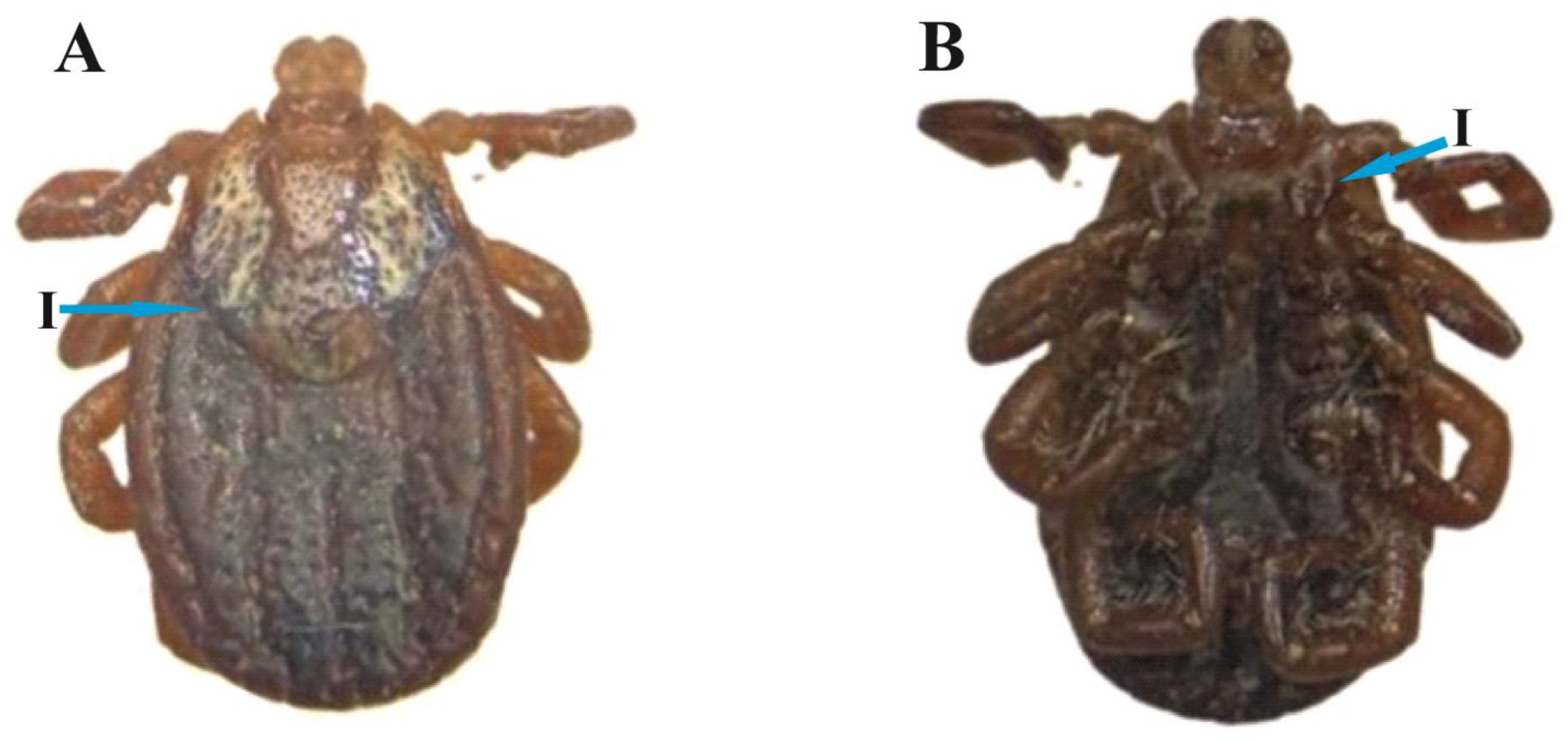
3.3. D. marginatus Female

Body idiosoma: The body is dark reddish and oval-shaped. The scutum is moderate-sized and sub-ovate in shape. The scutum posteriorly diverges at mid-position and gradually converges to a narrow-rounded position (Figure 3A(I)). The cervical groove is deep and curved inside. The alloscutum has inner deep grooves, with posterior–marginal depressions. Capitulum width is greater than length. The basis capitulum is broader as compared to the length. Punctuations are deep, not uniform, and irregularly distributed (Figure 3A). Coxa I is small with a paired spur (Figure 3B(I)).

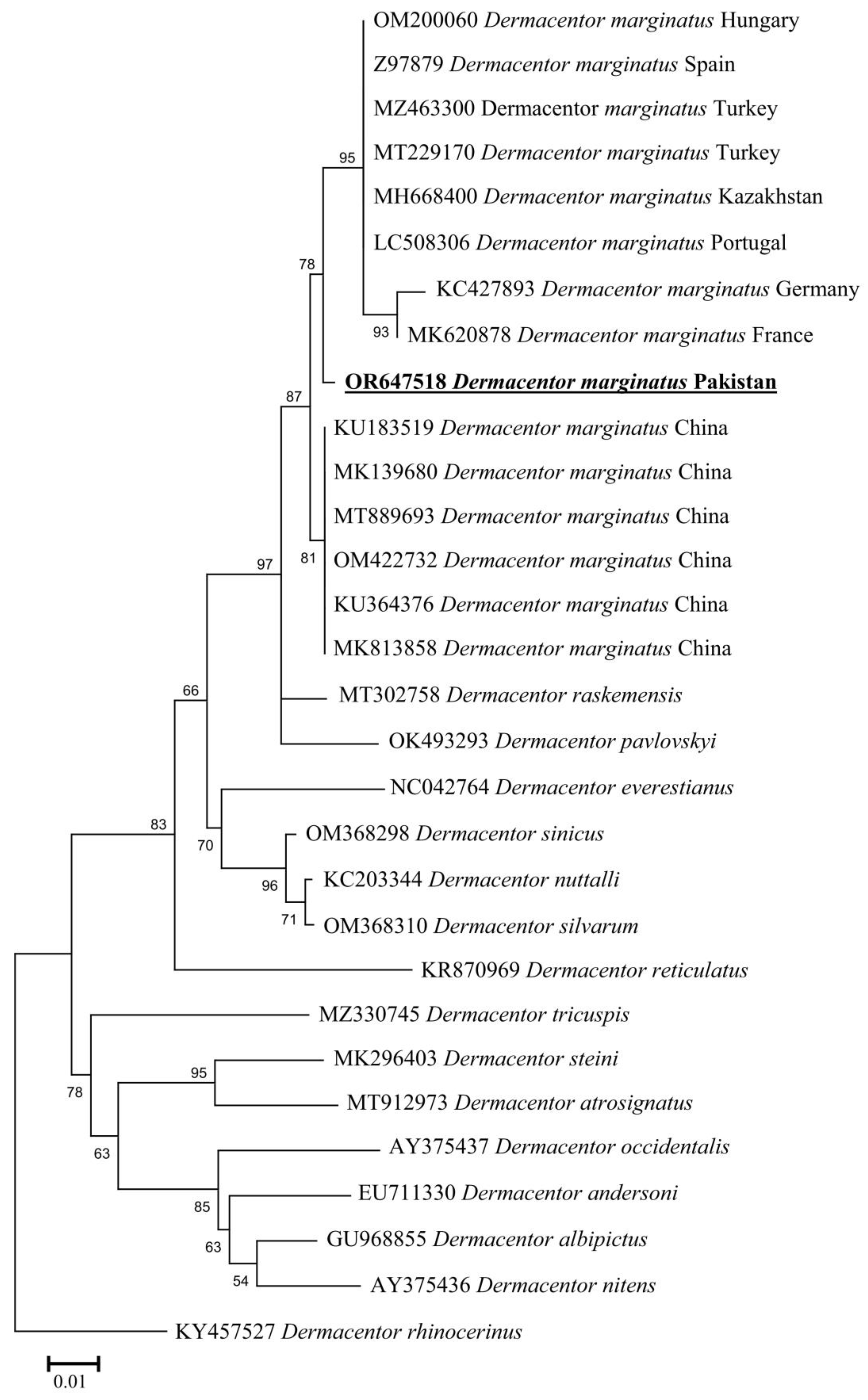
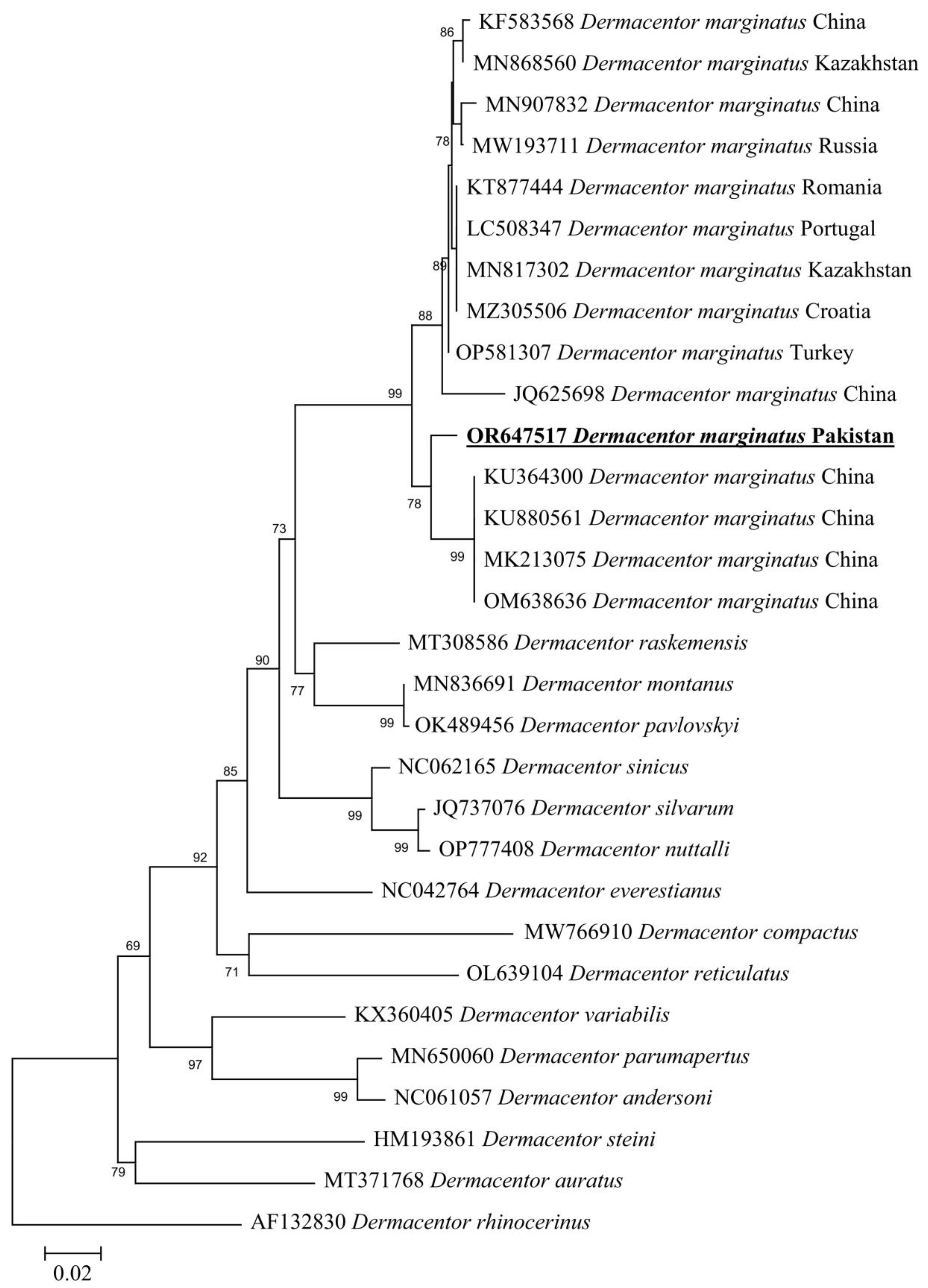
3.4. Molecular and Phylogenetic Analysis of D. marginatus

By the BLAST results, the 16S rDNA partial sequence showed a 97.28–98.85% maximum identity range with D. marginatus reported from various countries: China (98.85%—OM422732, 98.77%—KU183519, 98.61%—MK139680, 98.51%—KU364376, 98.34%—MT889693, and 98.13%—MK813858), Kazakhstan (97.93%—MH668400), Turkey (97.84%—MT229170), Portugal (97.82%—LC508306), Germany (96.79%—KC427893), Hungary (97.78%—OM200060), Turkey (97.74%—MZ463300), Spain (97.71%—Z97879), and France (97.28%—MK620878). On the other hand, the cox1 sequence showed a 95.82–98.03% maximum identity range with D. marginatus reported from different countries: Turkey (98.03%—OP581307), Croatia (97.87%—MZ305506), China (97.87%—KU364300, 97.86%—MK213075, 97.69%—KU880561, 97.65%—KF583568, 97.65%—OM638636, 97.51%—MN907832, and 95.82%—JQ625698), Kazakhstan (97.85%—MN817302 and 97.79%—MN868560), Portugal (97.70%—LC508347), Romania (97.61%—KT877444), and Russia (97.37%—MW193711). In the phylogenetic analysis, these 16S rDNA and cox1 sequences were clustered with D. marginatus reported from the Palearctic region (Figure 4 and Figure 5).

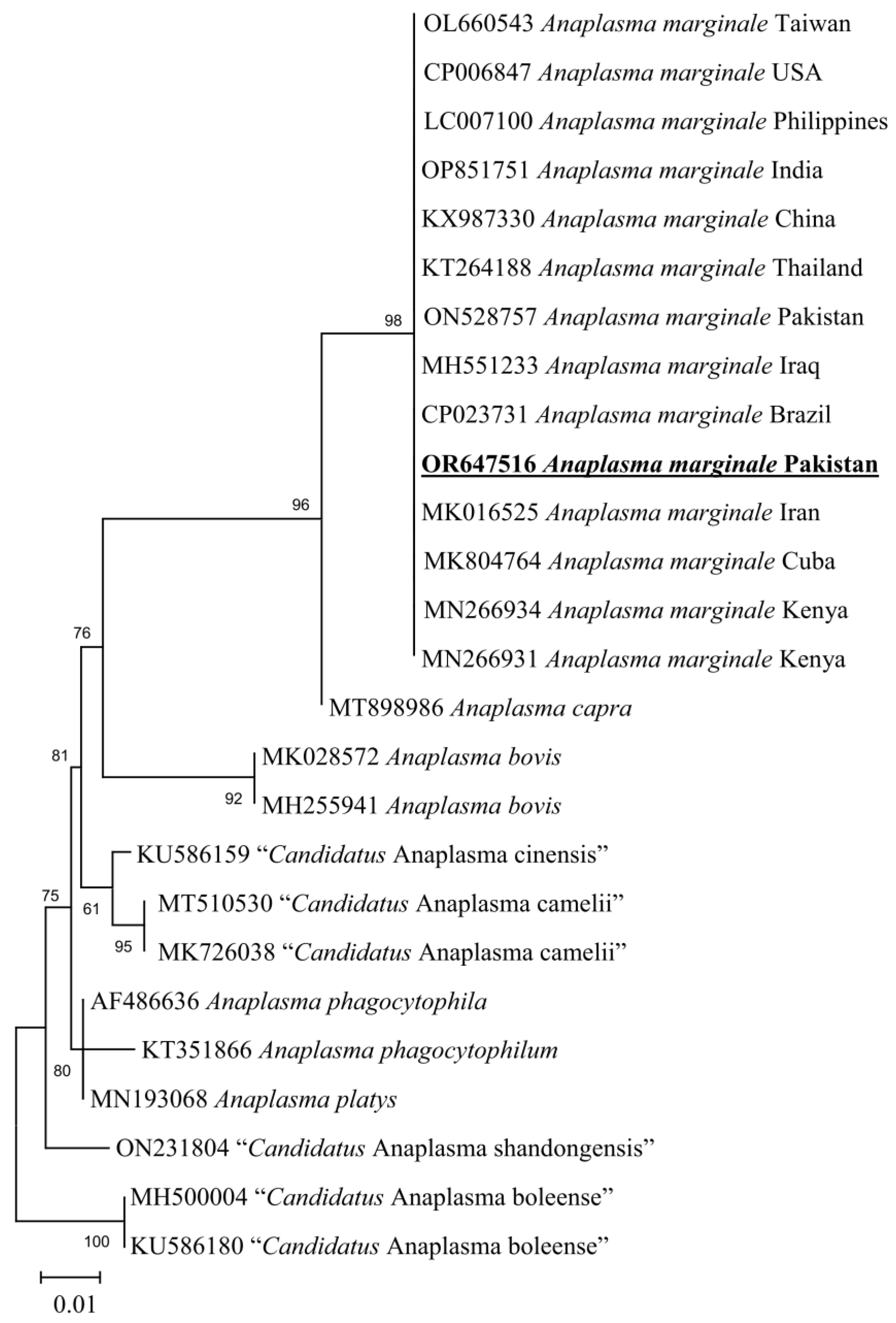
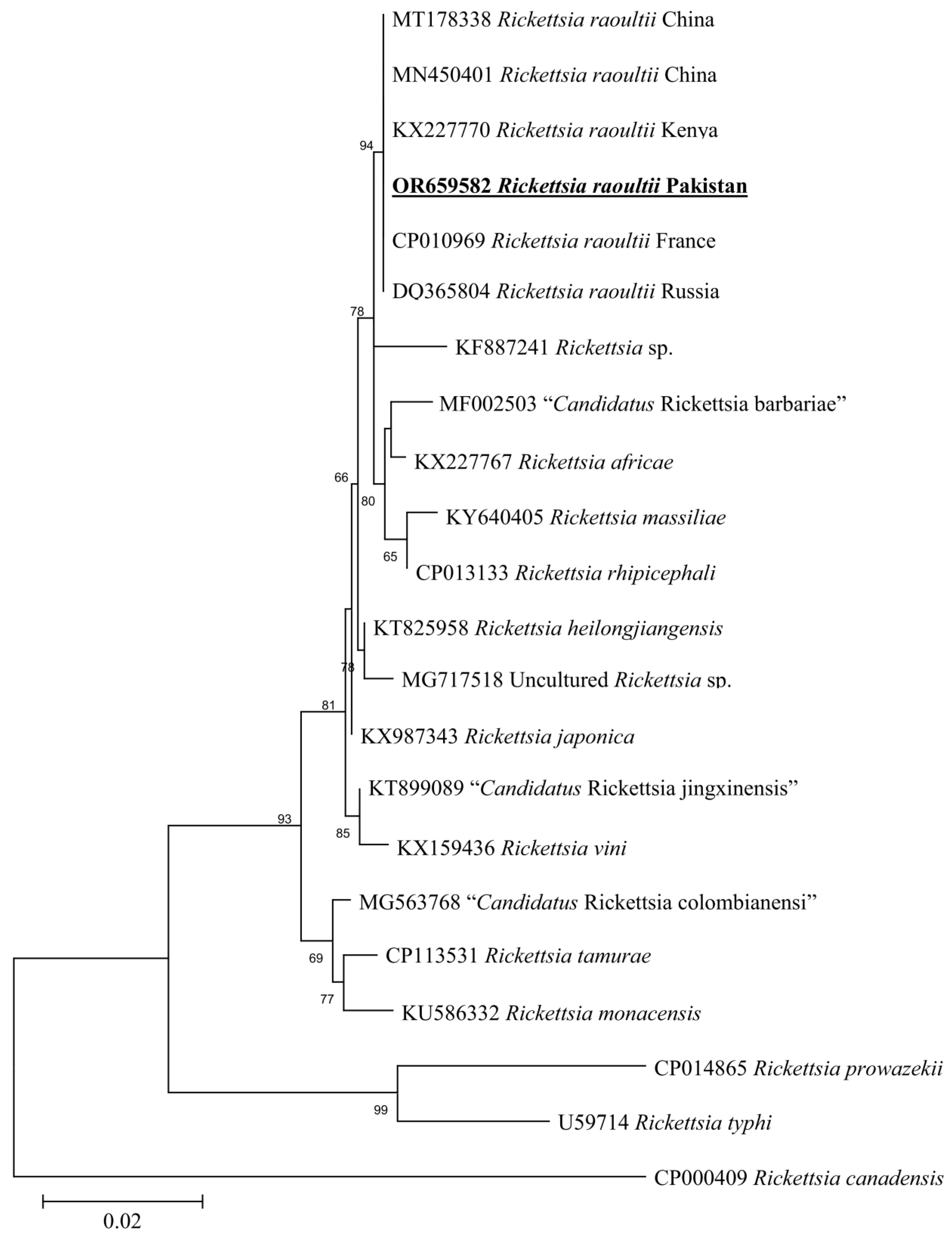
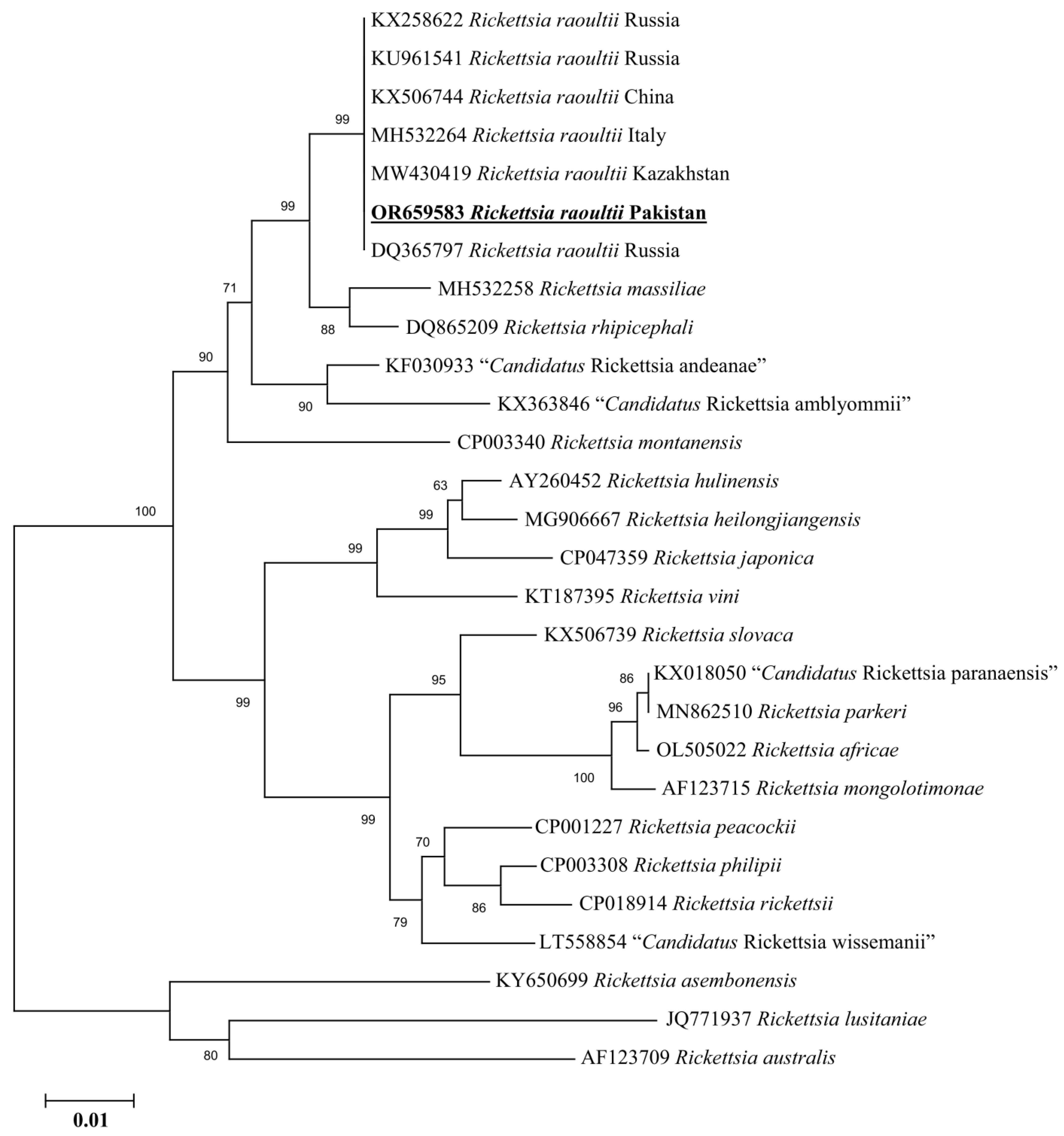
3.5. Molecular Screening and Phylogenetic Analysis of D. marginatus Associated Pathogens

Anaplasma marginale (2/18; 11.11%) and R. raoultii (2/18; 11.11%) were documented in D. marginatus ticks. The 16S rDNA sequence for Anaplasma sp. showed 100% identity with Anaplasma marginale and phylogenetically clustered with the same species reported from Pakistan (ON528757), Taiwan (OL660543), USA (CP006847), Philippines (LC007100), India (OP851751), China (KX987330), Thailand (KT264188), Iraq (MH551233), Brazil (CP023731), Iran (MK016525), Cuba (MK804764), and Kenya (MN266931 and MN266934) (Figure 6).
The rickettsial gltA sequence showed 100% identity with the Rickettsia raoultii reported from China (MT178338 and MN450401), Kenya (KX227770), France (CP010969), and Russia (DQ365804). Meanwhile, the rickettsial ompB sequence showed 100% identity with the Rickettsia raoultii from Russia (KX258622), Russia (KU961541), China (KX506744), Italy (MH532264), Kazakhstan (MW430419), and Russia (DQ365797). In the phylogenetic trees, these rickettsial gltA and ompB sequences were clustered with the corresponding species (Figure 7 and Figure 8).

4. Discussion

The genus Dermacentor is composed of 44 tick species, originated in Africa and spread to the New World through Palearctic zoogeographical regions [2,6,35]. One of the most diverse groups with fewer number of species [35], the systematic knowledge and evolutionary history of this genus is still poorly known [1,35,36]. Herein, ticks were collected from goats in the Hindu Kush Mountain Range at Badgoi pass and identified morphologically and phylogenetically as D. marginatus. This is the first confirmed host record and the preliminary phylogenetic position of D. marginatus from Pakistan. Despite being widely distributed throughout the Palearctic region, the D. marginatus tick has not yet been identified in Pakistan [6,24]. This raises concerns about D. marginatus whether it is considered an exotic or native species in the country. Environmentalists and public health professionals face an increasing concern about the spread of exotic ticks into novel ecosystems [37].
D. marginatus ticks are difficult to morphologically identify, as Dermacentor nuttalli, Dermacentor ushakovae, Dermacentor niveus, and Dermacentor silvarum were considered as synonyms; in this case, [6] these species were confirmed as D. marginatus species complex. The morphological description of the male and female D. marginatus has revealed that both sexes have white enamel on the scutum and conscutum [4]. The collected male tick during this study has reddish-black pigmentation and with no ornamentation on the conscutum; this phenomenon has been previously reported in Dermacentor ticks, i.e., Dermacentor albipictus [38].
Together with morphological identification, the genetic characterization of ticks has been considered as an important component in understanding tick systematic and evolutionary history [39]. In this study, we employed DNA barcoding for the precise identification of D. marginatus. The 16S rRNA and cox1 genes are important in marking tick phylogenies due to the availability of the dataset in GenBank for these sequences as well as containing hypervariable regions [40]. In the phylogenetic trees, the obtained 16S rDNA and cox1 sequences found a close resemblance with the sequences of D. marginatus reported from different countries.
Exponential rates of ticks and tick-borne diseases due to climate change have threatened global health. In Pakistan, serious attention to regular surveillance of ticks and tick-borne pathogens has not been paid yet. However, previous studies have highlighted various ticks as zoonotic risks regarding the detection of pathogens like “Candidatus Rickettsia shennongii”, Rickettsia massiliae, Rickettsia aeschlimannii, Rickettsia conorii, Rickettsia hoogstraalii, Coxiella burnetii, Borrelia sp., A. marginale, and Theileria annulata [41,42,43,44,45]. Although the occurrence of D. marginatus in Pakistan and the detection of A. marginale and R. raoultii in this tick are a serious concern, as previously mentioned, this species has been found to be involved in the transmission of various pathogens like Rickettsia slovaca, A. marginale Coxiella spp., Rickettsia spp., Shigella spp., and Francisella spp. [46]. To assist the public and veterinary health surveillance, the current study monitored the abundance and distribution of the utmost important goat-associated Dermacentor ticks in the highlands of the northern region of Pakistan. Moreover, the molecular detection of pathogens like R. raoultii and A. marginale highlights the zoonotic risks due to these parasites. Future research must address the gaps regarding the presence of D. marginatus in various topographic regions of Pakistan in order to understand whether this species is established or is invasive.

5. Conclusions

This study presents a comprehensive report on D. marginatus ticks and associated A. marginale and R. raoultii for the first time in Pakistan. Molecular identification and host record confirmed that the ticks of the genus Dermacentor mostly prefer cold climatic conditions. Furthermore, pathogens were detected that have a zoonotic threat to the livestock-holders of the study areas. Moreover, this study may assist in understanding the epidemiology and systematic history of Dermacentor species.

Author Contributions

A.A. (Abid Ali) designed this study. I.A., S.U. and A.A. (Abid Ali) collected the ticks. A.A. (Abid Ali), A.A. (Abdulaziz Alouffi), M.M.A., S.-C.C., C.-C.C., T.T., I.A., M.N. and S.U. performed molecular work and phylogenetic analyses and wrote the initial draft of the manuscript. All authors have read and agreed to the published version of the manuscript.

Funding

The researchers supporting project number (RSP2023R494), King Saud University, Riyadh, Saudi Arabia.

Institutional Review Board Statement

For the current study, ethical approval was obtained from the Advanced Studies and Research Board of the Faculty of Chemical and Life Sciences, with notification number: Dir/A&R/AWKUM/2022/9674, Abdul Wali Khan University Mardan, KP, Pakistan. Oral permissions were taken from the owners of animals or herds.

Informed Consent Statement

Not applicable.

Data Availability Statement

The datasets to support the conclusions of this article are given within the article.

Acknowledgments

We are thankful for the financial support offered by the Pakistan Science Foundation (PSF) and Higher Education Commission (HEC) of Pakistan. The researchers supporting project number (RSP2023R494), King Saud University, Riyadh, Saudi Arabia. We are thankful to Dmitry A. Apanaskevich for revising an earlier version of this manuscript.

Conflicts of Interest

The authors declare no conflict of interest.

References

  1. Guglielmone, A.A.; Robbins, R.G.; Apanaskevich, D.A.; Petney, T.N.; Estrada-Peña, A.; Horak, I.G. The Hard Ticks of the World; Springer: Dordrecht, The Netherlands, 2014; Volume 10, pp. 978–994. [Google Scholar]
  2. Lado, P.; Klompen, H. Evolutionary history of New World ticks of the genus Dermacentor (Ixodida: Ixodidae); the origin of D. variabilis. Biol. J. Linn. Soc. 2019, 127, 863–875. [Google Scholar] [CrossRef]
  3. Guzmán-Cornejo, C.; Robbins, R.G.; Guglielmone, A.A.; Montiel-Parra, G.; Rivas, G.; Pérez, T.M. The Dermacentor (Acari, Ixodida, Ixodidae) of Mexico: Hosts, geographical distribution and new records. Zookeys 2016, 569, 1–22. [Google Scholar] [CrossRef] [PubMed]
  4. Estrada-Peña, A.; Estrada-Peña, R. Notes on Dermacentor ticks: Redescription of D. marginatus with the synonymies of D. niveus; D. daghestanicus (Acari: Ixodidae). J. Med. Entomol. 1991, 28, 1–15. [Google Scholar] [CrossRef]
  5. Raad, M.; Azar, D.; Perotti, M.A. First report of the ticks Haemaphysalis punctata Canestrini et Fanzago, 1878, Haemaphysalis parva (Neumann, 1897) and Dermacentor marginatus (Sulzer, 1776) (Acari, Amblyommidae) from humans in Lebanon. Acta Parasitol. 2020, 65, 541–545. [Google Scholar] [CrossRef] [PubMed]
  6. Guglielmone, A.A.; Nava, S.; Robbins, R.G. Geographic distribution of the hard ticks (Acari: Ixodida: Ixodidae) of the world by countries and territories. Zootaxa 2023, 5251, 1–274. [Google Scholar] [CrossRef] [PubMed]
  7. Filippova, N.A.; Plaksina, M.A. Some aspects of intraspecific variability of the closely related species of the Dermacentor marginatus complex (Acari: Ixodidae) as demonstration of microevolutionary process. Parazitologiya 2005, 39, 337–364. [Google Scholar]
  8. Apanaskevich, D.A.; Apanaskevich, M.A. Description of a new Dermacentor (Acari: Ixodidae) species from Thailand and Vietnam. J. Med. Entomol. 2015, 52, 806–812. [Google Scholar] [CrossRef]
  9. Apanaskevich, D.A.; Apanaskevich, M.A.; Nooma, W.; Ahantarig, A.; Trinachartvanit, W. Reinstatement of Dermacentor tricuspis (Schulze, 1933) n. comb.; n. stat. (Acari: Ixodidae) as a valid species, synonymization of D. atrosignatus Neumann, 1906; description of a new species from Indonesia, Malaysia and Thailand. Syst. Parasitol. 2021, 98, 207–230. [Google Scholar] [CrossRef]
  10. Apanaskevich, D.A.; Apanaskevich, M.A. Description of two new species of Dermacentor Koch, 1844 (Acari: Ixodidae) from Oriental Asia. Syst. Parasitol. 2016, 93, 159–171. [Google Scholar] [CrossRef]
  11. Dhanda, V.; Kulkarni, S.M.; Pratt, P. Dermacentor raskemensis (Ixodoidea: Ixodidae), redescription and notes on ecology. J. Parasitol. 1971, 57, 1324–1329. [Google Scholar] [CrossRef]
  12. Filippova, N.A. Redescription of Dermacentor raskemensis Pomerantzev, 1946 (Ixodidae) a representative of the mountain fauna of the southern regions of the USSR and adjacent territories. Parazitologiia 1983, 17, 283–292. [Google Scholar] [PubMed]
  13. Apanaskevich, D.A. First description of the nymph and larva of Dermacentor raskemensis (Acari: Ixodidae), parasites of pikas and other small mammals in Central Asia. J. Med. Entomol. 2013, 50, 959–964. [Google Scholar] [CrossRef] [PubMed]
  14. Masala, G.; Chisu, V.; Satta, G.; Socolovschi, C.; Raoult, D.; Parola, P. Rickettsia slovaca from Dermacentor marginatus ticks in Sardinia, Italy. Ticks Tick Borne Dis. 2012, 3, 393–395. [Google Scholar] [CrossRef] [PubMed]
  15. Rubel, F.; Brugger, K.; Pfeffer, M.; Chitimia-Dobler, L.; Didyk, Y.M.; Leverenz, S.; Dautel, H.; Kahl, O. Geographical distribution of Dermacentor marginatus and Dermacentor reticulatus in Europe. Ticks Tick Borne Dis. 2016, 7, 224–233. [Google Scholar] [CrossRef] [PubMed]
  16. Bonnet, S.; De La Fuente, J.; Nicollet, P.; Liu, X.; Madani, N.; Blanchard, B.; Maingourd, C.; Alongi, A.; Torina, A.; Fernández de Mera, I.G.; et al. Prevalence of tick-borne pathogens in adult Dermacentor spp. ticks from nine collection sites in France. Vector Borne Zoonotic Dis. 2013, 13, 226–236. [Google Scholar] [CrossRef] [PubMed]
  17. Rubel, F.; Brugger, K.; Belova, O.A.; Kholodilov, I.S.; Didyk, Y.M.; Kurzrock, L.; García-Pérez, A.L.; Kahl, O. Vectors of disease at the northern distribution limit of the genus Dermacentor in Eurasia: D. reticulatus and D. silvarum. Exp. Appl. Acarol. 2020, 82, 95–123. [Google Scholar] [CrossRef] [PubMed]
  18. Parola, P.; Paddock, C.D.; Socolovschi, C.; Labruna, M.B.; Mediannikov, O.; Kernif, T.; Abdad, M.Y.; Stenos, J.; Bitam, I.; Fournier, P.E.; et al. Update on tick-borne rickettsioses around the world: A geographic approach. Clin. Microbiol. Rev. 2013, 26, 657–702. [Google Scholar] [CrossRef] [PubMed]
  19. Yan, P.; Qiu, Z.; Zhang, T.; Li, Y.; Wang, W.; Li, M.; Yu, Z.; Liu, J. Microbial diversity in the tick Argas japonicus (Acari: Argasidae) with a focus on Rickettsia pathogens. Med. Vet. Entomol. 2019, 33, 327–335. [Google Scholar] [CrossRef]
  20. Špitalská, E.; Štefanidesová, K.; Kocianová, E.; Boldiš, V. Rickettsia slovaca; Rickettsia raoultii in Dermacentor marginatus and Dermacentor reticulatus ticks from Slovak Republic. Exp. Appl. Acarol. 2012, 57, 189–197. [Google Scholar] [CrossRef]
  21. Remesar, S.; Díaz, P.; Portillo, A.; Santibáñez, S.; Prieto, A.; Díaz-Cao, J.M.; López, C.M.; Panadero, R.; Fernández, G.; Díez-Baños, P.; et al. Prevalence and molecular characterization of Rickettsia spp. in questing ticks from north-western Spain. Exp. Appl. Acarol. 2019, 79, 267–278. [Google Scholar] [CrossRef]
  22. Alkishe, A.; Cobos, M.E.; Osorio-Olvera, L.; Peterson, A.T. Ecological niche and potential geographic distributions of Dermacentor marginatus and Dermacentor reticulatus (Acari: Ixodidae) under current and future climate conditions. Web Ecol. 2022, 22, 33–45. [Google Scholar] [CrossRef]
  23. Nosek, J.; Sixl, W. Central-European ticks (Ixodoidea). Pomerantzev Mitt. Abt. Zool. Landesmus Joanneum 1972, 1, 480. [Google Scholar]
  24. Estrada-Peña, A.; Mihalca, A.D.; Petney, T.N. (Eds.) Ticks of Europe and North Africa: A Guide to Species Identification; Springer: Berlin/Heidelberg, Germany, 2018. [Google Scholar]
  25. Sambrook, J.; Fritsch, E.E.; Maniatis, T. Molecular Cloning: A Laboratory Manual, 2nd ed.; Cold Spring Harbor Laboratory Press: New York, NY, USA, 1989. [Google Scholar]
  26. Mangold, A.J.; Bargues, M.D.; Mas-Coma, S. Mitochondrial 16S rDNA sequences and phylogenetic relationships of species of Rhipicephalus and other tick genera among Metastriata (Acari: Ixodidae). Parasitol. Res. 1998, 84, 478–484. [Google Scholar] [CrossRef] [PubMed]
  27. Folmer, O.; Hoeh, W.R.; Black, M.B.; Vrijenhoek, R.C. Conserved primers for PCR amplification of mitochondrial DNA from different invertebrate phyla. Mol. Mar. Biol. Biotechnol. 1994, 3, 294–299. [Google Scholar] [PubMed]
  28. Inokuma, H.; Raoult, D.; Brouqui, P. Detection of Ehrlichia platys DNA in brown dog ticks (Rhipicephalus sanguineus) in Okinawa Island, Japan. J. Clin. Microbiol. 2000, 38, 4219–4221. [Google Scholar] [CrossRef] [PubMed]
  29. Labruna, M.B.; Whitworth, T.; Horta, M.C.; Bouyer, D.H.; McBride, J.W.; Pinter, A.; Popov, V.; Gennari, S.M.; Walker, D.H. Rickettsia species infecting Amblyomma cooperi ticks from an area in the state of Sao Paulo, Brazil, where Brazilian spotted fever is endemic. J. Clin. Microbiol. 2004, 42, 90–98. [Google Scholar] [CrossRef] [PubMed]
  30. Roux, V.; Raoult, D. Phylogenetic analysis of members of the genus Rickettsia using the gene encoding the outer-membrane protein rompB (ompB). Int. J. Syst. Evol. 2000, 50, 1449–1455. [Google Scholar] [CrossRef] [PubMed]
  31. Thompson, J.D.; Higgins, D.G.; Gibson, T.J. CLUSTAL W: Improving the sensensitivity of progressive multiple sequence alignments through sequence weighting, position-specific gap penalties and weight matrix choice. Nucleic Acids Res. 1994, 22, 4673–4680. [Google Scholar] [CrossRef]
  32. Hall, T.; Biosciences, I.; Carlsbad, C. BioEdit: An important software for molecular biology. GERF Bull. Biosci. 2011, 2, 60–61. [Google Scholar]
  33. Edgar, R.C. MUSCLE: Multiple sequence alignment with high accuracy and high throughput. Nucleic Acids Res. 2004, 32, 1792–1797. [Google Scholar] [CrossRef]
  34. Kumar, S.; Stecher, G.; Li, M.; Knyaz, C.; Tamura, K. MEGA X: Molecular evolutionary genetics analysis across computing platforms. Mol. Biol. Evol. 2018, 35, 1547. [Google Scholar] [CrossRef] [PubMed]
  35. Apanaskevich, D.A.; Vongphayloth, K.; Jeangkhwoa, P.; Chaloemthanetphong, A.; Ahantarig, A.; Apanaskevich, M.A.; Brey, P.T.; Lakeomany, K.; Trinachartvanit, W. Description of a new species of Dermacentor Koch, 1844 (Acari: Ixodidae) from the mountains of Laos and Thailand. Syst. Parasitol. 2020, 97, 347–355. [Google Scholar] [CrossRef] [PubMed]
  36. Guglielmone, A.A.; Petney, T.N.; Robbins, R.G. Ixodidae (Acari: Ixodoidea): Descriptions and redescriptions of all known species from 1758 to December 31, 2019. Zootaxa 2020, 4871, 1–322. [Google Scholar] [CrossRef] [PubMed]
  37. Bayliss, H.R.; Schindler, S.; Adam, M.; Essl, F.; Rabitsch, W. Evidence for changes in the occurrence, frequency or severity of human health impacts resulting from exposure to alien species in Europe: A systematic map. Environ. Evid. 2017, 6, 21. [Google Scholar] [CrossRef]
  38. Reynolds, S.; Hedberg, M.; Herrin, B.; Chelladurai, J.R.J. Analysis of the complete mitochondrial genomes of Dermacentor albipictus suggests a species complex. Ticks Tick Borne Dis. 2022, 13, 102038. [Google Scholar] [CrossRef] [PubMed]
  39. Barker, S.C.; Murrell, A. Phylogeny, evolution and historical zoogeography of yicks: A review of recent progress. Exp. Appl. Acarol. 2002, 28, 55–68. [Google Scholar] [CrossRef] [PubMed]
  40. Lv, J.; Wu, S.; Zhang, Y.; Chen, Y.; Feng, C.; Yuan, X.; Jia, G.; Deng, J.; Wang, C.; Wang, Q.; et al. Assessment of four DNA fragments (COI, 16S rDNA, ITS2, 12S rDNA) for species identification of the Ixodida (Acari: Ixodida). Parasit. Vectors 2014, 7, 93. [Google Scholar] [CrossRef]
  41. Shehla, S.; Ullah, F.; Alouffi, A.; Almutairi, M.M.; Khan, Z.; Tanaka, T.; Labruna, M.B.; Tsai, K.H.; Ali, A. Association of SFG Rickettsia massiliae and Candidatus Rickettsia shennongii with different hard ticks infesting livestock hosts. Pathogens 2023, 12, 1080. [Google Scholar] [CrossRef]
  42. Ali, A.; Obaid, M.K.; Almutairi, M.M.; Alouffi, A.; Numan, M.; Ullah, S.; Rehman, G.; Islam, Z.U.; Khan, S.B.; Tanaka, T. Molecular detection of Coxiella spp. in ticks (Ixodidae and Argasidae) infesting domestic and wild animals: With notes on the epidemiology of tick-borne Coxiella burnetii in Asia. Front. Microbiol. 2023, 14, 1229950. [Google Scholar] [CrossRef]
  43. Khan, M.; Islam, N.; Khan, A.; Islam, Z.U.; Muñoz-Leal, S.; Labruna, M.B.; Ali, A. New records of Amblyomma gervaisi from Pakistan, with detection of a reptile-associated Borrelia sp. Ticks Tick Borne Dis. 2022, 13, 102047. [Google Scholar] [CrossRef]
  44. Khan, Z.; Shehla, S.; Alouffi, A.; Kashif Obaid, M.; Zeb Khan, A.; Almutairi, M.M.; Numan, M.; Aiman, O.; Alam, S.; Ullah, S.; et al. Molecular survey and genetic characterization of Anaplasma marginale in ticks collected from livestock hosts in Pakistan. Animals 2022, 12, 1708. [Google Scholar] [CrossRef]
  45. Ullah, K.; Numan, M.; Alouffi, A.; Almutairi, M.M.; Zahid, H.; Khan, M.; Islam, Z.U.; Kamil, A.; Safi, S.Z.; Ahmed, H.; et al. Molecular characterization and assessment of risk factors associated with Theileria annulata infection. Microorganisms 2022, 10, 1614. [Google Scholar] [CrossRef]
  46. Zhang, Y.K.; Yu, Z.J.; Wang, D.; Bronislava, V.; Branislav, P.; Liu, J.Z. The bacterial microbiome of field-collected Dermacentor marginatus and Dermacentor reticulatus from Slovakia. Parasit. Vectors 2019, 12, 325. [Google Scholar] [CrossRef]
Figure 1. Tick collection site in the border region (Badgoi pass) of districts Dir Upper and Swat of the Hindu Kush Mountain Range.
Animals 13 03686 g001
Figure 2. D. marginatus male: (A) male dorsum (I: cervical groove), (B) posterior dorsum (I: lateral groove, II: festoons), (C) anterior dorsum (I: basis capitulum), (D) male venter (I: coxa IV spur), (E); anterior venter (I: capitulum ventrally, II: first coxa spurs), and (F); posterior venter (I: spiracle plate).
Animals 13 03686 g002
Figure 3. D. marginatus: (A) female dorsum (I: scutum shape) and (B) female venter (I: first coxa spurs).
Animals 13 03686 g003
Figure 4. Maximum-likelihood phylogenetic analysis of nucleotide sequences of 16S rDNA partial sequences for Dermacentor spp. The sequences are represented by their GenBank accession number followed by the name of species and countries (when applicable). The obtained sequence in the present study is indicated in bold and underlined (accession number: OR647518).
Animals 13 03686 g004
Figure 5. Maximum-likelihood phylogenetic analysis of nucleotide sequences of cox1 partial sequences for Dermacentor spp. The sequences are represented by their GenBank accession number followed by the name of species and countries (when applicable). The obtained sequence in the present study is indicated in bold and underlined (accession number: OR647517).
Animals 13 03686 g005
Figure 6. Maximum-likelihood phylogenetic analysis of nucleotide sequences of 16S rDNA partial sequences for Anaplasma spp. The sequences are represented by their GenBank accession number followed by the name of species and countries (when applicable). The obtained sequence in the present study is indicated in bold and underlined (accession number: OR647516).
Animals 13 03686 g006
Figure 7. Maximum-likelihood phylogenetic analysis of nucleotide sequences of gltA partial sequences for Rickettsia spp. The sequences are represented by their GenBank accession number followed by the name of species and countries (when applicable). The obtained sequence in the present study is indicated in bold and underlined (accession number: OR659582).
Animals 13 03686 g007
Figure 8. Maximum-likelihood phylogenetic analysis of nucleotide sequences of ompB partial sequences for Rickettsia spp. The sequences are represented by their GenBank accession number followed by the name of species and countries (when applicable). The obtained sequence in the present study is indicated in bold and underlined (accession number: OR659583).
Animals 13 03686 g008
Table 1. List of primers utilized for the amplification of target DNA sequences.
SamplesTarget GenesSequences (5′-3′)Size bpPCR ConditionsReferences
Ticks16S rRNA16S+1-CCGGTCTGAACTCAGATCAAGT
16S−1-CTCAATGATTTTTTAAATTGCTG
460 95 °C 3 min, 40 × (95 °C 30 s, 55 °C 60 s, 72 °C 1 min), 72 °C 7 min[26]
cox1HC02198-TAAACTTCAGGGTGACCAAAAAATCA
LCO1490-GGTCAACAAATCATAAAGATATTGG
71295 °C 30 s, 40 × (95 °C 30 s, 48 °C 30 s, 72 °C 1 min), 72 °C 5 min[27]
Pathogens16S rRNAEHR16SD-GGTACCYACAGAAGAAGTCC
EHR16SR-TAGCACTCATCGTTTACAGC
34495 °C 5 min, 35 × (95 °C 30 s, 55 °C 30 s, 72 °C 90 s), 72 °C 5 min[28]
gltACS-78 GCAAGTATCGGTGAGGATGTAAT
CS-323 GCTTCCTTAAAATTCAATAAATCAGGAT
40195 °C 3 min, 40 × (95 °C 15 s, 48 °C 30 s, 72 °C 30 s) 72 °C 7 min[29]
ompB120-M59-CCGCAGGGTTGGTAACTGC
120-807-CCTTTTAGATTACCGCCTAA
86295 °C 3 min, 40 × (95 °C 30 s, 50 °C 30 s, 68 °C 1 min 30 s), 68 °C 7 min[30]
Table 2. Table describing the comprehensive data regarding the collection of D. marginatus in Hindu Kush Mountain Range.
DistrictHostSexAge GroupTemperature and HumidityPlace of Collection and ElevationTick Species
Targeted Species:
D. marginatus
Accompanied Species:
H. montgomeryi
Dir UpperGoatFemaleAdult (above 1 year)12 °C, 78%Kund Banda, 3347 m2M2M, 1F *
Dir UpperGoatMaleYoung (above 6 months–1 year)13 °C, 78%Gaedar Banda, 3344 m1M, 2F3M
Dir UpperGoatMaleKid (below 6 months)13 °C, 78%Bend Banda, 3340 m2M4F
Dir UpperGoatFemaleYoung (above 6 months–1 year)15 °C, 78%Jan Shahi, 3340 m2M, 1F *1M, 3F
Dir UpperGoatFemaleAdult (above 1 year)13 °C, 78%Cherry Banda, 3349 m2M2M
Dir UpperGoatFemaleKid (below 6 months)14 °C, 78%Dand Banda, 3352 m1M4M, 1F *
Dir UpperGoatMaleYoung (above 6 months–1 year)14 °C, 78%Otalshai, 3348 m3M2M, 4F
SwatGoatMaleKid (below 6 months)15 °C, 78%Dirgal, 3340 m1M, 3F4M
SwatGoatFemaleAdult (above 1 year)15 °C, 78%Gorgal, 3345 m2M5F
SwatGoatFemaleYoung (above 6 months–1 year)15 °C, 78%Landai dara, 3336 m1M, 1F3M, 1F *
SwatGoatMaleAdult (above 1 year)15 °C, 78%Dasht e Liala, 3351 m2M2M, 3F *
Total11 goats5 males,
6 females
4 adults, 4 young, 3 kids 19M, 7F23M, 22F
M: male, F *: unfed female and F: fed female.
Disclaimer/Publisher’s Note: The statements, opinions and data contained in all publications are solely those of the individual author(s) and contributor(s) and not of MDPI and/or the editor(s). MDPI and/or the editor(s) disclaim responsibility for any injury to people or property resulting from any ideas, methods, instructions or products referred to in the content.

