
Effect of Different Vitrification Techniques on Viability and Apoptotic Index of Domestic Cat Testicular Tissue Cells
 , , , , , and
Abstract
:Simple Summary
Abstract
1. Introduction
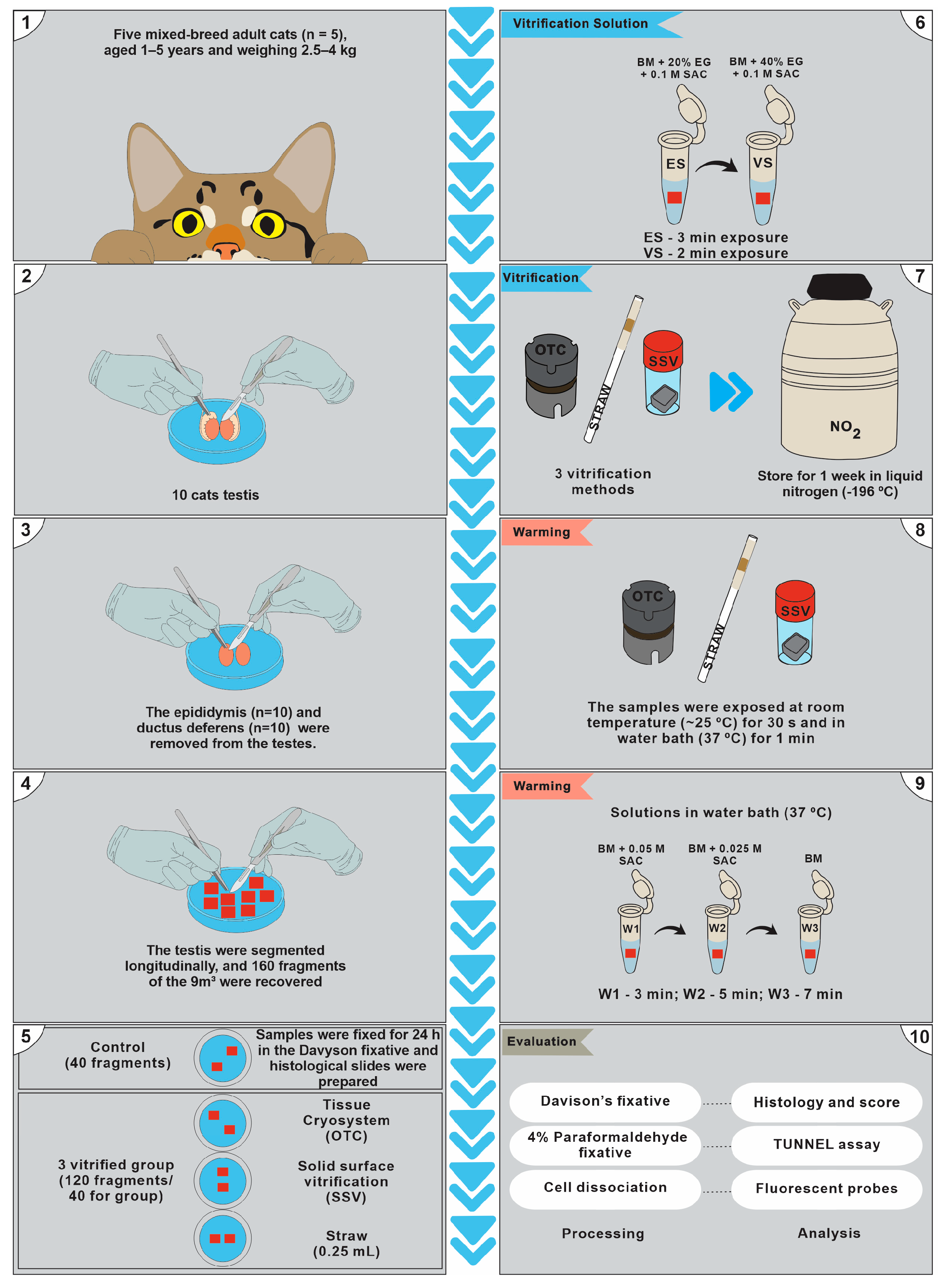
2. Materials and Methods
2.1. Ethics and Animal Selection
2.2. Testicular Biopsies Assessment
2.3. Experimental Design
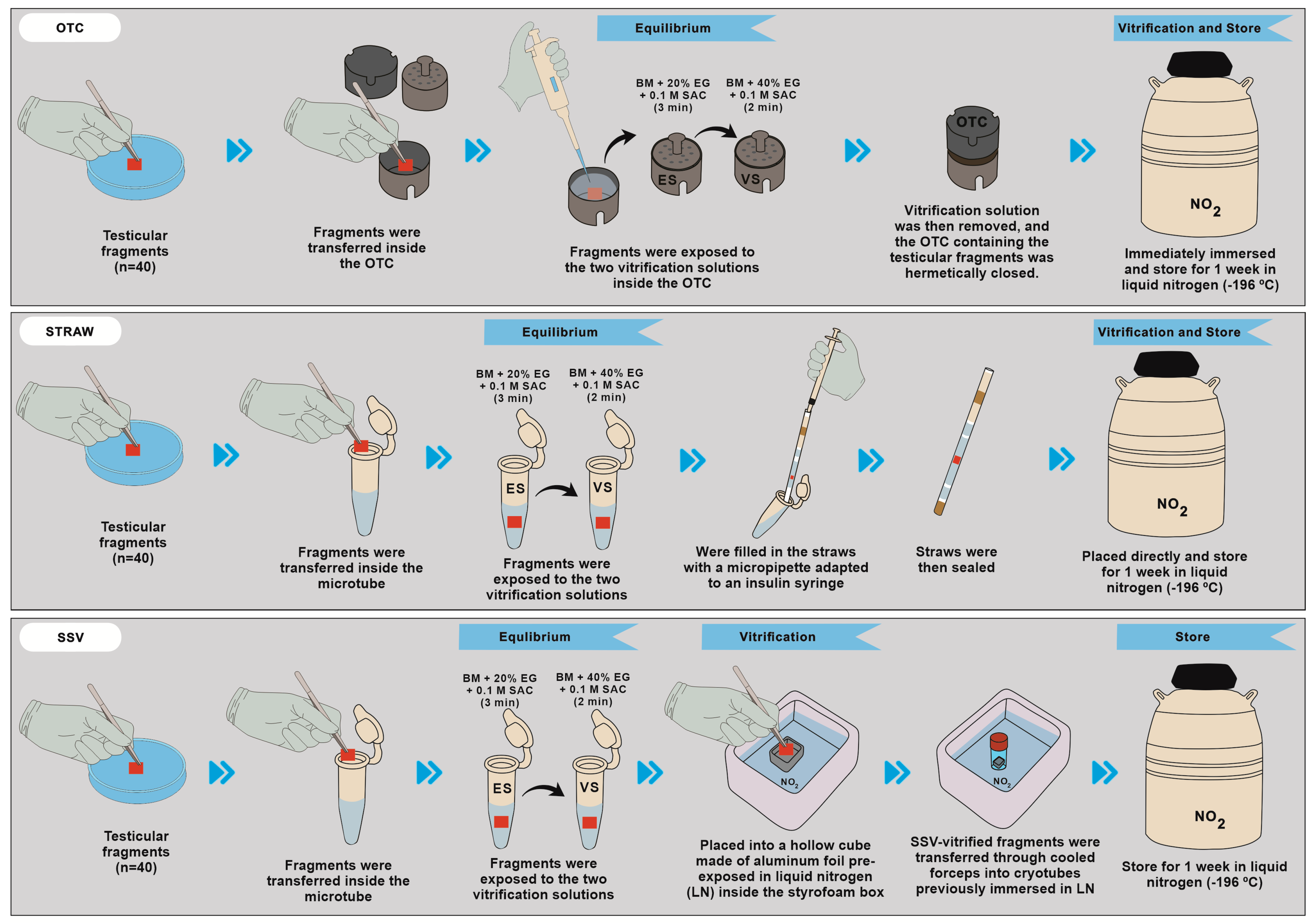
2.3.1. Vitrification and Warming
2.3.2. Ovarian Tissue Cryosystem (OTC)
2.3.3. Straw (STW)
2.3.4. Solid-Surface Vitrification (SSV)
2.3.5. Warming
2.4. Histological Analysis
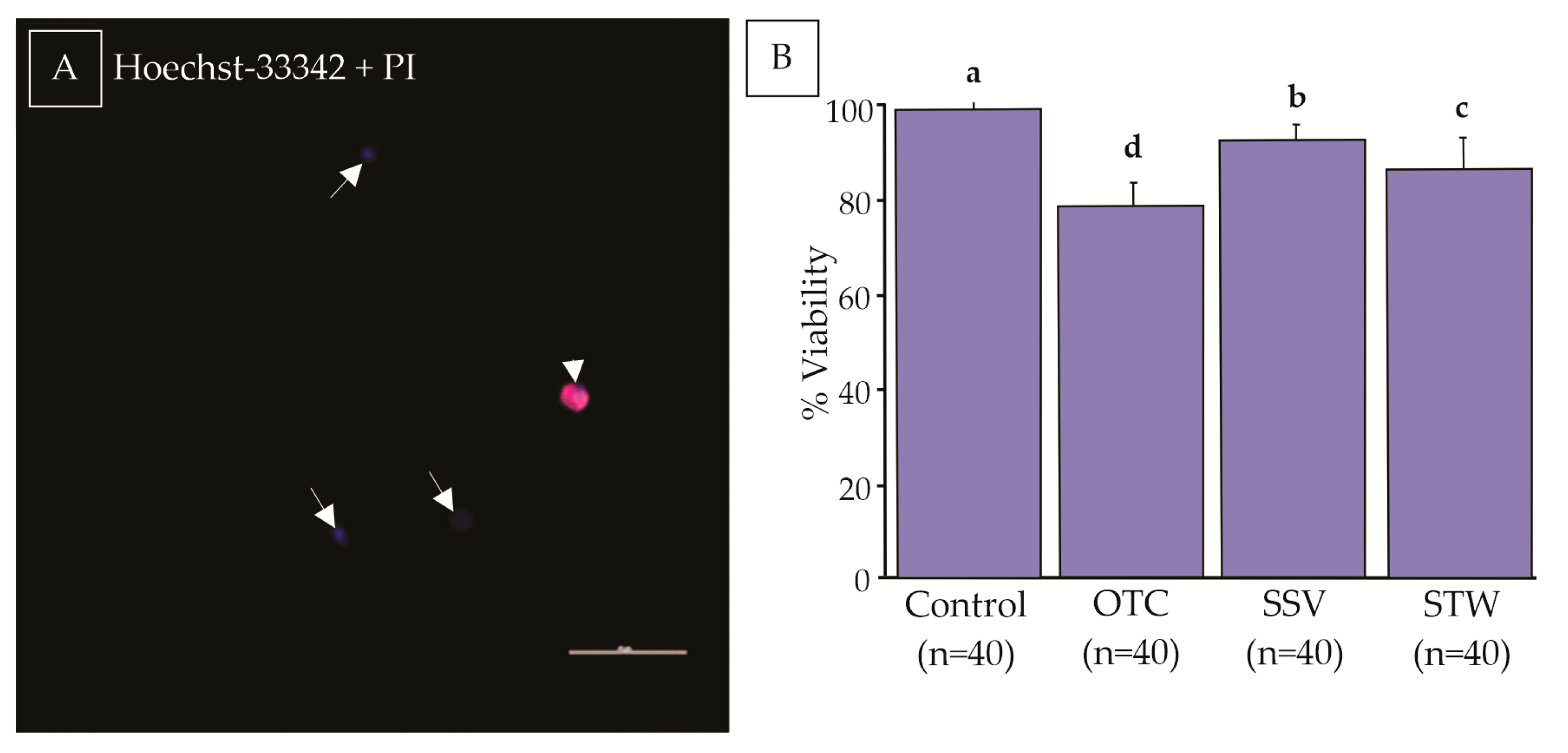
2.5. Cell Viability
2.6. Tunel Assay
2.7. Statistical Analysis
3. Results
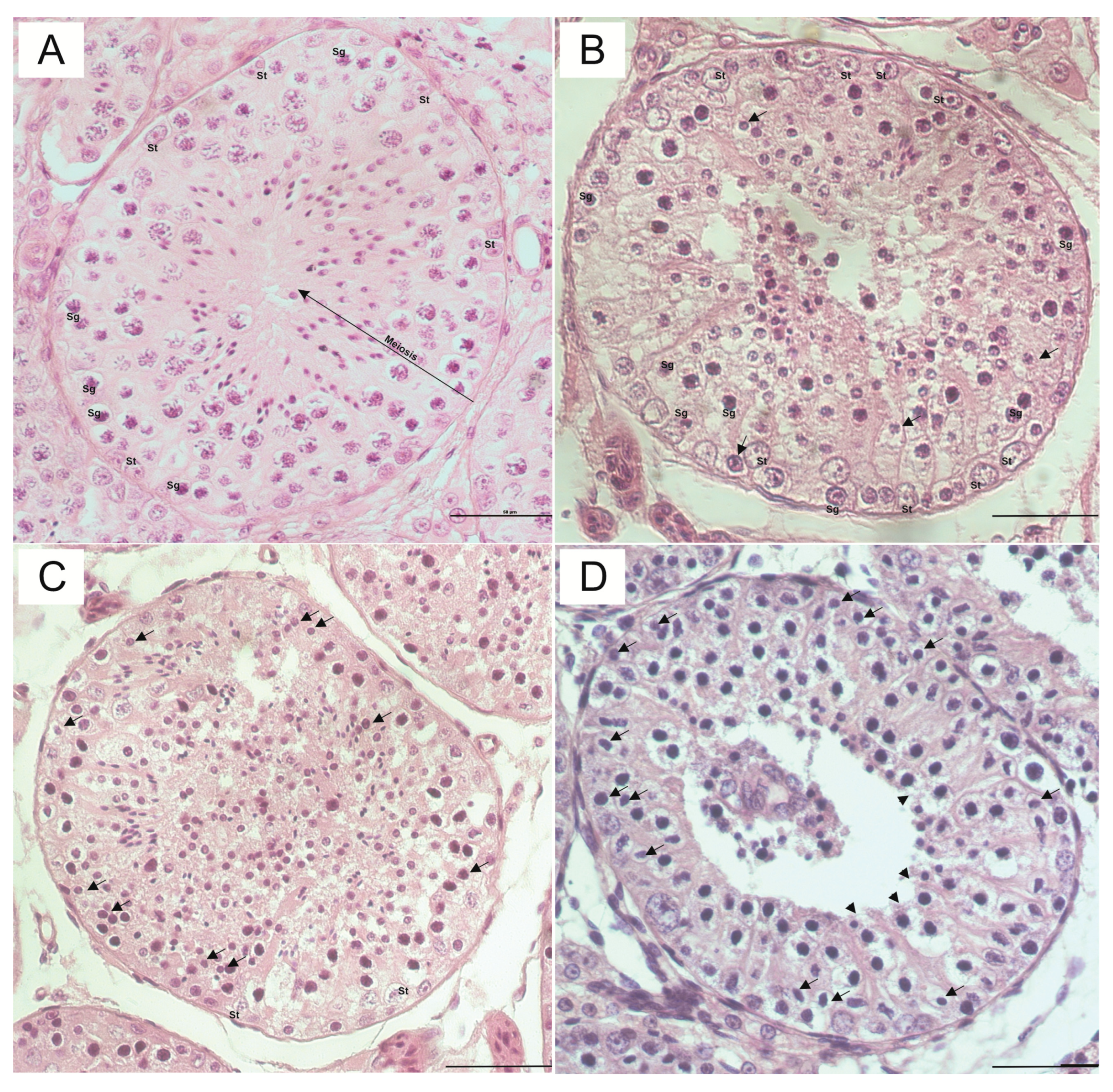
3.1. Histological Analysis
3.1.1. The Distinction between Sertoli Cells and Spermatogonial Nuclei
3.1.2. Nucleolar Visualization
3.1.3. Nuclear Condensation
3.1.4. Detachment of the Basement Membrane
3.1.5. Shrinkage of the Epithelium
3.2. Cell Viability
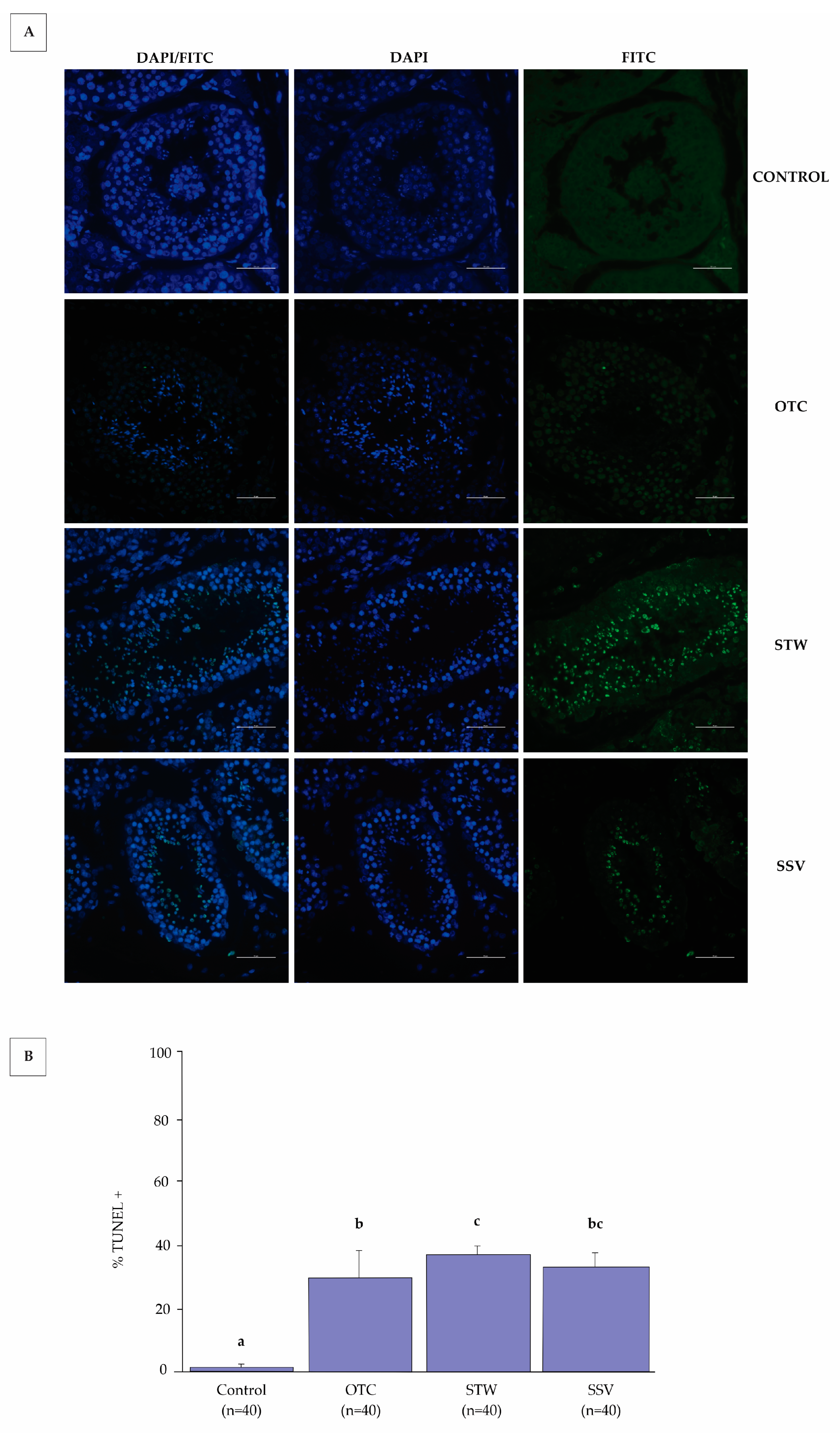
3.3. TUNEL Assay
4. Discussion
5. Conclusions
Author Contributions
Funding
Institutional Review Board Statement
Informed Consent Statement
Data Availability Statement
Conflicts of Interest
References
- Lima, D.B.C.; Silva, L.D.M.; Comizzoli, P. Influence of warming and reanimation conditions on seminiferous tubule morphology, mitochondrial activity, and cell composition of vitrified testicular tissues in the domestic cat model. PLoS ONE 2018, 13, 1–18. [Google Scholar] [CrossRef]
- Yokonishi, T.; Sato, T.; Komeya, M.; Katagiri, K.; Kubota, Y.; Nakabayashi, K.; Hata, K.; Inoue, K.; Ogonuki, N.; Ogura, A.; et al. Offspring production with sperm grown in vitro from cryopreserved testis tissues. Nat. Commun. 2014, 5, 1–6. [Google Scholar] [CrossRef]
- Yavin, S.; Arav, A. Measurement of essential physical properties of vitrification solutions. Theriogenology 2007, 67, 81–89. [Google Scholar] [CrossRef] [PubMed]
- Natalia, S. The domestic cat as a research model in the assisted reproduction procedures of wild felids. Postepy Biochem. 2021, 67, 362–369. [Google Scholar] [CrossRef]
- Buarpung, S.; Tharasanit, T.; Comizzoli, P.; Techakumphu, M. Feline spermatozoa from fresh and cryopreserved testicular tissues have comparable ability to fertilize matured oocytes and sustain the embryo development after intracytoplasmic sperm injection. Theriogenology 2013, 79, 149–158. [Google Scholar] [CrossRef]
- Abrishami, M.; Anzar, M.; Yang, Y.; Honaramooz, A. Cryopreservation of immature porcine testis tissue to maintain its developmental potential after xenografting into recipient mice. Theriogenology 2010, 73, 86–96. [Google Scholar] [CrossRef]
- Yamini, N.; Pourmand, G.; Amidi, F.; Salehnia, M.; Nejad, N.A.; Mougahi, S.M. Developmental potential of vitrified mouse testiculas tissue after ectopic transplantation. Cell J. 2016, 18, 74–82. [Google Scholar] [CrossRef]
- Teixeira, D.O.; Oliveira, E.S.; Fernandes, J.S.; Palomino, G.J.Q.; Tabosa, B.E.A.; Barbosa, H.T.S.; Pinheiro, B.Q.; Silva, L.D.M. Histological evaluation of testicles from prepubertal dogs submitted to vitrification with different cryoprotectant associations. RSD 2021, 10, 1–9. [Google Scholar] [CrossRef]
- Thuwanut, P.; Chatdarong, K. Cryopreservation of cat testicular tissues: Effects of storage temperature, freezing protocols and cryoprotective agents. Reprod. Domest. Anim. 2012, 47, 777–781. [Google Scholar] [CrossRef]
- Lima, D.B.C.; Silva, T.F.P.; Morais, G.B.; Aquino-Cortez, A.; Evangelista, J.S.A.M.; Xavier Júnior, F.A.F.; Viana, D.A.; Silva, L.D.M. Different associations of cryoprotectants for testicular tissue of prepubertal cats submitted to vitrification. Reprod. Domest. Anim. 2017, 52, 235–241. [Google Scholar] [CrossRef]
- Fernandes, J.S.; Tabosa, B.E.A.; Brito, B.F.; Silva, H.V.R.; Lima, D.B.C.; Silva, L.D.M. Influence of different warming temperatures on the vitrified testicular fragments from pre-pubertal cat. Reprod. Domest. Anim. 2021, 56, 1342–1348. [Google Scholar] [CrossRef]
- Macente, B.I.; Fonseca-Alves, C.E.; Magalhães, G.M.; Tavares, M.R.; Mansano, C.F.M.; Mouttham, L.; Apparício, M.; Toniollo, G.H.; Comizzoli, P. Influence of vitrification vevice, warming protocol, and subsequent in vitro culture on structural integrity of testicular fragments from adult domestic cats. Biopreserv. Biobank. 2022, 20, 392–400. [Google Scholar] [CrossRef]
- Ha, A.N.; Park, H.S.; Jin, J.I.; Lee, S.H.; Ko, D.H.; Lee, D.S.; White, K.L.; Kong, I.K. Postthaw survival of invitro-produced bovine blastocysts loaded onto the inner surface of a plastic vitrification straw. Theriogenology 2014, 81, 467–473. [Google Scholar] [CrossRef]
- Hu, H.; Ji, G.; Shi, X.; Liu, R.; Zhang, J.; Zhang, H.; Yuan, X.; Zhang, G.; Yuan, W.; Li, M. Comparison of rapid freezing versus vitrification for human sperm cryopreservation using sucrose in closed straw systems. Cell Tissue Bank. 2020, 21, 667–673. [Google Scholar] [CrossRef]
- Zhao, X.M.; Quan, G.B.; Zhou, G.; Hou, Y.P.; Zhu, S.E. Conventional freezing, straw, and open-pulled straw vitrification of mouse two pronuclear (2-PN) stage embryos. Anim. Biotechnol. 2007, 18, 203–212. [Google Scholar] [CrossRef]
- Sá, R.; Cremades, N.; Malheiro, I.; Sousa, M. Cryopreservation of human testicular diploid germ cell suspensions. Andrologia 2012, 44, 366–372. [Google Scholar] [CrossRef]
- Dinnyés, A.; Dai, Y.; Jiang, S.; Yang, X. High Developmental rates of vitrified bovine oocytes following parthenogenetic activation, in vitro fertilization, and somatic cell nuclear transfer. Biol. Reprod. 2000, 63, 513–518. [Google Scholar] [CrossRef]
- Curaba, M.; Verleysen, M.; Amorim, C.A.; Dolmans, M.M.; Langendonckt, A.; Hovatta, O.; Wyns, C.; Donnez, J. Cryopreservation of prepubertal mouse testicular tissue by vitrification. Fertil. Steril. 2011, 95, 1229–1234. [Google Scholar] [CrossRef]
- Carvalho, A.A.; Faustino, L.R.; Silva, C.M.G.; Castro, S.V.; Lopes, C.A.P.; Santos, R.R.; Báo, S.N.; Figueiredo, J.R.; Rodrigues, A.P.R. Novel wide-capacity method for vitrification of caprine ovaries: Ovarian Tissue Cryosystem (OTC). Anim. Reprod. Sci. 2013, 138, 220–227. [Google Scholar] [CrossRef]
- Faustino, L.R.; Carvalho, A.A.; Silva, C.M.G.; Rossetto, R.; Lopes, C.A.P.; Tilburg, M.F.; Carneiro, P.B.M.; Báo, S.N.; Moura, A.A.A.; Bordignon, V.; et al. Assessment of DNA damage in goat preantral follicles after vitrification of the ovarian cortex. Reprod. Fertil. Dev. 2015, 27, 440–448. [Google Scholar] [CrossRef]
- Campos, L.B.; Silva, A.M.; Praxedes, E.C.G.; Bezerra, L.G.P.; Lins, T.L.B.G.; Menezes, V.G.; Matos, M.H.T.; Lima, G.L.; Rodrigues, A.P.R.; Silva, A.R. Vitrification of collared peccary ovarian tissue using open or closed systems and different intracellular cryoprotectants. Cryobiology 2019, 91, 77–83. [Google Scholar] [CrossRef] [PubMed]
- Brandão, F.A.S.; Alves, K.A.; Brito, D.C.C.; Pereira, L.M.C.; Morais, G.B.; Ñaupas, L.V.S.; Souza, S.S.; Alves, B.G.; Rodrigues, A.P.R.; Teixeira, D.Í.A. Vitrification of canine ovarian tissue using the Ovarian Tissue Cryosystem (OTC) device. Reprod. Domest. Anim. 2021, 56, 1156–1161. [Google Scholar] [CrossRef] [PubMed]
- Brito, D.C.; Domingues, S.F.S.; Silva, J.K.; Wu, X.; Santos, R.R.; Pieczarka, J.C. Detrimental effect of phenol red on the vitrification of cat (Felis catus) ovarian tissue. Biopreserv. Biobank. 2016, 14, 17–22. [Google Scholar] [CrossRef] [PubMed]
- Santos, R.R.; Tharasanit, T.; Haeften, T.; Figueiredo, J.R.; Silva, J.R.V.; Hurk, R. Vitrification of goat preantral follicles enclosed in ovarian tissue by using conventional and solid-surface vitrification methods. Cell and Tissue Res. 2007, 327, 167–176. [Google Scholar] [CrossRef]
- Rahimi, G.; Isachenko, E.; Sauer, H.; Isachenko, V.; Wartenberg, M.; Hescheler, J.; Mallmann, P.; Nawroth, F. Effect of different vitrification protocols for human ovarian tissue on reactive oxygen species and apoptosis. Reprod. Fertil. Dev. 2003, 15, 343–349. [Google Scholar] [CrossRef]
- Milazzo, J.P.; Vaudreuil, L.; Cauliez, B.; Gruel, E.; Massé, L.; Mousset-Siméon, N.; Macé, B.; Rives, N. Comparison of conditions for cryopreservation of testicular tissue from immature mice. Hum. Reprod. 2008, 23, 17–28. [Google Scholar] [CrossRef]
- Rashidi, Z.; Azadbakht, M.; Khazaei, M. Hydrostatic pressure improves in-vitro maturation of oocytes derived from vitrified-warmed mouse ovaries. Iran J. Reprod. Med. 2012, 10, 257–264. [Google Scholar]
- Liebermann, J.; Nawroth, F.; Isachenko, V.; Isachenko, E.; Rahimi, G.; Tucker, M.J. Potential importance of vitrification in reproductive medicine. Biol. Reprod. 2002, 67, 1671–1680. [Google Scholar] [CrossRef]
- Brito, D.C.; Domingues, S.F.S.; Rodrigues, A.P.R.; Maside, C.; Lunardi, F.O.; Wu, X.; Figueiredo, J.R.; Pieczarka, J.C.; Santos, R.R. Cryopreservation of domestic cat (Felis catus) ovarian tissue: Comparison of two vitrification methods. Theriogenology 2018, 111, 69–77. [Google Scholar] [CrossRef]
- Patra, T.; Gupta, M.K. Cryopreservation of murine testicular Leydig cells by modified solid surface vitrification with supplementation of antioxidants. Cryobiology 2019, 88, 38–46. [Google Scholar] [CrossRef]
- Patra, T.; Gupta, M.K. Solid surface vitrification of goat testicular cell suspension enriched for spermatogonial stem cells. Cryobiology 2021, 104, 8–14. [Google Scholar] [CrossRef]
- Patra, T.; Pathak, D.; Gupta, M.K. Comparison of two culture methods during in vitro spermatogenesis of vitrified-warmed testis tissue: Organ culture vs. hanging drop culture. Cryobiology 2021, 100, 142–150. [Google Scholar] [CrossRef]
- Santos, R.R.; Amorim, C.; Cecconi, S.; Fassbender, M.; Imhof, M.; Lornage, J.; Paris, M.; Schoenfeldt, V.; Martinez-Madrid, B. Cryopreservation of ovarian tissue: An emerging technology for female germline preservation of endangered species and breeds. Anim. Reprod. Sci. 2010, 122, 151–163. [Google Scholar] [CrossRef]
- Xing, W.; Zhou, C.; Bian, J.; Montag, M.; Xu, Y.; Li, Y.; Li, T. Solid-surface vitrification is an appropriate and convenient method for cryopreservation of isolated rat follicles. Reprod. Biol. Endocrinol. 2010, 8, 1–9. [Google Scholar] [CrossRef]
- Said, T.M.; Paasch, U.; Glander, H.J.; Agarwal, A. Role of caspases in male infertility. Hum. Reprod. Update 2004, 10, 39–51. [Google Scholar] [CrossRef]
- Furuchi, T.; Masuko, K.; Nishimune, Y.; Obinata, M.; Matsui, Y. Inhibition of testicular germ cell apoptosis and differentiation in mice misexpressing Bel-2 in spermatogonia. Development 1996, 122, 1703–1709. [Google Scholar] [CrossRef]
- Smith, K.F.; Whitehouse, K.A.; Pedersen, A.B. The role of infectious diseases in biological conservation. Anim. Cons. 2009, 12, 1–12. [Google Scholar] [CrossRef]
- Bielanski, A.; Vajta, G. Risk of contamination of germplasm during cryopreservation and cryobanking in IVF units. Hum. Reprod. 2009, 24, 2457–2467. [Google Scholar] [CrossRef]
- Grout, B.W.W.; Morris, G.J. Contaminated liquid nitrogen vapour as a risk factor in pathogen transfer. Theriogenology 2009, 71, 1079–1082. [Google Scholar] [CrossRef]
- Elliott, G.D.; Wang, S.; Fuller, B.J. Cryoprotectants: A review of the actions and applications of cryoprotective solutes that modulate cell recovery from ultra-low temperatures. Cryobiology 2017, 76, 74–91. [Google Scholar] [CrossRef]


| Control | OTC | STW | SSV | |
|---|---|---|---|---|
| Spermatogonia/Sertoli nuclei distinction | 0.03 (±0.03) a | 0.6 (±0.12) b | 1.16 (±0.05) c | 1.2 (±0.19) c |
| Nucleolar visualization | 0 (±0.00) a | 0.6 (±0.53) b | 1 (±0.03) c | 0.5 (±0.14) b |
| Nuclear condensation | 0.2 (±0.08) a | 0.8 (±0.06) b | 1 (±0.00) b | 0.8 (±0.21) b |
| Basal membrane detachment | 0 (±0.00) a | 0.5 (±0.08) ab | 0.9 (±0.17) b | 1 (±0.49) b |
| Shrinkage of epithelium | 0.3 (±0.11) a | 2 (±0.00) c | 2 (±0.09) c | 1 (±0.35) b |
Disclaimer/Publisher’s Note: The statements, opinions and data contained in all publications are solely those of the individual author(s) and contributor(s) and not of MDPI and/or the editor(s). MDPI and/or the editor(s) disclaim responsibility for any injury to people or property resulting from any ideas, methods, instructions or products referred to in the content. |
© 2023 by the authors. Licensee MDPI, Basel, Switzerland. This article is an open access article distributed under the terms and conditions of the Creative Commons Attribution (CC BY) license (https://creativecommons.org/licenses/by/4.0/).
Share and Cite
Carvalho, J.V.G.d.; Soares, A.R.B.; Leão, D.L.; Reis, A.N.; Santos, R.R.; Rodrigues, A.P.R.; Domingues, S.F.S. Effect of Different Vitrification Techniques on Viability and Apoptotic Index of Domestic Cat Testicular Tissue Cells. Animals 2023, 13, 2768. https://doi.org/10.3390/ani13172768
Carvalho JVGd, Soares ARB, Leão DL, Reis AN, Santos RR, Rodrigues APR, Domingues SFS. Effect of Different Vitrification Techniques on Viability and Apoptotic Index of Domestic Cat Testicular Tissue Cells. Animals. 2023; 13(17):2768. https://doi.org/10.3390/ani13172768
Chicago/Turabian StyleCarvalho, Julyne Vivian Guimarães de, Airton R. B. Soares, Danuza L. Leão, Adriana N. Reis, Regiane R. Santos, Ana P. R. Rodrigues, and Sheyla F. S. Domingues. 2023. "Effect of Different Vitrification Techniques on Viability and Apoptotic Index of Domestic Cat Testicular Tissue Cells" Animals 13, no. 17: 2768. https://doi.org/10.3390/ani13172768