Article

Effect of CXCL17 on Subcutaneous Preadipocytes Proliferation in Goats

1
College of Animal & Veterinary Sciences, Southwest Minzu University, Chengdu 610041, China
2
Key Laboratory of Qinghai—Tibetan Plateau Animal Genetic Resource Reservation and Utilization, Ministry of Education, Southwest Minzu University, Chengdu 610041, China
*
Author to whom correspondence should be addressed.
Animals 2023, 13(11), 1757; https://doi.org/10.3390/ani13111757
Submission received: 31 March 2023 / Revised: 17 May 2023 / Accepted: 23 May 2023 / Published: 25 May 2023
(This article belongs to the Collection Small Ruminant Genetics and Breeding)

Abstract

:

Simple Summary

There are many existing studies related to goat adipose deposition. However, no studies have reported the role of the CXCL17 gene on adipose deposition in goats before. The aim of this study was to explore the effect of CXCL17 on goat subcutaneous preadipocytes proliferation and provide potential targets for goat-directed molecular breeding. The results showed that the exogenous expression of CXCL17 at different doses had different regulatory effects on the proliferation of subcutaneous preadipocytes of goats.

Abstract

The presence or absence of subcutaneous adipose accumulation will affect the energy storage, insulation resistance and metabolism of animals. Proliferation and differentiation of preadipocytes play a significant role in lipid deposition. The objective of this study was to clone the goat CXCL17 gene and investigate its potential functions on goat subcutaneous preadipocytes’ proliferation by gaining or losing function in vitro. The goat CXCL17 gene was cloned by Reverse Transcription-Polymerase Chain Reaction (RT-PCR) and bioinformatics analysis was performed. The expression of the CXCL17 gene in the different goat tissues and adipocytes at different differentiation stages was detected by real-time fluorescence quantitative PCR (qPCR). The results showed that the cloned sequence of goat CXCL17 gene is 728 bp and the CDS region is 357 bp, encoding 118 amino acids. CXCL17 protein is located in nucleus, cytoplasm, mitochondria and extracellular matrix. Tissue-expression profiles revealed that CXCL17 expressed in all of the examined tissues. In visceral tissues, the highest expression level was found in lung (p < 0.01); in muscle tissues, the highest CXCL17 expression level was found in the longissimus dorsi (p < 0.01) and in adipose tissues, the highest expression level was found in subcutaneous adipose (p <0.01). Compared with those cells before differentiation, CXCL17 expression levels upregulated at 48 h (p < 0.01), 72 h (p < 0.01), 120 h (p < 0.01) and downregulated at 96 h (p < 0.01). Furthermore, the results of crystal violet staining and semi-quantitative assay showed that transfection with 1 μg CXCL17 expression plasmid reduced the cell numbers in vitro. Meanwhile, the expression of CCND1 was significantly decreased. A similar consequence happened after interfering with CXCL17 expression. However, plasmid transfected with 2 μg pEGFPN1-CXCL17 increased the number of cells in vitro. These results suggest that CXCL17 is involved in the proliferation of goat subcutaneous preadipocytes.

1. Introduction

In humans, there are many diseases related to adipose tissue [1,2,3]. Subcutaneous fat (SAT), intramuscular fat (IMF) and visceral fat (VAT) constitute the mammalian adipose tissue. For meat-used animals, IMF and SAT are highly valued because they are important factors affecting meat quality [4]. Subcutaneous adipose tissue plays an important role in the stabilization of lipid metabolism balance and is the main site of lipid deposition [5]. lipid deposition can seriously affect their meat quality, meat flavor and carcass lean meat percentage [6,7]. Therefore, it is important to explore the metabolic characteristics and regulatory mechanism of subcutaneous adipose for the meat value of meat-used animals.
Chemokines (also known as chemotaxis hormone) is a small-molecule cytokine family of proteins that can collect leukocytes to clear foreign bodies such as foreign pathogens and plays an important role in inflammatory response [8]. By classifying the relative positions of the cysteine residues at the amino terminal (N end), the chemokines can be divided into four subgroups: CXC (major), CC (major), XC (minor) and CX3C (minor) [9]. According to the triplet glutamate-to-leucine-arginine sequence (ELR) before the first cysteine, the CXC subgroup can be further divided into ELR-positive (ELR +) and -negative (ELR −). CXC motif chemokine ligand 17 (CXCL17) has obvious angiogenesis-promoting effect and belongs to the ELR + class of CXC subgroup [10]. The CXCL17 gene localized on human chromosome 19q13.2 was found in 2006 and consisted of four exons. The precursor protein (DMC) contains 119 amino acid residues, six of which are cysteines. Human CXCL17 contains a signal peptide consisting of 22 amino acids [11].
CXCL17 can recruit monocytes, macrophages and mature and immature dendritic cells (DC), which is of great significance for the development of multiple cancer types [12,13,14,15,16,17,18]. Abnormal expression of CXCL17 in tumor cells can accumulate immature myeloid cells and promote cancer development by promoting vascular proliferation [19]. In addition, CXCL17 is an antibacterial mucosal chemokine, which can not only exert antibacterial activity through a peptide-mediated way to disrupt bacterial membrane, but also reduce the expression of type I collagen by upregulating the expression of matrix metalloproteinase 1 (MMP1) and miR-29 in fibroblasts, thus participating in the occurrence of systemic sclerosis (SSc) [20,21]. Moreover, CXCL17/CXCR8 signaling pathway plays an important role in mucosal immunity in the female genital tract [22]. CXCL17 is highly expressed in salivary glands and lacrimal glands of patients with primary Sjogren’s syndrome [23]. Since CXCL17 has previously been reported to be involved in cell proliferation, preadipocytes proliferation promotes lipid accumulation. Goat lipid deposition plays an important role in meat quality improvement. Tissue expression detection result showed that CXCL17 expression level was the highest in subcutaneous adipose tissue. Therefore, we speculated that it plays an important regulatory role in the process of subcutaneous fat deposition in goats.
The Jianzhou Daer goat is the second breed of goat bred independently in China and has high meat value. Subcutaneous adipose deposition can reduce the proportion of lean meat in the carcass and has a great impact on meat quality [24]. In this study, we cloned the CXCL17 coding sequence and analyzed its biological characteristics by the bioinformatics method. Real-time PCR was used to analyze the expression level of CXCL17 gene in different tissues and subcutaneous preadipocytes at different differentiation induction times. Moreover, we analyzed its effect on cell proliferation using gain-of-function and loss-of-function experiments. Therefore, this study aims to characterize the potential functions of CXCL17 on the proliferation of goat subcutaneous preadipocytes and provide a new theoretical basis for further study of lipid deposition in goat adipocytes.

2. Materials and Methods

2.1. Test Animals and Samples Collection

The test animals are one-year-old healthy Jianzhou Daer goats (n = 3) that were purchased from Sichuan Tiandi Yang Biological Engineering Co., Ltd., (Chengdu, China) and sampled immediately after slaughter. The samples included heart, liver, spleen, lung, kidney, large intestine, small intestine, rumen, longissimus dorsi, vastus, gluteus, abdominal adipose, subcutaneous adipose, mesenteric adipose, pericardial adipose, intermuscular adipose and visceral adipose tissue. The Institutional Animal Care and Use Committee of Southwest Minzu University (Chengdu, China) permitted this research project, and all animal experiments in this study were in line with animal ethical treatment.

2.2. Total RNA Extraction and Reverse Transcription

Total RNA was extracted from each tissue and cells by Trizol (TaKara, Dalian, China). Then, 1 μg RNA was reverse transcribed according to the instructions of reverse transcription kit (Thermo, Waltham, MA, USA) and stored at −20 °C.

2.3. Cloning of Coding Region of Goat CXCL17

For gene cloning, primers were designed according to the mRNA sequence of bovine CXCL17 gene in GenBank (accession number: NM_00101014862.2), and the sequence of primers were as follows: sense: 5′ CGCCCTCTAATGAGAATGCT 3′ antisense: 5′ AGCGTAAGGCTGTGTGAAGG 3′. Complementary DNA (cDNA) of goat cardiac tissue was used for PCR amplification. As described in the previous article in our laboratory, the positive bacterial solution was sent to Chengdu Qingke Biotechnology Co., Ltd. (Chengdu, China) for verification and sequencing [25].

2.4. Bioinformatics Analysis of Goat CXCL17 Coding Region

The analysis software and online tools of goat CXCL17 gene refer to Table 1, and the detailed analysis method is based on the relevant literature [26].

2.5. Induced Differentiation of Subcutaneous Preadipocytes

Primary culturing of subcutaneous goat preadipocytes was conducted according to the isolation and culture methods established in the laboratory [27]. Goat subcutaneous preadipocytes cultured to F3 were counted and inoculated into 12-well plates. When the cell confluence reached 80%, the complete medium was replaced with 50 μmol/L oleic acid induction solution (Sigma, Tokyo, Japan), and the fluid was changed every 2 d. Cells were collected at 0 h, 24 h, 48 h, 72 h, 96 h and 120 h.

2.6. Real-Time Fluorescence Quantitative PCR (qPCR)

According to the instructions of SYBR ® Premix Ex Taq TM (2×) kit (TaKara, Kusatsu, Shiga, Japan), the expression levels of CXCL17 and proliferation marker genes were determined using UXT as an internal reference gene. CXCL17 quantitative primers and expression vector construction primers were designed by cloned CXCL17 gene sequence, and other primers were designed by goat sequences in NCBI. Primer information is listed in Table 2.

2.7. Construction of Overexpression Plasmid and Synthesis of siRNA

For construction of overexpression plasmid, the sequence of goat CXCL17 gene (GenBank accession number: ON930036) was amplified by PCR. After detection by 1% agarose gel electrophoresis, the PCR product was purified and recycled. Then the PCR products and the pEGFP-N1 plasmid were digested by restriction enzymes (EcoR Ⅰ; TaKara, Dalian, China; Kpn Ⅰ; TaKara, Dalian, China) and ligated by T4 DNA Ligase (TaKara, Dalian, China) at 16 °C overnight. Transforming and coating plates were used. Then colonies were selected and identified by PCR, and sent to Chengdu Qingke Biotechnology Co., Ltd. (Qingke, Chengdu, China) for sequencing. After successful sequencing, we used plasmid extraction kits (TIANGEN, Beijing, China) to extract the plasmid and stored it at 4 °C.
For siRNAs, the siRNAs were synthesized by GenePharma (GenePharma, Shanghai, China). The sequence of siRNAS were as follows:
si-CXCL17: sense: 5′-CCAAGAAUGUGAGUGCCAATT-3′;
antisense: 5′-UUGGCACUCACAUUCUUGGTT-3′;
si-NC: sense: 5′-UUCUCCGAACGUGUCACGUTT-3′;
antisense: 5′-ACGUGACACGUUCGGAGAATT-3′.

2.8. Transfection

When goat subcutaneous preadipocytes were cultured to F3 generation and reached about 80% confluence, cells were planted into 6-well plates or 96-well plates. The control group was transfected with pEGFP-N1 (Vector) or siRNA (NC), and the experimental group was transfected with pEGFP-CXCL17 or siRNA-CXCL17, respectively.
Transfection system for overexpression (6-well plates): 1 μg/2 μg pEGFP-CXCL17/pEGFP-N1, 400 μL Opti-MEM (Gibco, Calabasas, CA, USA), 6 μL Hieff TransTM Liposomal Transfection Reagent (Yeasen, Shanghai, China).
Transfection system for interference (6-well plates): 6 μL (20 μM) siRNA-CXCL17/siRNA (NC), 400 μL Opti-MEM (Gibco, Calabasas, CA, USA), 6 μL Hieff TransTM Liposomal Transfection Reagent (Yeasen, Shanghai, China).
Transfection system for overexpression (96-well plates): 0.1 μg/0.2 μg pEGFP-CXCL17/pEGFP-N1, 20 μL Opti-MEM (Gibco, Calabasas, CA, USA), 3 μL Hieff TransTM Liposomal Transfection Reagent (Yeasen, Shanghai, China).
Transfection system for interference (96-well plates): 0.3 μL (1 μM) siRNA-CXCL17/siRNA (NC), 20 μL Opti-MEM (Gibco, Calabasas, CA, USA), 3 μL Hieff TransTM Liposomal Transfection Reagent (Yeasen, Shanghai, China).

2.9. MTT Assay

MTT experiments were performed to detect the cells’ viability. We planted goat subcutaneous preadipocytes in 96-well plates with a density of 3 × 10³ per well and cultured them according to the methods of previous articles in our laboratory [28]. Adding 10 µL MTT reagent (solarbio, Beijing, China) into each well of 96-well plates away from light, cells were cultured at 37 °C and 5% CO2 for 4 h. The absorbance at 490 nm was measured by using an enzyme-labeled instrument.
For crystal violet staining, goat subcutaneous preadipocytes were seeded in 96-well plates with a density of 3 × 10³ per well. Culture media was discarded after 0, 24, 48 and 72 h of cell culture. Then goat subcutaneous preadipocytes were washed twice with PBS and fixed with 4% formaldehyde for 30 min. Next, 4% formaldehyde was discarded, and cells were washed twice with PBS. Then, 200 μL of 0.1% Crystal violet-solution was added into each well to stain for 20 min. After discarding Crystal violet solution, cells were washed twice with PBS. The images of crystal violet staining were captured by fluorescence microscopy (Olympus, Tokyo, Japan).
For semi-quantification, 200 μL of 20% glacial acetic acid was added to each well of the 96-well plate for 15 min. Then the optical density at 490 nm was detected using an enzyme-labeled instrument.

2.10. Statistical Analysis

Quantitative PCR data were analyzed using the 2− ΔΔ Ct method, and the data were expressed as “mean ± standard deviation (Mean ± SD)”. The difference of the data was analyzed using the one-way ANOVA method, the multiple comparison t-test and the Student’s two-tailed t-test in GraphPad Prism 8.0 software. All experiments in this study including biological information analysis, qPCR, cell culture, MTT and semi-quantitative experiments were performed in triplicate. All experiments were statistically significant when p < 0.05. “*”: p < 0.05 indicates a significant difference and “**”: p < 0.01 indicates a very significant difference.

3. Results

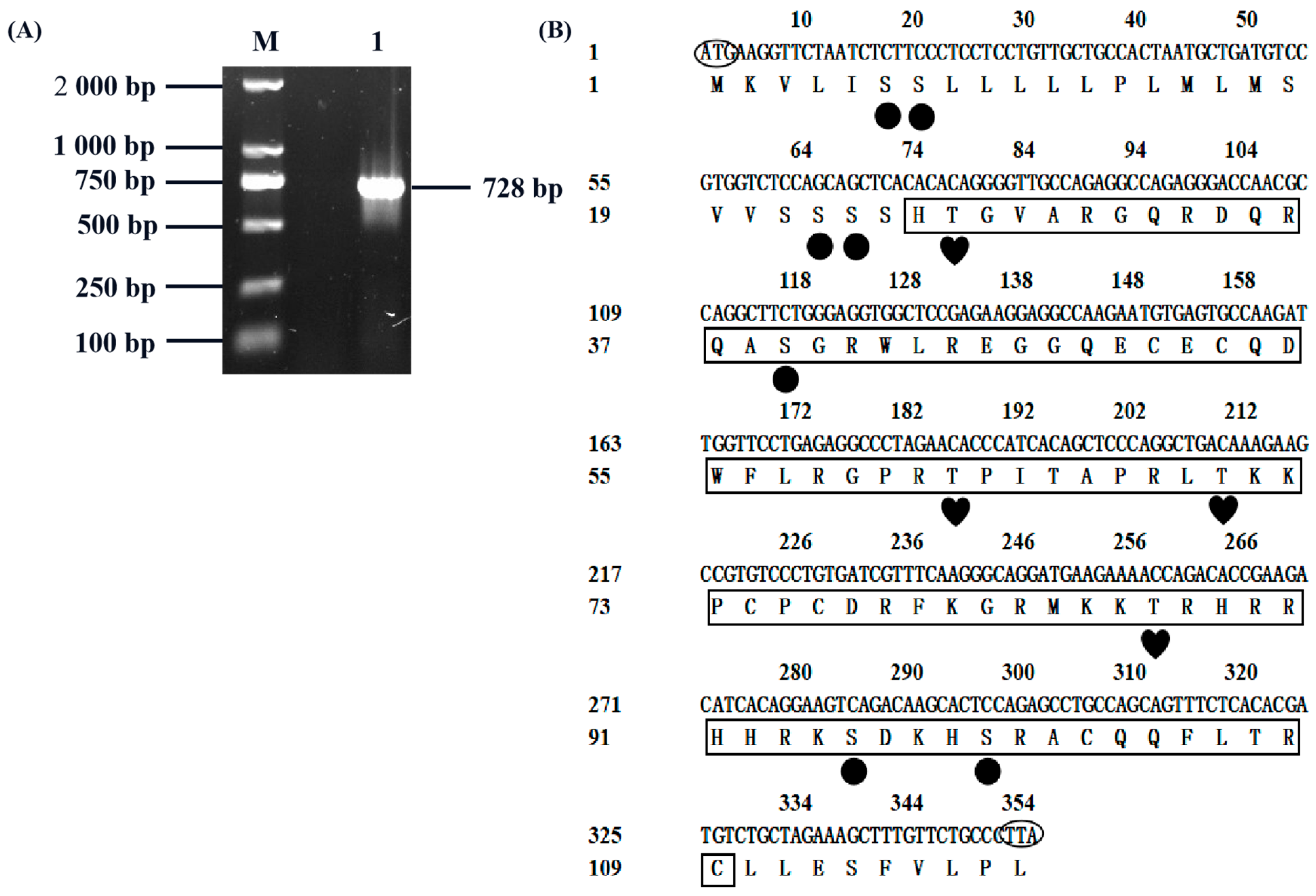
3.1. Goat CXCL17 Gene Cloning

In order to study the biological function of CXCL17 in goats, we cloned its coding sequence first. Goat heart tissue cDNA was used as the template to clone the CXCL17 gene, resulting in a specific band consistent with the size of the expected target fragment (Figure 1A). After sequencing, the obtained sequence was 728 bp long and sequence analysis indicated that the CXCL17 CDS region was 357 bp encoding 118 amino acids (Figure 1B). The GenBank accession number was ON930036.

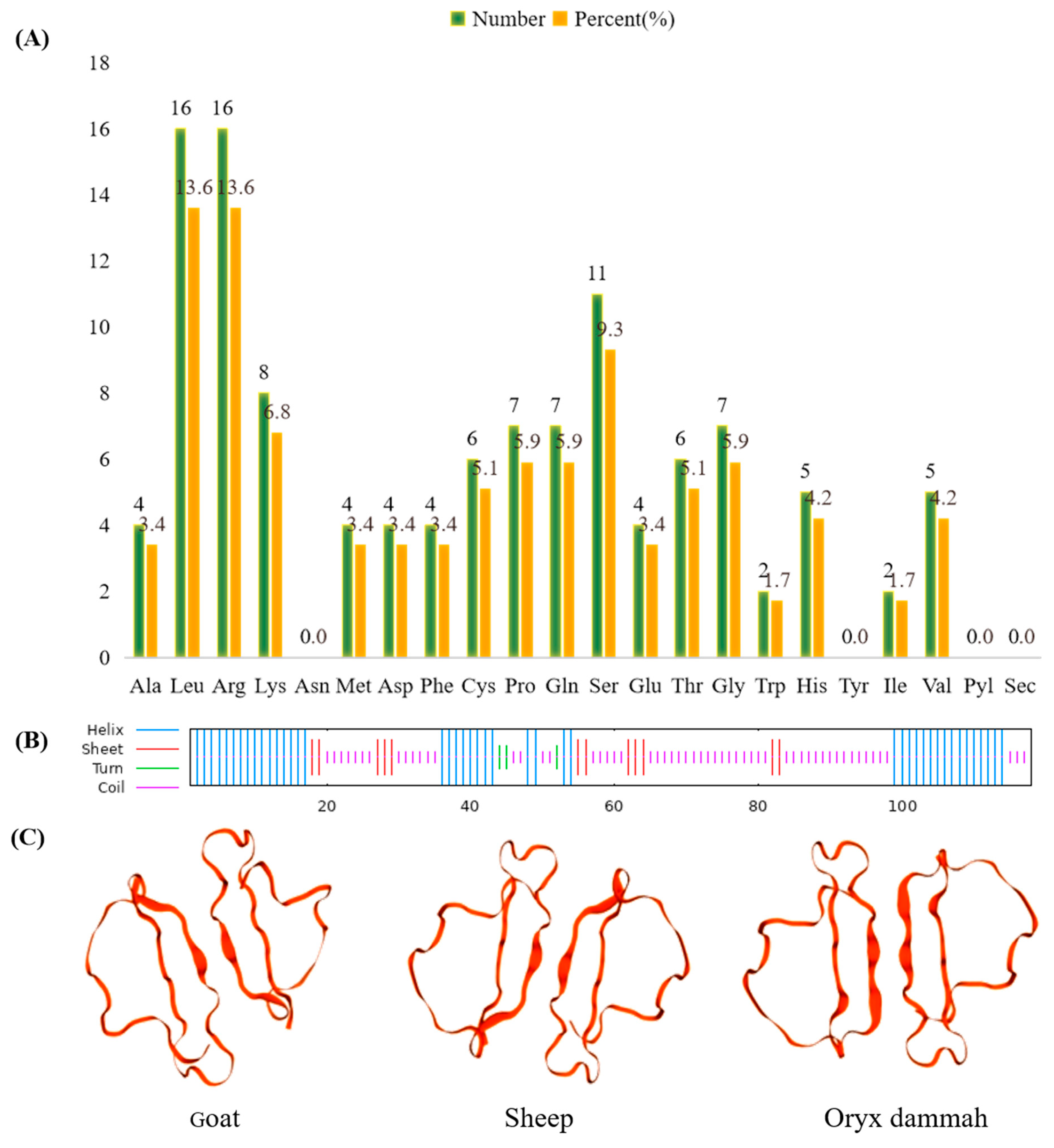
3.2. Protein Structure and Amino Acid Composition Analysis of Goat CXCL17

The primary structure and physicochemical properties of goat CXCL17 were analyzed by Ex PASy-ProParam. The protein molecular formula of CXCL17 is C 592 H 981 N 193 O 159 S 10, its relative molecular mass is 13,667.10 Da, the isoelectric point (pI) is 11.07, the total average of hydrophilic (GRAVY) is −0.582 and the instability index (II) is 57.27, indicating that the protein is an unstable basic hydrophilic protein. The content of various amino acids of the CXCL17 protein is shown in Figure 2A, with the highest content of arginine (Arg) and leucine (Leu), and the least content of isoleucine (Ile) and tryptophan (Trp), respectively. The aliphatic index of CXCL17 protein is 90.20 and the half-life is 30 h. The number of total residues of positive charge (Arg + Lys) and the total number of negative charge (Asp + Glu) are 24 and 8, respectively, indicating that the protein is positively charged.
According to the secondary structure prediction of goat CXCL17 using SOPMA, it was found that 57 amino acids (48.31%) had the highest proportion of random curling, 46 amino acids (38.98%) could form an α helix, 12 amino acids could form an extension chain (10.17%) and 3 amino acids (2.54%) could form a β turn (Figure 2B). SWISS-MODEL software was used to predict the tertiary structure models of goat and other species’ proteins and the results showed that the spatial structure of goat CXCL17 protein was extremely close to sheep and oryx dammah (Figure 2C).

3.3. Subcellular Localization, Signal Peptide Prediction, Protein Phosphorylation Site and Transmembrane Domain, Amino Acid Sequence Homology and Phylogenetic Tree Analysis

PSORT Ⅱ was used to predict subcellular localization of goat CXCL17. The result showed that 11.1% is located in the cytoplasmic matrix and nucleus, 33.3% in the mitochondria and 44.4% in the extracellular matrix (including cell membrane). SignalP-5.0 Server predicted signal peptide showed that CXCL17 has a signal peptide (Figure 3A). The NetPhos 3.1-predicted result showed that there were 11 phosphorylation sites, 7 serine phosphorylation sites and 4 threonine phosphorylation sites (Figure 3B). The CXCL17 transmembrane domain was predicted by TMHMM 2.0, which found that CXCL17 has a transmembrane domain (Figure 3C).
The comparison results of amino acid sequence homology showed that goat CXCL17 shared the highest similarity with ovis aries, and higher similarity with oryx dammah, bison, cervus elaphus and bos mutus (Figure 3D), indicating that CXCL17 protein was highly conserved among different species. The phylogenetic tree was constructed by MEGA 5.0 according to the amino acid sequence homology between goat and other species. CXCL17 of goat and ovis aries were in the same branch of the phylogenetic tree, indicating that goat CXCL17 and ovis aries were closely related (Figure 3E).

3.4. Analysis of Tissue and Temporal Expression Profile of CXCL17 Gene in Goats

UXT was used as a reference gene to determine CXCL17 expression level in various goat tissues, and qPCR detection results showed that CXCL17 expressed in all the examined tissues [29]. In visceral tissues, CXCL17 expression was highest in lung (p < 0.01), the next-highest expression tissue was rumen (p < 0.01), and low expression was observed in liver, small intestine, large intestine and spleen (Figure 4A). The muscle tissue expression profiles showed that CXCL17 expressed in longissimus dorsi, gluteus (p < 0.01) and vastus (p < 0.01) with a trend from high to low (Figure 4A). In adipose tissue, the expression of CXCL17 in subcutaneous adipose tissue was the highest (p < 0.01), followed by the higher expression in abdominal adipose (p < 0.01), and low expression in mesenteric adipose, intramuscular adipose and visceral adipose (Figure 4A). Because CXCL17 gene expression was the highest in subcutaneous adipose, we then speculated that it may play a vital regulatory role in lipid deposition of goat subcutaneous preadipocytes. The expression of CXCL17 gene during subcutaneous preadipocytes differentiation was determined by qPCR. The results showed that its expression was significantly upregulated at 48 h and 72 h (Figure 4B). Interestingly, the expression of CXCL17 gene was reduced to about half of the original expression level at 96 h of induced differentiation, and the expression level was significantly upregulated at 120 h of induced differentiation (Figure 4B). The above results indicated that CXCL17 may play different regulatory roles in different stages of differentiation of goat subcutaneous preadipocytes.

3.5. CXCL17 Expression Plasmid Construction

Preadipocytes proliferation is another factor affecting lipid deposition. To investigate the role of CXCL17 in goat subcutaneous preadipocytes proliferation, CXCL17 expression plasmid was constructed first. PCR amplifying results showed a correct strip location (Figure 5A). Primer information used to construct overexpressed vectors is shown in Table 2. The PCR product was connected to pEGFP-N1 vector after digestion, and the sequencing results were correct (Figure 5B). In order to clarify the localization of goat CXCL17, subcutaneous preadipocytes were transfected with exogenous pEGFP-CXCL17 plasmid and took fluorescence photographs. In contrast to pEGFP-N1, pEGFP-CXCL17 has many dispersed extracellular fluorescences, largely consistent with the subcellular localization results in 3.3 (Figure 5C). The results of overexpression efficiency showed that the expression level of CXCL17 gene was nearly 2 400 000-fold upregulated when compared with the control group (Figure 5D). In conclusion, the pEGFP-CXCL17 overexpression plasmid was constructed successfully.

3.6. Transfected Plasmid with Different Dose Showed Different Value-Added Effect

In order to study the effect of CXCL17 on subcutaneous preadipocytes proliferation, we transfected 1 μg expression plasmid into goat subcutaneous preadipocytes. The results of qPCR showed that the transfection efficiency was about a 4 000-fold change, when compared to the negative control (Figure 6A). The detection results of proliferation marker genes expression showed that CCND1 were downregulated in the CXCL17 overexpression group (Figure 6B).
To further verify the effect of CXCL17 on the proliferation of subcutaneous preadipocytes in goats, we performed crystal violet staining and semi-quantitative experiments. The number of cells in the CXCL17 group was significantly less than that in the control group at 72 h (Figure 6C). Furthermore, the semi-quantitative results were generally consistent with the crystal violet staining results (Figure 6D). We then examined the cell viability by MTT, and the result showed that CXCL17 overexpression significantly inhibited cell proliferation after 24 h–72 h transfection (Figure 6E). Eventually, the above results suggested that CXCL17 had a significant inhibitory effect on the proliferation of goat subcutaneous preadipocytes when transfected with a 1μg pEGFP-CXCL17 overexpression plasmid.
To more accurately understand the effect of CXCL17 overexpression on goat subcutaneous preadipocytes proliferation, we transfected 2 μg CXCL17 plasmid into these cells. We first examined the transfection efficiency, and CXCL17 was successfully overexpressed (Figure 7A). Subsequently, we detected the proliferation relative marker genes’ expression, and found the expression of CDK2 and CCND1 increased in the CXCL17 overexpression group (p < 0.05) (Figure 7B).
Then we performed crystal violet staining to determine whether the exogenous overexpression of CXCL17 has an effect on the cell numbers (Figure 7C). Meanwhile, from the semi-quantitative results in Figure 7D, there was significant difference in the cell numbers between the two groups at 72 h. The crystal violet staining result is generally in line with the semi-quantitative result, with significant differences in the cell number between the two groups at 72 h. We found that the cell viability in the overexpression group at 72 h (p < 0.01) was greater than that in the control group by MTT (Figure 7E). All the experimental results above showed that overexpression of high-dose CXCL17 could promote goat subcutaneous preadipocytes proliferation.

3.7. Knockdown of CXCL17 Inhibits Goat Subcutaneous Preadipocytes Proliferation

To further confirm the effect of CXCL17 on goat subcutaneous preadipocytes proliferation, three specific siRNA targets for CXCL17 had been synthesized. As shown in Figure S1, the interference efficiency of siRNA-2 is the strongest, so we choose siRNA-2 (named si-CXCL17) for the following studies. We examined the transfection efficiency of siRNA-2 again to check the correctness of the results, and the qPCR results indicated the expression of CXCL17 mRNA in si-CXCL17 group was significantly lower than that in NC group (Figure 8A). Meanwhile, we examined the mRNA expression level of marker genes associated with cell proliferation and found the expression of marker genes CCNE1 (p < 0.01) and CDK2 (p < 0.05) in the si-CXCL17 group was lower than the NC group (Figure 8B).
In addition to the above experiments, we performed crystal violet staining and found that the number of cells in the si-CXCL17 group was less than that in the NC group at 72 h (Figure 8C). The results are in Figure 8D, and the absorbance of the si-CXCL17 group was very much lower than the NC group at 72 h (p < 0.01). We also used MTT to analyze cell viability with the interference of CXCL17 and found that cell viability in the interference group was significantly lower than in the NC group at 24 h–72 h (p < 0.01) (Figure 8E). Combined with disrupting all experiments, we speculated that decreased expression of CXCL17 could inhibit subcutaneous preadipocytes proliferation in goats.

3.8. Analysis of CXCL17 Interaction Proteins

To study the mechanism of CXCL17 inhibiting the proliferation of subcutaneous preadipocytes, the STRING interaction database was used to predict the potential interaction of proteins. The result indicated that CXCL17 may interact with proteins such as GPR35, CCL17 and SNTN (Figure 9). Relevant studies showed that GPR35 knockdown will affect lipid accumulation in hepatocytes in mice [30]. Moreover, some studies reported that GPR35 is the receptor of CXCL17 [31]. So, we hypothesized that CXCL17 may affect subcutaneous adipose deposition by interacting with GPR35 to affect subcutaneous preadipocytes proliferation in goats.

4. Discussion

The human chemokine superfamily consists of 48 ligands and 20 receptors. They can regulate the migration of cells such as leukocytes and stem cells in vivo. Chemokines can promote the development of inflammatory response by clustering leukocytes in inflamed or injured tissues and by regulating leukocyte homeostasis and homing [32]. CXCL17 is the last chemokine identified and characterized so far, which can promote angiogenesis, so it is considered to be a vascular endothelial growth factor (VEGF) coregulatory chemokine [33]. CXCL17 has both homeostatic chemokines and inflammatory chemokines, which is the reason why it is considered as a “dual” chemokine. CXCL17 has many functions, including antibacterial activity and antiviral function, and is highly chemotropic to bone marrow cells [34]. Multiple cases indicate a dual effect of CXCL17 on human health, but the role of CXCL17 in goats has not been reported before.
In this study, the mRNA sequence of cattle CXCL17 gene was used as a template to design primers and clone goat coding sequence. Fluorescence localization revealed that transfected pEGFP-CXCL17 had much extracellular fluorescence, consistent with the CXCL17 precursor protein as a secreted protein [11]. The differential expression of goat CXCL17 in various tissues was determined by qPCR technique. We found that goat CXCL17 expressed in all the tissues examined. In visceral tissues, the expression of CXCL17 in lung was the highest (p < 0.01), followed in rumen (p < 0.01). Related studies have shown that CXCL17 plays an important role in the respiratory system-related diseases [17,20,35,36], which may explain the extremely high expression of CXCL17 in goats in the lung. According to the relevant study, CXCL17 not only plays an important role in the development and prognosis of gastric cancer, but is also related with some bacteria in the stomach [37,38,39]. So, we speculated that CXCL17 may affect its digestive function by affecting the internal flora in the rumen of goats. In addition, the expression level of goat CXCL17 in the liver is relatively high, and according to the results of related studies [14,39,40], we speculated that CXCL17 may be related to the immune function of goat liver. In adipose tissues, the expression level of CXCL17 was the highest in subcutaneous adipose and the lowest in pericardial adipose. Since our sample came from Jianzhou Daer goats, whether tissue expression detection experiments have the same results in other goat breeds is unknown and needs to be verified. Furthermore, our test results were a replication of three goats, and it is unknown whether increasing the sample size will affect the tissue expression profile of CXCL17 in goats.
Meanwhile, in this study we found that CXCL17 expression level increased in preadipocytes at 48 h, 72 h and 120 h. Consequently, we surmised CXCL17 may play a role in promoting the adipose deposition of subcutaneous preadipocytes at these stages. Interestingly, the CXCL17 expression level in preadipocytes was decreased at 96 h. It was speculated that the regulation of CXCL17 in subcutaneous adipose deposition changed from positive to negative. In conclusion, these results suggest that CXCL17 may play different regulatory roles in the different stages of adipose deposition of subcutaneous preadipocytes in goats. Relevant studies showed that CXCL10 and CXCL11 are potential predictive molecules for obesity onset and CXCL1 expression level is related to adipocyte differentiation [41,42]. It was also reported that CXCL-1 overexpression can reduce adipose accumulation in adipose tissue and, in the context of high glucose, the effect of adipocyte pyroptosis in different concentrations of CXCL14 and the effect of CXCL14 on adipocytes decreased first and then increased with time [43,44]. In addition, CXCL17 and CXCL14 are related in structure and function [45]. Based on these studies, we speculated that CXCL17 may be related to lipid deposition in goat subcutaneous preadipocytes.
The expression vector transfected with 1 μg inhibited the proliferation of goat subcutaneous preadipocytes, while the expression vector transfected with 2 μg promoted the proliferation of goat subcutaneous preadipocytes, interfered with CXCL17 expression and inhibited the proliferation of goat subcutaneous preadipocytes. These results indicated that different doses of CXCL17 had different effects on the proliferation of subcutaneous preadipocytes of goats. Related studies reported that low-dose recombinant CXCL17 reduced the number of spleen cells in mice, while high-dose recombinant CXCL17 increased the number of spleen cells in mice [19]. Different doses of CXCL17 showed different biological effects on the cells, which was consistent with the phenomenon of CXCL17 in goat subcutaneous preadipocytes in this study. Therefore, it is highly likely that CXCL17 needs to overcome the threshold before its inhibitory effect on the proliferation of subcutaneous preadipocytes of goats can be converted into promoting effect.

5. Conclusions

In conclusion, our data clarify CXCL17 may have a modulatory effect on subcutaneous preadipocytes proliferation. Low dose of exogenous CXCL17 expression inhibited the proliferation, while high dose of exogenous CXCL17 expression promoted goat subcutaneous preadipocytes proliferation. Interference with CXCL17 inhibited the proliferation of goat subcutaneous preadipocytes. These results indicate that CXCL17 is a novel candidate gene for adipose deposition in goats. This study will provide a new theoretical basis for further study of lipid deposition in goat adipocytes and provide a basis for enriching the molecular regulatory network and mechanism of goat CXCL17.

Supplementary Materials

The following supporting information can be downloaded at: https://www.mdpi.com/article/10.3390/ani13111757/s1, Figure S1: The knockdown efficiency detection of CXCL17 at mRNA, SiRNA-1, SiRNA-2 and SiRNA-3 are synthesized by Shanghai GenePharma Co., Ltd. (Shanghai, China), **: p < 0.01.

Author Contributions

Conceptualization, Y.L. (Yanyan Li); methodology, Y.L. (Yanyan Li), D.C.; software and formal analysis, G.L., X.M., F.W. and D.C.; validation, G.L., X.M. and F.W. and D.C.; writing-original draft preparation, G.L., X.M. and F.W.; writing-review and editing, Y.L. (Yanyan Li), Y.L. (Yaqiu Lin), Y.W. and W.L. All authors have read and agreed to the published version of the manuscript.

Funding

This research was funded by the National Natural Science Foundation of China (82002817), Foundation from Science and Technology Department of Sichuan Province (2022NSFSC0067) and the Fundamental Research Funds for the Central Universities, Southwest Minzu University (2021057).

Institutional Review Board Statement

The experimental protocols were approved by the Institutional Animal Care and Use Committee of Southwest Minzu University.

Informed Consent Statement

Not applicable.

Data Availability Statement

Not applicable.

Conflicts of Interest

The authors declare no conflict of interest.

References

  1. Yoon, H.; Shaw, J.L.; Haigis, M.C.; Greka, A. Lipid metabolism in sickness and in health: Emerging regulators of lipotoxicity. Mol. Cell 2021, 81, 3708–3730. [Google Scholar] [CrossRef] [PubMed]
  2. Lim, S.A.; Su, W.; Chapman, N.M.; Chi, H. Lipid metabolism in T cell signaling and function. Nat. Chem. Biol. 2022, 18, 470–481. [Google Scholar] [CrossRef] [PubMed]
  3. Oteng, A.-B.; Kersten, S. Mechanisms of Action of trans Fatty Acids. Adv. Nutr. Int. Rev. J. 2020, 11, 697–708. [Google Scholar] [CrossRef]
  4. Li, X.; Fu, X.; Yang, G.; Du, M. Review: Enhancing intramuscular fat development via targeting fibro-adipogenic progenitor cells in meat animals. Animal 2020, 14, 312–321. [Google Scholar] [CrossRef] [PubMed]
  5. Kou, J. Study on the Developmental Difference of Subcutaneous Adipose Tissue in Different Parts of Duck. Master’s Thesis, Sichuan Agricultural University, Chengdu, China, 2013. [Google Scholar]
  6. Anderson, F.; Pannier, L.; Pethick, D.; Gardner, G. Intramuscular fat in lamb muscle and the impact of selection for improved carcass lean meat yield. Animal 2015, 9, 1081–1090. [Google Scholar] [CrossRef]
  7. Liu, R.; Liu, X.; Bai, X.; Xiao, C.; Dong, Y. A Study of the Regulatory Mechanism of the CB1/PPARγ2/PLIN1/HSL Pathway for Fat Metabolism in Cattle. Front. Genet. 2021, 12, 631187. [Google Scholar] [CrossRef]
  8. Denisov, S.S. CXCL17: The Black Sheep in the Chemokine Flock. Front. Immunol. 2021, 12, 712897. [Google Scholar] [CrossRef]
  9. Guo, Y.J.; Ou, Z.L.; Shao, Z.M. CXCL17: A novel member of the CXC chemokine family. Chem. Life 2013, 33, 5. [Google Scholar] [CrossRef]
  10. Lee, W.-Y.; Wang, C.-J.; Lin, T.-Y.; Hsiao, C.-L.; Luo, C.-W. CXCL17, an orphan chemokine, acts as a novel angiogenic and anti-inflammatory factor. Am. J. Physiol. Metab. 2013, 304, E32–E40. [Google Scholar] [CrossRef]
  11. Xiao, S.; Xie, W.; Zhou, L. Mucosal chemokine CXCL17: What is known and not known. Scand. J. Immunol. 2020, 93, e12965. [Google Scholar] [CrossRef]
  12. Oka, T.; Sugaya, M.; Takahashi, N.; Takahashi, T.; Shibata, S.; Miyagaki, T.; Asano, Y.; Sato, S. CXCL17 Attenuates Imiquimod-Induced Psoriasis-like Skin Inflammation by Recruiting Myeloid-Derived Suppressor Cells and Regulatory T Cells. J. Immunol. 2017, 198, 3897–3908. [Google Scholar] [CrossRef]
  13. Hsu, Y.-L.; Yen, M.-C.; Chang, W.-A.; Tsai, P.-H.; Pan, Y.-C.; Liao, S.-H.; Kuo, P.-L. CXCL17-derived CD11b+Gr-1+ myeloid-derived suppressor cells contribute to lung metastasis of breast cancer through platelet-derived growth factor-BB. Breast Cancer Res. 2019, 21, 23. [Google Scholar] [CrossRef] [PubMed]
  14. Wang, L.; Li, H.; Zhen, Z.; Ma, X.; Yu, W.; Zeng, H.; Li, L. CXCL17 promotes cell metastasis and inhibits autophagy via the LKB1-AMPK pathway in hepatocellular carcinoma. Gene 2018, 690, 129–136. [Google Scholar] [CrossRef] [PubMed]
  15. Ohlsson, L.; Hammarström, M.-L.; Lindmark, G.; Hammarström, S.; Sitohy, B. Ectopic expression of the chemokine CXCL17 in colon cancer cells. Br. J. Cancer 2016, 114, 697–703. [Google Scholar] [CrossRef]
  16. Yu, J.L.; Ruan, J.Y.; Li, S.H.; He, B.J.; Zhu, P.Y.; Xue, X.Y.; Zou, R.M.; Lin, K.Z. Expression of chemokine CXCL17 and its clinical relevance in gastric carcinoma. Chin. J. Pathophysiol. 2016, 32, 1022–1026. [Google Scholar]
  17. Liu, W.; Xie, X.; Wu, J. Mechanism of lung adenocarcinoma spine metastasis induced by CXCL17. Cell Oncol. 2020, 43, 311–320. [Google Scholar] [CrossRef]
  18. Hiraoka, N.; Yamazaki–Itoh, R.; Ino, Y.; Mizuguchi, Y.; Yamada, T.; Hirohashi, S.; Kanai, Y. CXCL17 and ICAM2 Are Associated with a Potential Anti-Tumor Immune Response in Early Intraepithelial Stages of Human Pancreatic Carcinogenesis. Gastroenterology 2011, 140, 310–321.e4. [Google Scholar] [CrossRef]
  19. Matsui, A.; Yokoo, H.; Negishi, Y.; Endo-Takahashi, Y.; Chun, N.A.L.; Kadouchi, I.; Suzuki, R.; Maruyama, K.; Aramaki, Y.; Semba, K.; et al. CXCL17 Expression by Tumor Cells Recruits CD11b+Gr1highF4/80− Cells and Promotes Tumor Progression. PLoS ONE 2012, 7, e44080. [Google Scholar] [CrossRef]
  20. Burkhardt, A.M.; Tai, K.P.; Flores-Guiterrez, J.P.; Vilches-Cisneros, N.; Kamdar, K.; Barbosa-Quintana, O.; Valle-Rios, R.; Hevezi, P.A.; Zuñiga, J.; Selman, M.; et al. CXCL17 Is a Mucosal Chemokine Elevated in Idiopathic Pulmonary Fibrosis That Exhibits Broad Antimicrobial Activity. J. Immunol. 2012, 188, 6399–6406. [Google Scholar] [CrossRef]
  21. Shimada, S.; Makino, K.; Jinnin, M.; Sawamura, S.; Kawano, Y.; Ide, M.; Kajihara, I.; Makino, T.; Fukushima, S.; Ihn, H. CXCL17-mediated downregulation of type I collagen via MMP1 and miR-29 in skin fibroblasts possibly contributes to the fibrosis in systemic sclerosis. J. Dermatol. Sci. 2020, 100, 183–191. [Google Scholar] [CrossRef]
  22. Srivastava, R.; Hernández-Ruiz, M.; Khan, A.A.; Fouladi, M.A.; Kim, G.J.; Ly, V.T.; Yamada, T.; Lam, C.; Sarain, S.A.B.; Boldbaatar, U.; et al. CXCL17 Chemokine–Dependent Mobilization of CXCR8+CD8+ Effector Memory and Tissue-Resident Memory T Cells in the Vaginal Mucosa Is Associated with Protection against Genital Herpes. J. Immunol. 2018, 200, 2915–2926. [Google Scholar] [CrossRef] [PubMed]
  23. Hernández-Ruiz, M.; Zlotnik, A.; Llorente, L.; Hernandez-Molina, G. Markedly high salivary and lacrimal CXCL17 levels in primary Sjögren’s syndrome. Jt. Bone Spine 2018, 85, 379–380. [Google Scholar] [CrossRef] [PubMed]
  24. Schumacher, M.; DelCurto-Wyffels, H.; Thomson, J.; Boles, J. Fat Deposition and Fat Effects on Meat Quality—A Review. Animals 2022, 12, 1550. [Google Scholar] [CrossRef] [PubMed]
  25. Wang, R.L.; Li, Y.Y.; Lin, Y.Q.; Chen, D.; Sheng, X.; Zhao, N.; Liu, W. Cloning and expression characteristic analysis of goat ST13 gene. Chin. J. Biotech. 2022, 38, 2959–2973. [Google Scholar] [CrossRef]
  26. Sheng, X.Q.; Zhao, N.; Lin, Y.Q.; Chen, D.S.; Wang, R.L.; Li, Y.Y. Cloning and expression analysis of goat ZNF32. Biotechnol. Bull. 2022, 38, 300–311. [Google Scholar] [CrossRef]
  27. Lin, S.; Lin, Y.Q.; Zhu, J.J.; Xu, Q.; Zhao, Y.; Ji, Y.D.; Wang, Y. Effect of Wnt10b on the expression of preadipocytes differentiation related genes in goat. Acta Vet. Zootech. Sin. 2018, 49, 685–692. [Google Scholar]
  28. Chen, D.; Wang, R.; Sheng, X.; Zhao, N.; Lin, Y.; Wang, Y.; Zhu, J.; Li, Y. PDZK1-interacting protein 1(PDZK1IP1) promotes subcutaneous preadipocyte proliferation in goats. Anim. Biotechnol. 2022, 1–11. [Google Scholar] [CrossRef]
  29. Chi, Y.D.; Wang, Y.; Hu, M. Screening of Internal Reference Genes in Different Tissues and Organs of Goats. Genom. Appl. Biol. 2020, 39, 561–567. [Google Scholar] [CrossRef]
  30. Lin, L.-C.; Quon, T.; Engberg, S.; Mackenzie, A.E.; Tobin, A.B.; Milligan, G. G Protein-Coupled Receptor GPR35 Suppresses Lipid Accumulation in Hepatocytes. ACS Pharmacol. Transl. Sci. 2021, 4, 1835–1848. [Google Scholar] [CrossRef]
  31. Maravillas-Montero, J.L.; Burkhardt, A.M.; Hevezi, P.A.; Carnevale, C.D.; Smit, M.J.; Zlotnik, A. Cutting Edge: GPR35/CXCR8 Is the Receptor of the Mucosal Chemokine CXCL17. J. Immunol. 2015, 194, 29–33. [Google Scholar] [CrossRef]
  32. Hernández-Ruiz, M.; Othy, S.; Herrera, C.; Nguyen, H.-T.; Arrevillaga-Boni, G.; Catalan-Dibene, J.; Cahalan, M.D.; Zlotnik, A. Cxcl17 -/- mice develop exacerbated disease in a T cell-dependent autoimmune model. J. Leukoc. Biol. 2019, 105, 1027–1039. [Google Scholar] [CrossRef]
  33. Choreño-Parra, J.A.; Thirunavukkarasu, S.; Zúñiga, J.; Khader, S.A. The protective and pathogenic roles of CXCL17 in human health and disease: Potential in respiratory medicine. Cytokine Growth Factor Rev. 2020, 53, 53–62. [Google Scholar] [CrossRef] [PubMed]
  34. Choreño-Parra, J.A.; Jiménez-Álvarez, L.A.; Ramírez-Martínez, G.; Sandoval-Vega, M.; Salinas-Lara, C.; Sánchez-Garibay, C.; Luna-Rivero, C.; Hernández-Montiel, E.M.; Fernández-López, L.A.; Cabrera-Cornejo, M.F.; et al. CXCL17 Is a Specific Diagnostic Biomarker for Severe Pandemic Influenza A(H1N1) That Predicts Poor Clinical Outcome. Front. Immunol. 2021, 12, 633297. [Google Scholar] [CrossRef]
  35. Zhang, K.; Liang, Y.; Feng, Y.; Wu, W.; Zhang, H.; He, J.; Hu, Q.; Zhao, J.; Xu, Y.; Liu, Z.; et al. Decreased epithelial and sputum miR-221-3p associates with airway eosinophilic inflammation and CXCL17 expression in asthma. Am. J. Physiol. Cell Mol. Physiol. 2018, 315, L253–L264. [Google Scholar] [CrossRef] [PubMed]
  36. Korbecki, J.; Kojder, K.; Kapczuk, P.; Kupnicka, P.; Gawrońska-Szklarz, B.; Gutowska, I.; Chlubek, D.; Baranowska-Bosiacka, I. The Effect of Hypoxia on the Expression of CXC Chemokines and CXC Chemokine Receptors—A Review of Literature. Int. J. Mol. Sci. 2021, 22, 843. [Google Scholar] [CrossRef] [PubMed]
  37. Li, Y.Z. The Involvement of CXCL17-GPR35 in the Stepwise Process of Gastic Cancer Development and Its Related Finction Analysis. Master’s Thesis, China Medical University, Taiwan, China, 2021. [Google Scholar] [CrossRef]
  38. Zhang, Z.S. The Correlation between Chemokine CXCL17 and Proliferation and Proliferation of Gastric Cancer Cells. Master’s Thesis, Zhejiang University, Hangzhou, China, 2019. [Google Scholar] [CrossRef]
  39. Li, S.Z.; Xiao, S.Y.; Xue, Y.; Zhou, L.Y. Characterization of gastric microbiota in patients eradicated Helicobacter pylori and its relationship with CXCL17. Chin. J. Gastroenterol. Hepatol. 2021, 30, 883–892. [Google Scholar]
  40. Li, L.; Yan, J.; Xu, J.; Liu, C.-Q.; Zhen, Z.-J.; Chen, H.-W.; Ji, Y.; Wu, Z.-P.; Hu, J.-Y.; Zheng, L.; et al. CXCL17 Expression Predicts Poor Prognosis and Correlates with Adverse Immune Infiltration in Hepatocellular Carcinoma. PLoS ONE 2014, 9, e110064. [Google Scholar] [CrossRef]
  41. Kochumon, S.; Al Madhoun, A.; Al-Rashed, F.; Azim, R.; Al-Ozairi, E.; Al-Mulla, F.; Ahmad, R. Adipose tissue gene expression of CXCL10 and CXCL11 modulates inflammatory markers in obesity: Implications for metabolic inflammation and insulin resistance. Ther. Adv. Endocrinol. Metab. 2020, 11, 902. [Google Scholar] [CrossRef]
  42. Ma, F.F.; Cao, D.D.; Li, W.; Tang, R.Q.; Ouyang, S.R.; Wu, J.X. Expression and significance of chemokine CXCL1 in the adipose tissue of mice with obesity. J. Xinxiang Med. Univ. 2016, 33, 849–851. [Google Scholar]
  43. Pedersen, L.; Hojman, P. Muscle-to-organ cross talk mediated by myokines. Adipocyte 2012, 1, 164–167. [Google Scholar] [CrossRef]
  44. Hou, L.N.; Liu, J.; Li, Y.L.; Sun, Z.; Zhang, L.L.; Wang, Z.Q. Effect of C-X-C motif chemokine ligand 14 on adipocytes pyroptosis in high glucose environment. Med. Sci. J. Cent. South China 2022, 50, 7–12. [Google Scholar] [CrossRef]
  45. Pisabarro, M.T.; Leung, B.; Kwong, M.; Corpuz, R.; Frantz, G.D.; Chiang, N.; Vandlen, R.; Diehl, L.J.; Skelton, N.; Kim, H.S.; et al. Cutting Edge: Novel Human Dendritic Cell- and Monocyte-Attracting Chemokine-Like Protein Identified by Fold Recognition Methods. J. Immunol. 2006, 176, 2069–2073. [Google Scholar] [CrossRef] [PubMed]
Figure 1. Cloning of CXCL17 gene in goat. (A) Amplification of CXCL17 coding region in goat; M: DL2000 DNA marker, 1: PCR products obtained in the amplification of the CXCL17 coding region. (B) The sequences of open reading frame and deduced amino acid of CXCL17 coding region in goat; Circles: Serine phosphorylation sites, Hearts: Threonine phosphorylation sites, Boxes: CXCL17 functional domains, ATG: The start codon, TTA: The stop codon.
Animals 13 01757 g001
Figure 2. Sequence analysis of the CXCL17 gene in goats. (A) Amino acid composition of goat CXCL17 protein. (B) Prediction of secondary structure of goat CXCL17 protein. (C) Prediction of CXCL17 protein tertiary structure in goat and other species.
Animals 13 01757 g002
Figure 3. The subcellular localization, signal peptide prediction, protein phosphorylation site, transmembrane domain, amino acid sequence homology and phylogenetic tree analysis of goat CXCL17. (A) Prediction of CXCL17 protein signal peptide. (B) Prediction of CXCL17 protein phosphorylation site. (C) Prediction of CXCL17 protein transmembrane helices structure. (D) Homology information of goat CXCL17 amino acid sequence. (E) The phylogenetic tree of CXCL17.
Animals 13 01757 g003
Figure 4. The relative expression of CXCL17 gene in goat during subcutaneous preadipocytes differentiation and the expression profile of CXCL17 gene in different tissues of goat. **: p < 0.01. (A) The expression profile of CXCL17 in 14 tissues of goat. (B) The temporal expression profile of CXCL17. All experiments in our study were performed at least in triplicate.
Animals 13 01757 g004
Figure 5. Over expression of CXCL17 gene in goat. (A) Results of CDS amplification of goat CXCL17 gene, M: DL2000 DNA marker, 1: Target strip in CDS region of goat CXCL17 gene. (B) Comparison of sequencing results with CDS region of CXCL17 gene, round boxes indicate the base to prevent the insertion of frameshift mutation, and square boxes indicate restriction sites. (C) Fluorescence localization of CXCL17 protein in goat preadipocytes cultured in vitro (100×). (D) The overexpression efficiency of CXCL17 gene, the transfection dose of expression plasmid was 3 μg, **: p < 0.01.
Animals 13 01757 g005
Figure 6. Overexpression of low-dose CXCL17 inhibits goat subcutaneous preadipocytes proliferation. (A) Overexpression efficiency of CXCL17 gene when transfected with 1 μg overexpression plasmid; **: p < 0.01. (B) Expression of genes associated with proliferation; The mRNA levels of CCNE1, CCND1, CDK2 and PCNA in control and experimental group; **: p < 0.01. (C) Overexpression of 1 μg overexpression plasmid inhibits goat subcutaneous preadipocytes proliferation by crystal violet staining analysis; the images are representative of the results obtained (100×). All experiments in our study were performed at least in triplicate (D) Semi-quantification was used to examine the cell number; *: p < 0.05. (E) MTT was used to check cell viability; **: p < 0.01.
Animals 13 01757 g006
Figure 7. Overexpression of high-dose CXCL17 promotes goat subcutaneous preadipocytes proliferation. (A) Overexpression efficiency of CXCL17 gene when transfected with 2 μg overexpression plasmid; **: p < 0.01. (B) Expression of genes associated with proliferation; The mRNA levels of CCNE1, CCND1, CDK2 and PCNA in control and experimental group, *: p < 0.05. (C) 2 μg overexpression plasmid inhibits goat subcutaneous preadipocytes proliferation by crystal violet staining analysis; The images are representative of the results obtained (100×). (D) Semi-quantification was used to examine the cell number; *: p < 0.05. (E) MTT was used to check cell viability; **: p < 0.01.
Animals 13 01757 g007
Figure 8. Knockdown of CXCL17 inhibits goat subcutaneous preadipocytes proliferation. (A) The knockdown efficiency of si-CXCL17 at mRNA; **: p < 0.01 (B) Expression of genes associated with proliferation; The mRNA levels of CCNE1, CCND1, CDK2 and PCNA in control and experimental group, **: p < 0.01, *: p < 0.05. (C) knockdown of CXCL17 inhibits goat subcutaneous preadipocytes proliferation by crystal violet staining analysis; The images are representative of the results obtained (100×). (D) Semi-quantification was used to examine the cell number; *: p < 0.05. (E) MTT was used to check cell viability; **: p < 0.01, *: p < 0.05.
Animals 13 01757 g008
Figure 9. Interaction diagram of goat CXCL17 protein.
Animals 13 01757 g009
Table 1. Contents and tools of Bioinformatics analysis.
Analytical ContentsAnalytical Software or Online Tools
Nucleotide sequence alignmentDNAMAN
Amino acid sequence translationORF Finder (NCBI)
Prediction of phosphorylation sitesNetPhos 3.1
Prediction of signal peptide sitesSignalP-5.0 Server
Prediction of transmembrane domainTMHMM
Subcellular localization analysisPSORT Ⅱ
Prediction of secondary structure proteinSOPMA
Prediction of protein tertiary structureSWISS-MODEL
Protein–protein interaction analysisSTRING
Phylogenetic tree constructionMEGA 5.05
Conserved Domain analysisConserved Domain (NCBI)
Physical and chemical properties and primary structure analysisEx PASy-ProParam
Table 2. Primer information.
Primer NamesPrimer Sequence (5′→3′) *Product Length (bp)Tm (T/°C)Purpose
CXCL17F: 5′ CCTGTTGCTGCCACTAATGC 3′
R: 5′ GTGATGTCTTCGGTGTCTGGT 3′
25060qPCR
CXCL17F: 5′ CTAGAATTCTGATGAAGGTTCTAATCTCTTCCC 3′ 1
R: 5′ CGGGGTACCACTAAGGGCAGAACAAAGCTT 3′
35760plasmid construction
UXTF: 5′ GCAAGTGGATTTGGGCTGTAAC 3′
R: 5′ ATGGAGTCCTTGGTGAGGTTGT 3′
18060qPCR
CCNE1F: 5′ CTCCCTGATTCCCACACCTG 3′
R: 5′ CATAAGATGCTTGTCCCTCA3′
19360qPCR
PCNAF: 5′ AGTGGAGAACTTGGAAATGGAA 3′
R: 5′ GAGACAGTGGAGTGGCTTTTGT 3′
15460qPCR
CCND1F: 5′ TGAACTACCTGGACCGCT 3′
R: 5′ CAGGTTCCACTTGAGTTTGT 3′
21260qPCR
CDK2F: 5′ GCCAGGAGTTACTTCTATGC 3′
R: 5′ TGGAAGAAAGGGTGAGCC 3′
18060qPCR
* F: Sense primer; R: Antisense primer. 1 The double underlined lines are restriction sites, and the single underlined are bases protecting the restriction sites.
Disclaimer/Publisher’s Note: The statements, opinions and data contained in all publications are solely those of the individual author(s) and contributor(s) and not of MDPI and/or the editor(s). MDPI and/or the editor(s) disclaim responsibility for any injury to people or property resulting from any ideas, methods, instructions or products referred to in the content.

