Article

The Novel-miR-659/SPP1 Interaction Regulates Fat Deposition in Castrated Male Pigs

1
Department of Animal Genetics and Breeding, National Engineering Laboratory for Animal Breeding, MOA Laboratory of Animal Genetics and Breeding, Beijing Key Laboratory for Animal Genetic Improvement, College of Animal Science and Technology, China Agricultural University, Beijing 100193, China
2
School of Life Sciences, University of Chinese Academy of Sciences, Beijing 100049, China
3
Sanya Institute of China Agricultural University, Sanya 572025, China
*
Author to whom correspondence should be addressed.
These authors contributed equally to this work.
Animals 2022, 12(8), 944; https://doi.org/10.3390/ani12080944
Submission received: 28 January 2022 / Revised: 20 March 2022 / Accepted: 31 March 2022 / Published: 7 April 2022

Abstract

:

Simple Summary

Castration is a standard method for eliminating boar taint in industrial hog production, but it also causes enormous fat accumulation in the carcass. Secreted phosphoprotein 1 (SPP1) was selected to investigate its functions on the regulation of adipose deposition based on our previous data. In the present study, SPP1 overexpression and interference bidirectionally verified that SPP1 inhibited adipogenic differentiation of porcine bone marrow mesenchymal stem cells (pBMSCs). Testosterone-treated cell models were used to simulate the androgen status of intact pigs, and testosterone addition influenced SPP1 mRNA levels during the differentiation of pBMSCs. Moreover, we identified novel-miR-659 and targeted the 3′ untranslated region of SPP1 based on bioinformatics analysis and dual-luciferase assays, and found that the novel-miR-659 upregulation promoted adipogenesis while novel-miR-659 downregulation suppressed adipogenesis in pBMSCs detected by Oil Red O staining and adipogenic markers. Collectively, the interaction between novel-miR-659 and SPP1 can regulate adipose accumulation in castrated male pigs. Our data provide a theoretical basis for further study on the fat deposition mechanism caused by castration.

Abstract

Castration is usually used to remove boar taint in commercial pork production, but the adipose accumulation was increased excessively, which affected the meat quality of pigs. Based on our previous study, secreted phosphoprotein 1 (SPP1) was significantly differentially expressed between castrated and intact male pigs. However, the role of SPP1 in regulating adipose growth and fat storage caused by castration is unknown. In this study, SPP1 was identified to inhibit adipogenesis by the expression of adipogenic markers PPARγ and FABP4 as well as Oil red staining assay during differentiation of porcine bone marrow mesenchymal stem cells (pBMSCs). Subsequently, testosterone was used to treat pBMSCs to simulate the androgen status of intact pigs. Compared with the control groups without testosterone, the SPP1 expression in the testosterone group was markedly increased in the late stage of pBMSCs differentiation. Furthermore, novel-miR-659 was predicted by TargetScan and miRDB to target SPP1 and verified through a dual-luciferase reporter assay. Oil Red O staining assay indicated that novel-miR-659 overexpression significantly promoted adipogenesis, whereas novel-miR-659 inhibition suppressed adipogenesis. The expressions of adipogenic markers PPARγ and FABP4 showed the same tendency. Taken together, our study found that the targeted interaction between novel-miR-659 and SPP1 is involved in regulation of fat deposition in castrated male pigs.

1. Introduction

Adipose tissue is a loose connective tissue, which is primarily made up of adipocyte and plays a significant function in energy storage. Moreover, adipose tissue is an active endocrine organ that could secrete a variety of factors that regulate adipogenesis via paracrine signals [1]. Obesity is a global health issue [2], which is linked to an increased risk of death from diseases such as type 2 diabetes, hyperlipidemia, and steatohepatitis [3]. Current studies have shown that sex hormones also influence the metabolism and deposition of adipose tissue [4]. Multiple studies have shown that androgens such as testosterone and dihydrotestosterone could stimulate steatolysis and release energy, and that low testosterone causes obesity in animals, implying that androgen is a principal player in visceral adiposity [5,6]. Traditional surgical castration and immunocastration based on synthetic gonadoliberin were typical procedures for reducing boar taint in industrial pork production, both of which have been widely reported to promote the fat deposition in male pigs since they dramatically limit the secretion of androgens [7,8,9,10,11,12]. Castrated boars have rapid fat deposition, similar organ size, metabolic characteristics, cardiovascular system, and high homology with humans; therefore, it is the best animal model for studying human androgen-deficiency-related disorders [13,14,15]. However, the underlying molecular basis between androgen and fat deposition in organisms is unknown.
Secreted phosphoprotein 1 (SPP1), also known as Osteopontin (OPN), was originally discovered in osteoblasts and functions similarly to BMPs proteins [16]. SPP1 is a secreted extracellular matrix (ECM) glycoprotein implicated in a wide range of pathogenesis. including insulin resistance, inflammation, tumor metastasis, and bone remodeling [17,18], and can be used as a biomarker for cancer and other intractable diseases [19,20]. SPP1 has a critical role in bone mineralization and calcification inhibition. In addition, SPP1 is highly expressed in normal, nonmineralized epithelial tissues such as kidneys, livers, lactating breast, immune cells, and salivary glands [21,22]. SPP1 is divided into two types in mammals, based on its structure and function: secreted-SPP1 (sOPN) and intracellular-SPP1 (iOPN) [23,24]. Currently, a lot of research has displayed that SPP1 is overexpressed in obese people, implying that SPP1 may play a significant regulatory function in energy homeostasis [25,26,27]. Numerous studies have identified that SPP1 is synthesized by adipocytes, and both adipocytes and adipose tissue are the main targets for SPP1 function [26,28]. Additionally, high levels of SPP1 are related to fat deposit, lipid accumulation, and inflammation in adipose tissue, and SPP1 expression rose markedly in visceral adipose tissue in obese individuals [29,30]. Recently, SPP1 can accelerate the development of white preadipocytes into brown adipocytes via CD44-dependent pathways [31,32,33], and the interaction of SPP1 with integrin αv/β1 inhibits the adipogenesis of mesenchymal stem cells (MSCs) [34]. It suggests that SPP1 has varied roles in the regulation of fat deposition, depending on the circumstances.
Other recent studies have also shown that the SPP1 gene has an effect on fat deposition [35,36]. Castration is known to significantly reduce androgen levels in pigs [7,37]. SPP1 was significantly differentially expressed between castrated and intact male pigs based on our previous data, and we hypothesized that the expression of SPP1 may be regulated by androgen. Therefore, we aimed to study the relationship between the SPP1 gene and obesity caused by androgen deficiency. Our research contributes to the development of a molecular basis for androgen regulation of fat metabolism.

2. Materials and Methods

2.1. Cell Culture and Differentiation

pBMSCs were cultivated in a medium (DMEM, 10% FBS and 1% Ps) at 37 °C in a 5% CO2 humidified environment. When the cell density was 100% confluent, pBMSCs were incubated in an adipocyte differentiation medium containing 0.5 mM 3-isobutyl-1-methylxanthine (IBMX), 60 mM indomethacin, 100 nM dexamethasone, and 10 mg/mL insulin for 3 days. Next, they were changed to an adipocyte maintenance medium with 10 mg/mL insulin for another 1 day. This was performed three times.

2.2. Plasmid Constructs, RNA Interference, and Cell Transfection

SPP1 CDS was amplified by PCR using full length SPP1 as a template, and subcloned into the PCDH vector. The sequences used here are as follows: sense (5′-TGACCTCCATAGAAGATT ATGAGAATTGCAGTGATAGCC-3′) and antisense (5′-TAAATTCGAATTCGCTAGTCAGTTGATCTCAGAAGACGC-3′). SiRNA was provided from GenePharma (Jiangsu, China); its sequence information is described in Table S1. Furthermore, cell transfection was conducted according to the instructions of Lipofectamine 2000 (Invitrogen, Carlsbad, CA, USA). In short, pBMSCs were plated in the 6-well plate, and allowed to grow to 90% confluence. The cells were incubated for 1 h with the Opti-MEM, and then transfected with 4 ug of the PCDH-SPP1 vector, while 4 ug of the PCDH empty vector was used as a control by Lipofectamine 2000 (Invitrogen, Carlsbad, CA, USA). However, the transfection concentration of the SPP1 gene wild type and mutant 3′UTR plasmid was 200 ng. Regarding RNA interference, pBMSCs were transfected with 100 nM siRNA when they grew to 90% confluency. The Opti-MEM was replaced by the complete DMEM, containing 10% FBS at five hours after transfection. Forty eight hours after transfection, the cells were harvested and detected by qPCR.

2.3. Testosterone Test

Our previous study found that 100 nM testosterone has a significant inhibitory effect on the adipogenic differentiation of pBMSCs. Therefore, we added 100 nM of testosterone during the differentiation of pBMSCs, and added the same volume of methanol and testosterone to the control group. Given that pBMSCs need 2 weeks to differentiate into mature adipocytes, we chose 18 days as the induction differentiation time of pBMSCs.

2.4. Oil Red O Staining

The Oil Red O was used to stain adipocytes, as previously described [33]. The cells were fixed by 10% formaldehyde for 30 min. Next, the formaldehyde-free cells were rinsed with 60% isopropanol for 10 s. After that, Oil Red O solution was used to stain the cells for 30 min. At last, the colored cells were inspected under a microscope.

2.5. Real-Time PCR

The total RNA was separated from cells using TRIzol® reagent and reverse transcription to synthesize cDNA. Quantitative PCR was carried out according to the instructions of Real-Time System. β-actin served as a housekeeping gene. Primer Premier 5.0 software was used to create the primer sequences for quantitative PCR (Table S2).

2.6. Dual Luciferase Reporter Assay

SPP1-3′UTR wild type (SPP1-3′UTR-WT) and mutant plasmid (SPP1-3′UTR-MUT) were synthesized by Beijing Genomics Institution (BGI, Beijing, China), and miRNA mimics were provided by GenePharma. In the present study, 100 nM miR-659 mimics or mimics control and 200 ng plasmids (SPP1-3′UTR-WT or SPP1-3′UTR-MUT) were co-transfected into 293T cells with Lipofectamine 2000. Then, 48 h after transfection, a Dual-Glo luciferase reporter gene was used to observe luciferase activities. Finally, the ratio of the Renilla fluorescence to the firefly fluorescence was calculated.

2.7. Statistical Analysis

All data were repeated at least three times in each experiment and showed as the mean ± SEM. Differences were analyzed by using t-test and one-way ANOVA associated with GraphPad Prism 8.0.1 software. * p < 0 05 was regarded as significant.

3. Result

3.1. Adipogenic Differentiation of pBMSCs

Bone marrow mesenchymal stem cells (BMSCs) were deemed to be good candidate cells for exploring de novo generation of adipocyte due to their easy availability. To validate the adipogenic differentiation ability of pBMSCs, lipid droplet accumulation of pBMSCs were visualized by Oil Red O staining at different time points. Results showed that there was no obvious droplet formation during the first 6 days, and multitudinous intracellular lipid droplets did not appear until 12 days later. Furthermore, the buildup of lipid droplets grew with the prolongation of differentiation time (Figure 1A). Additionally, the expression of adipogenic-specific genes PPARγ and FABP4 were analyzed by qRT-PCR. The PPARγ was expressed at the early stage of pBMSCs differentiation while the expression of FABP4 increased over time (Figure 1B), which was consistent with previous reports [38]. These results demonstrated the successful differentiation of pBMSCs into adipocytes.

3.2. SPP1 Overexpression Suppressed the Adipogenic Differentiation of pBMSCs

Overexpression of the SPP1 gene was performed by transfecting PCDH-SPP1 into pBMSCs (Figure 2A). Then the Oil Red O staining assay and analysis of the expressions of adipogenesis marker factors were used for validating the functions of SPP1 in pBMSCs. The results showed that after 14 days of induction differentiation, the pBMSCs containing the PCDH-SPP1 vector displayed a significant decrease in accumulation of lipid droplets compared with the PCDH empty vector group (Figure 2B). Meanwhile, the expression of PPARγ and FABP4 were also significantly lower in the PCDH-SPP1 group than in the PCDH empty vector group (Figure 2C) (p < 0.01). These results demonstrated that the overexpression of SPP1 inhibited the adipogenic differentiation of pBMSCs.

3.3. Inhibition of SPP1 Promoted the Adipogenic Differentiation of pBMSCs

To further explore the role of SPP1 on pBMSCs differentiation, three pairs of SPP1 siRNA (si-212-SPP1, si-810-SPP1, and si-925-SPP1) were designed and transfected into pBMSCs. The quantitative PCR displayed that si-925-SPP1 had the best inhibitory effect (Figure 3A). Oil Red O staining results revealed that treatment with the si-925-SPP1 group accumulated great quantities of fat droplets, which was contrasted with the negative control (NC) group after 14 days of induction differentiation (Figure 3B). The mRNA levels of PPARγ and FABP4 were significantly upregulated after si-925-SPP1 transfection (Figure 3C). These results demonstrated that the inhibition of SPP1 could promote the adipogenic differentiation of pBMSCs.

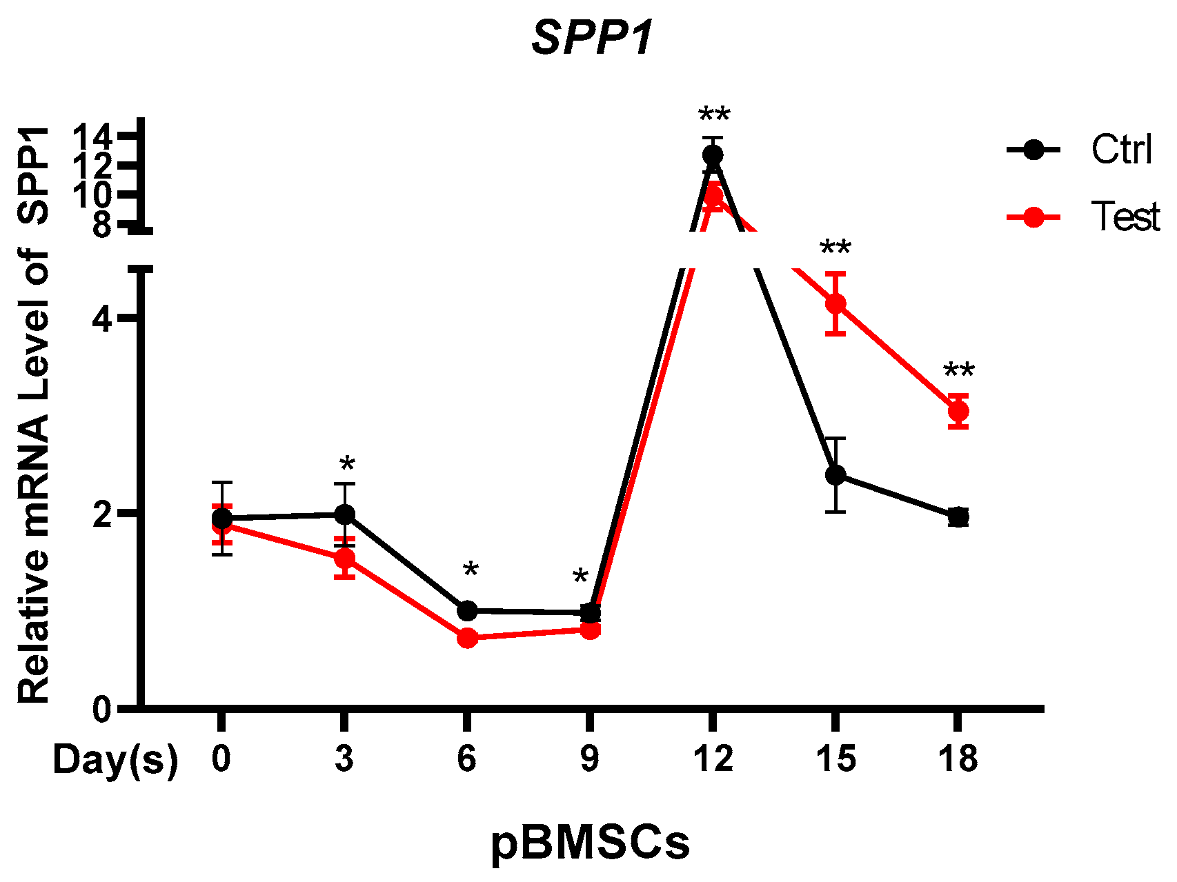
3.4. Testosterone Regulates the Expression of SPP1

This study tested whether the differential expression of SPP1 in castrated and intact male pigs was caused by testosterone. In vitro, pBMSCs were treated with testosterone to simulate the androgen status of intact pigs. In contrast with the control group, the expression of SPP1 in the testosterone group was slightly decreased at the early stage of pBMSCs differentiation. However, the SPP1 expression levels in the testosterone group was markedly higher than those in the control group at the adipose deposition stage (after 12 days) (Figure 4), which was consistent with the function of SPP1 in pBMSCs. The results indicated that the expression of SPP1 was regulated by testosterone.

3.5. Novel-miR-659 Promoted Adipogenesis of pBMSCs by Targeting SPP1

Our previous study indicated that SPP1 is a potential target of novel-miR-659 by TargetScan and miRDB software. To verify the targeting relationship between novel-miR-659 and SPP1, two plasmids containing either the WT or MUT 3′-UTR of SPP1 were constructed (Figure 5A). Subsequently, cotransfection of novel-miR-659 mimics or mimics NC into 293T cells was performed. Notably, novel-miR-659 mimics markedly decreased the activity of WT SPP1 compared with the mimics NC, but no significant changes were noted for the MUT (Figure 5B). Meanwhile, we determined the expression profile of novel-miR-659 in pBMSCs during adipogenesis induced by an adipogenic medium at different time points. This trend was the opposite of the expression trend of SPP1 in adipogenesis of pBMSCs (Figure S1). These findings indicate that novel-miR-659 can directly target the 3′-UTR of SPP1.
To further investigate the function of novel-miR-659 on pBMSCs differentiation, novel-miR-659 mimics and inhibitors were transfected into pBMSCs. Transfection efficiency of novel-miR-659 and expression of SPP1 were validated at day 14 of induction by qRT-PCR. The expression of SPP1 decreased with novel-miR-659 upregulation and increased with novel-miR-659 knockdown, indicating that the expression trend of the SPP1 gene was opposite to that of novel-miR-659 (Figure S2). After 14 days of induction Oil Red O staining, results showed that novel-miR-659 mimics promoted the formation of fat droplets while novel-miR-659 inhibitors inhibited the formation of fat droplets compared with mimics and inhibitors NC, respectively (Figure 5C). Quantitative PCR revealed that novel-miR-659 mimics significantly improved the expression of adipogenesis marker PPARγ and FABP4, whereas the novel-miR-659 inhibitor-treated group expressed lower amounts of these mRNAs (Figure 5D,E). Collectively, these results manifested that novel-miR-659 promoted adipogenesis of pBMSCs by repressing SPP1.

4. Discussion

Obesity is a global health issue, and androgen insufficiency has been linked to male obesity in animals. Our previous study identified differentially expressed mRNAs in backfat and abdominal fat tissues from intact and castrated full-sib male pigs, and found that SPP1 was related to fat deposition. Some researchers have confirmed that blockage of SPP1 expression facilitated MSCs adipogenic development but suppressed osteoclast formation [26]. Furthermore, Veronica et al. found that progenitor cells lacking SPP1 were more efficient in adipogenic differentiation [27]. Additionally, Tang et al. displayed that SPP1 prevents ASCs from becoming adipogenic by connecting to the external receptors’ integrin αν/β1 and CD44 [28]. In line with these previously published findings, our studies of in vitro experiments revealed that pBMSCs from SPP1 silence (or SPP1 overexpression) were more (or less) effective in lipogenesis than NC controls, as identified by enhanced (or receded) fat-droplet accumulation (Figure 2 and Figure 3), indicating that SPP1 was a negative regulator of adipogenesis [31].
It has been reported that SPP1 expression in epididymis main cells may be influenced by circulating testosterone [39]. Likewise, our results also revealed that the expression of SPP1 was affected by testosterone, and the SPP1 mRNA level was suppressed by the testosterone in the early stage of pBMSCs differentiation. However, testosterone promoted the SPP1 expression during the later stages of pBMSCs differentiation (Figure 4), which was in keeping with the inhibition of SPP1 on lipid deposition. The differentiation process from MSCs to mature adipocytes is generally considered to go through two stages. The first stage is called commitment, where MSCs are converted into preadipocytes. The preadipocyte is the intermediate stage between MSCs and adipocytes, and it can differentiate into adipocytes. The second stage, named terminal differentiation, is the transformation of preadipocytes into mature adipocytes. Preadipocytes are not morphologically distinguishable from adipocytes and lack specific markers to distinguish between the two stages [40]. Therefore, we believe that testosterone may play different roles in the control of SPP1 expression at different stages of pBMSCs differentiation.
To learn more about the role of SPP1 in the control of fat deposition. novel-miR-659 directly targeted the 3′UTR of SPP1, as shown in the dual-luciferase reporter assay (Figure 5B). Both novel-miR-659 overexpression and inhibition experiments demonstrated that novel-miR-659 promoted adipose deposition of pBMSCs by suppressing SPP1 (Figure 5C–E). The novel-miR-659 is a novel miRNA in pigs, and its relationship with fat deposition is rarely studied. Recent studies have shown that miR-659 was obesity-specific, being expressed only in amnion from obese women than control women [41]. This is consistent with our findings that miR-659 promoted the accumulation of lipid droplets in pBMSCs.

5. Conclusions

In summary, our results show that SPP1 negatively regulated adipogenic differentiation of pBMSCs, and the SPP1 expression in adipose tissue of pigs is regulated by testosterone and novel-miR-659. The interaction between novel-miR-659 and SPP1 coregulate fat deposition in castrated male pigs. These data expand our mechanistic understanding of fat deposition in pigs caused by testosterone deficiency.

Supplementary Materials

The following supporting information can be downloaded at: https://www.mdpi.com/article/10.3390/ani12080944/s1, Table S1. SiRNA and miRNA sequences; Table S2. Primers used for real-time quantitative PCR; Figure S1. SPP1 is a potential target of novel-miR-659; Figure S2. Transfection effect of novel-miR-659.

Author Contributions

Formal analysis, L.X. and Q.X.; Investigation, L.X., Q.X. and X.L.; Project administration, M.F.; Resources, X.L., S.C., Y.L. and S.H.; Writing—original draft, L.X., Q.X. and M.F. All authors have read and agreed to the published version of the manuscript.

Funding

This work was supported by the 2020 Research Program of Sanya Yazhou Bay Science and Technology City (Grant No. SKJC-2020-02-007); the National Natural Science Foundation of China (32072700); the National Key Research and Development Program of China (2021YFF1000603); the China Agriculture Research System of MOF and MARA(CARS-36); and the talent cultivation and developmental support program of China Agricultural University.

Institutional Review Board Statement

Not applicable.

Informed Consent Statement

Not applicable.

Data Availability Statement

The data are in the supplement.

Acknowledgments

The authors are grateful to Jinming Huang and Gang Liu for assistance with sample collection.

Conflicts of Interest

The authors declare no conflict of interest.

References

  1. Kershaw, E.E.; Flier, J.S. Adipose tissue as an endocrine organ. J. Clin. Endocrinol. Metab. 2004, 89, 2548–2556. [Google Scholar] [CrossRef] [PubMed]
  2. Lu, P.; Zhang, F.C.; Qian, S.W.; Li, X.; Cui, Z.M.; Dang, Y.J.; Tang, Q.Q. Artemisinin derivatives prevent obesity by inducing browning of WAT and enhancing BAT function. Cell Res. 2016, 26, 1169–1172. [Google Scholar] [CrossRef]
  3. Berrington, D.G.A.; Hartge, P.; Cerhan, J.R.; Flint, A.J.; Hannan, L.; Macinnis, R.J.; Moore, S.C.; Tobias, G.S.; Anton-Culver, H.; Freeman, L.B.; et al. Body-mass index and mortality among 1.46 million white adults. N. Engl. J. Med. 2010, 363, 2211–2219. [Google Scholar] [CrossRef] [PubMed] [Green Version]
  4. Zerradi, M.; Dereumetz, J.; Boulet, M.; Tchernof, A. Androgens, body fat Distribution and Adipogenesis. Curr. Obes. Rep. 2014, 3, 396–403. [Google Scholar] [CrossRef] [PubMed]
  5. Wang, J.; Hua, L.; Chen, J.; Zhang, J.; Bai, X.; Gao, B.; Li, C.; Shi, Z.; Sheng, W.; Gao, Y.; et al. Identification and characterization of long non-coding RNAs in subcutaneous adipose tissue from castrated and intact full-sib pair Huainan male pigs. BMC Genom. 2017, 18, 542. [Google Scholar] [CrossRef] [PubMed] [Green Version]
  6. Grossmann, M. Low Testosterone in Men with Type 2 Diabetes: Significance and Treatment. J. Clin. Endocrinol. Metab. 2011, 96, 2341–2353. [Google Scholar] [CrossRef] [PubMed]
  7. Christoffersen, B.O.; Gade, L.P.; Golozoubova, V.; Svendsen, O.; Raun, K. Influence of castration-induced testosterone and estradiol deficiency on obesity and glucose metabolism in male Göttingen minipigs. Steroids 2010, 75, 676–684. [Google Scholar] [CrossRef]
  8. Wang, K.; Li, W.; Bai, Y.; Yang, W.; Ling, Y.; Fang, M. ssc-miR-7134-3p regulates fat accumulation in castrated male pigs by targetingMARK4 gene. Int. J. Biol. Sci. 2017, 13, 189–197. [Google Scholar] [CrossRef] [PubMed] [Green Version]
  9. Li, M.; Bjorntorp, P. Effects of testosterone on triglyceride uptake and mobilization in different adipose tissues in male rats in vivo. Obes. Res. 1995, 3, 113–119. [Google Scholar] [CrossRef]
  10. Lundstrom, K.; Matthews, K.R.; Haugen, J.E. Pig meat quality from entire males. Animal 2009, 3, 1497–1507. [Google Scholar] [CrossRef] [PubMed] [Green Version]
  11. Bonneau, M. Use of entire males for pig meat in the European Union. Meat Sci. 1998, 49, S257–S272. [Google Scholar] [CrossRef]
  12. Poklukar, K.; Andek-Potokar, M.; Vrecl, M.; Batorek-Lukač, N.; Fazarinc, G.; Kress, K.; Weiler, U.; Stefanski, V.; Akrlep, M. The effect of immunocastration on adipose tissue deposition and composition in pigs. Animal 2021, 15, 100118. [Google Scholar] [CrossRef] [PubMed]
  13. Gispert, M.; Angels, O.M.; Velarde, A.; Suarez, P.; Perez, J.; Font, I.F.M. Carcass and meat quality characteristics of immunocastrated male, surgically castrated male, entire male and female pigs. Meat Sci. 2010, 85, 664–670. [Google Scholar] [CrossRef] [PubMed]
  14. Saintilan, R.; Sellier, P.; Billon, Y.; Gilbert, H. Genetic correlations between males, females and castrates for residual feed intake, feed conversion ratio, growth rate and carcass composition traits in Large White growing pigs. J. Anim. Breed. Genet. 2012, 129, 103–106. [Google Scholar] [CrossRef] [PubMed]
  15. Wolf, E.; Braun-Reichhart, C.; Streckel, E.; Renner, S. Genetically engineered pig models for diabetes research. Transgenic Res. 2014, 23, 27–38. [Google Scholar] [CrossRef] [PubMed]
  16. Friedenstein, A.J.; Gorskaja, J.F.; Kulagina, N.N. Fibroblast precursors in normal and irradiated mouse hematopoietic organs. Exp. Hematol. 1976, 4, 267–274. [Google Scholar] [PubMed]
  17. Jürets, A.; Le Bras, M.; Staffler, G.; Stein, G.; Leitner, L.; Neuhofer, A.; Tardelli, M.; Turkof, E.; Zeyda, M.; Stulnig, T.M. Inhibition of Cellular Adhesion by Immunological Targeting of Osteopontin Neoepitopes Generated through Matrix Metalloproteinase and Thrombin Cleavage. PLoS ONE 2016, 11, e148333. [Google Scholar] [CrossRef] [PubMed] [Green Version]
  18. Inoue, M.; Shinohara, M.L. Intracellular osteopontin (iOPN) and immunity. Immunol. Res. 2011, 49, 160–172. [Google Scholar] [CrossRef] [PubMed] [Green Version]
  19. Wolak, T. Osteopontin–A multi-modal marker and mediator in atherosclerotic vascular disease. Atherosclerosis 2014, 236, 327–337. [Google Scholar] [CrossRef]
  20. Bandopadhyay, M.; Bulbule, A.; Butti, R.; Chakraborty, G.; Ghorpade, P.; Ghosh, P.; Gorain, M.; Kale, S.; Kumar, D.; Kumar, S.; et al. Osteopontin as a therapeutic target for cancer. Expert Opin. Ther. Tar. 2014, 18, 883–895. [Google Scholar] [CrossRef] [PubMed]
  21. Staines, K.A.; Macrae, V.E.; Farquharson, C. The importance of the SIBLING family of proteins on skeletal mineralisation and bone remodelling. J. Endocrinol. 2012, 214, 241–255. [Google Scholar] [CrossRef] [Green Version]
  22. Lund, S.A.; Giachelli, C.M.; Scatena, M. The role of osteopontin in inflammatory processes. J. Cell Commun. Signal. 2009, 3, 311–322. [Google Scholar] [CrossRef] [PubMed] [Green Version]
  23. Jang, J.; Kim, J. Improved Cellular Response of Osteoblast Cells Using Recombinant Human Osteopontin Protein Produced by Escherichia coli. Biotechnol. Lett. 2005, 27, 1767–1770. [Google Scholar] [CrossRef] [PubMed]
  24. Denhardt, D.T.G.X. Osteopontin: A protein with diverse functions. FASEB J. 1993, 15, 1475–1482. [Google Scholar] [CrossRef]
  25. Kiefer, F.W.; Zeyda, M.; Todoric, J.; Huber, J.; Geyeregger, R.; Weichhart, T.; Aszmann, O.; Ludvik, B.; Silberhumer, G.R.; Prager, G.; et al. Osteopontin Expression in Human and Murine Obesity: Extensive Local Up-Regulation in Adipose Tissue but Minimal Systemic Alterations. Endocrinology 2008, 149, 1350–1357. [Google Scholar] [CrossRef] [PubMed] [Green Version]
  26. Gómez-Ambrosi, J.; Catalán, V.; Ramírez, B.; Rodríguez, A.; Colina, I.; Silva, C.; Rotellar, F.; Mugueta, C.; Gil, M.J.; Cienfuegos, J.A.; et al. Plasma Osteopontin Levels and Expression in Adipose Tissue Are Increased in Obesity. J. Clin. Endocrinol. Metab. 2007, 92, 3719–3727. [Google Scholar] [CrossRef] [PubMed] [Green Version]
  27. Kahles, F.; Findeisen, H.M.; Bruemmer, D. Osteopontin: A novel regulator at the cross roads of inflammation, obesity and diabetes. Mol. Metab. 2014, 3, 384–393. [Google Scholar] [CrossRef] [PubMed]
  28. Kiefer, F.W.; Neschen, S.; Pfau, B.; Legerer, B.; Neuhofer, A.; Kahle, M.; Hrabé De Angelis, M.; Schlederer, M.; Mair, M.; Kenner, L.; et al. Osteopontin deficiency protects against obesity-induced hepatic steatosis and attenuates glucose production in mice. Diabetologia 2011, 54, 2132–2142. [Google Scholar] [CrossRef] [PubMed] [Green Version]
  29. Chapman, J.; Miles, P.D.; Ofrecio, J.M.; Neels, J.G.; Yu, J.G.; Resnik, J.L.; Wilkes, J.; Talukdar, S.; Thapar, D.; Johnson, K.; et al. Osteopontin is required for the early onset of high fat diet-induced insulin resistance in mice. PLoS ONE 2010, 5, e13959. [Google Scholar] [CrossRef] [PubMed] [Green Version]
  30. Nomiyama, T.; Perez-Tilve, D.; Ogawa, D.; Gizard, F.; Zhao, Y.; Heywood, E.B.; Jones, K.L.; Kawamori, R.; Cassis, L.A.; Tschöp, M.H.; et al. Osteopontin mediates obesity-induced adipose tissue macrophage infiltration and insulin resistance in mice. J. Clin. Investig. 2007, 117, 2877–2888. [Google Scholar] [CrossRef]
  31. Wang, M.; Guo, Y.; Zhou, Y.; Yuan, W.; Li, H.; Xiong, S.; Wang, K.; Zeng, G.; Huang, Q. Secreted-Osteopontin Contributes to Brown Adipogenesis In Vitro via a CD44-Dependent Pathway. Horm. Metab. Res. 2019, 51, 741. [Google Scholar] [CrossRef] [PubMed]
  32. Lu, Y.; Xu, Y.; Yuan, W.; Wang, M.; Zhou, Y.; Chen, K.; Huang, Q. Downregulation of osteopontin inhibits browning of white adipose tissues through PI3K-AKT pathway in C57BL/6 mice. Eur. J. Pharmacol. 2020, 866, 172822. [Google Scholar] [CrossRef] [PubMed]
  33. Zhong, X.; Shen, X.; Wen, J.; Kong, Y.; Chu, J.; Yan, G.; Li, T.; Liu, D.; Wu, M.; Zeng, G.; et al. Osteopontin-induced brown adipogenesis from white preadipocytes through a PI3K-AKT dependent signaling. Biochem. Biophys. Res. Commun. 2015, 459, 553–559. [Google Scholar] [CrossRef] [PubMed]
  34. Chen, Q.; Shou, P.; Zhang, L.; Xu, C.; Zheng, C.; Han, Y.; Li, W.; Huang, Y.; Zhang, X.; Shao, C.; et al. An Osteopontin-Integrin Interaction Plays a Critical Role in Directing Adipogenesis and Osteogenesis by Mesenchymal Stem Cells. Stem Cells 2014, 32, 327–337. [Google Scholar] [CrossRef] [PubMed] [Green Version]
  35. Moreno-Viedma, V.; Tardelli, M.; Zeyda, M.; Sibilia, M.; Burks, J.D.; Stulnig, T.M. Osteopontin-deficient progenitor cells display enhanced differentiation to adipocytes. Obes. Res. Clin. Pract. 2018, 12, 277–285. [Google Scholar] [CrossRef]
  36. Fitter, S.; Zannettino, A.C.W. Osteopontin in the pathophysiology of obesity: Is Opn a fat cell foe? Obes. Res. Clin. Pract. 2018, 12, 249–250. [Google Scholar] [CrossRef]
  37. Cai, Z.; Zhang, L.; Chen, M.; Jiang, X.; Xu, N. Castration-induced changes in microRNA expression profiles in subcutaneous adipose tissue of male pigs. J. Appl. Genet. 2014, 55, 259–266. [Google Scholar] [CrossRef]
  38. Bai, C.; Chen, S.; Gao, Y.; Shan, Z.; Guan, W.; Ma, Y. Multi-lineage potential research of bone marrow mesenchymal stem cells from Bama miniature pig. J. Exp. Zool. Part B Mol. Dev. Evol. 2015, 324, 671–685. [Google Scholar] [CrossRef]
  39. Shoelson, S.E. Inflammation and insulin resistance. J. Clin. Investig. 2006, 116, 1793–1801. [Google Scholar] [CrossRef]
  40. Zuk, P.A.; Zhu, M.; Ashjian, P.; De Ugarte, D.A.; Huang, J.I.; Mizuno, H.; Alfonso, Z.C.; Fraser, J.K.; Benhaim, P.; Hedrick, M.H. Human Adipose Tissue Is a Source of Multipotent Stem Cells. Mol. Biol. Cell 2002, 12, 4279–4295. [Google Scholar] [CrossRef]
  41. Nardelli, C.; Iaffaldano, L.; Ferrigno, M.; Labruna, G.; Maruotti, G.M.; Quaglia, F.; Capobianco, V.; Di Noto, R.; Del, V.L.; Martinelli, P.; et al. Characterization and predicted role of the microRNA expression profile in amnion from obese pregnant women. Int. J. Obes. 2014, 38, 466–469. [Google Scholar] [CrossRef] [PubMed]
Figure 1. Adipogenic differentiation of pBMSCs. (A) Oil Red O was used to stain the pBMSCs, differentiated on day 0 (A-a), 3 (A-b), 6 (A-c), 9 (A-d), 12 (A-e), 15 (A-f), and 18 (A-g) (bar = 200 μm). (B) PPARγ and FABP4 mRNA levels on the relevant days were examined by qRT-PCR.
Animals 12 00944 g001
Figure 2. SPP1 overexpression suppressed the adipogenic differentiation of pBMSCs. (A) pBMSCs were transfected with the PCDH empty vector (PCDH) or the SPP1 overexpression vector (PCDH-SPP1), and SPP1 expression was measured by qRT-PCR. (B) The transfected pBMSCs were induced to differentiate for 14 days and lipid droplets were observed by Oil Red O staining (bar = 200 μm). (C) Adipose marker factors PPARγ and FABP4 mRNA expression of pBMSCs harboring PCDH or PCDH-SPP1 were detected by qRT-PCR. ***, p < 0.001; **, p < 0.01.
Animals 12 00944 g002
Figure 3. Inhibition of SPP1 promoted the adipogenic differentiation of pBMSCs. (A) pBMSCs were transfected with siRNA (si-212-SPP1, si-810-SPP1, and si-925-SPP1) or negative control, and to assay-interfering efficiency by expression of SPP1 two days after transfection. (B) Lipid droplet accumulation of pBMSCs transfected with si-925-SPP1 and negative control were verified by Oil Red O staining induction at 14 days (bar = 200 μm). (C) The mRNA levels of PPARγ and FABP4 in the si-925-SPP1 group and the negative control group were confirmed by qRT-PCR. ***, p < 0.001; **, p < 0.01; *, p < 0.05.
Animals 12 00944 g003
Figure 4. Testosterone regulates the expression of SPP1. The mRNA expression of SPP1 was detected by PCR at a different stage of pBMSCs differentiation. Ctrl, control group. Test, testosterone group. **, p < 0.01; *, p < 0.05.
Animals 12 00944 g004
Figure 5. Novel-miR-659 promoted adipogenesis of pBMSCs by targeting SPP1. (A) The interaction binding site of novel-miR-659 and SPP1 3′UTR was predicted by TargetScan and miRDB. (B) Novel-miR-659 directly targeted the 3′UTR of SPP1, normalized Renilla fluorescence activity forty eight hours following cotransfection of novel-miR-659 mimics and mimics NC with SPP1-3′UTR-WT and SPP1-3′UTR-MUT. (C) Adipogenesis of pBMSCs transfected with mimics/inhibitors NC or miR-659 mimics/inhibitors were stained by Oil Red O at 14 days after differentiation (bar = 200 μm). (D,E) Expressions of PPARγ and FABP4 in mimics NC, miR-659 mimics, inhibitors NC, and miR-659 inhibitors groups were detected by qPCR on day 14 after induction. **, p < 0.01; *, p < 0.05.
Animals 12 00944 g005aAnimals 12 00944 g005b
Publisher’s Note: MDPI stays neutral with regard to jurisdictional claims in published maps and institutional affiliations.

