
Effect of a Probiotic Mixture in Captive Cheetahs (Acinonyx Jubatus) with Gastrointestinal Symptoms—A Pilot Study †

 ,
,  , , , and
Abstract
:Simple Summary
Abstract
1. Introduction
2. Materials and Methods
2.1. Study Population
2.2. Verification of Helicobacter spp. Presence
2.3. Clinical Evaluation
2.4. Probiotics Administration
2.5. Statistical Analysis
3. Results
4. Discussion
5. Conclusions
Author Contributions
Funding
Institutional Review Board Statement
Informed Consent Statement
Data Availability Statement
Acknowledgments
Conflicts of Interest
References
- Durant, S.M.; Mitchell, N.; Groom, R.; Pettorelli, N.; Ipavec, A.; Jacobson, A.P.; Woodroffe, R.; Bohm, M.; Hunter, L.T.; Becker, M.S.; et al. The global decline of cheetah Acinonyx jubatus and what it means for conservation. Proc. Natl. Acad. Sci. USA 2017, 114, 528–533. [Google Scholar] [CrossRef] [PubMed] [Green Version]
- Munson, L. Diseases of captive cheetahs (Acinonyx jubatus): Results of the cheetah research council pathology survey, 1989–1992. Zoo Biol. 1993, 12, 105–124. [Google Scholar] [CrossRef]
- Bolton, L.A.; Munson, L. Glomerulosclerosis in captive cheetahs (Acinonyx jubatus). Vet. Pathol. 1999, 36, 14–22. [Google Scholar] [CrossRef] [PubMed] [Green Version]
- Terio, K.A.; Munson, L.; Marker, L.; Aldridge, B.M.; Solnick, J.V. Comparison of Helicobacter spp. in Cheetahs (Acinonyx jubatus) with and without gastritis. J. Clin. Microbiol. 2005, 43, 229–234. [Google Scholar] [CrossRef] [PubMed] [Green Version]
- Lane, E.; Lobetti, R.; Burroughs, R. Treatment with omeprazole, metronidazole, and amoxicillin in captive South African cheetahs (Acinonyx jubatus) with spiral bacteria infection and gastritis. J. Zoo Wildl. Med. 2004, 35, 15–19. [Google Scholar] [CrossRef]
- Wack, R.F.; Eaton, K.A.; Kramer, L.W. Treatment of gastritis in cheetahs (Acinonyx jubatus). J. Zoo Wildl. Med. 1997, 28, 260–266. [Google Scholar]
- Terio, K.A.; Mitchell, E.; Walzer, C.; Schmidt-Küntzel, A.; Marker, L.; Citino, S. Diseases Impacting Captive and Free-Ranging Cheetahs. In Cheetahs: Biology and Conservation; Elsevier: Amsterdam, The Netherlands, 2018; pp. 349–364. [Google Scholar] [CrossRef]
- Alanis, A.J. Resistance to antibiotics: Are we in the post-antibiotic era? Arch. Med. Res. 2005, 36, 697–705. [Google Scholar] [CrossRef]
- Di Cerbo, A.; Pezzuto, F.; Palmieri, L.; Rottigni, V.; Iannitti, T.; Palmieri, B. Clinical and experimental use of probiotic formulations for management of end-stage renal disease: An update. Int. Urol. Nephrol. 2013, 45, 1569–1576. [Google Scholar] [CrossRef]
- Amara, A.A.; Shibl, A. Role of Probiotics in health improvement, infection control and disease treatment and management. Saudi. Pharm. J. 2015, 23, 107–114. [Google Scholar] [CrossRef] [Green Version]
- Rossi, G.; Cerquetella, M.; Scarpona, S.; Pengo, G.; Fettucciari, K.; Bassotti, G.; Jergens, A.E.; Suchodolski, J.S. Effects of probiotic bacteria on mucosal polyamines levels in dogs with IBD and colonic polyps: A preliminary study. Benef. Microbes 2018, 9, 247–255. [Google Scholar] [CrossRef]
- Rossi, G.; Jergens, A.; Cerquetella, M.; Berardi, S.; Di Cicco, E.; Bassotti, G.; Pengo, G.; Suchodolski, J.S. Effects of a probiotic (SLAB51) on clinical and histologic variables and microbiota of cats with chronic constipation/megacolon: A pilot study. Benef. Microbes 2018, 9, 101–110. [Google Scholar] [CrossRef] [PubMed]
- Ji, J.; Yang, H. Using Probiotics as Supplementation for Helicobacter pylori Antibiotic Therapy. Int. J. Mol. Sci. 2020, 21, 1136. [Google Scholar] [CrossRef] [PubMed] [Green Version]
- Homan, M.; Orel, R. Are probiotics useful in Helicobacter pylori eradication? World J. Gastroenterol. 2015, 21, 10644–10653. [Google Scholar] [CrossRef] [PubMed]
- Lv, Z.; Wang, B.; Zhou, X.; Wang, F.; Xie, Y.; Zheng, H.; Lv, N. Efficacy and safety of probiotics as adjuvant agents for Helicobacter pylori infection: A meta-analysis. Exp. Ther. Med. 2015, 9, 707–716. [Google Scholar] [CrossRef] [PubMed] [Green Version]
- Vitini, E.; Alvarez, S.; Medina, M.; Medici, M.; de Budeguer, M.V.; Perdigon, G. Gut mucosal immunostimulation by lactic acid bacteria. Biocell 2000, 24, 223–232. [Google Scholar] [PubMed]
- Sgouras, D.; Maragkoudakis, P.; Petraki, K.; Martinez-Gonzalez, B.; Eriotou, E.; Michopoulos, S.; Kalantzopoulos, G.; Tsakalidou, E.; Mentis, A. In vitro and in vivo inhibition of Helicobacter pylori by Lactobacillus casei strain Shirota. Appl. Environ. Microbiol. 2004, 70, 518–526. [Google Scholar] [CrossRef] [Green Version]
- Erickson, K.L.; Hubbard, N.E. Probiotic immunomodulation in health and disease. J. Nutr. 2000, 130, 403S–409S. [Google Scholar] [CrossRef]
- Boyanova, L.; Gergova, G.; Markovska, R.; Yordanov, D.; Mitov, I. Bacteriocin-like inhibitory activities of seven Lactobacillus delbrueckii subsp. bulgaricus strains against antibiotic susceptible and resistant Helicobacter pylori strains. Lett. Appl. Microbiol. 2017, 65, 469–474. [Google Scholar] [CrossRef]
- De Melo Pereira, G.V.; de Oliveira Coelho, B.; Junior, A.I.M.; Thomaz-Soccol, V.; Soccol, C.R. How to select a probiotic? A review and update of methods and criteria. Biotechnol. Adv. 2018, 36, 2060–2076. [Google Scholar] [CrossRef]
- Di Cerbo, A.; Palmieri, B.; Aponte, M.; Morales-Medina, J.C.; Iannitti, T. Mechanisms and therapeutic effectiveness of lactobacilli. J. Clin. Pathol. 2016, 69, 187–203. [Google Scholar] [CrossRef]
- Ojetti, V.; Bruno, G.; Ainora, M.E.; Gigante, G.; Rizzo, G.; Roccarina, D.; Gasbarrini, A. Impact of Lactobacillus reuteri Supplementation on Anti-Helicobacter pylori Levofloxacin-Based Second-Line Therapy. Gastroenterol. Res. Pract 2012, 2012, 740381. [Google Scholar] [CrossRef] [PubMed] [Green Version]
- Mukai, T.; Asasaka, T.; Sato, E.; Mori, K.; Matsumoto, M.; Ohori, H. Inhibition of binding of Helicobacter pylori to the glycolipid receptors by probiotic Lactobacillus reuteri. FEMS Immunol. Med. Microbiol. 2002, 32, 105–110. [Google Scholar] [CrossRef] [PubMed] [Green Version]
- Midolo, P.D.; Lambert, J.R.; Hull, R.; Luo, F.; Grayson, M.L. In vitro inhibition of Helicobacter pylori NCTC 11637 by organic acids and lactic acid bacteria. J Appl. Bacteriol. 1995, 79, 475–479. [Google Scholar] [CrossRef] [PubMed]
- Borruel, N.; Casellas, F.; Antolin, M.; Llopis, M.; Carol, M.; Espiin, E.; Naval, J.; Guarner, F.; Malagelada, J.R. Effects of nonpathogenic bacteria on cytokine secretion by human intestinal mucosa. Am. J. Gastroenterol. 2003, 98, 865–870. [Google Scholar] [CrossRef]
- Lorca, G.L.; Wadstrom, T.; Valdez, G.F.; Ljungh, A. Lactobacillus acidophilus autolysins inhibit Helicobacter pylori in vitro. Curr. Microbiol. 2001, 42, 39–44. [Google Scholar] [CrossRef]
- Francavilla, R.; Lionetti, E.; Castellaneta, S.P.; Magista, A.M.; Maurogiovanni, G.; Bucci, N.; De Canio, A.; Indrio, F.; Cavallo, L.; Ierardi, E.; et al. Inhibition of Helicobacter pylori infection in humans by Lactobacillus reuteri ATCC 55730 and effect on eradication therapy: A pilot study. Helicobacter 2008, 13, 127–134. [Google Scholar] [CrossRef]
- Mack, D.R.; Ahrne, S.; Hyde, L.; Wei, S.; Hollingsworth, M.A. Extracellular MUC3 mucin secretion follows adherence of Lactobacillus strains to intestinal epithelial cells in vitro. Gut 2003, 52, 827–833. [Google Scholar] [CrossRef] [Green Version]
- Marcial, G.; Villena, J.; Faller, G.; Hensel, A.; de Valdez, G.F. Exopolysaccharide-producing Streptococcus thermophilus CRL1190 reduces the inflammatory response caused by Helicobacter pylori. Benef. Microbes 2017, 8, 451–461. [Google Scholar] [CrossRef]
- Charteris, W.P.; Kelly, P.M.; Morelli, L.; Collins, J.K. Selective detection, enumeration and identification of potentially probiotic Lactobacillus and Bifidobacterium species in mixed bacterial populations. Int. J. Food Microbiol. 1997, 35, 1–27. [Google Scholar] [CrossRef]
- Bielecka, M.; Biedrzycka, E.; Biedrzycka, E.; Smoragiewicz, W.; Smieszek, M. Interaction of Bifidobacterium and Salmonella during associated growth. Int. J. Food Microbiol. 1998, 45, 151–155. [Google Scholar] [CrossRef]
- Chitapanarux, T.; Thongsawat, S.; Pisespongsa, P.; Leerapun, A.; Kijdamrongthum, P. Effect of Bifidobacterium longum on PPI-based triple therapy for eradication of Helicobacter pylori: A randomized, double-blind placebo-controlled study. J. Funct. Foods 2015, 13, 289–294. [Google Scholar] [CrossRef]
- Szajewska, H.; Horvath, A.; Kolodziej, M. Systematic review with meta-analysis: Saccharomyces boulardii supplementation and eradication of Helicobacter pylori infection. Aliment. Pharmacol. Ther. 2015, 41, 1237–1245. [Google Scholar] [CrossRef] [PubMed]
- Gotteland, M.; Poliak, L.; Cruchet, S.; Brunser, O. Effect of regular ingestion of Saccharomyces boulardii plus inulin or Lactobacillus acidophilus LB in children colonized by Helicobacter pylori. Acta Paediatr. 2005, 94, 1747–1751. [Google Scholar] [CrossRef] [PubMed]
- Koeppel, K.N.; Bertschinger, H.; van Vuuren, M.; Picard, J.; Steiner, J.; Williams, D.; Cardwell, J. The use of a probiotic in captive cheetahs (Acinonyx jubatus). J. South Afr. Vet. Assoc. 2006, 77, 127–130. [Google Scholar] [CrossRef] [PubMed] [Green Version]
- Camargo, P.L.; Alfieri, A.A.; Bracarense, A.P.; Menoli, R.; Spinosa, S.R.; Hagiwara, M.K. Use of polymerase chain reaction and enzymatic cleavage in the identification of Helicobacter spp. in gastric mucosa of human beings from North Parana, Brazil. Memórias Do Inst. Oswaldo Cruz 2003, 98, 265–268. [Google Scholar] [CrossRef] [Green Version]
- Germani, Y.; Dauga, C.; Duval, P.; Huerre, M.; Levy, M.; Pialoux, G.; Sansonetti, P.; Grimont, P.A. Strategy for the detection of Helicobacter species by amplification of 16S rRNA genes and identification of H. felis in a human gastric biopsy. Res. Microbiol. 1997, 148, 315–326. [Google Scholar] [CrossRef]
- Jergens, A.E.; Crandell, J.M.; Evans, R.; Ackermann, M.; Miles, K.G.; Wang, C. A clinical index for disease activity in cats with chronic enteropathy. J. Vet. Intern. Med. 2010, 24, 1027–1033. [Google Scholar] [CrossRef]
- Azer, S.A.; Akhondi, H. Gastritis; StatPearls: Treasure Island, FL, USA, 2022. Available online: https://www.ncbi.nlm.nih.gov/books/NBK544250/ (accessed on 24 January 2022).
- Eaton, K.A.; Radin, M.J.; Kramer, L.; Wack, R.; Sherding, R.; Krakowka, S.; Fox, J.G.; Morgan, D.R. Epizootic gastritis associated with gastric spiral bacilli in cheetahs (Acinonyx jubatus). Vet. Pathol. 1993, 30, 55–63. [Google Scholar] [CrossRef]
- Lam, S.K.; Talley, N.J. Report of the 1997 Asia Pacific Consensus Conference on the management of Helicobacter pylori infection. J. Gastroenterol. Hepatol. 1998, 13, 1–12. [Google Scholar] [CrossRef]
- Citino, S.B.; Munson, L. Efficacy and long-term outcome of gastritis therapy in cheetahs (Acinonyx jubatus). J. Zoo Wildl. Med. 2005, 36, 401–416. [Google Scholar] [CrossRef]
- Safavi, M.; Sabourian, R.; Foroumadi, A. Treatment of Helicobacter pylori infection: Current and future insights. World J. Clin. Cases 2016, 4, 5–19. [Google Scholar] [CrossRef] [PubMed]
- Johnson-Henry, K.C.; Mitchell, D.J.; Avitzur, Y.; Galindo-Mata, E.; Jones, N.L.; Sherman, P.M. Probiotics reduce bacterial colonization and gastric inflammation in H. pylori-infected mice. Dig. Dis. Sci. 2004, 49, 1095–1102. [Google Scholar] [CrossRef] [PubMed]
- Hsieh, P.S.; Tsai, Y.C.; Chen, Y.C.; Teh, S.F.; Ou, C.M.; King, V.A. Eradication of Helicobacter pylori infection by the probiotic strains Lactobacillus johnsonii MH-68 and L. salivarius ssp. salicinius AP-32. Helicobacter 2012, 17, 466–477. [Google Scholar] [CrossRef] [PubMed]
- Eslami, M.; Yousefi, B.; Kokhaei, P.; Moghadas, A.J.; Moghadam, B.S.; Arabkari, V.; Niazi, Z. Are probiotics useful for therapy of Helicobacter pylori diseases? Comp. Immunol. Microbiol. Infect. Dis. 2019, 64, 99–108. [Google Scholar] [CrossRef] [PubMed]


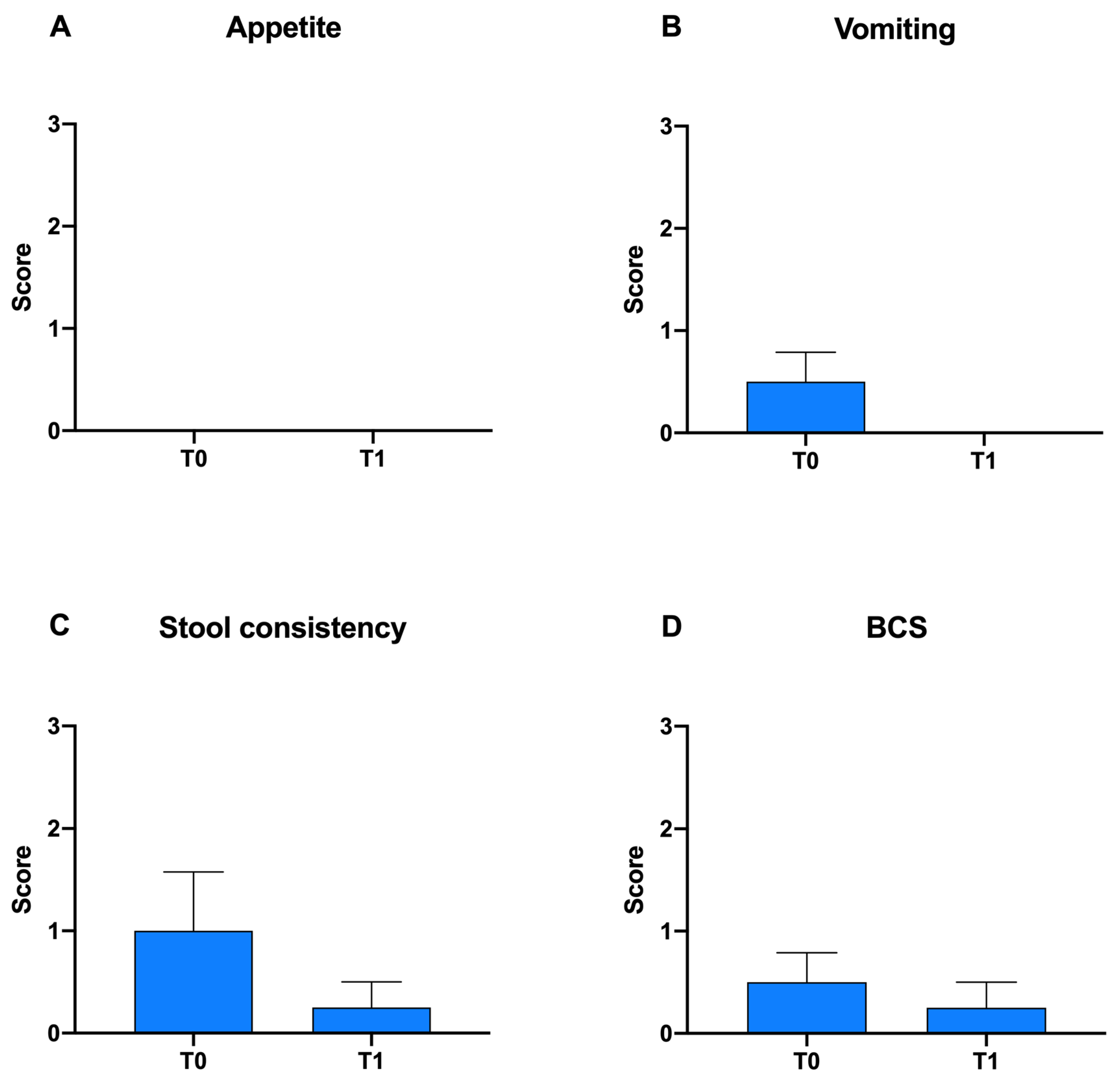
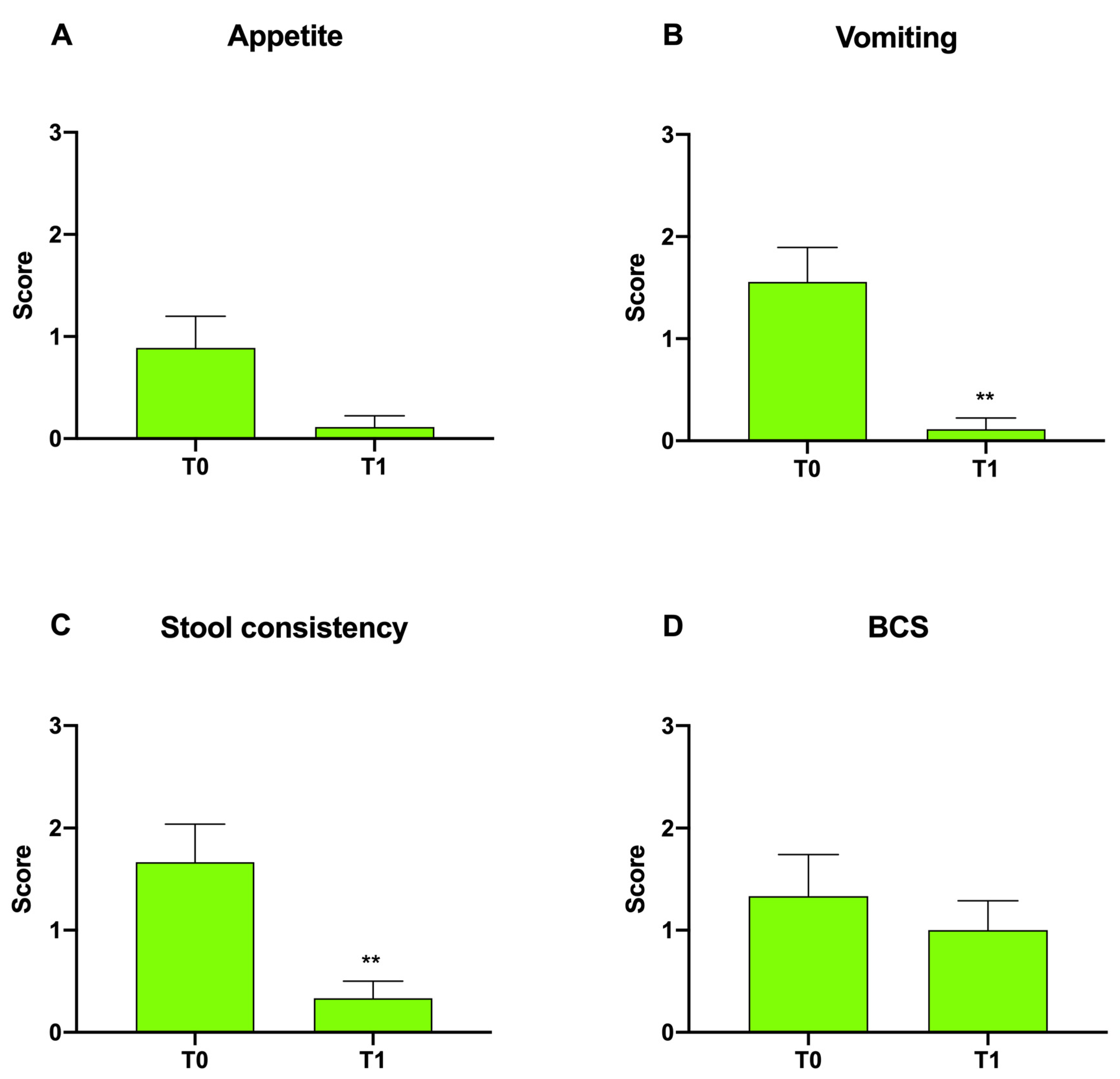
| Score | Appetite | Vomiting | Stool Consistency | Weight Loss |
|---|---|---|---|---|
| 0 | Normal | None | Normal: well-formed feces | None |
| 1 | Slight decrease | Mild (once a week) | Normal: well-formed feces | Mild (<5%) |
| 2 | Moderate decrease | Moderate (twice a week) | Very soft, moderately increased frequency | Moderate (5–10%) |
| 3 | Severe decrease | Severe (>2–3 times a week) | Watery diarrhea | Severe (>10%) |
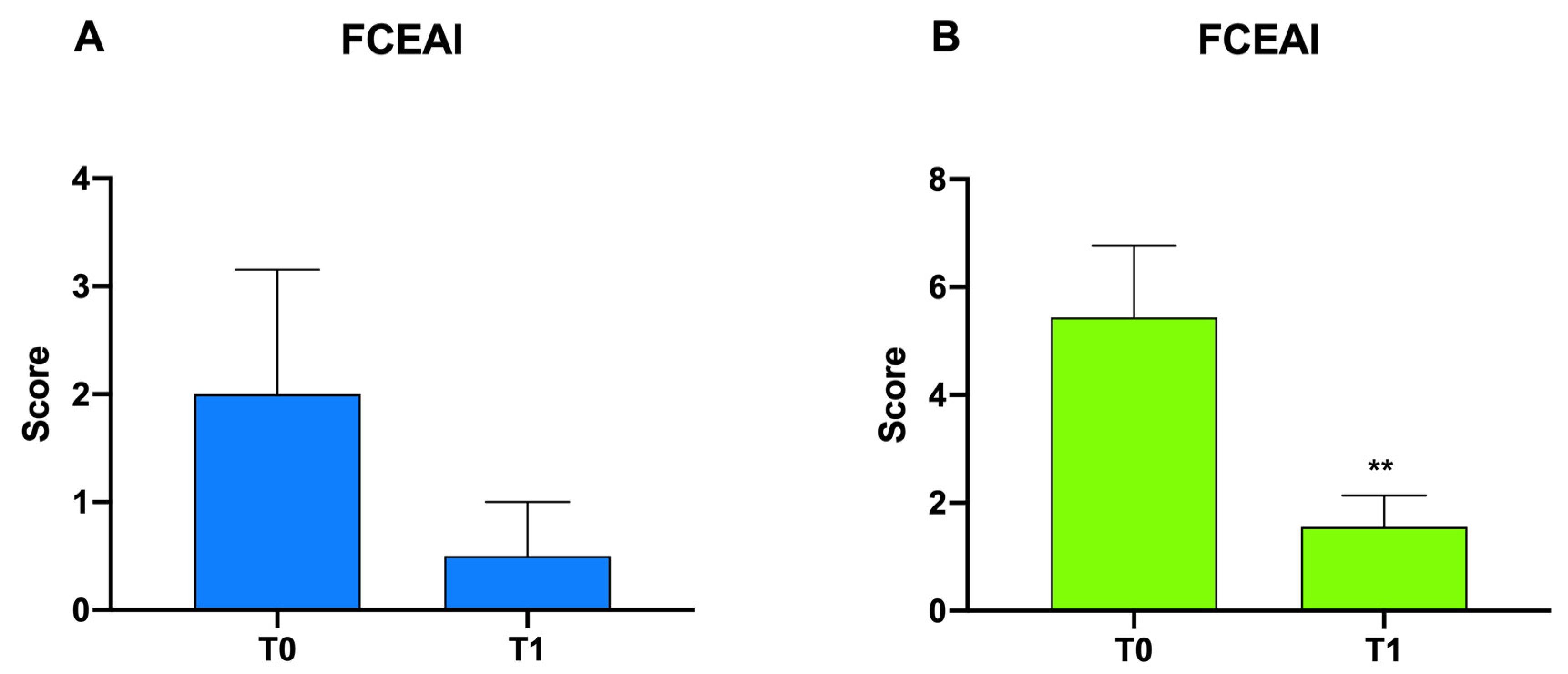
| Cheetah | Appetite | Vomiting | Stool Consistency | BCS | Simplified FCEAI Score | |||||
|---|---|---|---|---|---|---|---|---|---|---|
| T0 | T1 | T0 | T1 | T0 | T1 | T0 | T1 | T0 | T1 | |
| 1 | 0 | 0 | 1 | 0 | 2 | 0 | 1 | 0 | 4 | 0 |
| 2 | 0 | 0 | 1 | 0 | 2 | 1 | 1 | 1 | 4 | 2 |
| 3 | 0 | 0 | 0 | 0 | 0 | 0 | 0 | 0 | 0 | 0 |
| 4 | 0 | 0 | 0 | 0 | 0 | 0 | 0 | 0 | 0 | 0 |
| Mean values | 0.00 | 0.00 | 0.50 | 0.00 | 1.00 | 0.25 | 0.50 | 0.25 | 2.00 | 0.50 |
| Cheetah | Appetite | Vomiting | Stool Consistency | BCS | Simplified FCEAI Score | |||||
|---|---|---|---|---|---|---|---|---|---|---|
| T0 | T1 | T0 | T1 | T0 | T1 | T0 | T1 | T0 | T1 | |
| 1 | 2 | 0 | 3 | 0 | 3 | 1 | 3 | 2 | 11 | 3 |
| 2 | 2 | 0 | 3 | 0 | 3 | 1 | 3 | 2 | 11 | 3 |
| 3 | 0 | 0 | 2 | 0 | 2 | 0 | 2 | 1 | 6 | 1 |
| 4 | 0 | 0 | 1 | 0 | 1 | 0 | 0 | 0 | 2 | 0 |
| 5 | 0 | 0 | 1 | 0 | 0 | 0 | 0 | 0 | 1 | 0 |
| 6 | 0 | 0 | 1 | 0 | 1 | 0 | 0 | 0 | 2 | 0 |
| 7 | 1 | 0 | 1 | 0 | 1 | 0 | 1 | 1 | 4 | 1 |
| 8 | 1 | 0 | 0 | 0 | 1 | 0 | 1 | 1 | 3 | 1 |
| 9 | 2 | 1 | 2 | 1 | 3 | 1 | 2 | 2 | 9 | 5 |
| Mean values | 0.89 | 0.11 | 1.56 | 0.11 | 1.67 | 0.33 | 1.33 | 1.00 | 5.44 | 1.56 |
Publisher’s Note: MDPI stays neutral with regard to jurisdictional claims in published maps and institutional affiliations. |
© 2022 by the authors. Licensee MDPI, Basel, Switzerland. This article is an open access article distributed under the terms and conditions of the Creative Commons Attribution (CC BY) license (https://creativecommons.org/licenses/by/4.0/).
Share and Cite
Mangiaterra, S.; Schmidt-Küntzel, A.; Marker, L.; Di Cerbo, A.; Piccinini, R.; Guadagnini, D.; Turba, M.E.; Berardi, S.; Galosi, L.; Preziuso, S.; et al. Effect of a Probiotic Mixture in Captive Cheetahs (Acinonyx Jubatus) with Gastrointestinal Symptoms—A Pilot Study. Animals 2022, 12, 395. https://doi.org/10.3390/ani12030395
Mangiaterra S, Schmidt-Küntzel A, Marker L, Di Cerbo A, Piccinini R, Guadagnini D, Turba ME, Berardi S, Galosi L, Preziuso S, et al. Effect of a Probiotic Mixture in Captive Cheetahs (Acinonyx Jubatus) with Gastrointestinal Symptoms—A Pilot Study. Animals. 2022; 12(3):395. https://doi.org/10.3390/ani12030395
Chicago/Turabian StyleMangiaterra, Sara, Anne Schmidt-Küntzel, Laurie Marker, Alessandro Di Cerbo, Renato Piccinini, Davide Guadagnini, Maria Elena Turba, Sara Berardi, Livio Galosi, Silvia Preziuso, and et al. 2022. "Effect of a Probiotic Mixture in Captive Cheetahs (Acinonyx Jubatus) with Gastrointestinal Symptoms—A Pilot Study" Animals 12, no. 3: 395. https://doi.org/10.3390/ani12030395