Article

Effects of Dietary Supplementation with Vitamin A on Antioxidant and Intestinal Barrier Function of Broilers Co-Infected with Coccidia and Clostridium perfringens

Hubei Key Laboratory of Animal Nutrition and Feed Science, Engineering Research Center of Feed Protein Resources on Agricultural By-Products, Wuhan Polytechnic University, Ministry of Education, Wuhan 430023, China
*
Authors to whom correspondence should be addressed.
These authors contributed equally to this work and share first author.
Animals 2022, 12(23), 3431; https://doi.org/10.3390/ani12233431
Submission received: 28 October 2022 / Revised: 24 November 2022 / Accepted: 3 December 2022 / Published: 5 December 2022
(This article belongs to the Special Issue Precision Feeding and Nutrition in Poultry Production)

Abstract

:

Simple Summary

Coccidia and C. perfringens are the main accomplices in broiler necrotizing enteritis (NE), and NE causes oxidative stress and intestinal damage in broilers, which will result in huge economic losses to the poultry industry. Vitamin A (VA) is an essential micronutrient of the diet, and the beneficial effects of VA on vision, growth, and development have been intensively investigated. At present, the effects of VA on intestinal barrier and antioxidant functions in broilers have not been systematically reported. The present study was conducted to investigate the effects of dietary supplementation with vitamin A on the antioxidant and intestinal barrier function of broilers co-infected with coccidia and C. perfringens (CCP). Our outcomes showed that dietary VA might help a little with intestinal barrier function; nonetheless, it failed to alleviate the negative effects of CCP on the antioxidant function in broilers. Our study has guiding significance for the dose of VA in the diet of broilers, and it might arouse readers’ strong interest.

Abstract

Necrotic enteritis (NE) impairs poultry production and causes great economic loss. The nutritional regulation of diets has the potential to alleviate NE. The present study was conducted to investigate the effects of dietary supplementation with vitamin A (VA) on the antioxidant and intestinal barrier function of broilers co-infected with coccidia and C. perfringens (CCP). In a 2 × 2 factorial arrangement, 336 one-day-old Ross 308 broilers were divided into four treatments with two levels of VA (0 or 12,000 IU/kg) and challenged with or without CCP. The animal trial lasted for 42 days. The results showed that dietary supplemental VA improved body weight gain (BWG) and the feed intake (FI), and the FI was negatively affected by CCP. Additionally, the levels of catalase (CAT) in the serum, total superoxide dismutase (T-SOD), and CAT in the jejunum and glutathione peroxidase (GSH-Px) in the liver decreased with the CCP challenge (p < 0.05). The mRNA levels of SOD, CAT, GSH-Px1, and GSH-Px3 in the liver and jejunum were upregulated by the CCP challenge (p < 0.05). In addition, the level of serum diamine oxidase (DAO), and the mRNA level of ZO-1 were also upregulated with the CCP challenge. Dietary supplementation with VA contributed to the intestinal villi height and the mRNA level of Mucin-2 in the jejunum (p < 0.05). Additionally, dietary VA had the ability to alleviate the upregulation of SOD in the liver and SOD, CAT, GSH-Px1, GSH-Px3, ZO-1, and claudin-1 in the jejunum with the CCP challenge (p < 0.05). However, the mRNA level of GSH-Px3 and the levels of SOD in the liver and jejunum were downregulated with the VA supplementation in the diet. In conclusion, dietary VA improved the growth performance and the intestinal barrier function; nonetheless, it failed to alleviate the negative effects of CCP on the antioxidant function in broilers.

1. Introduction

Some pathogenic microorganisms and stimulators are prone to causing oxidative stress and intestinal damage in poultry production. A large number of reactive oxygen species (ROS) are produced under oxidative stress, which raises the levels of malondialdehyde (MDA) and myeloperoxidase (MPO) in birds and leads to tissue damage [1,2,3]. Additionally, ROS could also induce an inflammatory response by activating nuclear factor κB (NF-κB) [4]. The intestine is the main site for the digestion and absorption of nutrients, and it is also the largest immune organ of the body. A healthy intestine is a material mechanism for the high production of poultry [5]; while oxidative stress and inflammatory responses cause intestinal damage in poultry, an injured intestine opens the door for pathogenic microorganisms and stimulators, which will have negative effects on the growth performance and health of poultry [6]. Coccidia and C. perfringens are the main accomplices in necrotic enteritis (NE) in poultry, and NE causes the abnormal expression of antioxidant enzymes and peroxide products [7]. In addition, intestinal barrier-related proteins such as tight-junction proteins might be overexpressed in response to inflammation [8,9], and an excessive inflammatory response downregulates their expression and destroys the intestinal barrier [10]. The oxidative stress and intestinal damage induced by NE has negative effects on animal performance, which results in huge economic losses for the poultry industry.
Vitamin A (VA) is an essential micronutrient of the diet, and its beneficial effects on vision, growth and development have been intensively investigated [11]. In addition, it was demonstrated in a number of studies that VA has the potential to improve the antioxidant and intestinal barrier function. It has been suggested that VA enhances the bioavailability of selenium, which improves the activities of glutathione peroxidase (GSH-Px) [12]. Li also reported that carotenoid has the direct power of antioxidative activities [13]. Another study demonstrated that VA has the ability to maintain the integrity of the epithelium and mucosa [14]. Additionally, some studies suggested that a lack of VA in the diet might provide an opportunity for NE and other intestinal diseases in broilers [11,15]. At present, the effects of VA on NE in broilers have not been fully characterized. Taking the above studies into account, VA might alleviate the adverse effects of NE on broilers via improving antioxidant and barrier function.
We believed it would be very interesting to investigate the effects of VA on NE caused by coccidia and C. perfringens in broilers. In the present study, coccidia and C. perfringens were used to establish an NE model in broilers to investigate the effects of dietary supplementation with VA on the antioxidant and intestinal barrier function of NE birds.

2. Materials and Methods

2.1. Experimental Design and Diets

All experimental procedures used were approved by the Animal Care and Use Committee of Wuhan Polytechnic University.
A 2 × 2 factorial design was implemented in which broilers were challenged with or without coccidia and C. perfringens (CCP), and their diets were supplemented with or without VA. A total of 336 healthy one-day-old Ross 308 broilers with similar initial body weights were randomly assigned to four groups. There were 7 replicates in each group, and 12 birds (6 males and 6 females) in each replicate. The birds in the control (CTR) and CCP groups were fed a basal diet without supplementation of VA, and the basal diet was formulated accorded to the feeding standard of China (Ministry of Agriculture of China, 2004) and NRC requirements (National Research Council, 1994) (Table 1). The birds in the VA and VA + CCP groups were fed basal diets supplemented with 12,000 IU/kg of VA. Additionally, the birds in the CCP and VA + CCP groups were challenged with coccidia on day 14 of the trial, and treated with 1 mL 1 × 10⁸ CFU/mL C. perfringens from day 18 to day 20 of the trial. The C. perfringens (CVCC2030) was purchased from the China Veterinary Microbial Culture Collection and Management Center (Beijing, China), and coccidia from Foshan Zhengdian Biotechnology Co., Ltd., (Foshan, Guangdong, China) was used in the present study. The quadrivalent anti-coccidiosis vaccine consisted of 1 × 10⁵ oocysts of the E. acervuline strain PAHY as well as 5 × 10⁴ oocysts of the E. necatrix strain PNHZ, E. tenella strain PTMZ, and E. maxima strain PMHY. Every bird was recommended to be inoculated with 1100 ± 110 sporulated oocysts according to the instructions, and the dose used in the present study was 20 times that of the recommended. The birds in the CTR and VA groups were treated with an equal volume of saline at the same time. All birds were raised in cages and had free access to feed and water. A 24 h light regime was carried out throughout the animal trial, and the room temperature was controlled at 30–33 °C in the first week, and then it decreased by 2–3 °C in each week until reaching 22 °C, and this temperature was kept till the end of trial. The animal trial lasted for 42 days. The broilers were weighed at days 1, 13, 21, and 42 of the trial, and the body weight gain (BWG), feed intake (FI), and feed conversion ratio (FCR) were measured. On days 28 and 35, 1 male and 1 female broiler in each replicate were selected to harvest the blood from the underwing vein, and then those birds were injected intravenously with 50 mg/kg body weight (BW) sodium pentobarbital and quickly slaughtered for sample collection. The blood was collected and centrifuged at 3000× g at 4 °C for 10 min to obtain the serum. The liver and jejunum were collected and frozen in liquid nitrogen, and then they were transferred to a −80 °C freezer for storage.

2.2. Antioxidative Enzymes and Peroxidation Products

A total of 0.1 g liver or jejunum was placed in a 1.5 mL centrifuge tube, and 0.9 mL saline was added. Then, the samples were homogenized at 4 °C. The mixture was centrifuged at 3500× g at 4 °C for 15 min to collect the supernatants. Commercial assay kits were purchased from Nanjing Jiancheng Institute of Biological Engineering, Nanjing, China. The levels of total antioxidant capacity (T-AOC), catalase (CAT), hydrogen peroxide (H2O2), glutathione peroxidase (GSH-Px), malondialdehyde (MDA), and total superoxide dismutase (T-SOD) in the serum, liver, and jejunum were measured according to the manufacturer’s instructions.

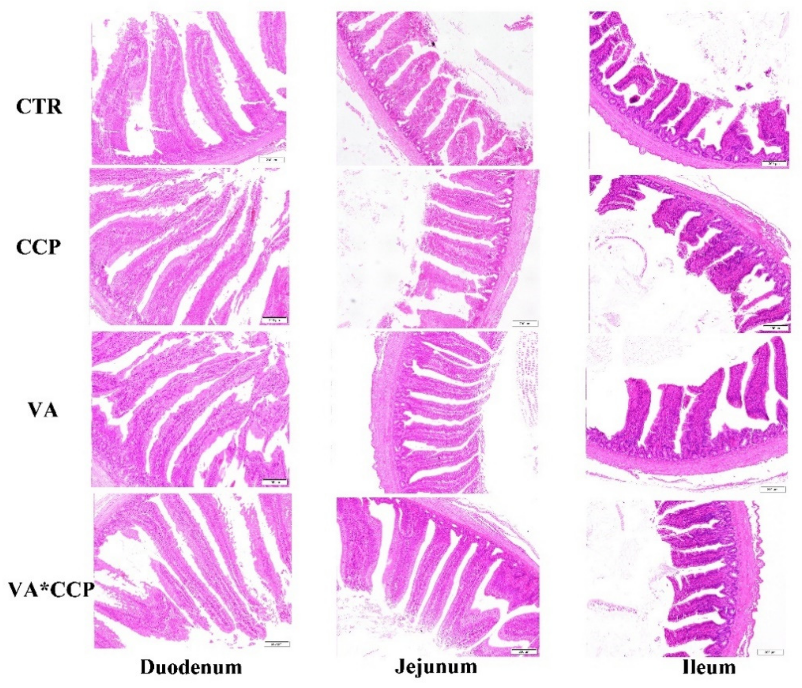
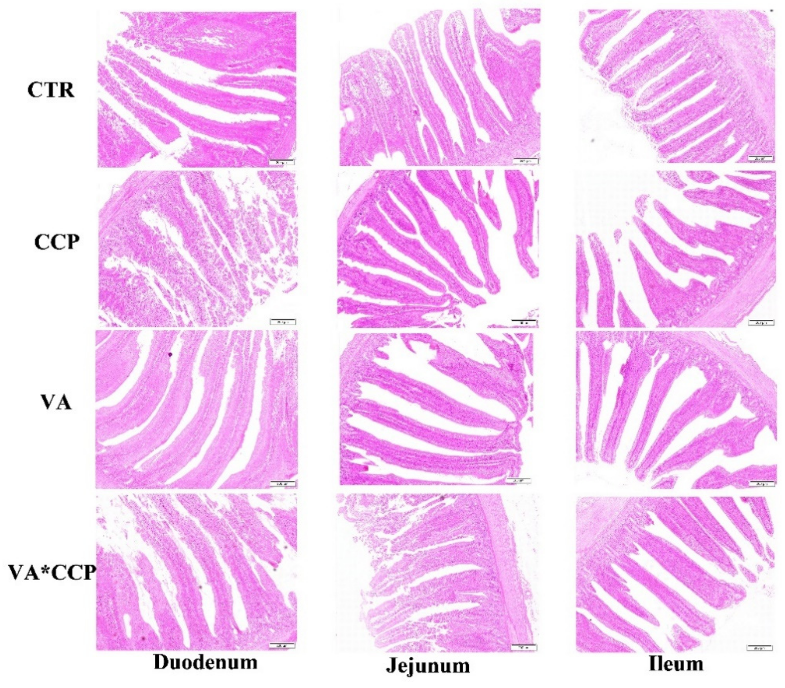
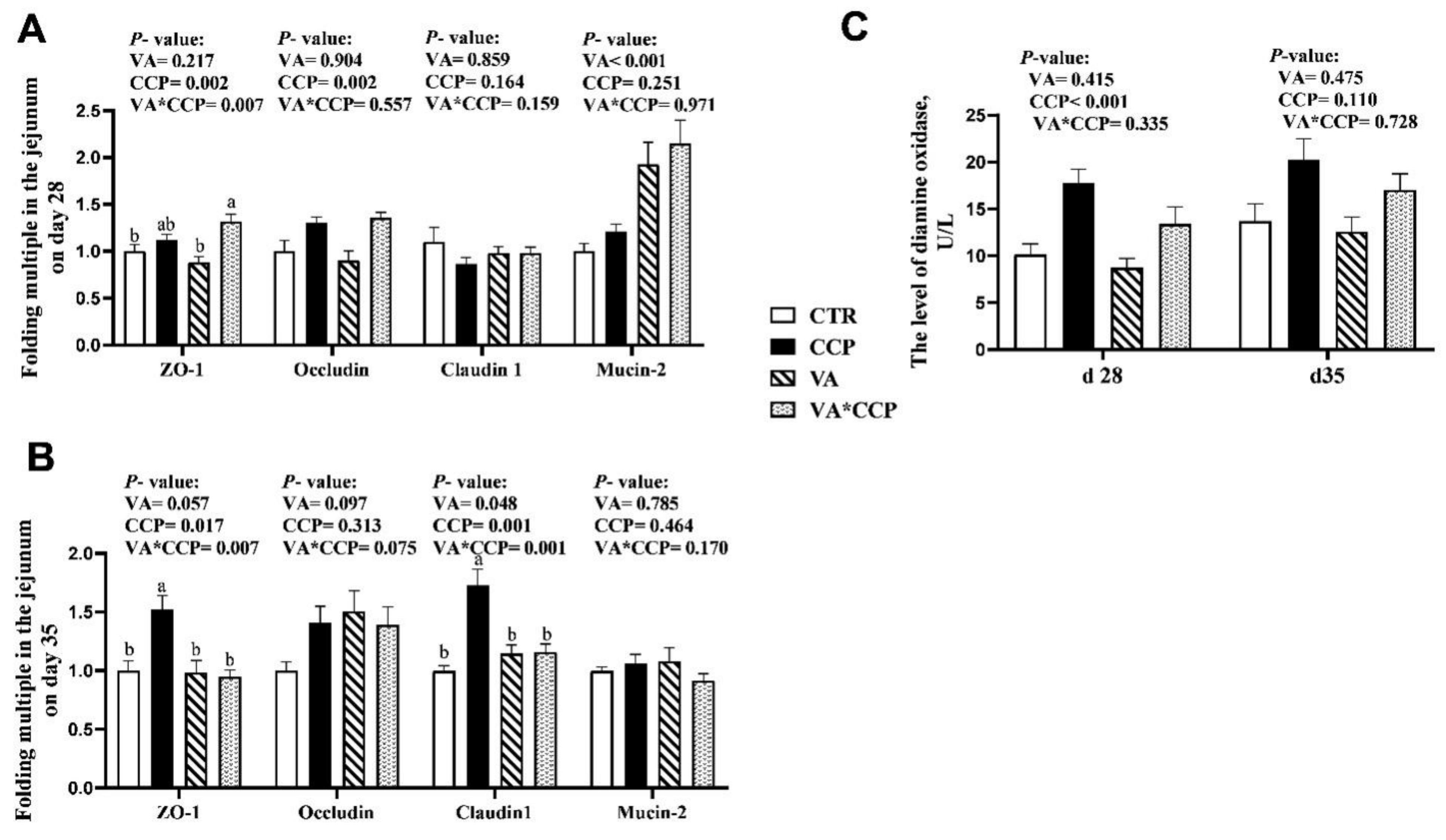
2.3. Serum Diamine Oxidase and Intestinal Morphology

The level of diamine oxidase (DAO) in the serum was determined using commercially available kits from the Nanjing Jiancheng Institute of Biological Engineering, Jiangsu, China. Approximately 1 cm of the duodenum, jejunum, and ileum were collected and placed in a 4% paraformaldehyde solution, and then the intestinal tissue was dehydrated, transparent, waxed, and embedded, after which a slicer was used to cut the tissue to a thickness of 3–5 microns. Finally, the sections were stained with hematoxylin and eosin. A previous study [16] was used to measure the villus height (VH) and crypt depth (CD). Briefly, the intestinal sections of 14 birds (7 males and 7 females) in each treatment were selected, and for each intestinal section, 10 morphologically intact intestinal villi were selected for measurements. These 10 values were averaged and used as the result of this intestinal segment for subsequent analysis. The Olymps BX-41TF microscope was used for observation and a thousand-screen high-definition color pathology graphic analysis system was used to measure the VH and CD. The ratio of the VH to CD (VH/CD) was calculated.

2.4. Transcription Level of Genes in the Liver and Jejunum

Approximately 0.1 g of hepatic and jejunal sample was put in a 1.5 mL RNA-free centrifuge tube, and 1 mL Trizol reagent (Invitrogen Life Technologies, Carlsbad, CA, USA) was added. Then, the mixture was homogenized at 4 °C. The total RNA was extracted according to the method described by Hou et al. [17]. The concentration and purity of the total RNA was quantified by measuring its optical density at 260 and 280 nm with the spectrophotometer Nano Drop® ND-2000 UV-VIS (Thermo Scientific, Wilmington, DE, USA). The RNA integrity was verified by agarose gel electrophoresis, and then the total RNA was reverse transcribed to obtain the cDNA followed with the protocol of the gDNA Eraser (Takara Biotechnology (Dalian) Co., Ltd., Dalian, China). A quantitative real-time PCR assay was performed with the 7500-fluorescence detection system (Applied Biosystems, Foster City, CA, USA). The SYBR Premix Ex Taq kit (Takara Biotechnology (Dalian) Co., Ltd., Dalian, China), the cDNA of each sample, and primers were used in its performance. The PCR reaction program was as follows: an initial denaturation step set at 95 °C for 30 s, 40 cycles at 95 °C for 5 s, and an annealing and extension temperature at 60 °C for 34 s. The PCR products from each primer pair were subjected to a melting curve analysis and subsequent agarose gel electrophoresis. The gene expression was quantified using the comparative threshold cycle method [18], and the data are expressed relative to the CTR group. The mRNA levels of the nuclear factor erythroid 2-related factor 2 (Nrf-2), superoxide dismutase 1 (SOD-1), catalase (CAT), glutathione peroxidase 1 (GSH-px1), and glutathione peroxidase 3 (GSH-px3) in the liver and jejunum were measured. Additionally, the transcription levels of claudin-1, occludin, zonula occludens-1 (ZO-1), and mucin-2 in the jejunum were detected. β-Actin was used as an internal reference gene, and the primer sequences of each gene are described in Table 2.

2.5. Statistical Analysis

The general linear model program in SPSS 23.0 software (SPSS Inc., Chicago, IL, USA) was used to analyze the data. Two-way ANOVA was performed in a 2 × 2 factor arrangement to analyze the effects of the CCP challenge and VA supplementation as well as the interaction of these two factors. When interactive effects were observed, one-way ANOVA and Tukey’s multiple comparisons were used. p < 0.05 was considered to be significant. GraphPad Prism 8.0 software (GraphPad software, LLC, San Diego, CA, USA) was used to graph the data.

3. Results

3.1. Growth Performance

A dietary supplemental of 12,000 IU/kg VA was also able to elevate the BWG and FI from day 14 to day 20 of the trial (p < 0.05) (Table 3), and the FI from day 21 to day 42 of the trial decreased with the coccidia and C. perfringens challenge.

3.2. Antioxidative Enzymes and Peroxidation Products

The activity of CAT in the serum decreased on day 35 with the coccidia and C. perfringens challenge (p < 0.05) (Table 4). Dietary supplementation with 12,000 IU/kg VA raised the level of MDA on day 28 and decreased the level of H2O2 on day 35 in the serum (p < 0.05). In the liver, the levels of T-AOC and MDA increased, and the activity of GSH-Px decreased on day 28 with the coccidia and C. perfringens challenge (p < 0.05) (Table 5). Dietary VA failed to alleviate these indices, and the T-AOC level and T-SOD activity in the liver dropped on day 35 with VA supplementation in the diet (p < 0.05). Dietary VA might have a negative effect on the antioxidant function of broilers, a phenomenon that was also found in the jejunum. The T-AOC level and T-SOD activity on day 28 and the activities of CAT and T-SOD on day 35 decreased with VA addition (p < 0.05) (Table 6). However, dietary VA decreased the contents of H2O2 and MDA on day 35 (p < 0.05). The T-SOD activity on day 28 as well as the T-SOD and CAT activities on day 35 decreased with the coccidia and C. perfringens challenge (p < 0.05).

3.3. The mRNA Levels of Antioxidant-Related Genes

Dietary supplementation with VA downregulated the mRNA levels of GSH-Px1 and GSH-Px3 in the liver on days 28 and 35 (p < 0.05) (Table 7). The mRNA levels of SOD and CAT in the liver on day 35 were upregulated by the CCP challenge (p < 0.05). Dietary VA could alleviate the upregulation of SOD in the liver in the CCP-challenged broilers. The mRNA levels of Nrf-2, SOD, CAT, GSH-Px1, and GSH-Px3 in the jejunum on day 35 were upregulated by the CCP challenge (p < 0.05) (Table 8). Dietary VA was able to alleviate the upregulation of SOD, CAT, GSH-Px1, and GSH-Px3 in the CCP-challenged broilers (p < 0.05). The mRNA level of CAT in the jejunum on day 35 was upregulated by VA addition in the diet (p < 0.05), whereas the transcript level of GSH-Px3 was downregulated with the VA supplementation (p < 0.05).

3.4. Intestinal Barrier Function

The statistical parameters of the intestinal tract were not significantly affected by the CCP challenge; however, the surface of the intestinal tract showed roughness, and the intestinal tract was scattered and varied in shape and size (p > 0.05) (Table 9 and Table 10) (Figure 1 and Figure 2). Dietary supplementation with VA was able to raise the villi height (VH) of the duodenum, jejunum, and ileum on day 28 (Table 9) and the VH of ileum on day 35 (p < 0.05) (Table 10). In the VA treated, the intestinal tract was uniform in size and neat in direction. Fluff indicated smoothness (Figure 1 and Figure 2). Co-infection with CCP elevated the mRNA levels of occludin and ZO-1 in the jejunum on day 28. Additionally, the transcript levels of ZO-1 and claudin-1 were also upregulated on day 35 (p < 0.05) (Figure 3A,B). Interestingly, dietary VA could alleviate the upregulation of ZO-1 and claudin-1 on day 35, and the mRNA level of mucin-2 was upregulated on day 28 with VA supplementation in the diet. In addition, the level of DAO in the serum on day 28 was increased by the CCP challenge (p < 0.05) (Figure 3C). Dietary VA also failed to alleviate this index.

4. Discussion

NE is a multifactorial inflammatory disease of the intestine, and C. perfringens is one of the main causes [19], whereas with C. perfringens it only occurs in concert with other factors that cause intestinal damage, for example, a coccidia infection and the wheat basal diet [20,21]. In the present study, C. perfringens and coccidia were used to establish a model of NE. The antioxidant function of NE-infected broilers was weakened, and the intestinal barrier function was impaired [22,23]. The antioxidant system and the pro-oxidative system were maintained at a stable level, and some adverse factors, such as pathogens, viruses, and other stressors, could disturb this balance. At the same time, the body will have an oxidative stress response. Accordingly, some peroxidation products, such as ROS, malondialdehyde (MDA), and myeloperoxidase (MPO), will be increased [24]. In this study, the levels of MDA in the liver and jejunum were increased by the CCP challenge, and it was only observed for a short time after infection. Antioxidant enzymes, such as T-SOD, CAT, and GSH-Px, have the ability to mitigate the elevated peroxidation products [25]. The total antioxidant capacity (T-AOC) is regarded as the ability to maintain the balance of the antioxidant and peroxidation systems [26]. In the present study, co-infection with coccidia and C. perfringens decreased the activities of CAT, T-SOD, and GSH-Px. These results demonstrate that the combined infection of coccidia and C. perfringens caused oxidative stress. This is basically consistent with previous studies [7]. We observed that dietary supplementation with 12,000 IU/kg VA failed to alleviate the oxidative stress of NE-infected broilers. Moreover, the activities of T-SOD and GSH-Px in the liver decreased with VA supplementation in the diet. This study suggests that the activities of SOD, CAT, and GSH-Px significantly increased with 10 mg/kg β-carotene, which is a VA precursor. Conversely, 20 mg/kg of dietary β-carotene decreased the activities of SOD and GSH-Px [27]. The dual effects of VA on antioxidant function may be related to its dose. This illuminated for us that a dose of 12,000 IU/kg VA might be detrimental to the antioxidant function of broilers, and it was worthy of further study.
The Kelch-like ECH-associated protein 1 (Keap-1)-Nrf-2 system is a major regulatory pathway against oxidative stress. Keap-1 and Nrf-2 are tightly tied together under normal conditions, and they are disassembled with stressor challenge. Then, Nrf-2 is translocated into the nucleus to initiate the transcription of antioxidant genes, such as SOD, CAT, and GSH-Px [28]. In the present study, we observed that the mRNA levels of Nrf-2, SOD, CAT, GSH-Px1, and GSH-Px3 in the jejunum were upregulated by the CCP challenge. Additionally, the transcript levels of SOD and CAT in the liver were also upregulated. Our outcomes suggest that the antioxidant system of broilers was activated to respond to the infection by CCP. In addition, dietary VA was able to alleviate the upregulation of antioxidant enzyme genes with CCP challenge. However, the transcript level of GSH-Px3 was downregulated with VA supplementation in the diet. Although VA was helpful to the antioxidant capacity of the NE-infected broilers at the gene expression level, the outcomes of the antioxidant enzymes and their products suggest that a lower dose of VA than 12,000 IU/kg should be recommended. It is worth mentioning that these changes in antioxidant enzyme genes were not significant for a short period of time after the infection. Interestingly, it was significant for 2 weeks after the infection. A study suggested that the growth performance of broilers infected with NE was negatively affected, and it was only observed for a short period of time after infection [10]. A mechanism of self-compensating growth and homeostasis maintenance may be activated after infection [29].
Diamine oxidase (DAO) is an intracellular enzyme that catalyzes diamine in the mucosal or ciliated epithelial cells of the mammalian small intestine. It contributes to protecting the intestinal mucosa via promoting cell repair [30]. An early enteral nutrition study demonstrated that the concentration of DAO in plasma was raised sharply when the intestinal mucosa was damaged [31]. In the present study, the level of DAO in the plasma was elevated with the CCP challenge. A study suggested that in the intestinal damage model, the DAO level in the intestine was highly negatively correlated with it in the serum [17]. This evidence indicates that a combined infection with CCP caused intestinal damage in broilers. An intact intestinal structure is the basis for the intestinal tract to exert some physiological functions. The degree of development of the intestinal morphology could be evaluated by the indices of VH, CD, and VH/CD [32]. In our study, the surface of the intestinal tract challenged with CCP showed roughness, and the intestinal tract was scattered and varied in shape and size. However, the levels of VH, CD, and VH/CD were not affected by the CCP challenge. This is inconsistent with our expectations. To explain the observed phenomenon, we might consider that a wheat basal diet should be required in the establishment of an NE model using CCP infections. This might make the intestinal damage observation more significant. Intriguingly, dietary supplementation with 12,000 IU/kg VA contributed to the VH of intestine. A study also implicated that dietary β-carotene raised the intestinal VH [33]. This might be one of the reasons why VA was useful for the growth performance of broilers in the present study.
Tight junction proteins between intestinal cells help maintain the intestinal barrier function. ZO-1, claudin-1, and occludin play key roles in regulating the barrier function of intestinal epithelium [34]. Some studies demonstrated that the transcriptional levels of genes encoding tight junction proteins are upregulated under inflammatory conditions [8,9]. In the present study, co-infection with CCP also upregulated the mRNA levels of occludin, ZO-1, and claudin-1 in the intestine. A possible explanation is that the inflammation activated the intestinal self-protection mechanism, and the high expression of tight junctions was used to protect the intestine from pathogen stimulation. Interestingly, dietary supplementation with 12,000 IU/kg VA could alleviate the upregulation of ZO-1 and claudin-1 with CCP challenge. In addition, the mRNA level of mucin-2 was upregulated with VA addition, which is consistent with another study [15]. Mucin-2 secreted by goblet cells adheres to the surface of the intestinal villi and is considered the first physical barrier of the intestine [35]. Symbiotic bacteria in the intestinal tract generally colonize the outer layer of the intestinal mucosa. Some probiotics stimulate goblet cells to secrete mucin, which strengthens the mucin layer to protect the intestinal tract from an invasion of pathogenic bacteria [36]. In the present study, dietary supplemental VA might be useful for the proliferation of some probiotics and the proliferation and differentiation of intestinal goblet cells of broilers. Further research is needed to investigate this issue. Taking the beneficial effects of VA on intestinal VH and the mRNA levels of tight junction proteins into account, dietary supplementation with 12,000 IU/kg VA might offer exciting opportunities for the intestinal barrier function of NE-infected broilers.

5. Conclusions

Dietary supplementation with 12,000 IU/kg VA might have a negative effect on the antioxidant function of broilers. However, it had great potential to alleviate the adverse effect of co-infection with CCP on intestinal barrier function and promoted intestinal development.

Author Contributions

B.D. and S.G. designed the study; P.L. wrote the manuscript; P.L., C.L. (Chengao Liu), J.N., Y.Z., C.L. (Changwu Li) and Z.Z. helped collect and analyze the experimental results; B.D., S.G. and P.L. participated in the writing and revision of the manuscript. All authors have read and agreed to the published version of the manuscript.

Funding

This research was funded by the National Key R&D Program of China (grant number: 2016YFD0501202), the National Natural Science Foundation of China (grant number: 32172757), the National Science Foundation of Hubei Province (grant number: 2021CFB559), and the Open Project of Hubei Key Laboratory of Animal Nutrition and Feed Science (grant number: 201902).

Institutional Review Board Statement

All animal procedures used in the present study were performed in compliance with Hubei Provincial Regulations for Laboratory Animals (011043145-029-2013-000009) and were approved by the Institutional Animal Care and Use Committee of Wuhan Polytechnic University (number: WPU201904001).

Informed Consent Statement

Informed consent was obtained from all subjects involved in the study.

Data Availability Statement

The datasets produced and/or analyzed during the current study are available from the corresponding author upon reasonable request.

Acknowledgments

All authors would like to thank all of the volunteers for their commitment and patience during the study.

Conflicts of Interest

The authors declare no conflict of interest.

References

  1. Appiah, M.O.; He, B.; Lu, W.; Wang, J. Antioxidative effect of melatonin on cryopreserved chicken semen. Cryobiology 2019, 89, 90–95. [Google Scholar] [CrossRef]
  2. Balogh, K.; Kovesi, B.; Zandoki, E.; Kulcsar, S.; Ancsin, Z.; Erdelyi, M.; Dobolyi, C.; Bata-Vidacs, I.; Inotai, K.; Szekeres, A.; et al. Effect of sterigmatocystin or aflatoxin contaminated feed on lipid peroxidation and glutathione redox system and expression of glutathione redox system regulatory genes in broiler chicken. Antioxidants 2019, 8, 201. [Google Scholar] [CrossRef] [PubMed] [Green Version]
  3. Yang, D.; Zhao, C.; Zhang, M.; Zhang, S.; Zhai, J.; Gao, X.; Liu, C.; Lv, X.; Zheng, S. Changes in oxidation-antioxidation function on the thymus of chickens infected with reticuloendotheliosis virus. BMC Vet. Res. 2020, 16, 483. [Google Scholar] [CrossRef] [PubMed]
  4. Chen, D.; Ning, F.; Zhang, J.; Tang, Y.; Teng, X. NF-kappaB pathway took part in the development of apoptosis mediated by miR-15a and oxidative stress via mitochondrial pathway in ammonia-treated chicken splenic lymphocytes. Sci. Total Environ. 2020, 729, 139017. [Google Scholar] [CrossRef] [PubMed]
  5. Khan, S.; Moore, R.J.; Stanley, D.; Chousalkar, K.K. The gut microbiota of laying hens and its manipulation with prebiotics and probiotics to enhance gut health and food safety. Appl. Environ. Microbiol. 2020, 86, e00600-20. [Google Scholar] [CrossRef]
  6. Li, Q.; Wan, G.; Peng, C.; Xu, L.; Yu, Y.; Li, L.; Li, G. Effect of probiotic supplementation on growth performance, intestinal morphology, barrier integrity, and inflammatory response in broilers subjected to cyclic heat stress. Anim. Sci. J. 2020, 91, e13433. [Google Scholar] [CrossRef]
  7. Yu, C.; Tong, Y.; Li, Q.; Wang, T.; Yang, Z. Trans-anethole ameliorates intestinal injury through activation of Nrf2 signaling pathway in subclinical necrotic enteritis-induced broilers. Front. Vet. Sci. 2022, 9, 877066. [Google Scholar] [CrossRef]
  8. Memon, F.U.; Yang, Y.; Lv, F.; Soliman, A.M.; Chen, Y.; Sun, J.; Wang, Y.; Zhang, G.; Li, Z.; Xu, B.; et al. Effects of probiotic and Bidens pilosa on the performance and gut health of chicken during induced Eimeria tenella infection. J. Appl. Microbiol. 2021, 131, 425–434. [Google Scholar] [CrossRef]
  9. Zhang, H.; Chen, Y.; Chen, Y.; Li, Y.; Jia, P.; Ji, S.; Zhou, Y.; Wang, T. Dietary pterostilbene supplementation attenuates intestinal damage and immunological stress of broiler chickens challenged with lipopolysaccharide. J. Anim. Sci. 2020, 98, skz373. [Google Scholar] [CrossRef]
  10. Liu, D.; Guo, Y.; Wang, Z.; Yuan, J. Exogenous lysozyme influences Clostridium perfringens colonization and intestinal barrier function in broiler chickens. Avian Pathol. 2010, 39, 17–24. [Google Scholar] [CrossRef]
  11. Howell, J.M.; Thompson, J.N. Observations on the lesions in vitamin A deficient adult fowls with particular reference to changes in bone and central nervous system. Br. J. Exp. Pathol. 1967, 48, 450–454. [Google Scholar]
  12. Wang, Y.; Li, L.; Gou, Z.; Chen, F.; Fan, Q.; Lin, X.; Ye, J.; Zhang, C.; Jiang, S. Effects of maternal and dietary vitamin A on growth performance, meat quality, antioxidant status, and immune function of offspring broilers. Poult. Sci. 2020, 99, 3930–3940. [Google Scholar] [CrossRef]
  13. Li, R.; Yang, Y.; Hong, P.; Zhang, Z.; Li, L.; Hui, J.; Zheng, X. Beta-carotene attenuates weaning-induced apoptosis via inhibition of PERK-CHOP and IRE1-JNK/p38 MAPK signalling pathways in piglet jejunum. J. Anim. Physiol. Anim. Nutr. 2020, 104, 280–290. [Google Scholar] [CrossRef]
  14. Pattanakitsakul, P.; Chongviriyaphan, N.; Pakakasama, S.; Apiwattanakul, N. Effect of vitamin A on intestinal mucosal injury in pediatric patients receiving hematopoietic stem cell transplantation and chemotherapy: A quasai-randomized trial. BMC Res. Notes 2020, 13, 464. [Google Scholar] [CrossRef]
  15. Hui, J.; Li, L.; Li, R.; Wu, M.; Yang, Y.; Wang, J.; Fan, Y.; Zheng, X. Effects of supplementation with beta-carotene on the growth performance and intestinal mucosal barriers in layer-type cockerels. Anim. Sci. J. 2020, 91, e13344. [Google Scholar] [CrossRef]
  16. Li, P.; Gao, M.; Fu, J.; Yan, S.; Liu, Y.; Mahmood, T.; Lv, Z.; Guo, Y. Dietary soya saponin improves the lipid metabolism and intestinal health of laying hens. Poult. Sci. 2022, 101, 101663. [Google Scholar] [CrossRef]
  17. Hou, Y.; Wang, L.; Zhang, W.; Yang, Z.; Ding, B.; Zhu, H.; Liu, Y.; Qiu, Y.; Yin, Y.; Wu, G. Protective effects of N-acetylcysteine on intestinal functions of piglets challenged with lipopolysaccharide. Amino Acids 2012, 43, 1233–1242. [Google Scholar] [CrossRef]
  18. Heid, C.A.; Stevens, J.; Livak, K.J.; Williams, P.M. Real time quantitative PCR. Genome Res. 1996, 6, 986–994. [Google Scholar] [CrossRef] [Green Version]
  19. Latorre, J.D.; Adhikari, B.; Park, S.H.; Teague, K.D.; Graham, L.E.; Mahaffey, B.D.; Baxter, M.; Hernandez-Velasco, X.; Kwon, Y.M.; Ricke, S.C.; et al. Evaluation of the epithelial barrier function and ileal microbiome in an established necrotic enteritis challenge model in broiler chickens. Front. Vet. Sci. 2018, 5, 199. [Google Scholar] [CrossRef]
  20. Guo, S.; Xi, Y.; Xia, Y.; Wu, T.; Zhao, D.; Zhang, Z.; Ding, B. Dietary Lactobacillus fermentum and Bacillus coagulans supplementation modulates intestinal immunity and microbiota of broiler chickens challenged by Clostridium perfringens. Front. Vet. Sci. 2021, 8, 680742. [Google Scholar] [CrossRef]
  21. La Ragione, R.M.; Woodward, M.J. Competitive exclusion by Bacillus subtilis spores of Salmonella enterica serotype Enteritidis and Clostridium perfringens in young chickens. Vet. Microbiol. 2003, 94, 245–256. [Google Scholar] [CrossRef] [PubMed]
  22. Tang, Y.; Zhang, X.; Wang, Y.; Guo, Y.; Zhu, P.; Li, G.; Zhang, J.; Ma, Q.; Zhao, L. Dietary ellagic acid ameliorated Clostridium perfringens-induced subclinical necrotic enteritis in broilers via regulating inflammation and cecal microbiota. J. Anim. Sci. Biotechnol. 2022, 13, 47. [Google Scholar] [CrossRef] [PubMed]
  23. Zhang, B.; Lv, Z.; Li, H.; Guo, S.; Liu, D.; Guo, Y. Dietary l-arginine inhibits intestinal Clostridium perfringens colonisation and attenuates intestinal mucosal injury in broiler chickens. Br. J. Nutr. 2017, 118, 321–332. [Google Scholar] [CrossRef] [PubMed] [Green Version]
  24. Jelic, M.D.; Mandic, A.D.; Maricic, S.M.; Srdjenovic, B.U. Oxidative stress and its role in cancer. J. Cancer Res. Ther. 2021, 17, 22–28. [Google Scholar] [CrossRef] [PubMed]
  25. He, L.; He, T.; Farrar, S.; Ji, L.; Liu, T.; Ma, X. Antioxidants maintain cellular redox homeostasis by elimination of reactive oxygen species. Cell. Physiol. Biochem. 2017, 44, 532–553. [Google Scholar] [CrossRef]
  26. Zhou, M.; Xu, W.; Wang, J.; Yan, J.; Shi, Y.; Zhang, C.; Ge, W.; Wu, J.; Du, P.; Chen, Y. Boosting mTOR-dependent autophagy via upstream TLR4-MyD88-MAPK signalling and downstream NF-kappa B pathway quenches intestinal inflammation and oxidative stress injury. eBio Med. 2018, 35, 345–360. [Google Scholar]
  27. Sun, Z.Y.; Peng, T.; Zhang, M.J.; Cheng, W.U.; Luo, Y.X. Effects of β-Carotenes on Blood Component and Activities of SOD, GSH-PX and CAT in Mice. Prog. Vet. Med. 2007, 28, 25–28. (In Chinese) [Google Scholar]
  28. Gall, T.K.; Tomljanovic, M.; Jaganjac, M.; Matijevic, G.T.; Cipak, G.A.; Milkovic, L.; Borovic, S.S.; Buttari, B.; Profumo, E.; Saha, S.; et al. Oxidative stress and cancer heterogeneity orchestrate NRF2 roles relevant for therapy response. Molecules 2022, 27, 1468. [Google Scholar] [CrossRef]
  29. Leeson, S.; Caston, L.; Summers, J.D. Broiler response to energy or energy and protein dilution in the finisher diet. Poult. Sci. 1996, 75, 522–528. [Google Scholar] [CrossRef]
  30. Bragg, L.E.; Thompson, J.S.; West, W.W. Intestinal diamine oxidase levels reflect ischemic injury. J. Surg. Res. 1991, 50, 228–233. [Google Scholar] [CrossRef]
  31. Guo, Y.; Li, H.; Liu, Z.; Li, C.; Chen, Y.; Jiang, C.; Yu, Y.; Tian, Z. Impaired intestinal barrier function in a mouse model of hyperuricemia. Mol. Med. Rep. 2019, 20, 3292–3300. [Google Scholar] [CrossRef] [Green Version]
  32. Lv, Z.; Dai, H.; Wei, Q.; Jin, S.; Wang, J.; Wei, X.; Yuan, Y.; Yu, D.; Shi, F. Dietary genistein supplementation protects against lipopolysaccharide-induced intestinal injury through altering transcriptomic profile. Poult. Sci. 2020, 99, 3411–3427. [Google Scholar] [CrossRef]
  33. Cucco, M.; Guasco, B.; Malacarne, G.; Ottonelli, R. Effects of beta-carotene supplementation on chick growth, immune status and behaviour in the grey partridge, Perdix perdix. Behav. Process. 2006, 73, 325–332. [Google Scholar] [CrossRef]
  34. Dokladny, K.; Zuhl, M.N.; Moseley, P.L. Intestinal epithelial barrier function and tight junction proteins with heat and exercise. J. Appl. Physiol. 2016, 120, 692–701. [Google Scholar] [CrossRef]
  35. Liu, C.; Chen, C.; Yang, F.; Li, X.; Cheng, L.; Song, Y. Phytic acid improves intestinal mucosal barrier damage and reduces serum levels of proinflammatory cytokines in a 1,2-dimethylhydrazine-induced rat colorectal cancer model. Br. J. Nutr. 2018, 120, 121–130. [Google Scholar] [CrossRef] [Green Version]
  36. Amiri, N.; Afsharmanesh, M.; Salarmoini, M.; Meimandipour, A.; Hosseini, S.A.; Ebrahimnejad, H. Nanoencapsulation (in vitro and in vivo) as an efficient technology to boost the potential of garlic essential oil as alternatives for antibiotics in broiler nutrition. Animal 2021, 15, 100022. [Google Scholar] [CrossRef]
Figure 1. The intestinal morphological parameters on day 28. CTR = control group; CCP = co-infected with coccidia and C. perfringens group; VA = dietary supplementation with 12,000 IU/kg VA group; VA + CCP = dietary supplementation with 12,000 IU/kg VA and co-infected with coccidia and C. perfringens group. Each image has a 200 μm scale, and the image is magnified 100 times.
Animals 12 03431 g001
Figure 2. The intestinal morphological parameters on day 35. CTR = control group; CCP = co-infected with coccidia and C. perfringens group; VA = dietary supplementation with 12,000 IU/kg VA group; VA* CCP = VA × CCP = dietary supplementation with 12,000 IU/kg VA and co-infected with coccidia and C. perfringens group. Each image has a 200 μm scale, and the image is magnified 100 times.
Animals 12 03431 g002
Figure 3. Transcription levels of jejunal barrier-related genes and serum DAO levels: (A,B) results of the jejunum barrier genes on days 28 and 35, respectively; (C) results of serum DAO. a, b Different pillar without common superscripts differ significantly (p < 0.05). Data are presented as the mean ± standard deviation. CTR = control group; CCP = co-infected with coccidia and C. perfringens group; VA = dietary supplementation with 12,000 IU/kg VA group; VA + CCP = dietary supplementation with 12,000 IU/kg VA and co-infected with coccidia and C. perfringens group.
Animals 12 03431 g003
Table 1. The feed ingredient composition and nutrient levels, on an air-dried basis.
Items
(%, Unless Otherwise Indicated)
Starter Period
(Days 0 to 21)
Grower Period
(Days 22 to 42)
Ingredient
Corn51.5957.68
Soybean meal40.7835.15
Soybean oil3.443.66
Dicalcium phosphate1.921.33
Limestone1.161.26
Sodium chloride0.350.35
DL-methionine0.260.13
Choline chloride0.250.20
Trance mineral premix 10.200.20
Vitamin premix 20.050.04
Calculated nutrient levels
Crude protein21.5019.50
ME (MJ/kg)12.2212.56
Ca1.001.00
Available P0.450.35
Lysine1.171.04
Methionine + cystine0.900.72
1 The trace mineral premix supplied the following per kilogram of diet: copper, 8 mg; iron, 80 mg; zinc, 75 mg; manganese, 100 mg; selenium, 0.15 mg; iodine, 0.35 mg. 2 The vitamin premix supplied the following per kilogram of diet: vitamin D, 32,500 IU; vitamin E, 30 IU; vitamin K, 32.65 mg; vitamin B1, 2 mg; vitamin B2, 6 mg; vitamin B12, 0.025 mg; biotin, 0.0325 mg; folic acid, 1.25 mg; pantothenic acid, 12 mg; nicotinic acid, 50 mg.
Table 2. The primer sequences list 1.
Gene Name Primer Sequence (5′ to 3′)NCBI Number
β-actinForwardACTCTGGTGATGGTGTTACNM 205518
ReverseGGCTGTGATCTCCTTCTG
Nrf-2ForwardATCACCTCTTCTGCACCGAANM 205117
ReverseGCTTTCTCCCGCTCTTTCTG
SOD-1ForwardGGTGCTCACTTTAATCCTGNM 205064
ReverseCTACTTCTGCCACTCCTCC
CATForwardGGTTCGGTGGGGTTGTCTTTNM_001031215.2
ReverseCACCAGTGGTCAAGGCATCT
GSH-Px1ForwardGACCAACCCGCAGTACATCANM_001277853.2
ReverseGAGGTGCGGGCTTTCCTTTA
GSH-Px3ForwardAAGTGCCAGGTGAACGGGAAGGNM 001163232
ReverseAGGGCTGTAGCGGCGGAAAG
Claudin-1ForwardCATACTCCTGGGTCTGGTTGGTAY750897
ReverseGACAGCCATCCGCATCTTCT
OccludinForwardACGGCAGCACCTACCTCAAD21837.1
ReverseGGGCGAAGAAGCAGATGAG
ZO-1ForwardCTTCAGGTGTTTCTCTTCCTCCTCXM_413773
ReverseCTGTGGTTTCATGGCTGGATC
Mucin-2ForwardTTCATGATGCCTGCTCTTGTGXM_421035
ReverseCCTGAGCCTTGGTACATTCTTGT
1 The primers were designed and synthesized by Shanghai Sangon Bioengineering Co., Ltd., Shanghai, China. Nrf-2 = nuclear factor erythroid 2-related factor 2; SOD-1 = superoxide dismutase 1; CAT = catalase; GSH-Px1 and GSH-Px3 = glutathione peroxidase 1 and 3. Claudin-1, occludin, and ZO-1 were tight junction proteins.
Table 3. The growth performance of broilers after the challenge (days 14 to 42).
ItemDays 14 to 20Days 21 to 42
BWG (g)FI (g)FCRBWG (g)FI (g)FCR
CTR176.2302.31.7551307.61966.41.506
CCP236.4368.61.5581391.82125.21.529
VA186.9289.11.58213061878.31.444
VA × CCP240.1432.61.7981388.32019.11.456
SEM7.2016.600.0776.0032.500.02
Main
effect
VA (−)181.2 b296.2 b1.6751306.81925.71.478
VA (+)238.2 a400.6 a1.67813902072.11.492
CCP (−)206.3335.51.6561349.72045.8 a1.517
CCP (+)215.6366.41.6981350.31954.1 b1.45
p-ValueVA0.0080.0010.9470.7980.5310.741
CCP0.4810.3370.8180.5420.0200.193
VA × CCP0.7040.1500.1670.8560.9880.912
a, b Means in the same row without common superscripts differ significantly (p < 0.05). CTR = control group; CCP = co-infected with coccidia and C. perfringens group; VA = dietary supplementation with 12,000 IU/kg VA group; VA × CCP = dietary supplementation with 12,000 IU/kg VA and co-infected with coccidia and C. perfringens group; BWG = body weight gain; FI = feed intake; FCR = feed conversion ratio; SEM = standard error of the mean.
Table 4. Effect of vitamin A on the levels of antioxidant enzymes and peroxidation products in the serum of CCP-challenged broilers.
Itemd 28d 35
CATH2O2MDAGSH-pxT-SODCATH2O2MDAGSH-pxT-SOD
U/mg Protnmol/mg ProtU/mg ProtU/mg Protnmol/mg ProtU/mg Prot
CTR1.3040.942.131493.9290.571.1724.472.761543.70101.59
CCP1.1034.232.171398.1398.830.6723.003.181641.23102.87
VA0.7837.482.721556.6196.031.0717.633.121544.98105.19
VA × CCP0.7738.292.761461.46108.170.3919.033.301493.25103.79
SEM0.6912.120.70208.2813.790.585.060.54361.4911.19
Main
effect
VA (−)1.2037.592.15 a1446.0294.700.9223.74 a2.971592.47102.23
VA (+)0.7737.882.74 b1509.03102.100.7318.33 b3.211519.11104.49
CCP (−)1.0439.212.421525.2693.30 b1.12 a21.052.941544.34103.39
CCP (+)0.9336.262.471429.79103.50 a0.53 b21.023.241567.24103.33
p-ValueVA0.0900.9280.0180.3450.0730.286<0.0010.1580.5370.541
CCP0.6710.3710.8550.1560.0150.0030.9810.0830.8470.987
VA × CCP0.7000.2550.9890.9960.6310.6070.3060.4780.5300.715
a, b Means in the same row without common superscripts differ significantly (p < 0.05). CTR = control group; CCP = co-infected with coccidia and C. perfringens group; VA = dietary supplementation with 12,000 IU/kg VA group; VA × CCP = dietary supplementation with 12,000 IU/kg VA and co-infected with coccidia and C. perfringens group; Prot = protein; SEM = standard error of the mean.
Table 5. Effect of vitamin A on the levels of antioxidant enzymes and peroxidation products in the liver of CCP-challenged broilers.
Itemd 28d 35
T-AOCCATH2O2MDAGSH-pxT-SODT-AOCCATH2O2MDAGSH-pxT-SOD
mMU/mg Protnmol/mg ProtU/mg ProtmMU/mg Protnmol/mg ProtU/mg Prot
CTR1.18 b5.6933.551.01187.431187.101.0810.4848.401.11189.971284.11 b
CCP1.43 a5.4331.031.29147.731223.181.0410.1143.971.03187.121452.47 a
VA1.41 a5.0644.771.00145.661210.100.9510.5046.481.25219.301123.94 c
VA × CCP1.44 a4.7141.561.10115.651195.110.907.9742.561.16216.901088.80 c
SEM0.141.767.860.3041.83105.340.162.6712.390.2735.79198.04
Main
effect
VA (−)1.30 b5.5632.29 b1.15167.58 a1205.141.06 a10.2946.181.07188.55 b1368.29 a
VA (+)1.43 a4.8843.16 a1.05130.65 b1202.610.93 b9.2444.521.20218.10 a1106.37 b
CCP (−)1.30 b5.3739.161.01 b166.54 a1198.601.0110.4947.441.18204.641204.03
CCP (+)1.43 a5.0736.301.20 a131.69 b1209.150.979.0443.261.09202.011270.63
p-ValueVA<0.0010.161<0.0010.173<0.0010.9300.0070.2160.6700.1110.007<0.001
CCP<0.0010.5250.0610.0170.0010.7150.3270.0920.2890.2710.8010.127
VA × CCP<0.0010.9310.8170.2610.6110.3780.8800.2060.9480.9350.9830.022
a, b, c Means in the same row without common superscripts differ significantly (p < 0.05). CTR = control group; CCP = co-infected with coccidia and C. perfringens group; VA = dietary supplementation with 12,000 IU/kg VA group; VA × CCP = dietary supplementation with 12,000 IU/kg VA and co-infected with coccidia and C. perfringens group; Prot = protein; SEM = standard error of the mean.
Table 6. Effect of vitamin A on the levels of antioxidant enzymes and peroxidation products in the jejunum of CCP-challenged broilers.
Itemd 28d 35
T-AOCCATH2O2MDAGSH-pxT-SODT-AOCCATH2O2MDAGSH-pxT-SOD
mMU/mg Protnmol/mg ProtU/mg ProtmMU/mg Protnmol/mg ProtU/mg Prot
CTR0.871.624.74 a1.92 b41.601274.410.451.513.890.6821.321166.59
CCP0.860.982.93 b11.31 a38.65997.070.531.354.860.5221.08789.92
VA0.381.254.42 a2.52 b24.80953.010.531.313.240.3818.05849.46
VA × CCP0.371.284.54 a1.05 b26.71779.950.570.633.340.4520.79555.99
SEM0.260.851.374.7811.54323.980.100.711.210.298.61313.54
Main
effect
VA (−)0.86 a1.303.83 b6.61 a40.12 a1135.74 a0.49 b1.43 a4.37 a0.60 a21.20978.25 a
VA (+)0.38 b1.264.48 a1.79 b25.75 b866.48 b0.55 a0.97 b3.29 b0.42 b19.42702.72 b
CCP (−)0.621.444.58 a2.22 b33.201113.71 a0.49 b1.41 a3.570.5319.681008.02 a
CCP (+)0.611.133.73 b6.18 a32.68888.51 b0.55 a0.99 b4.100.4920.94672.95 b
p-ValueVA<0.0010.8760.049<0.001<0.0010.0010.0120.010<0.0010.0200.461<0.001
CCP0.6960.1870.011<0.0010.8410.0040.0170.0200.0650.5340.602<0.001
VA × CCP0.8870.1530.004<0.0010.3460.4920.5480.1400.1290.1190.5350.501
a, b Means in the same row without common superscripts differ significantly (p < 0.05). CTR = control group; CCP = co-infected with coccidia and C. perfringens group; VA = dietary supplementation with 12,000 IU/kg VA group; VA × CCP = dietary supplementation with 12,000 IU/kg VA and co-infected with coccidia and C. perfringens group; Prot = protein; SEM = standard error of the mean.
Table 7. Effect of vitamin A on mRNA levels of antioxidant-related genes in the liver of CCP-challenged broilers.
Itemd 28d 35
Nrf-2SOD1CATGSH-px1GSH-px3Nrf-2SOD1CATGSH-px1GSH-px3
CTR1.031.031.041.02 b1.06 a1.121.05 b1.041.011.05
CCP1.391.201.111.00 b1.01 a0.901.61 a1.751.330.80
VA1.621.191.330.57 c0.42 b1.110.81 b0.990.680.82
VA × CCP1.621.401.341.47 a1.33 a1.320.91 b1.300.810.63
SEM0.500.410.430.460.610.510.410.580.360.29
Main
effect
VA (−)1.21 b1.111.071.011.041.011.33 a1.401.17 a0.92 a
VA (+)1.62 a1.301.341.020.881.220.86 b1.140.75 b0.72 b
CCP (−)1.331.111.180.79 b0.74 b1.120.93 b1.02 b0.85 b0.93 a
CCP (+)1.511.301.231.23 a1.17 a1.111.26 a1.53 a1.07 a0.71 b
p-ValueVA0.0080.1560.0540.9170.3500.242<0.0010.120<0.0010.045
CCP0.2250.1420.738<0.0010.0180.9680.0020.0030.0380.026
VA × CCP0.2280.8780.842<0.0010.0090.2280.0220.2250.3380.753
a, b, c Means in the same row without common superscripts differ significantly (p < 0.05). CTR = control group; CCP = co-infected with coccidia and C. perfringens group; VA = dietary supplementation with 12,000 IU/kg VA group; VA × CCP = dietary supplementation with 12,000 IU/kg VA and co-infected with coccidia and C. perfringens group; SEM = standard error of the mean.
Table 8. Effect of vitamin A on mRNA levels of antioxidant-related genes in the jejunum of CCP-challenged broilers.
Itemd 28d 35
Nrf-2SOD1CATGSH-px1GSH-px3Nrf-2SOD1CATGSH-px1GSH-px3
CTR1.071.021.031.01 b1.001.091.03 b1.04 c1.04 b1.06 b
CCP0.891.290.910.90 b0.992.191.42 a1.76 a2.14 a1.72 a
VA0.510.910.761.00 b1.441.031.04 bc1.84 ab1.36 b0.69 bc
VA × CCP0.651.150.871.40 a1.461.220.84 c1.30 bc1.33 b0.60 c
SEM0.430.340.280.340.500.890.360.550.620.63
Main
effect
VA (−)0.98 a1.150.970.95 b0.99 b1.641.22 a1.401.591.39 a
VA (+)0.58 b1.030.811.20 a1.45 a1.130.94 b1.571.350.64 b
CCP (−)0.790.97 b0.891.001.221.06 b1.031.441.20 a0.87
CCP (+)0.771.22 a0.891.151.221.70 a1.131.531.74 b1.16
p-ValueVA0.0040.2460.0940.0140.0030.0530.0040.2410.128<0.001
CCP0.8520.020.9560.1340.9870.0160.3270.5630.0020.058
VA × CCP0.2120.8550.2190.0100.8920.080.004<0.001 0.0010.016
a, b, c Means in the same row without common superscripts differ significantly (p < 0.05). CTR = control group; CCP = co-infected with coccidia and C. perfringens group; VA = dietary supplementation with 12,000 IU/kg VA group; VA × CCP = dietary supplementation with 12,000 IU/kg VA and co-infected with coccidia and C. perfringens group; SEM = standard error of the mean.
Table 9. Effect of vitamin A on the intestinal morphology of broiler chickens co-infected with CCP on day 28.
ItemDuodenumJejunumIleum
VH (μm)CD (μm)VH/CDVH (μm)CD (μm)VH/CDVH (μm)CD (μm)VH/CD
CTR864.0293.499.57490.2264.897.65274.1160.234.55
CCP930.27101.329.89527.6269.757.64303.1974.374.40
VA969.9893.7410.44596.6283.547.95406.8366.436.00
VA × CCP974.7294.7610.18648.3892.937.69345.2867.635.11
SEM101.3916.561.66135.0732.051.81146.0821.271.51
Main
effect
VA (−)897.14 b97.409.73508.92 b67.32 b7.64288.65 b67.304.47 b
VA (+)972.35 a94.2510.31622.50 a88.24 a7.82376.05 a67.035.55 a
CCP (−)917.0093.6110.00543.4274.227.80340.4763.335.27
CCP (+)952.4998.0410.03588.0081.347.66324.2371.004.75
p-ValueVA0.0170.5050.2200.0020.0190.7270.0370.9650.011
CCP0.2430.3500.9510.2030.4120.7940.6920.2160.204
VA × CCP0.3110.4720.5450.8360.7930.8070.2710.2960.368
a, b Means in the same row without common superscripts differ significantly (p < 0.05). CTR = control group; CCP = co-infected with coccidia and C. perfringens group; VA = dietary supplementation with 12,000 IU/kg VA group; VA × CCP = dietary supplementation with 12,000 IU/kg VA and co-infected with coccidia and C. perfringens group; SEM = standard error of the mean.
Table 10. Effect of vitamin A on intestinal morphology of broiler chickens co-infected with CCP on day 35.
ItemDuodenumJejunumIleum
VH (μm)CD (μm)VH/CDVH (μm)CD (μm)VH/CDVH (μm)CD (μm)VH/CD
CTR819.86103.858.21719.4988.158.58519.1196.535.45
CCP846.28103.358.47791.6091.359.49568.8294.486.09
VA902.6196.799.78713.6190.078.13762.7596.458.38
VA × CCP818.8893.929.18686.2983.198.52706.42109.516.51
SEM141.0022.222.32110.4515.231.45328.8918.783.57
Main
effect
VA (−)833.07103.608.34755.5589.759.04543.97 b95.515.77
VA (+)860.7495.369.48699.9586.638.33734.58 a102.987.44
CCP (−)861.23100.329.00716.5589.118.36640.9396.496.91
CCP (+)832.5898.638.83738.9587.279.01637.62102.006.30
p-ValueVA0.4990.2110.0940.1120.4770.0800.0480.1650.106
CCP0.4840.7960.7960.5160.6760.1080.9720.3040.550
VA × CCP0.1820.8560.5170.1540.2530.5230.5740.1610.222
a, b Means in the same row without common superscripts differ significantly (p < 0.05). CTR = control group; CCP = co-infected with coccidia and C. perfringens group; VA = dietary supplementation with 12,000 IU/kg VA group; VA × CCP = dietary supplementation with 12,000 IU/kg VA and co-infected with coccidia and C. perfringens group; SEM = standard error of the mean.
Publisher’s Note: MDPI stays neutral with regard to jurisdictional claims in published maps and institutional affiliations.

