Article

Seroprevalence of Specific Antibodies to Toxoplasma gondii, Neospora caninum, and Brucella spp. in Sheep and Goats in Egypt

by
Ragab M. Fereig
1,*,
Gamal Wareth
2,
Hanan H. Abdelbaky
3,
Amira M. Mazeed
4,
Mohamed El-Diasty
5,
Adel Abdelkhalek
6,
Hassan Y. A. H. Mahmoud
7,
Alsagher O. Ali
7,
Abdelrahman El-tayeb
8,
Abdullah F. Alsayeqh
9 and
Caroline F. Frey
10,*
1
Division of Internal Medicine, Department of Animal Medicine, Faculty of Veterinary Medicine, South Valley University, Qena 83523, Egypt
2
Friedrich-Loeffler Institut, Institute of Bacterial Infections and Zoonosis, Naumburger Str. 96a, 07743 Jena, Germany
3
Doctor of Veterinary Sciences, Veterinary Clinic, Veterinary Directorate, Qena 83523, Egypt
4
Department of Infectious Disease, Faculty of Veterinary Medicine, Arish University, Arish 45516, Egypt
5
Agricultural Research Center (ARC), Animal Health Research Institute-Mansoura Provincial Lab (AHRI-Mansoura), P.O. Box 264-Giza, Cairo 12618, Egypt
6
School of Veterinary Medicine, Badr University, Cairo 11829, Egypt
7
Division of Infectious Diseases, Department of Animal Medicine, Faculty of Veterinary Medicine, South Valley University, Qena 83523, Egypt
8
Educational Veterinary Hospital, Faculty of Veterinary Medicine, South Valley University, Qena 83523, Egypt
9
Department of Veterinary Medicine, College of Agriculture and Veterinary Medicine, Qassim University, Buraidah 51452, Saudi Arabia
10
Institute of Parasitology, Department of Infectious Diseases and Pathobiology, Vetsuisse-Faculty, University of Bern, Länggassstrasse 122, CH-3012 Bern, Switzerland
*
Authors to whom correspondence should be addressed.
Animals 2022, 12(23), 3327; https://doi.org/10.3390/ani12233327
Submission received: 8 November 2022 / Revised: 18 November 2022 / Accepted: 25 November 2022 / Published: 28 November 2022
(This article belongs to the Collection Diseases of Small Ruminants)

Abstract

:

Simple Summary

Toxoplasma gondii, Neospora caninum and Brucella species are widely distributed infectious agents that induce severe clinical illness in numerous farm animals. In addition T. gondii and Brucella spp. are of public health concern because of their zoonotic potentials. In Egypt and other countries, toxoplasmosis, neosporosis, and brucellosis are three well-known etiological agents of infectious abortion in many farm animals including sheep and goats. Inaccurate diagnosis of the abortion cause results in great economic losses because of application of inappropriate control measures. In the current study, we provided the first report on the comparative seroprevalence of toxoplasmosis, neosporosis and brucellosis in sheep and goats in Egypt based on antibody detection. We found that the overall seroprevalence rate was the highest for T. gondii. Nevertheless, only antibodies to Brucella spp. were associated with recent abortion in sheep and goats. Our study provides solid data on the existence of abortifacient agents in the common small ruminants in Egypt. Accordingly, these data can be exploited in developing more efficient control policies against such serious infections.

Abstract

Toxoplasmosis, neosporosis, and brucellosis are devastating diseases causing infectious abortion and, therefore, substantial economic losses in farm animals. Toxoplasmosis and neosporosis are caused by the intracellular protozoan parasites Toxoplasma gondii (T. gondii) and Neospora caninum (N. caninum), respectively. Brucellosis is a bacterial disease caused by numerous Brucella species in multiple hosts. Toxoplasmosis and brucellosis are also considered foodborne zoonotic diseases. In the current study, specific antibodies to T. gondii and N. caninum, in addition to those to Brucella spp., were detected to gain a better understanding of the epidemiological situation for these three pathogens. Sheep and goat sera from Egypt (n = 360) of animals with and without a history of abortion were tested using commercial ELISAs. Seropositivity rates of 46.1%, 11.9%, and 8.6% for T. gondii, N. caninum, and Brucella spp., respectively, were revealed. Mixed infections with T. gondii and Brucella spp. (4.4%), T. gondii and N. caninum (4.2%), N. caninum and Brucella spp. (1.4%), and even some triple infections (0.6%) have been observed. Animals with a history of abortion had a significantly higher seroprevalence for Brucella spp. infection than those without abortion (12.6%; 28/222 vs. 2.2%; 3/138) (p = 0.0005; Odds ratio = 1.9–21.8), while none of the other pathogens showed a similar effect. This result suggests brucellosis as a possible cause of abortion in the study population. However, the high seroprevalence for T. gondii and N. caninum revealed in our study warrants further investigations.

1. Introduction

Toxoplasma gondii (T. gondii) is an intracellular protozoan parasite that affects almost all warm-blooded animals, including cats as definitive hosts as well as sheep, goats, pigs, and humans as intermediate hosts. The vertical transmission from dams to offspring, ingestion of food or water contaminated with oocysts excreted by infected cats, or ingestion of uncooked or undercooked meat containing tissue cysts are the major modes of transmission of T. gondii [1].
Neospora caninum (N. caninum) is a closely related protozoan parasite to T. gondii and affects dogs as final and intermediate hosts and numerous animal species, particularly cattle and sheep as intermediate ones. Neosporosis is characterized clinically by neurological disorders and reproductive problems in dogs and mainly abortion in other intermediate hosts. Transmission is remarkably similar to T. gondii and occurs either by horizontal ingestion of oocysts or tissue cysts or vertically from infected dams to their offspring [1,2]. Up-to-date, clinical neosporosis in humans has not been confirmed yet, despite the detection of specific antibodies in serum samples of human origin [3]. However, a recent study found N. caninum DNA in human umbilical cord blood samples but not in the respective placentas [4].
Brucella (B.) spp., Gram-negative coccobacillus bacteria, are also common aborting agents in multiple animal species, especially sheep, goats, cattle, buffaloes, and pigs. Brucella includes many species, of which B. abortus, B. melitensis, and B. suis are the most common among farm animals. Brucella melitensis is the most important one in sheep and goats, and abortion mainly occurs in case of infection of pregnant ewes or goats [5]. Transmission of Brucella organisms usually occurs through direct or indirect contact with infected animals or infected materials or ingestion of contaminated feed or water [6].
Although T. gondii and N. caninum are protozoan parasites and Brucella spp. are bacteria, they possess several similarities in aspects of infection and pathogenesis. All these pathogens are intracellular microorganisms that evade the killing by macrophages and even use them to reach their target tissues [7,8,9,10,11].
A placental and/or fetal infection caused by T. gondii, N. caninum, or B. melitensis in pregnant sheep and goats may result in fetal death, abortion, or stillbirth, as well as severe placentitis in the case of brucellosis [12,13,14,15,16,17]. In addition, Brucella spp. cause a decrease in progesterone production and an increase in estrogen levels, resulting in premature delivery [18]. Abortion induced by the three above-mentioned infectious agents is a major cause of substantial economic losses in livestock production. Financial losses have been estimated for abortions in sheep caused by T. gondii to amount to over EUR 100/abortion in Spain [19], and for abortions in sheep and goats caused by Brucella sp. to amount to over USD 120 million annually in India [20]. Brucellosis and toxoplasmosis not only cause enormous economic losses, but they also are of substantial public health concern [1,21,22]. Brucellosis is mainly a serious occupational hazard (characterized by variable clinical signs ranging from fever to miscarriage [23,24,25]) for people in contact with infected animals or contaminated animal products, byproducts, or utensils, such as farm workers, animal owners or dealers, veterinarians, and abattoir or laboratory workers.
Numerous animal species, including sheep and goats, are susceptible to toxoplasmosis, neosporosis, and brucellosis in Egypt [26,27,28,29,30]. However, simultaneous investigation of antibodies to T. gondii, N. caninum, and Brucella spp. was not conducted before in Egypt, particularly in sheep and goats. Thus, this study aimed to estimate the prevalence of these infections in sheep and goats from various regions of Egypt and assess correlations with the abortion history of the sampled animals.

2. Materials and Methods

2.1. Ethical Statement

This study was conducted according to instructions established by the “Research Board” of the Faculty of Veterinary Medicine, South Valley University, Qena, Egypt. The protocols were approved by the Research Code of Ethics at South Valley University number 36 (RCOE-36). Blood samples were collected by a group of highly trained veterinarians and staff after consultation with the officials and animal owners.

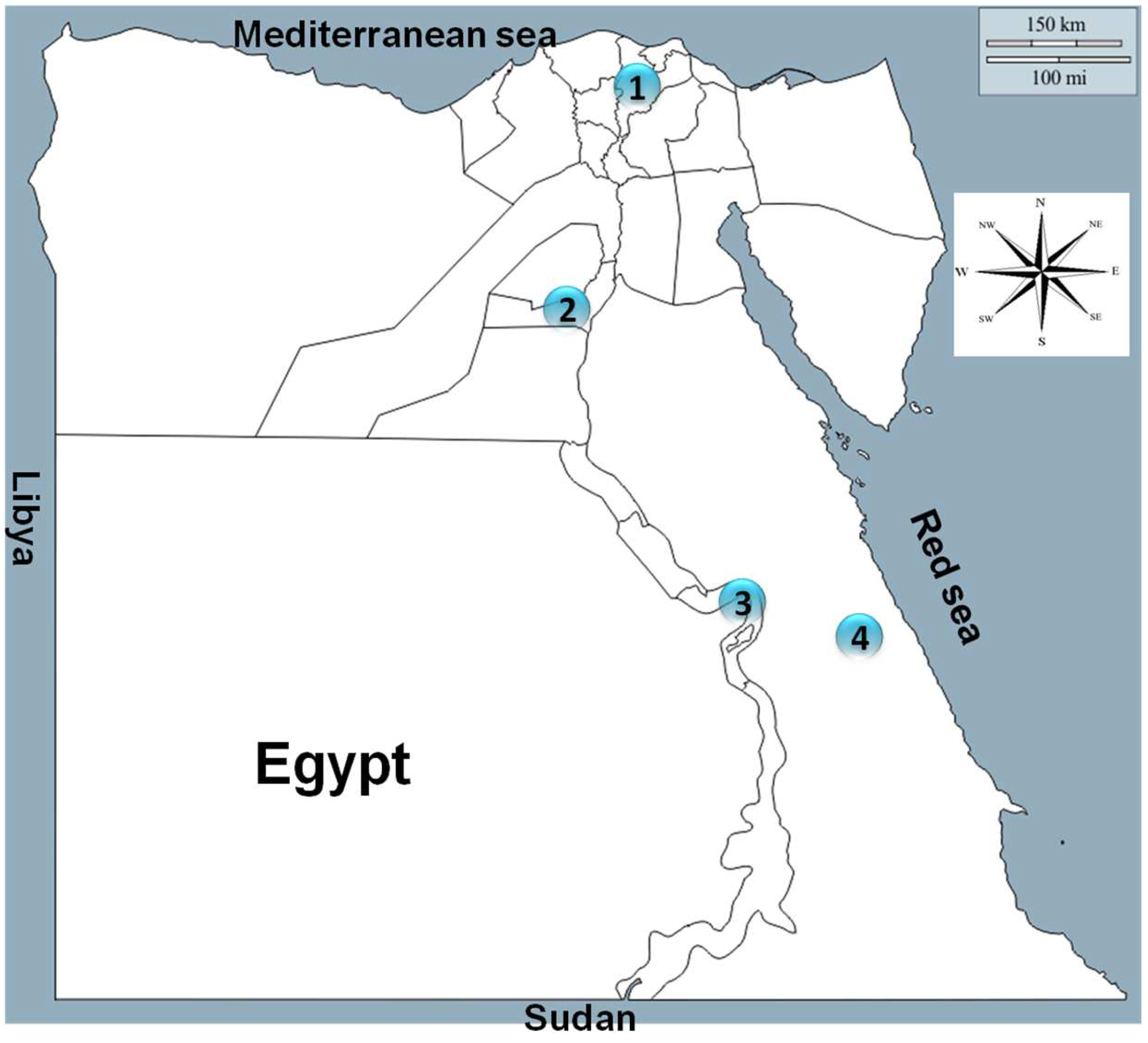
2.2. Description of the Animals and Regions of the Study

The present study was conducted in four governorates representing the northern part (Dakahlia), middle part (Beni Suef), southern part (Qena), and eastern part (Red Sea) of Egypt (Figure 1).
The study animals were indigenous sheep and goats kept in farms (more than fifty animals) or small holdings (<50 animals/farm). Samples were collected at various time points from May 2016 to November 2021 (Table 1). All sheep and goats included in this study were older than 6 months and mostly females. None of the sampled animals was vaccinated against Brucella infection, as confirmed by animal owners. A total of 360 serum samples were collected randomly from sheep (n = 239) and goats (n = 121) with or without a history of abortion. Sera collected from animals with a history of abortion were confined mostly to medium to large farms (50 to 200 heads/farm) and collected during the first few months after abortions occurred. The selection of samples for the study was designed to cover a large time span (5 years) and a wide geographical range (approximately 1000 kilometers apart) in order to give a reliable estimation of the epidemiological situation of the three pathogens. Furthermore, flocks with and without a history of abortion were included, while vaccination for Brucella was an exclusion criterium. Within the selected flocks, samples were collected randomly from apparently healthy animals.

2.3. Sample Collection and Serum Preparation

Approximately 5 mL of whole blood samples were collected by venipuncture from the jugular vein using sterile disposable plain vacutainer tubes and needles. The blood samples were kept in an upward position and allowed to clot at room temperature for 2–3 h. The samples were transported on ice in an icebox to our laboratory at the Faculty of Veterinary Medicine, South Valley University, Egypt. Sera were transferred into 1.5 mL Eppendorf tubes after centrifugation at 10,000× g for 15 min [30] and stored at −20 °C until use.

2.4. Serological Diagnosis of T. gondii, N. caninum, and Brucella Species

In order to detect antibodies to T. gondii, the serum samples were analyzed with the indirect multi-species ELISA for toxoplasmosis (ID.vet, Grabels, France) according to the manufacturer’s instructions. Serum samples and controls were diluted 1:10. The optical density (OD) obtained was used to calculate the percentage of sample (S) to positive (P) ratio (S/P%) for each of the test samples according to the following formula: S/P (%) = (OD sample − OD negative control)/(OD positive control − OD negative control) × 100. Samples with an S/P% less than 40% were considered negative; if the S/P% was between 40% and 50%, the result was considered doubtful and considered positive if the S/P% was greater than 50%.
For antibodies to N. caninum, serum samples were analyzed with the competitive multi-species ELISA for neosporosis (ID.vet, Grabels, France). Serum samples and controls were diluted 1:2. The ODs obtained were used to calculate the percentage of sample (S) to negative (N) ratio (S/N%) for each of the test samples according to the following formula: S/N (%) = OD sample/OD negative control × 100. Samples with an S/N% greater than 60% were considered negative; if the S/N% was between 50% and 60%, the result was considered doubtful, and it was considered positive if the S/N% was less than 50%.
Concerning Brucella spp., serum samples were analyzed with the indirect multi-species ELISA for brucellosis (ID.vet, Grabels, France). Serum samples and controls were diluted 1:20. The ODs obtained were used to calculate the percentage of sample (S) to positive (P) ratio (S/P %) for each of the test samples according to the following formula: S/P (%) = (OD sample − OD negative control)/(OD positive control − OD negative control) × 100. Samples with an S/P% less than 110% were considered negative; if the S/P% was between 110% and 120%, the result was considered doubtful, and it was considered positive if the S/P% was greater than or equal to 120%.
The ODs of all ELISA results were read at 450 nm and measured with an Infinite® F50/Robotic ELISA reader (Tecan Group Ltd., Männedorf, Switzerland). More details for the commercial kits used are available in Table 2.

2.5. Statistical Analysis

The significance of the differences in the prevalence rates and risk factors was analyzed with Chi-square (Pearson) test, 95% confidence intervals (including continuity correction), and odds ratios using an online statistical website www.vassarstats.net (accessed on 9 August 2022). p-values and odds ratio were also confirmed with GraphPad Prism version 5 (GraphPad Software Inc., La Jolla, CA, USA). The results were considered significant when the p-value was <0.05 or highly significant when the p-value was <0.0001.

3. Results

The highest overall seroprevalence was found for T. gondii (46.1%; 166/360), subdivided as 35.6% (85/239) in sheep and 66.9% (81/121) in goats (Table 3). For N. caninum, a seroprevalence of 11.9%, 15.5%, and 5% in all animals, sheep, and goats, respectively, was found. Antibodies to Brucella spp. were detected in 8.6%, 10.5%, and 5% of all animals, sheep, and goats, respectively. Regarding mixed infection, seropositive rates in the three categories (all, sheep, and goats) were reported for T. gondii and N. caninum (4.2%, 4.2%, and 4.1%), and T. gondii and Brucella spp. (4.4%, 4.6%, and 4.1%), respectively. Mixed N. caninum and Brucella spp. infections were only recorded in 2.1% (5/239) of the sheep, leading to an overall rate of 1.4% (5/360). In addition, two sheep from northern Egypt (Dakhalia) were seropositive for all three infectious agents (T. gondii, N. caninum, and Brucella spp.), resulting in a seroprevalence of 0.8% (2/239) in sheep and 0.6% (2/360) for all tested animals (Table 3).
Possible associations between antibodies to T. gondii, N. caninum and Brucella spp. and animal species (sheep and goat), location (Dakahlia, Beni Suef, Qena, and Red Sea governorates), and abortion history (yes or no) were investigated. For T. gondii, goats were more likely to be seropositive, with a prevalence of 66.9% in goats compared to 35.6% in sheep (odds ratio (OR) 3.7, p = < 0.0001). Moreover, the location was regarded as a predisposing factor for T. gondii infection with a higher prevalence in Qena (66.1%; OR = 15.9; p ≤ 0.0001), Beni Suef (43.9%; OR = 6.4; p = 0.0004), and Dakahlia (32.4%; OR = 3.9; p = 0.0051) compared to the seroprevalence in Red Sea (10.9%) set as a reference. History of abortion was not associated with antibodies against T. gondii (p = 0.2435) (Table 4).
Regarding risk factor analysis for N. caninum, the seroprevalence in sheep (13.1%) was significantly higher than that in goats (5%) (OR = 0.3; p = 0.0036). Regarding the location, the prevalence in Dakahlia (16.2%; OR = 2.7; p = 0.0130) and Red Sea (23.9%; OR = 4.3; p = 0.0009), but not Beni Suef (7.3%; OR = 1.4; p = 0.6391), were significantly higher than that of Qena (6.8%). The history of abortion was not associated with antibodies to N. caninum (p = 0.8624) (Table 5).
For Brucella risk factor assessment, the seroprevalence in sheep (10.5%) was higher than that in goats (5%), although this was not statistically significant (OR = 0.5; p = 0.0787). Samples collected from Dakahlia had a significantly higher seroprevalence (18.9%; OR = 4.5; p = 0.0002) than those from Beni Suef (4.9%; OR = 1; p = 0.9873), Red Sea (0%; OR = 0.2; p = 0.1243), or from Qena (4.9%). Other than for T. gondii and N. caninum, the history of recent abortion was correlated with antibodies against Brucella spp. Animals with a history of recent abortion had a significantly higher seroprevalence (12.6%; OR = 6.5; p = 0.0005) than sheep and goats without a history of abortion (2.2%) (Table 6).

4. Discussion

Toxoplasma gondii, N. caninum, and Brucella spp. are professional abortifacient infectious agents in several animal species and different countries. Studies on the seroprevalence of all three pathogens and possible association with abortion history in sheep and goats have not yet been conducted in Egypt. Brucellosis is quite a common occupational disease among veterinarians and animal handlers in Egypt, and it is usually regarded as the first cause of infectious abortion in ruminants, especially in late gestation, and complicated by retention of the placenta [23]. These assumptions are supported by studies and official investigations, which diagnosed brucellosis as a cause of abortion in many abortion outbreaks [26,31,32]. However, our study of comparative seroprevalence of T. gondii, N. caninum and Brucella species in small ruminants had two main drivers: First, most of the reported brucellosis investigations were performed using screening tests, such as the Rose Bengal test and serum agglutination test, not confirmatory tests such as complement fixation test or ELISA. Second, numerous previous studies have recorded a high prevalence of T. gondii and N. caninum antibodies in many farm animals in Egypt, including in small ruminants, and therefore their contribution to abortions might currently be underestimated [27,28,30].
Our study indeed found the highest seroprevalence against T. gondii among all three tested pathogens, thus confirming previous studies from Egypt that found seroprevalences between 28.7% and 98.4% in small ruminants [33,34,35]. In our study, goats were significantly more affected (66.9%) than sheep (35.6%), and a region with a lower seroprevalence was also identified (Red Sea, eastern Egypt, compared to northern, middle, and southern Egypt). These findings confirmed earlier results from Egypt in that region and animal species have an effect on T. gondii seroprevalence [35], and also other studies found similar effects, namely Magalhães et al. [36] and Rêgo et al. [37], in Brazil; Dong et al. in China [38]; and Suazo-Cortez et al. in Mexico [39]. As for N. caninum, the seroprevalence in our study was 11.9% overall, 15.5% in sheep, and 5% in goats, respectively. Our results compared nicely with previous reports from sheep in Egypt: 8.6% reported by Selim et al. [30], and 36.1% reported by El-Ghaysh et al. [40]. Moreover, for this parasite, animal species and location had significant effects on the seroprevalence, with sheep being more affected than goats and northern and eastern Egypt being more affected than middle and southern Egypt. Other authors have also reported significant effects of location and animal species on N. caninum seroprevalence in small ruminants [30,41,42,43].
Of all three pathogens tested, seroprevalence against Brucella spp. was the lowest, with 8.6% overall, 10.5% in sheep, and 5% in goats, respectively. Other authors found prevalences varying between 7.2% and 56.7% for sheep and goats in Egypt [29,32,44,45]. In our study, there was a trend of higher seropositivity in sheep compared to goats, but it was not significant. Northern Egypt, however, did have a significantly higher seroprevalence than the other regions tested. Similar factors were found before by other authors from Egypt [46,47,48] and elsewhere [49,50,51].
Arguably, the most interesting association revealed in this study was the seropositivity against Brucella spp. and a history of abortion. Interestingly, no such association was found between antibodies against T. gondii or N. caninum and recent abortions. A study conducted on pregnant ewes in Iraq found that ewes seroconverting to Brucella were 2.9 times more likely to lose their pregnancy than those remaining seronegative. In the same study, seroconverting to T. gondii had no significant impact on the loss of pregnancy [52]. Similarly, in another region of the world, only about 1.6% of lost lambs were attributed to T. gondii in a prospective cohort study in primiparous ewes from southern Australia, albeit at an overall much lower seroprevalence than in our study [53]. Moreover, in a study performed with breeding ewes from Mexico, seroprevalence for T. gondii (61.96%) and N. caninum (15.22%) was similar to our study, and T. gondii seroprevalence was not associated with a history of abortion, but N. caninum seroprevalence was [54]. However, although our results suggest an involvement of Brucella in the abortions, they do not negate the role of T. gondii or N. caninum as aborting agents. Numerous previous reports have documented toxoplasmosis [1,19,55] and neosporosis [56,57,58] as causes of abortion in sheep and goats.
The high prevalence of anti-Brucella antibodies in animals with an abortion history was correlated to the highest seropositivity in Dakahlia governorate in the Nile Delta region, northern Egypt. This area is endemic for different Brucella species, including B. abortus, the causative agent of bovine brucellosis, and B. melitensis, the main cause of ovine and caprine brucellosis [26,29]. In addition, the case history of abortion in that region demonstrated that some aborting ewes and does have experienced late abortion and retained placenta, both common signs of Brucella abortion [12,23]. In contrast, Neospora abortion can occur at any time with a special preference for mid-gestation, and usually, no clinical signs or lesions are evident in aborting dams or in the placenta or fetus [16]. In the case of toxoplasmosis, early abortion and fetal resorption are mostly noticed, and similarly to neosporosis, no abnormalities in the dams, fetus, or placenta are usually observed [21].
However, to obtain a better understanding of the causes of abortions of small ruminants in Egypt, fetuses, placentas, and serum samples of the mothers should be submitted for laboratory analyses. Moreover, our data provide insights into the current epidemiological situation for Brucella spp. and T. gondii, both important zoonotic species, and N. caninum, all three of which cause losses to the livestock industry. More efficient control programs will have to be established in the future based on a good understanding of the real situation, which can only be gained by investigations such as this one.
Due to the limitations in the number, content, and distribution of studies focusing on the relevant theme worldwide, including Egypt, we could not perform a comprehensive comparison with our study. Additionally, higher numbers of samples are required from sheep, goats, and other species and geographic locations to establish an accurate assessment of the main causes of abortions among our tested pathogens.

5. Conclusions

Abortion in farm animals, including sheep and goats, is a major and direct cause of enormous economic losses in animal production sectors. Toxoplasmosis, neosporosis, and brucellosis are well-known causes of infectious abortions in sheep and goats in Egypt and other countries. Misevaluation of the current situation of these infections as causes of abortion would jeopardize the livestock industry in Egypt. This study is the first to establish a comparison between toxoplasmosis, neosporosis, and brucellosis based on seroprevalence in sheep and goats in Egypt. Seroprevalence for T. gondii was highest, followed by N. caninum and Brucella spp. Animal species and location were considered as risk factors for T. gondii and N. caninum infections, while only the location was a significant factor for Brucella spp. infection. Importantly, antibodies to Brucella spp. were significantly higher in aborting than in non-aborting animals, a feature not observed for the other two pathogens.

Author Contributions

Conceptualization and design, R.M.F. and C.F.F.; experiments, formal analysis, investigation, R.M.F., H.H.A., H.Y.A.H.M., G.W. and C.F.F.; resources and shared materials, R.M.F., G.W., H.H.A., A.M.M., M.E.-D., A.A., H.Y.A.H.M., A.O.A., A.E.-t., A.F.A. and C.F.F.; writing—original draft, R.M.F., G.W. and H.H.A.; writing—review and editing, C.F.F. Project administration and funding acquisition; R.M.F., C.F.F. All authors have read and agreed to the published version of the manuscript.

Funding

This study did not receive any special funds.

Institutional Review Board Statement

The protocols were approved by the Research Code of Ethics at South Valley University number 36 (RCOE-36).

Informed Consent Statement

Not applicable.

Data Availability Statement

All data generated and analyzed during this study are included in this published article. Raw data supporting the findings of this study are available from the corresponding author on request.

Acknowledgments

We thank all veterinarians who helped in collecting blood samples from tested animals and the animal owners for their cooperation in providing animals and the required data and information of each animal. We would like to appreciate the great help of our colleagues at the Department of Animal Medicine, Faculty of Veterinary Medicine, South Valley University, Qena, for their cooperation and technical assistance. We also provide special thanks to Veterinarian Fatma Al-Zahraa Khaled Ahmed for her technical assistance in ELISA experiments.

Conflicts of Interest

The authors declare no conflict of interest.

References

  1. Dubey, J.P. Toxoplasmosis of Animals and Humans, 2nd ed.; CRC Press: Boca Raton, FL, USA, 2010; pp. 1–313. [Google Scholar]
  2. Lindsay, D.S.; Dubey, J.P. Neosporosis, Toxoplasmosis, and Sarcocystosis in Ruminants: An Update. Vet. Clin. N. Am. Food Anim. Pract. 2020, 36, 205–222. [Google Scholar] [CrossRef]
  3. Tranas, J.; Heinzen, R.A.; Weiss, L.M.; McAllister, M.M. Serological evidence of human infection with the protozoan Neospora caninum. Clin. Diagn. Lab. Immunol. 1999, 6, 765–767. [Google Scholar] [CrossRef] [PubMed] [Green Version]
  4. Duarte, P.O.; Oshiro, L.M.; Zimmermann, N.P.; Csordas, B.G.; Dourado, D.M.; Barros, J.C.; Andreott, R. Serological and molecular detection of Neospora caninum and Toxoplasma gondii in human umbilical cord blood and placental tissue samples. Sci. Rep. 2020, 10, 9043. [Google Scholar] [CrossRef]
  5. Rossetti, C.A.; Maurizio, E.; Rossi, U.A. Comparative review of brucellosis in small domestic ruminants. Front. Vet. Sci. 2022, 9, 887671. [Google Scholar] [CrossRef]
  6. Godfroid, J.; Bosman, P.P.; Herr, S.; Bishop, G.C. Bovine Brucellosis. In Infectious Diseases of Livestock; Coetzer, J.A.W., Thompson, G., Tustin, R.C., Eds.; Oxford University Press: Cape Town, South Africa, 2004; pp. 1510–1527. [Google Scholar]
  7. Arellano-Reynoso, B.; Lapaque, N.; Salcedo, S.; Briones, G.; Ciocchini, A.E.; Ugalde, R.; Moreno, E.; Moriyón, I.; Gorvel, J.P. Cyclic beta-1,2-glucan is a Brucella virulence factor required for intracellular survival. Nat. Immunol. 2005, 6, 618–625. [Google Scholar] [CrossRef] [PubMed]
  8. Celli, J. Surviving inside a macrophage: The many ways of Brucella. Res. Microbiol. 2006, 157, 93–98. [Google Scholar] [CrossRef] [PubMed]
  9. Zhao, Y.; Marple, A.H.; Ferguson, D.J.; Bzik, D.J.; Yap, G.S. Avirulent strains of Toxoplasma gondii infect macrophages by active invasion from the phagosome. Proc. Natl. Acad. Sci. USA 2014, 111, 6437–6442. [Google Scholar] [CrossRef] [Green Version]
  10. García-Sánchez, M.; Jiménez-Pelayo, L.; Horcajo, P.; Regidor-Cerrillo, J.; Ólafsson, E.B.; Bhandage, A.K.; Barragan, A.; Werling, D.; Ortega-Mora, L.M.; Collantes-Fernández, E. Differential responses of bovine monocyte-derived macrophages to infection by Neospora caninum isolates of high and low virulence. Front. Immunol. 2019, 10, 915. [Google Scholar] [CrossRef]
  11. Fereig, R.M.; Nishikawa, Y. From signaling pathways to distinct immune responses: Key factors for establishing or combating Neospora caninum infection in different susceptible hosts. Pathogens 2020, 9, 384. [Google Scholar] [CrossRef]
  12. Samartino, L.E.; Enright, F.M. Pathogenesis of abortion of bovine brucellosis. Comp. Immunol. Microbiol. Infect. Dis. 1993, 16, 95–101. [Google Scholar] [CrossRef] [PubMed]
  13. Buxton, D. Protozoan infections (Toxoplasma gondii, Neospora caninum and Sarcocystis spp.) in sheep and goats: Recent advances. Vet. Res. 1998, 29, 289–310. [Google Scholar] [PubMed]
  14. Buxton, D.; McAllister, M.M.; Dubey, J.P. The comparative pathogenesis of neosporosis. Trends Parasitol. 2002, 18, 546–552. [Google Scholar] [CrossRef] [PubMed]
  15. Dubey, J.P.; Schares, G. Neosporosis in animals—The last five years. Vet. Parasitol. 2011, 180, 90–108. [Google Scholar] [CrossRef]
  16. Porto, W.J.; Regidor-Cerrillo, J.; Kim Pde, C.; Benavides, J.; Silva, A.C.; Horcajo, P.; Oliveira, A.A.; Ferre, I.; Mota, R.A.; Ortega-Mora, L.M. Experimental caprine neosporosis: The influence of gestational stage on the outcome of infection. Vet. Res. 2016, 47, 29. [Google Scholar] [CrossRef] [PubMed] [Green Version]
  17. González-Warleta, M.; Castro-Hermida, J.A.; Calvo, C.; Pérez, V.; Gutiérrez-Expósito, D.; Regidor-Cerrillo, J.; Ortega-Mora, L.M.; Mezo, M. Endogenous transplacental transmission of Neospora caninum during successive pregnancies across three generations of naturally infected sheep. Vet. Res. 2018, 49, 106. [Google Scholar] [CrossRef] [PubMed] [Green Version]
  18. Wang, X.; Lin, P.; Li, Y.; Xiang, C.; Yin, Y.; Chen, Z.; Du, Y.; Zhou, D.; Jin, Y.; Wang, A. Brucella suis vaccine strain 2 induces endoplasmic reticulum stress that affects intracellular replication in goat trophoblast cells In Vitro. Front. Cell. Infect. Microbiol. 2016, 6, 19. [Google Scholar] [CrossRef] [Green Version]
  19. Gutiérrez-Expósito, D.; Tejerina, F.; Gutiérrez, J.; Fernández-Escobar, M.; Ortega-Mora, L.M.; Mantecón, A.R.; Dagleish, M.P.; Pérez, V.; Benavides, J. Direct economic losses of Toxoplasma gondii abortion outbreaks in two Spanish sheep flocks. Vet. Parasitol. Reg. Stud. Rep. 2021, 26, 100623. [Google Scholar] [CrossRef] [PubMed]
  20. Singh, B.B.; Dhand, N.K.; Gill, J.P. Economic losses occurring due to brucellosis in Indian livestock populations. Prev. Vet. Med. 2015, 119, 211–215. [Google Scholar] [CrossRef] [PubMed] [Green Version]
  21. Dubey, J.P. Toxoplasmosis in sheep—The last 20 years. Vet. Parasitol. 2009, 163, 1–14. [Google Scholar] [CrossRef] [PubMed]
  22. Refai, M. Incidence and control of brucellosis in the Near East region. Vet. Microbiol. 2002, 90, 81–110. [Google Scholar] [CrossRef] [PubMed]
  23. Acha, N.P.; Szyfres, B. Zoonoses and Communicable Diseases Common to Man and Animals, 3rd ed.; Pan American Organization (PAHO): Washington, DC, USA, 2003; Volume 1. [Google Scholar]
  24. Khan, M.Y.; Mah, M.W.; Memish, Z.A. Brucellosis in pregnant women. Clin. Infect. Dis. 2001, 32, 1172–1177. [Google Scholar] [CrossRef] [PubMed]
  25. Young, E.J. Human brucellosis. Rev. Infect. Dis. 1983, 5, 821–842. [Google Scholar] [CrossRef] [PubMed]
  26. Wareth, G.; Hikal, A.; Refai, M.; Melzer, F.; Roesler, U.; Neubauer, H. Animal brucellosis in Egypt. Infect. Dev. Ctries 2014, 8, 1365–1373. [Google Scholar] [CrossRef] [PubMed]
  27. Rouatbi, M.; Amairia, S.; Amdouni, Y.; Boussaadoun, M.A.; Ayadi, O.; Al-Hosary, A.A.T.; Rekik, M.; Ben Abdallah, R.; Aoun, K.; Darghouth, M.A.; et al. Toxoplasma gondii infection and toxoplasmosis in North Africa: A review. Parasite 2019, 26, 6. [Google Scholar] [CrossRef] [PubMed] [Green Version]
  28. Abbas, I.E.; Villena, I.; Dubey, J.P. A review on toxoplasmosis in humans and animals from Egypt. Parasitology 2020, 147, 135–159. [Google Scholar] [CrossRef] [PubMed]
  29. El-Diasty, M.; El-Said, R.; Abdelkhalek, A. Seroprevalence and molecular diagnosis of sheep brucellosis in Dakahlia governorate, Egypt. Ger. J. Vet. Res. 2021, 1, 34–39. [Google Scholar] [CrossRef]
  30. Selim, A.; Khater, H.; Almohammed, H.I. A recent update about seroprevalence of ovine neosporosis in Northern Egypt and its associated risk factors. Sci. Rep. 2021, 11, 14043. [Google Scholar] [CrossRef]
  31. Wareth, G.; Melzer, F.; Tomaso, H.; Roesler, U.; Neubauer, H. Detection of Brucella abortus DNA in aborted goats and sheep in Egypt by real-time PCR. BMC Res. Notes 2015, 8, 212. [Google Scholar] [CrossRef] [Green Version]
  32. Abdelbaset, A.E.; Abushahba, M.F.N.; Hamed, M.I.; Rawy, M.S. Sero-diagnosis of brucellosis in sheep and humans in Assiut and El-Minya governorates, Egypt. Int. J. Vet. Sci. Med. 2018, 6, S63–S67. [Google Scholar] [CrossRef] [PubMed] [Green Version]
  33. Ghoneim, N.H.; Shalaby, S.I.; Hassanain, N.A.; Zeedan, G.S.; Soliman, Y.A.; Abdalhamed, A.M. Comparative study between serological and molecular methods for diagnosis of toxoplasmosis in women and small ruminants in Egypt. Foodborne Pathog. Dis. 2010, 7, 17–22. [Google Scholar] [CrossRef] [PubMed]
  34. Younis, E.E.; Abou-Zeid, N.Z.; Zakaria, M.; Mahmoud, M.R. Epidemiological studies on toxoplasmosis in small ruminants and equines in Dakahlia governorate, Egypt. Assiut Vet. Med. J. 2015, 61, 22–31. [Google Scholar]
  35. Fereig, R.M.; Mahmoud, H.Y.A.H.; Mohamed, S.G.A.; AbouLaila, M.R.; Abdel-Wahab, A.; Osman, S.A.; Zidan, S.A.; El-Khodary, S.A.; Mohamed, A.E.A.; Nishikawa, Y. Seroprevalence and epidemiology of Toxoplasma gondii in farm animals in different regions of Egypt. Vet. Parasitol. Reg. Stud. Reports. 2016, 3–4, 1–6. [Google Scholar] [CrossRef] [PubMed]
  36. Magalhães, F.J.; Ribeiro-Andrade, M.; Alcântara, A.M.; Pinheiro, J.W.; Júnior Sena, M.J.; Porto, W.J.; Vieira, R.F.; Mota, R.A. Risk factors for Toxoplasma gondii infection in sheep and cattle from Fernando de Noronha Island, Brazil. Rev. Bras. Parasitol. Vet. 2016, 25, 511–515. [Google Scholar] [CrossRef] [PubMed]
  37. Rêgo, W.M.F.; Paula, N.R.O.; Vitor, R.W.A.; Silva, R.A.B.; Diniz, B.L.M.; Sousa, M.M.; Coelho, W.A.C.; Profirio, K.P.P.; Pinheiro, R.R.P.; Alves, F.S.F.; et al. Risk factors for Toxoplasma gondii infection in goats and sheep raised in the State of Piauí in northeast Brazil. Small Rumin. Res. 2016, 141, 17–23. [Google Scholar] [CrossRef]
  38. Dong, H.; Su, R.; Lu, Y.; Wang, M.; Liu, J.; Jian, F.; Yang, Y. Prevalence, risk factors, and genotypes of Toxoplasma gondii in food animals and humans (2000–2017) from China. Front. Microbiol. 2018, 9, 2108. [Google Scholar] [CrossRef] [PubMed]
  39. Suazo-Cortez, R.; Martínez-Herrera, D.I.; Pardío-Sedas, V.T.; Cruz-Vázquez, C.R.; Morales-Álvarez, J.F.; Sánchez-Viveros, G.; Galindo-Tovar, M.E. Seroprevalence and risk factors associated with Toxoplasma gondii infection in sheep of Veracruz State, southeast Mexico. Vet. Res. Forum. 2020, 11, 77–81. [Google Scholar] [CrossRef] [PubMed]
  40. El-Ghayash, K.F.; Hilali, M.; Nassar, A.M. Serological diagnosis of Neospora caninum infection in some domestic animals from Egypt. Vet. Med. J. Giza 2003, 51, 355–361. [Google Scholar]
  41. Gazzonis, A.L.; Garcia, G.A.; Zanzani, S.A.; Mora, L.M.O.; Invernizzi, A.; Manfred, M.T. Neospora caninum infection in sheep and goats from north-eastern Italy and associated risk factors. Small Rumin. Res. 2016, 140, 7–12. [Google Scholar] [CrossRef]
  42. Gharekhani, J.; Yakhchali, M.; Esmaeilnejad, B.; Mardani, K.; Majidi, G.; Sohrabi, A.; Berahmat, R.; Hazhir Alaei, M. Seroprevalence and risk factors of Neospora caninum and Toxoplasma gondii in small ruminants in Southwest of Iran. Arch. Razi Inst. 2018, 73, 305–310. [Google Scholar] [CrossRef]
  43. Udonsom, R.; Supanta, J.; Tanglakmankhong, O.; Ngoenphisutsin, K.; Nishikawa, Y.; Fereig, R.M.; Jirapattharasate, C. Toxoplasma gondii and Neospora caninum prevalence and risk factors on goat farms in Kanchanaburi province, Thailand. Vet. Integr. Sci. 2021, 19, 65–74. [Google Scholar] [CrossRef]
  44. Hegazy, Y.M.; Moawad, A.; Osman, S.; Ridler, A.; Guitian, J. Ruminant brucellosis in the Kafr El Sheikh Governorate of the Nile Delta, Egypt: Prevalence of a neglected zoonosis. PLoS. Negl. Trop. Dis. 2011, 5, e944. [Google Scholar] [CrossRef] [PubMed]
  45. Nagati, S.F.; Hassen, S.K. Diagnosis of Brucella infection in sheep and goat and evaluation of the associated practices in animal contacts. Am. J. Infec. Dis. Microbiol. 2016, 4, 95–101. [Google Scholar] [CrossRef]
  46. Mahboub, H.D.; Helal, M.A.; Abd Eldaim, M.A.; Abd El-Razek, E.M.; Elsify, A.M. Seroprevalence of abortion causing agents in Egyptian sheep and goat breeds and their effects on the animal’s performance. J. Agric. Sci. 2013, 5, 9. [Google Scholar] [CrossRef]
  47. Selim, A.; Gaber, A.; Moustafa, A. Diagnosis of brucellosis in ruminants in Kafr El-Sheikh governorate, Egypt. Int. J. Adv. Res. 2015, 3, 2320–5407. [Google Scholar]
  48. Nayel, M.; Ibrahim, R.; Zaghawa, A. Seroprevalence and associated risk factors of brucellosis among sheep, goats and camels in Northwestern Coastal area of Egypt. J. Curr. Vet. Res. 2020, 2, 25–34. [Google Scholar] [CrossRef]
  49. Ogugua, A.J.; Akinseye, V.O.; Ayoola, M.C.; Oyesola, O.O.; Shima, F.K.; Tijjani, A.O.; Musa, A.N.; Adesokan, H.K.; Perrett, L.; Taylor, A.; et al. Seroprevalence and risk factors of brucellosis in goats in selected states in Nigeria and the public health implications. Afr. J. Med. Med. Sci. 2014, 43 (Suppl. 1), 121–129. [Google Scholar]
  50. Alhamada, A.G.; Habib, I.; Barnes, A.; Robertson, I. Risk factors associated with Brucella seropositivity in sheep and goats in Duhok province, Iraq. Vet. Sci. 2017, 4, 65. [Google Scholar] [CrossRef] [PubMed] [Green Version]
  51. Gompo, T.R.; Shah, R.; Tiwari, I.; Gurung, Y.B. Sero-epidemiology and associated risk factors of brucellosis among sheep and goat population in the southwestern Nepal: A comparative study. BMC Vet. Res. 2021, 17, 132. [Google Scholar] [CrossRef]
  52. Al Hamada, A.; Habib, I.; Bruce, M.; Barnes, A.; Robertson, I.D. Seroconversion to Brucella spp. and Toxoplasma gondii in sheep and goats in Dohuk province, Iraq and its association with pregnancy loss. Animals 2021, 11, 836. [Google Scholar] [CrossRef]
  53. Clune, T.; Lockwood, A.; Hancock, S.; Thompson, A.N.; Bruce, M.; Beetson, S.; Campbell, A.J.; Glanville, E.; Brookes, D.; Trengove, C.; et al. Toxoplasma gondii is not an important contributor to poor reproductive performance of primiparous ewes from southern Australia: A prospective cohort study. BMC Vet. Res. 2022, 18, 109. [Google Scholar] [CrossRef] [PubMed]
  54. Alcalá-Gómez, J.; Medina-Esparza, L.; Vitela-Mendoza, I.; Cruz-Vázquez, C.; Quezada-Tristán, T.; Gómez-Leyva, J.F. Prevalence and risk factors of Neospora caninum and Toxoplasma gondii infection in breeding ewes from central western Mexico. Trop. Anim. Health Prod. 2022, 54, 225. [Google Scholar] [CrossRef]
  55. Henker, L.C.; Silveira Flores Vogel, F.; Santana de Cecco, B.; Ribeiro Dos Santos, I.; Roman, I.J.; D’ambroso Fernandes, F.; Genro Cony, F.; Petinatti Pavarini, S.; Driemeier, D. Abortion outbreak in a sheep flock caused by Toxoplasma gondii clonal type III. Parasitol. Res. 2022, 121, 2633–2639. [Google Scholar] [CrossRef]
  56. Dubey, J.P.; Lindsay, D.S. Neospora caninum induced abortion in sheep. J. Vet. Diagn. Invest. 1990, 2, 230–233. [Google Scholar] [CrossRef] [PubMed] [Green Version]
  57. Pinto, A.P.; Bacha, F.B.; Santos, B.S.; Driemeier, D.; Antoniassi, N.A.B.; de Sá Ribas, N.L.K.; Lemos, R.A.A. Sheep abortion associated with Neospora caninum in Mato Grosso do Sul, Brazil. Pesq. Vet. Bras. 2012, 32, 8. [Google Scholar] [CrossRef]
  58. Varaschin, M.S.; Hirsch, C.; Wouters, F.; Nakagaki, K.Y.; Guimarães, A.M.; Santos, D.S.; Bezerra, P.S., Jr.; Costa, R.C.; Peconick, A.P.; Langohr, I.M. Congenital neosporosis in goats from the State of Minas Gerais, Brazil. Korean J. Parasitol. 2012, 50, 63–67. [Google Scholar] [CrossRef] [PubMed]
Figure 1. Geographic locations of the tested regions: 1—Dakahlia governorate (northern Egypt); 2—Beni Suef governorate (middle Egypt); 3—Qena governorate (southern Egypt); Red Sea governorate (eastern Egypt).
Animals 12 03327 g001
Table 1. Sample information and history of abortion.
Sample Group Time of CollectionSpeciesNumberPlace of CollectionType of Breeding SystemHistory of Abortion
Group 12016Sheep111DakahliaFarmYes
Group 22016Sheep
Goat
12
29
Beni SuefFarmYes
Group 32021Sheep70QenaFarmYes
Group 42018Goat92QenaSmallholdersNo
Group 52016Sheep46Red SeaSmallholdersNo
Total2016–2021Sheep and goat360VariousFarm and smallholderVarious
Table 2. Commercially available ELISA test kits used for detecting antibodies to T. gondii, N. caninum, and Brucella spp.
Infectious AgentELISA Test KitManufacturerAntigenConjugateSensitivity *Specificity *
Toxoplasma gondiiID Screen® Toxoplasmosis Indirect Multi-speciesID.vet Innovative Diagnostics, Grabels, FranceP30 antigenAnti-multi-species IgG-HRP98.36%
(CI 95% #: 95.29–99.44%)
99.42%
(CI 95%: 98.8–100%)
Neospora caninumID Screen® Neospora caninum competition MultispeciesID.vet Innovative Diagnostics, Grabels, FrancePurified extract of Neospora caninumAnti-N. caninum-HRP
(detect IgG or IgM)
100%
(CI 95%:
98.8–100%)
100%
(CI 95%: 99.63–100%)
Brucella species ID Screen® Brucellosis Serum Indirect MultispeciesID.vet Innovative Diagnostics, Grabels, FranceLPS of Brucella species Anti-multi-species-IgG-HRP 100%
(CI 95%:
89.57–100%)
99.74%
(CI 95%: 99.24–99.91%)
* The sensitivity and specificity of the diagnostic kits were provided by the manufacturer of the kits. # CI 95%; 95% confidence interval.
Table 3. Seroprevalence of T. gondii, N. caninum, Brucella spp., and mixed infections in sheep and goats from Egypt.
Type of InfectionAnimal SpeciesNo. of TestedNo. of Negative (%)No. of Doubtful (%)No. of Positive (%)95% CI *
T. gondiiSheep
Goat
Both
239
121
360
142 (59.4)
37 (30.6)
179 (49.7)
12 (5)
3 (2.5)
15 (4.2)
85 (35.6)
81 (66.9)
166 (46.1)
29.6–42
57.7–75.1
40.9–51.4
N. caninumSheep
Goat
Both
239
121
360
191 (79.9)
113 (93.4)
304 (84.4)
11 (4.6)
2 (1.6)
13 (3.6)
37 (15.5)
6 (5)
43 (11.9)
11.3–20.8
2–10.9
8.9–15.9
Brucella species #Sheep
Goat
Both
239
121
360
206 (86.2)
112 (92.6)
318 (88.3)
8 (3.3)
3 (2.5)
11 (3.1)
25 (10.5)
6 (5)
31 (8.6)
7–15.2
2–10.9
6–12.1
T. gondii + N. caninumSheep
Goat
Both
239
121
360
219 (91.6)
114 (94.2)
333 (92.5)
10 (4.2)
2 (1.7)
12 (3.3)
10 (4.2)
5 (4.1)
15 (4.2)
2.1–7.8
1.5–9.8
2.4–6.9
T. gondii + Brucella speciesSheep
Goat
Both
239
121
360
227 (95)
114 (94.2)
341 (94.7)
1 (0.4)
2 (1.7)
3 (0.8)
11 (4.6)
5 (4.1)
16 (4.4)
2.4–8.3
1.5–9.8
2.6–7.3
N. caninum + Brucella speciesSheep
Goat
Both
239
121
360
230 (96.2)
120 (99.2)
350 (97.2)
4 (1.7)
1 (0.8)
5 (1.4)
5 (2.1)
0
5 (1.4)
0.8–5.1
0–3.8
0.5–3.4
T. gondii + N. caninum + Brucella speciesSheep
Goat
Both
239
121
360
234 (97.9)
120 (99.2)
354 (98.3)
3 (1.3)
1 (0.8)
4 (1.1)
2 (0.8)
0
2 (0.6)
0.2–3.3
0–3.8
0.1–2.2
* 95% CI calculated according to the method described by (http://vassarstats.net/ accessed on 9 August 2022); # the kit can detect antibodies against Brucella abortus, B. melitensis, and B. suis.
Table 4. Risk factors for T. gondii antibodies in sheep and goats in Egypt.
Analyzed FactorNo. of TestedNo. of Negative (%)No. of Positive (%)OR (95% CI) *p-Value #
Animal species
Sheep239154 (64.4)85 (35.6)Ref.Ref.
Goat12140 (33.1)81 (66.9)3.7 (2.3–5.8)<0.0001
Geographical location
Dakahlia (Northern Egypt)11175 (67.6)36 (32.4)3.9 (1.4–10.8)0.0051
Beni Suef (Middle Egypt)4123 (56.1)18 (43.9)6.4 (2.1–19.6)0.0004
Qena (Southern Egypt)16255 (33.9)107 (66.1)15.9 (6–42.7)<0.0001
Red Sea (Eastern Egypt)4641 (89.1)5 (10.9)Ref.Ref.
Abortion history
Yes222125 (56.3)97 (43.7)0.8 (0.5–1.2)0.2435
No13869 (50)69 (50)Ref.Ref.
* Odds ratio at 95% confidence interval as calculated by http://vassarstats.net/ (accessed on 9 August 2022); # p-value was evaluated by Chi-square test (Pearson test) using online statistics software http://vassarstats.net/ (accessed on 9 August 2022) and GraphPad Prism version 5; Ref.; is the value that used as a reference.
Table 5. Risk factors for N. caninum antibodies in sheep and goats in Egypt.
Analyzed FactorNo. of TestedNo. of Negative (%)No. of Positive (%)OR (95% CI) *p-Value #
Animal species
Sheep239202 (86.9)37 (13.1)Ref.Ref.
Goat121115 (95)6 (5)0.3 (0.1–0.7)0.0036
Geographical location
Dakahlia (Northern Egypt)11193 (83.8)18 (16.2)2.7 (1.2–5.9)0.0130
Beni Suef (Middle Egypt)4138 (92.7)3 (7.3)1.4 (0.4–5.2)0.6391
Qena (Southern Egypt)162151 (93.2)11 (6.8)Ref.Ref.
Red Sea (Eastern Egypt)4635 (76.1)11 (23.9)4.3 (1.7–10.8)0.0009
Abortion history
Yes222196 (88.3)26 (11.7)0.9 (0.5–1.8)0.8624
No138121 (87.7)17 (12.3)Ref.Ref.
* Odds ratio at 95% confidence interval as calculated by http://vassarstats.net/ (accessed on 9 August 2022); # p-value was evaluated by Chi-square test (Pearson test) using online statistics software http://vassarstats.net/ (accessed on 9 August 2022) and GraphPad Prism version 5; Ref.; value used as a reference.
Table 6. Risk factors for Brucella spp. antibodies in sheep and goats in Egypt.
Analyzed FactorNo. of TestedNo. of Negative (%)No. of Positive (%)OR (95% CI) *p-Value #
Animal species
Sheep239214 (89.5)25 (10.5)Ref.Ref.
Goat121115 (95)6 (5)0.5 (0.2–1.1)0.0787
Geographical location
Dakahlia (Northern Egypt)11190 (81.1)21 (18.9)4.5 (1.9–10.6)0.0002
Beni Suef (Middle Egypt)4139 (95.1)2 (4.9)1 (0.2–4.8)0.9873
Qena (Southern Egypt)162154 (95.1)8 (4.9)Ref.Ref.
Red Sea (Eastern Egypt)464600.2 (0–3.5)0.1243
Abortion history
Yes222194 (87.4)28 (12.6)6.5 (1.9–21.8)0.0005
No138135 (97.8)3 (2.2)Ref.Ref.
* Odds ratio at 95% confidence interval as calculated by http://vassarstats.net/ (accessed on 9 August 2022); # p-value was evaluated by Chi-square test (Pearson test) using online statistics software http://vassarstats.net/ (accessed on 9 August 2022) and GraphPad Prism version 5; Ref.; value used as a reference.
Publisher’s Note: MDPI stays neutral with regard to jurisdictional claims in published maps and institutional affiliations.

