Article

Development and Preliminary Evaluation of a Nanoparticle-Assisted PCR Assay for the Detection of Cryptosporidium parvum in Calves

1
College of Veterinary Medicine, Northwest A&F University, Xianyang 712100, China
2
State Key Laboratory of Veterinary Etiological Biology, Key Laboratory of Veterinary Parasitology of Gansu Province, Lanzhou Veterinary Research Institute, Chinese Academy of Agricultural Sciences, Lanzhou 730046, China
*
Author to whom correspondence should be addressed.
These authors contributed equally to this work.
Animals 2022, 12(15), 1953; https://doi.org/10.3390/ani12151953
Submission received: 29 May 2022 / Revised: 26 July 2022 / Accepted: 28 July 2022 / Published: 1 August 2022
(This article belongs to the Collection Advances in Parasite Epidemiology and Population Genetics)

Abstract

:

Simple Summary

Accurate and rapid detection of Cryptosporidium parvum is useful for the prevention and control of cryptosporidiosis in humans and animals. This study developed a nano-PCR assay for the rapid detection of C. parvum in calves for the first time, and it was ten-fold more sensitive than the normal PCR assay and had no cross-reaction with other common gastrointestinal pathogens. Further analyses of faecal samples of calves indicated potential usage of the nano-PCR assay in clinical settings.

Abstract

C. parvum is an important diarrheal pathogen in humans and animals, especially in young hosts. To accurately and rapidly detect C. parvum infection in calves, we established a nano-PCR assay targeting the cgd3_330 gene for the specific detection of C. parvum. This nano-PCR assay was ten times more sensitive than that of the normal PCR assay by applying the same primers and did not cross-react with C. andersoni, C. bovis, C. ryanae, Balantidium coli, Enterocytozoon bieneusi, Giardia lamblia, and Blastocystis sp. To further test the nano-PCR in clinical settings, a total of 20 faecal samples from calves were examined by using the nano-PCR, the normal PCR, and the nested PCR assays. The positive rates were 30% (6/20), 30% (6/20), and 25% (5/20) for the nano-PCR, the normal PCR, and the nested PCR assays, respectively, indicating that the nano-PCR and the normal PCR assays had the same positive rate (30%). Taken together, the present study could provide a candidate method for the specific detection of C. parvum infection in calves in clinical settings.

1. Introduction

Bovine cryptosporidiosis is an important disease caused by the zoonotic protozoan Cryptosporidium spp., with the clinical syndromes of diarrhea, fever, and massive fluid loss in the gastrointestinal tracts, especially in young animals [1]. Although adult cattle infected with Cryptosporidium spp. usually exhibit asymptomatic shedding of oocysts, calves often suffer severely fatal diarrhea [2]. Of over 40 valid Cryptosporidium species reported, four species are commonly detected in cattle, namely C. parvum, C. andersoni, C. ryanae, and C. bovis. Cryptosporidium parvum was found to be predominant in calves of the suckling period, C. andersoni was mostly found in adult and yearling cattle, while C. bovis and C. ryanae were usually detected in post-weaned calves [2,3,4,5,6]. Among them, C. parvum has been identified as the major pathogenic and zoonotic pathogen, endangering the health of humans and the development of the breeding industry [7]. However, there have been no effective vaccines or drugs for the prevention and control of C. parvum infection or cryptosporidiosis.
Accurate and rapid diagnosis is of great significance to the prevention and control of parasitic diseases. To date, several methods for detecting C. parvum infection have been developed, including microscopic examination, immunological assays, and nucleic acid-based detection methods. Of them, traditional microscopic examination is the most common technique to detect C. parvum oocysts, which are easily recognized in fresh stools by microscopic examination [8]. However, this method is time-consuming, lacks sensitivity and requires a well-trained examiner. Immunological methods have several advantages over microscopic examination in terms of sensitivity and specificity. In some western countries, there are several commercially available kits for detecting C. parvum, including enzyme immunoassay, immunofluorescence assay, and immune-chromatography test formats [9]. However, this approach is easily contaminated, and costly. Nucleic acid-based detection methods have been widely used for the detection of C. parvum with high sensitivity and specificity. For example, a nested polymerase chain reaction (nested PCR) targeting the gp60 gene can effectively identify C. parvum and its subtypes [10,11,12]. However, nested PCR can increase the operational complexity as well as susceptibility to contamination [13]. Thus, it is urgently needed to establish a rapid, user-friendly, and reliable assay to detect C. parvum infection.
With the development of nanotechnology, nanoparticles, including carbon nanotubes, silver nanoparticles, titanium oxide nanoparticles, zinc oxide nanoparticles, quantum dots, and gold nanoparticles (AuNPs), have been used in almost all fields of science, and nano-PCR has become a novel type of PCR technique and is more sensitive than normal PCR [14]. Previous studies have shown that nanoparticles could bind to single-strand DNA and play a similar role to the single-strand binding protein, significantly reducing mismatches between primers and templates, improving the specificity of the PCR reaction, and effectively reducing non-specific amplification [15,16]. Furthermore, using AuNPs as a PCR additive could increase detection sensitivity by 5- to 10-fold in standard PCR [17]. Recently, some nano-PCR methods have been applied for the detection of viruses, bacteria, and parasites [18,19,20,21,22,23]. Previously, one nano-PCR was established for the detection of Cryptosporidium infection in several animals. This nano-PCR was found to be sensitive and specific and had potential application in the detection of Cryptosporidium infection in clinical settings [22]. However, it could not distinguish between those pathogenic Cryptosporidium species, especially for C. parvum, an important diarrheal pathogen threatening the health of humans and animals. Therefore, we developed and assessed a sensitive and species-specific nano-PCR diagnostic method for the detection of C. parvum infection in this study.

2. Materials and Methods

2.1. Parasites, Clinical Samples, and gDNA Samples

In this study, the oocysts of C. parvum were purified, isolated, and preserved in our lab. Clinical faecal samples of calves were previously collected from a dairy cattle farm in Yangling, Shaanxi province, and stored in our laboratory at 4 °C. Genomic DNA (gDNA) samples were isolated from oocysts of C. parvum and clinical faecal samples using a commercial kit as described [24]. Those gDNA samples for C. andersoni, C. bovis, C. ryanae, Balantidium coli, Enterocytozoon bieneusi, Blastocystis sp., and Giardia lamblia were from previous studies [25,26,27,28].

2.2. PCR Primers

Through comparative genomic analysis, a gene, namely cgd3_330, would have the potential to differentiate C. parvum from the remaining three common Cryptosporidium species (C. andersoni, C. ryanae, and C. bovis) in cattle. Two specific primers (Table 1) based on the cgd3_330 gene of C. parvum for the normal PCR and the nano-PCR were designed and synthesized as reported [22].

2.3. Nested PCR

Nested PCR was used to detect C. parvum infection in faecal samples based on the reported primers as shown in Table 1 [10]. The protocol of nested PCR to amplify the gp60 gene was the same as in the previous report [10].

2.4. Optimization of the Normal PCR Assay for C. parvum

The normal PCR to obtain the cgd3_330 gene was conducted in a 12 μL reaction mixture. The initial PCR reaction system and conditions were the same as in the previous study [22]. On this basis, we optimized the annealing temperature and the concentration of MgCl2. The annealing temperatures ranged from 50 °C to 60 °C and the concentration of MgCl2 ranged from 0.42 mM to 2.92 mM. All the products were verified under a UV transilluminator after electrophoresis.

2.5. Optimization of the Nano-PCR Assay for C. parvum

Nano-PCR reaction system was carried out in a 12 μL reaction mixture containing 6 μL 2 × Nano-QPCR buffer plus AuNPs (Catalog no. NHS20-3; Shanghai Hushi Medicine Technology Co., Ltd., Shanghai, China), 10 pmol each primer, 1 U Taq enzyme mix (Shanghai Hushi Medicine Technology Co., Ltd., Shanghai, China), and 1 μL gDNA template. The initial reaction condition was also the same as in the previous study [22]. The annealing temperature (50–60 °C) and primer amounts (2–14 pmol) were then optimized. All the products were visualized under a UV transilluminator after 1% agarose gel electrophoresis.

2.6. Sensitivities of the Normal PCR and the Nano-PCR Assays

Ten-fold serial dilutions of one gDNA sample of C. parvum (102 ng/μL) were used to analyze the sensitivities of the normal PCR and the nano-PCR assays, with ddH2O used as a negative control. All the products were verified using a UV transilluminator after electrophoresis.

2.7. Specificities of the Normal PCR and the Nano-PCR Assays

The gDNA samples of C. parvum (102 ng/μL), C. andersoni (29 ng/μL), C. bovis (35 ng/μL), C. ryanae (32 ng/μL), B. coli (24 ng/μL), E. bieneusi (31 ng/μL), G. lamblia (18 ng/μL), and Blastocystis sp. (23 ng/μL) were used to verify the specificities of the established detection methods.

2.8. Assessment of the Nano-PCR in Faecal Samples

Twenty faecal samples were used to assess the accuracy of the nano-PCR assay. Meanwhile, the nested PCR and the normal PCR were performed on these samples for comparison, respectively. The representative products were sent for sequencing (Sangon Biotech (Shanghai) Co., Ltd., Shanghai, China).

3. Results

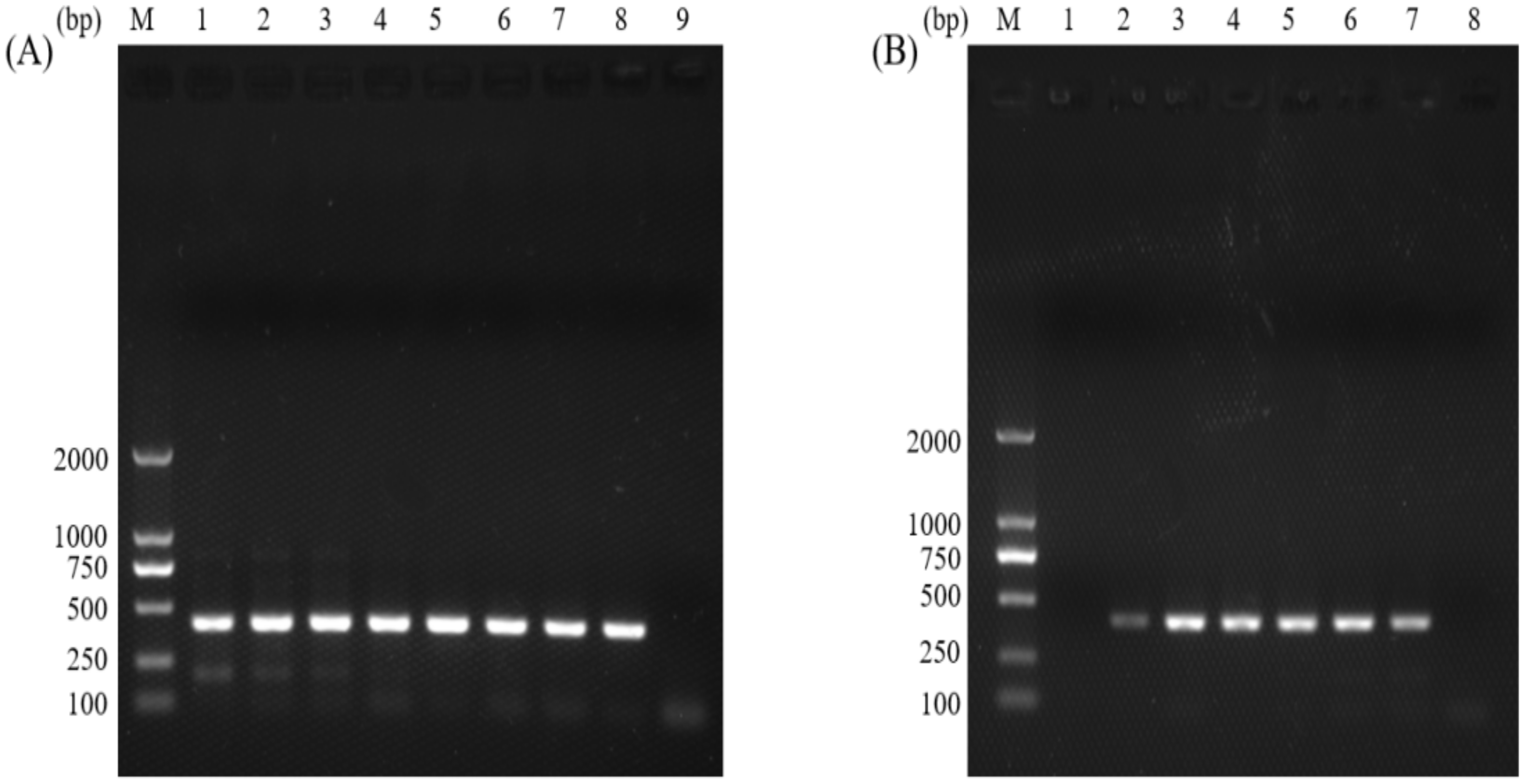
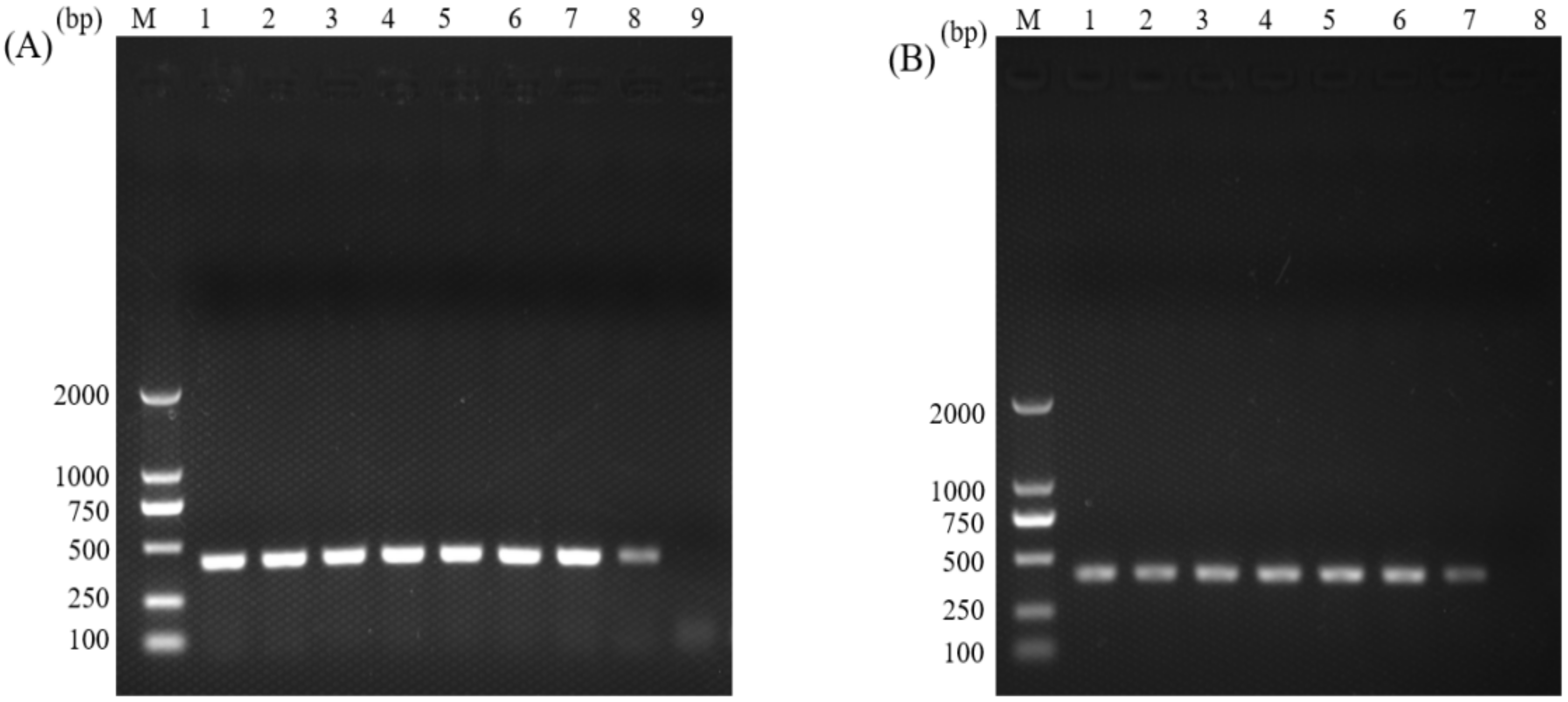
3.1. Optimization of the Normal PCR and the Nano-PCR for C. parvum

The gDNA samples from C. parvum oocysts were used as templates in the normal PCR and the nano-PCR assays, and the target fragment was about 410 bp in length. The optimal annealing temperature for normal PCR was 56 °C (Figure 1A), and the concentration of MgCl2 was 1.25 mM (Figure 1B). For nano-PCR, the optimal annealing temperature was 56 °C (Figure 2A), and the concentration of primers was 10 pmol (Figure 2B).

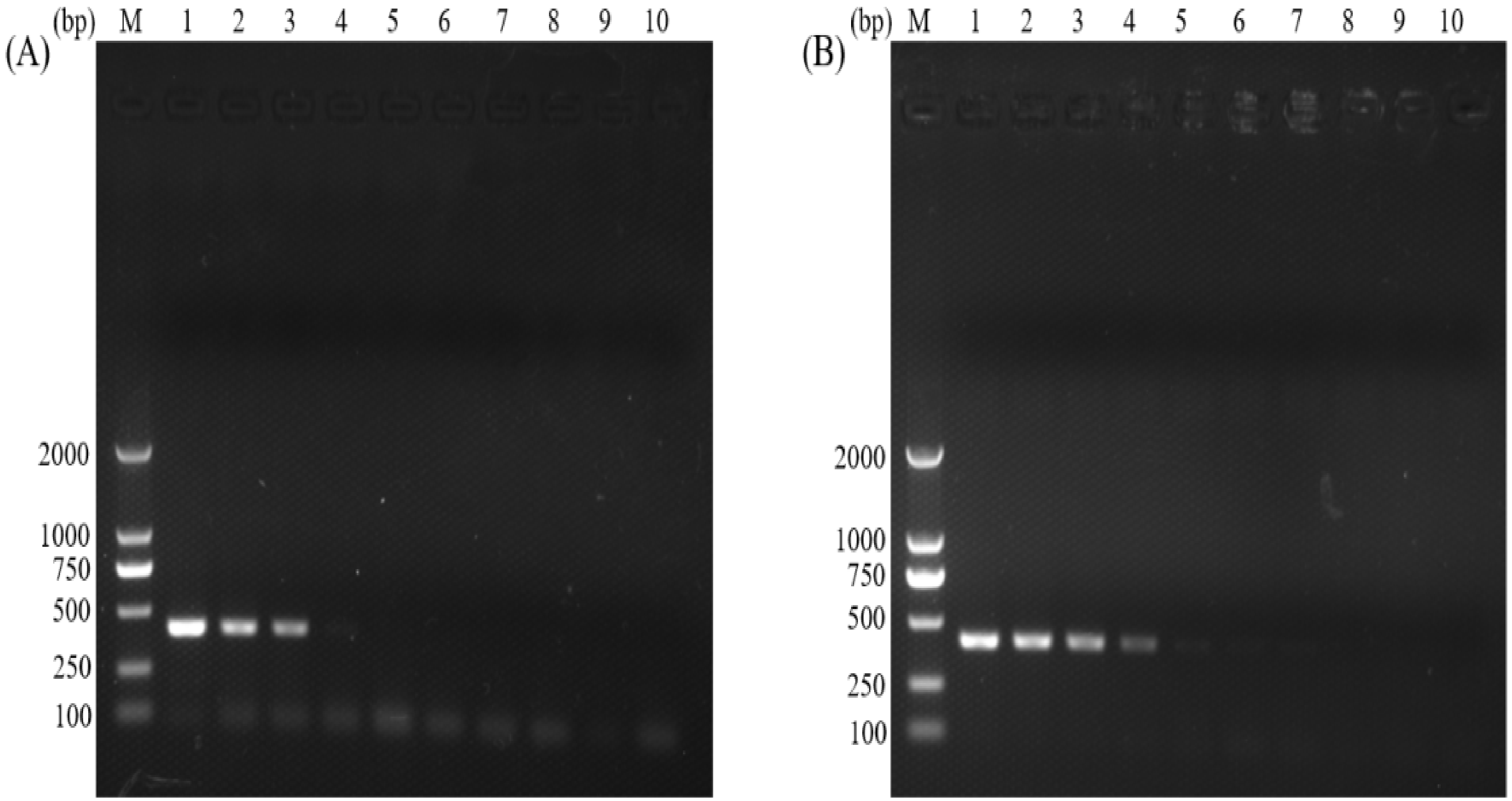
3.2. Sensitivities of the Normal PCR and the Nano-PCR Assays

The sensitivities of the normal PCR and the nano-PCR assays were analyzed by the ten-fold serial dilutions of one gDNA sample of C. parvum. The results showed that the nano-PCR was ten-fold more sensitive than the normal PCR, with a detection limit of 1.02 ng and 102 pg for the normal PCR (Figure 3A) and the nano-PCR (Figure 3B), respectively.

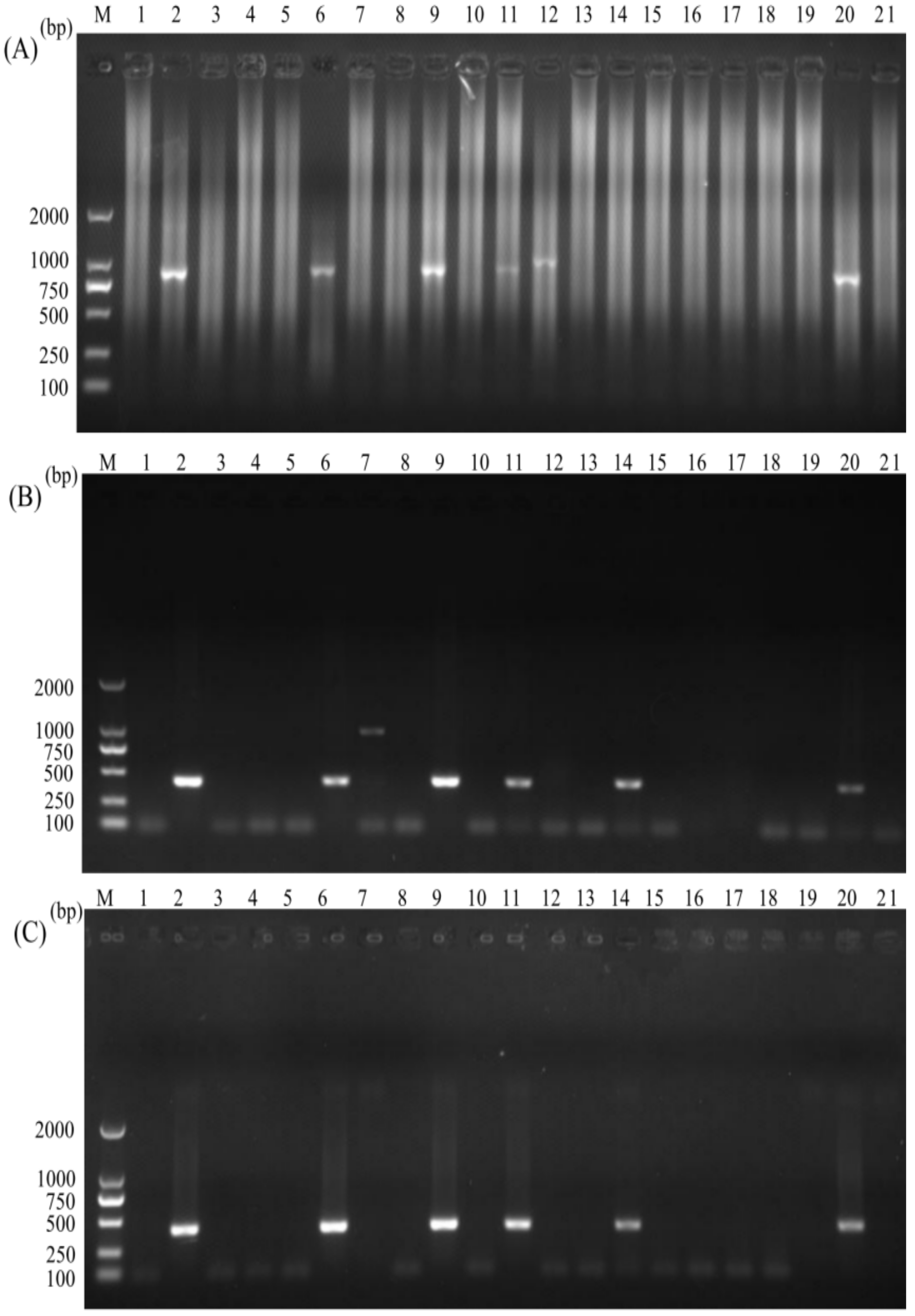
3.3. Specificities of the Normal PCR and the Nano-PCR Assays

The specificities of the normal PCR and the nano-PCR assays were assessed by testing the gDNA samples of eight intestinal pathogens. In the nano-PCR, the specific band was only found in a gDNA sample of C. parvum (Figure 4A). Similarly, normal PCR could amplify the target band in the gDNA sample of C. parvum. Although a gDNA sample of E. bieneusi could also be amplified, this non-specific band had a larger size than the specific band of C. parvum so that these two species could be identified in the normal PCR (Figure 4B). The results showed that both the normal PCR and the nano-PCR had good specificity in detecting C. parvum.

3.4. Assessment of the Nano-PCR for C. parvum in Clinical Settings

Twenty clinical samples of calves were determined for infection of C. parvum using nested PCR, normal PCR, and nano-PCR, respectively. Among those clinical samples, 25% (5/20), 30% (6/20), and 30% (6/20) were positive for C. parvum infection by using the nested PCR (Figure 5A), the normal PCR (Figure 5B), and the nano-PCR (Figure 5C). The sample in the lane 12 in the nested PCR was likely infected with other Cryptosporidium species but not C. parvum since the band was larger than that of other positive samples. The sample in the lane 7 in the normal PCR might be infected with other pathogens but not C. parvum since the band was nearly two-fold larger than that of other positive samples, and we have tried to sequence the amplicon but failed. The amplicon in the lane 14 was negative in the nested PCR but positive in the normal PCR and the nano-PCR was sent to Sangon Biotech for sequencing. The sequence was identified to be C. parvum for the amplicon in the lane 14 in the nano-PCR.

4. Discussion

Limitations of microscopic and immunological techniques resulted in the involvement of nucleic acid-based detection methods (e.g., PCR, nested PCR, qPCR, ddPCR, and LAMP) for the detection of C. parvum [29,30,31]. Compared to microscopic examination and immunological assays, the molecular techniques were found to be more sensitive and specific [32]. The nested PCR based on the gp60 locus has been widely used for detecting and subtyping Cryptosporidium, but it is time-consuming, easily contaminated, and needs to be combined with RFLP or sequencing for detection of C. parvum [33,34]. The qPCR assay was sensitive, specific, and reproducible for detecting C. parvum and significantly improved laboratory workflow and turnaround times, but it was dependent on the accuracy of the standard curve and required expensive instruments [35,36]. The ddPCR was an emerging method for detecting Cryptosporidium infection, which provided absolute quantitation without the use of calibration curves [37]. However, the template copy numbers of ddPCR were less than those of qPCR, and the average cost per sample was nearly two times higher than that of qPCR [37]. LAMP was an accurate, rapid, and sensitive method used for detecting Cryptosporidium species, but it was easily polluted by aerosol, which commonly exists in the environment [38,39]. In the present study, a novel, rapid, and sensitive nano-PCR assay was developed for the specific detection of C. parvum in faecal samples of calves.
Several useful molecular markers, e.g., 18S rRNA [40] and gp60 [12] genes, have been widely applied in the accurate detection of Cryptosporidium infection, significantly contributing to the development of Cryptosporidium infection and cryptosporidiosis molecular epidemiology. Previously, we compared the nano-PCR, the normal PCR, and the nested PCR for detecting Cryptosporidium infection based on the 18S rRNA gene. The results indicated that the nano-PCR was 100-fold more sensitive than the normal PCR, and the nano-PCR showed a higher detection rate than the nested PCR, reflecting the potential superiority of the nano-PCR in the detection of Cryptosporidium infection in clinical settings compared with the nested PCR [22]. However, due to the high conservatism of these gene loci among Cryptosporidium species, detection methods applying these loci are usually unable to effectively identify Cryptosporidium at the species level. Through comparative genomic analysis, the cgd3_330 gene was recognized as a potential marker for the detection of C. parvum in cattle, and no homologous gene sequences of the cgd3_330 gene were found for the other three Cryptosporidium species (C. bovis, C. ryanae, and C. andersoni) commonly found in cattle based on results from BLASTN and BLAST analyses in CryptoDB and NCBI, respectively. Thus, we used the cgd3_330 gene of C. parvum in a nano-PCR assay for accurate detection of C. parvum in faecal samples of calves.
The developed nano-PCR method in the present study was ten-fold more sensitive than the normal PCR, with a detection rate of 30% (6/20). Meanwhile, this method was specific for detection of C. parvum infection, and no cross-reaction was found with the other seven common pathogens in the faecal samples of calves, including C. ryanae, C. andersoni, and C. bovis. We have sequenced the 18S rRNA gene of the three Cryptosporidium species negative for amplification of the cgd3_330 gene of C. parvum. They were identified to be C. bovis, C. ryanae, and C. andersoni, respectively, by BLAST analyses and phylogenetic analyses (Supplementary Materials: Figure S1). Meanwhile, we also used the gp60 locus, a common recognized genetic marker for zoonotic Cryptosporidium species, in detecting clinical samples, and samples positive for both markers were all identified to be C. parvum. Interestingly, a clinical sample in the lane 14 that was negative for amplification of the gp60 gene was also positive in the nano-PCR based on the cgd3_330 gene, and sequencing verified that this sample was infected with C. parvum. Taking these together, the nano-PCR targeting the cgd3_330 gene could detect more positive samples than the nested PCR targeting the gp60 gene but needs to be verified in future studies.
Recently, the outbreaks of diarrhea caused by C. parvum have led to hundreds of deaths in calves on several farms in China, causing great economic losses to the farms [41,42]. With the application of the nano-PCR, we could rapidly identify C. parvum infection early, and the targeted interventions, such as isolating and treating infected animals, and sterilizing the environment, could be conducted to block the transmission of C. parvum among animals and minimize animal death and economic losses.

5. Conclusions

The present study established an efficient nano-PCR assay targeting the cgd3-330 gene for specific detection of C. parvum in calves, and it could effectively detect C. parvum in a small scale of clinical samples. This nano-PCR assay showed superiority in detection of C. parvum infection compared with the normal PCR and the nested PCR techniques. Further studies are needed to test the nano-PCR on large-scale clinical samples from diverse hosts and geographical areas.

Supplementary Materials

The following are available online at https://www.mdpi.com/article/10.3390/ani12151953/s1. Figure S1: Phylogenetic tree generated by the Neighbor-Joining method using partial sequences of the 18S rRNA gene of four Cryptosporidium species in the present study and representative sequences of other Cryptosporidium species available in the GenBankTM. The sequences obtained in this study are marked with red triangles. Bootstrap values (>70) are indicated at the nodes. Scale bar indicates 0.02 nucleotide substitutions/site.

Author Contributions

Conceptualization, G.Z.; methodology, Q.Y., X.Y., Y.W. and J.S.; software and formal analysis, J.W.; validation and investigation, S.H.; data curation, Y.W.; writing—original draft preparation, Q.Y.; writing—review and editing, X.Y. and Q.Y.; visualization, X.Y. and Q.Y.; supervision, G.Z. and J.S.; project administration, G.Z. All authors have read and agreed to the published version of the manuscript.

Funding

This work was funded by grants from the Open Funds of the State Key Laboratory of Veterinary Aetiological Biology, Lanzhou Veterinary Research Institute, Chinese Academy of Agricultural Sciences (SKLVEB2020KFKT015), the National Natural Science Foundation of China (32072890), and the National Key Research and Development Program of China (2017YFD0501305).

Institutional Review Board Statement

This study was conducted under the approval and instructions of the ethics committee of Northwest A&F University (DY2021036) and the animal ethics requirements of the People’s Republic of China.

Informed Consent Statement

Not applicable.

Data Availability Statement

Data is contained within the article.

Conflicts of Interest

The authors declare no conflict of interest.

References

  1. de Graaf, D.C.; Vanopdenbosch, E.; Ortega-Mora, L.M.; Abbassi, H.; Peeters, J.E. A review of the importance of cryptosporidiosis in farm animals. Int. J. Parasitol. 1999, 29, 1269–1287. [Google Scholar] [CrossRef]
  2. Santín, M. Cryptosporidium and Giardia in ruminants. Vet. Clin. N. Am. Food Anim. Pract. 2020, 36, 223–238. [Google Scholar] [CrossRef]
  3. Santín, M.; Trout, J.M.; Xiao, L.; Zhou, L.; Greiner, E.; Fayer, R. Prevalence and age-related variation of Cryptosporidium species and genotypes in dairy calves. Vet. Parasitol. 2004, 122, 103–117. [Google Scholar] [CrossRef] [PubMed]
  4. Fayer, R.; Santín, M.; Trout, J.M.; Greiner, E. Prevalence of species and genotypes of Cryptosporidium found in 1–2-year-old dairy cattle in the eastern United States. Vet. Parasitol. 2006, 135, 105–112. [Google Scholar] [CrossRef] [PubMed]
  5. Santín, M.; Trout, J.M.; Fayer, R. A longitudinal study of cryptosporidiosis in dairy cattle from birth to 2 years of age. Vet. Parasitol. 2008, 155, 15–23. [Google Scholar] [CrossRef]
  6. Wang, Y.; Cao, J.; Chang, Y.; Yu, F.; Zhang, S.; Wang, R.; Zhang, L. Prevalence and molecular characterization of Cryptosporidium spp. and Giardia duodenalis in dairy cattle in Gansu, northwest China. Parasite 2020, 27, 62. [Google Scholar] [CrossRef]
  7. Thomson, S.; Hamilton, C.A.; Hope, J.C.; Katzer, F.; Mabbott, N.A.; Morrison, L.J.; Innes, E.A. Bovine cryptosporidiosis: Impact, host-parasite interaction and control strategies. Vet. Res. 2017, 48, 42. [Google Scholar] [CrossRef] [Green Version]
  8. Khurana, S.; Sharma, P.; Sharma, A.; Malla, N. Evaluation of Ziehl-Neelsen staining, auramine phenol staining, antigen detection enzyme linked immunosorbent assay and polymerase chain reaction, for the diagnosis of intestinal cryptosporidiosis. Trop. Parasitol. 2012, 2, 20–23. [Google Scholar] [CrossRef] [PubMed]
  9. Koehler, A.V.; Jex, A.R.; Haydon, S.R.; Stevens, M.A.; Gasser, R.B. Giardia/giardiasis-a perspective on diagnostic and analytical tools. Biotechnol. Adv. 2014, 32, 280–289. [Google Scholar] [CrossRef]
  10. Alves, M.; Xiao, L.; Sulaiman, I.; Lal, A.A.; Matos, O.; Antunes, F. Subgenotype analysis of Cryptosporidium isolates from humans, cattle, and zoo ruminants in Portugal. J. Clin. Microbiol. 2003, 41, 2744–2747. [Google Scholar] [CrossRef] [PubMed] [Green Version]
  11. Xiao, L.; Ryan, U.M. Cryptosporidiosis: An update in molecular epidemiology. Curr. Opin. Infect. Dis. 2004, 17, 483–490. [Google Scholar] [CrossRef] [PubMed] [Green Version]
  12. Xiao, L. Molecular epidemiology of cryptosporidiosis: An update. Exp. Parasitol. 2010, 124, 80–89. [Google Scholar] [CrossRef] [PubMed]
  13. Balatbat, A.B.; Jordan, G.W.; Tang, Y.J.; Silva, J., Jr. Detection of Cryptosporidium parvum DNA in human feces by nested PCR. J. Clin. Microbiol. 1996, 34, 1769–1772. [Google Scholar] [CrossRef] [Green Version]
  14. Demming, A. The state of research after 25 years of nanotechnology. Nanotechnology 2014, 25, 010201. [Google Scholar] [CrossRef]
  15. Ali, Z.; Jin, G.; Hu, Z.; Wang, Z.; Khan, M.A.; Dai, J.; Tang, Y. A review on nanoPCR: History, mechanism and applications. J. Nanosci. Nanotechnol. 2018, 18, 8029–8046. [Google Scholar] [CrossRef] [PubMed]
  16. Gedda, M.R.; Madhukar, P.; Shukla, A.; Mudavath, S.L.; Srivastava, O.N.; Singh, O.P.; Sundar, S. Nanodiagnostics in leishmaniasis: A new frontiers for early elimination. Wiley Interdiscip. Rev. Nanomed. Nanobiotechnol. 2021, 13, e1675. [Google Scholar] [CrossRef]
  17. Li, M.; Lin, Y.C.; Wu, C.C.; Liu, H.S. Enhancing the efficiency of a PCR using gold nanoparticles. Nucleic Acids Res. 2005, 33, e184. [Google Scholar] [CrossRef] [Green Version]
  18. Gabriel, S.; Rasheed, A.K.; Siddiqui, R.; Appaturi, J.N.; Fen, L.B.; Khan, N.A. Development of nanoparticle-assisted PCR assay in the rapid detection of brain-eating amoebae. Parasitol. Res. 2018, 117, 1801–1811. [Google Scholar] [CrossRef] [PubMed]
  19. Rehman, A.; Sarwar, Y.; Raza, Z.A.; Hussain, S.Z.; Mustafa, T.; Khan, W.S.; Ghauri, M.A.; Haque, A.; Hussain, I. Metal nanoparticle assisted polymerase chain reaction for strain typing of Salmonella typhi. Analyst 2015, 140, 7366–7372. [Google Scholar] [CrossRef] [PubMed]
  20. Zhu, Y.; Liang, L.; Luo, Y.; Wang, G.; Wang, C.; Cui, Y.; Ai, X.; Cui, S. A sensitive duplex nanoparticle-assisted PCR assay for identifying porcine epidemic diarrhea virus and porcine transmissible gastroenteritis virus from clinical specimens. Virus Genes 2017, 53, 71–76. [Google Scholar] [CrossRef] [PubMed]
  21. Liu, Z.; Li, J.; Liu, Z.; Li, J.; Li, Z.; Wang, C.; Wang, J.; Guo, L. Development of a nanoparticle-assisted PCR assay for detection of bovine respiratory syncytial virus. BMC Vet. Res. 2019, 15, 110. [Google Scholar] [CrossRef] [PubMed]
  22. Yin, Y.L.; Wang, Y.; Lai, P.; Yao, Q.; Li, Y.; Zhang, L.X.; Yang, X.; Song, J.K.; Zhao, G.H. Establishment and preliminary application of nanoparticle-assisted PCR assay for detection of Cryptosporidium spp. Parasitol. Res. 2021, 120, 1837–1844. [Google Scholar] [CrossRef] [PubMed]
  23. Luan, Q.; Jiang, Z.; Wang, D.; Wang, S.; Yin, Y.; Wang, J. A sensitive triple nanoparticle-assisted PCR assay for detection of fowl adenovirus, infectious bursal disease virus and chicken anemia virus. J. Virol. Methods 2022, 303, 114499. [Google Scholar] [CrossRef]
  24. Zhao, S.S.; Li, Y.H.; Zhang, Y.; Zhou, Q.; Jing, B.; Xu, C.Y.; Zhang, L.X.; Song, J.K.; Qi, M.; Zhao, G.H. Multilocus genotyping of Giardia duodenalis in Bactrian camels (Camelus bactrianus) in China. Parasitol. Res. 2020, 119, 3873–3880. [Google Scholar] [CrossRef] [PubMed]
  25. Li, Y.H.; Yao, Q.; Dong, H.P.; Wang, S.S.; Chen, R.R.; Song, J.K.; Yan, W.C.; Zhao, G.H. Molecular characterization of Balantioides coli in pigs from Shaanxi province, northwestern China. Parasitol. Res. 2020, 119, 3075–3081. [Google Scholar] [CrossRef] [PubMed]
  26. Yang, X.; Li, Y.H.; Wang, Y.X.; Wang, J.W.; Lai, P.; Li, Y.; Song, J.K.; Qi, M.; Zhao, G.H. Molecular characterization of Blastocystis sp. in Camelus bactrianus in Northwestern China. Animals 2021, 11, 3016. [Google Scholar] [CrossRef]
  27. Wang, S.S.; Li, J.Q.; Li, Y.H.; Wang, X.W.; Fan, X.C.; Liu, X.; Li, Z.J.; Song, J.K.; Zhang, L.X.; Zhao, G.H. Novel genotypes and multilocus genotypes of Enterocytozoon bieneusi in pigs in northwestern China: A public health concern. Infect. Genet. Evol. 2018, 63, 89–94. [Google Scholar] [CrossRef]
  28. Wang, X.T. Study on Population Structure of Cryptosporidium spp. and Giardia lamblia in in Calves from Partial Areas of Shaanxi. Master Thesis, Northwest A&F University, Xianyang, China, 2017. (In Chinese). [Google Scholar]
  29. Adeyemo, F.E.; Singh, G.; Reddy, P.; Stenström, T.A. Methods for the detection of Cryptosporidium and Giardia: From microscopy to nucleic acid based tools in clinical and environmental regimes. Acta Trop. 2018, 184, 15–28. [Google Scholar] [CrossRef] [PubMed]
  30. Destura, R.V.; Rohani, C.B.; Ma, J.; Sevilleja, J.E.A.D. Advancing Cryptosporidium diagnostics from bench to bedside. Curr. Trop. Med. Rep. 2015, 2, 150–160. [Google Scholar] [CrossRef] [Green Version]
  31. Cunha, F.S.; Peralta, R.H.S.; Peralta, J.M. New insights into the detection and molecular characterization of Cryptosporidium with emphasis in Brazilian studies: A review. Rev. Inst. Med. Trop. Sao Paulo 2019, 61, e28. [Google Scholar] [CrossRef] [PubMed]
  32. Mesa, L.E.; Manrique, R.; Muskus, C.; Robledo, S.M. Test accuracy of polymerase chain reaction methods against conventional diagnostic techniques for Cutaneous Leishmaniasis (CL) in patients with clinical or epidemiological suspicion of CL: Systematic review and meta-analysis. PLoS Negl. Trop. Dis. 2020, 14, e0007981. [Google Scholar] [CrossRef] [PubMed] [Green Version]
  33. Ezzaty Mirhashemi, M.; Zintl, A.; Grant, T.; Lucy, F.E.; Mulcahy, G.; De Waal, T. Comparison of diagnostic techniques for the detection of Cryptosporidium oocysts in animal samples. Exp. Parasitol. 2015, 151–152, 14–20. [Google Scholar] [CrossRef] [PubMed] [Green Version]
  34. Xiao, L.; Bern, C.; Limor, J.; Sulaiman, I.; Roberts, J.; Checkley, W.; Cabrera, L.; Gilman, R.H.; Lal, A.A. Identification of 5 types of Cryptosporidium parasites in children in Lima, Peru. J. Infect. Dis. 2001, 183, 492–497. [Google Scholar] [CrossRef] [PubMed] [Green Version]
  35. Taylor, S.C.; Laperriere, G.; Germain, H. Droplet digital PCR versus qPCR for gene expression analysis with low abundant targets: From variable nonsense to publication quality data. Sci. Rep. 2017, 7, 2409. [Google Scholar] [CrossRef] [Green Version]
  36. Hadfield, S.J.; Robinson, G.; Elwin, K.; Chalmers, R.M. Detection and differentiation of Cryptosporidium spp. in human clinical samples by use of real-time PCR. J. Clin. Microbiol. 2011, 49, 918–924. [Google Scholar] [CrossRef] [PubMed] [Green Version]
  37. Yang, R.; Paparini, A.; Monis, P.; Ryan, U. Comparison of next-generation droplet digital PCR (ddPCR) with quantitative PCR (qPCR) for enumeration of Cryptosporidium oocysts in faecal samples. Int. J. Parasitol. 2014, 44, 1105–1113. [Google Scholar] [CrossRef] [PubMed] [Green Version]
  38. Bakheit, M.A.; Torra, D.; Palomino, L.A.; Thekisoe, O.M.; Mbati, P.A.; Ongerth, J.; Karanis, P. Sensitive and specific detection of Cryptosporidium species in PCR-negative samples by loop-mediated isothermal DNA amplification and confirmation of generated LAMP products by sequencing. Vet. Parasitol. 2008, 158, 11–22. [Google Scholar] [CrossRef]
  39. Karanis, P.; Thekisoe, O.; Kiouptsi, K.; Ongerth, J.; Igarashi, I.; Inoue, N. Development and preliminary evaluation of a loop-mediated isothermal amplification procedure for sensitive detection of Cryptosporidium oocysts in fecal and water samples. Appl. Environ. Microbiol. 2007, 73, 5660–5662. [Google Scholar] [CrossRef] [PubMed] [Green Version]
  40. Nichols, R.A.B.; Campbell, B.M.; Smith, H.V. Identification of Cryptosporidium spp. oocysts in United Kingdom noncarbonated natural mineral waters and drinking waters by using a modified nested PCR-restriction fragment length polymorphism assay. Appl. Environ. Microbiol. 2003, 69, 4183–4189. [Google Scholar] [CrossRef] [Green Version]
  41. Li, N.; Wang, R.; Cai, M.; Jiang, W.; Feng, Y.; Xiao, L. Outbreak of cryptosporidiosis due to Cryptosporidium parvum subtype IIdA19G1 in neonatal calves on a dairy farm in China. Int. J. Parasitol. 2019, 49, 569–577. [Google Scholar] [CrossRef]
  42. Li, N.; Zhao, W.; Song, S.; Ye, H.; Chu, W.; Guo, Y.; Feng, Y.; Xiao, L. Diarrhoea outbreak caused by coinfections of Cryptosporidium parvum subtype IIdA20G1 and rotavirus in pre-weaned dairy calves. Transbound. Emerg. Dis. 2022; in press. [Google Scholar] [CrossRef] [PubMed]
Figure 1. Optimization of the annealing temperature (A) and the concentration of MgCl2 (B) of the normal PCR assay. (A) lane M: DL2000 DNA Marker (TakaRa); lane 1: 50 °C; lane 2: 50.5 °C; lane 3: 52 °C; lane 4: 54 °C; lane 5: 56 °C; lane 6: 58 °C; lane 7: 59.5 °C; lane 8: 60 °C; lane 9: negative control. (B) lane M: DL2000 DNA Marker (TakaRa); lane 1: 0.42 mM; lane 2: 0.83 mM; lane 3: 1.25 mM; lane 4: 1.67 mM; lane 5: 2.08 mM; lane 6: 2.5 mM; lane 7: 2.92 mM; lane 8: negative control.
Animals 12 01953 g001
Figure 2. Optimization of the annealing temperature (A) and the primer amounts (B) of the nano-PCR assay. (A) lane M: DL2000 DNA Marker (TakaRa); lane 1: 50 °C; lane 2: 50.5 °C; lane 3: 52 °C; lane 4: 54 °C; lane 5: 56 °C; lane 6: 58 °C; lane 7: 59.5 °C; lane 8: 60 °C; lane 9: negative control. (B) lane M: DL2000 DNA Marker (TakaRa); lane 1: 2 pmol; lane 2: 4 pmol; lane 3: 6 pmol; lane 4: 8 pmol; lane 5: 10 pmol; lane 6: 12 pmol; lane 7: 14 pmol; lane 8: negative control.
Animals 12 01953 g002
Figure 3. A serial ten-fold dilution of gDNA samples from C. parvum oocysts was used to analyze sensitivities of the normal PCR (A) and the nano-PCR (B) assays. Lane M: DL2000 DNA Marker (TakaRa); lane 1: 102 ng; lane 2: 10.2 ng; lane 3: 1.02 ng; lane 4: 102 pg; lane 5: 10.2 pg; lane 6: 1.02 pg; lane 7: 102 fg; lane 8: 10.2 fg; lane 9: 1.02 fg; lane 10: negative control.
Animals 12 01953 g003
Figure 4. Specificities of the nano-PCR (A) and the normal PCR (B) assays. Lane M: DL2000 DNA Marker (TaKaRa); lane 1: gDNA sample of C. parvum; lane 2: gDNA sample of C. andersoni; lane 3: gDNA sample of C. bovis; lane 4: gDNA sample of C. ryanae; lane 5: gDNA sample of Blastocystis sp.; lane 6: gDNA sample of G. lamblia; lane 7: gDNA sample of E. bieneusi; lane 8: gDNA sample of B. coli; lane 9: negative control.
Animals 12 01953 g004
Figure 5. Detection of C. parvum in clinical samples by using the nested PCR (A), the normal PCR (B) and the nano-PCR assays (C). Lane M: DL2000 DNA Marker (TaKaRa); lanes 1–20: faecal samples of calves; lane 21: negative control.
Animals 12 01953 g005
Table 1. Sequences of the primers used in this study.
PCR AssaysPrimer NamesSequences (5′–3′)Product Sizes (bp)
Normal PCR
/Nano-PCR
cgd3_330-FAGTGGTTACAGGTGGGATGAGT~413
cgd3_330-RGCGAGTTTCCTTGATTCATAGC
Nested PCRgp60-F1TTACTCTCCGTTATAGTCTCC~915
gp60-R1GGAAGGAACGATGTATCTGA
gp60-F2TCCGCTGTATTCTCAGCC~800
gp60-R2GCAGAGGAACCAGCATC
Publisher’s Note: MDPI stays neutral with regard to jurisdictional claims in published maps and institutional affiliations.

