Article

CYP19A1 May Influence Lambing Traits in Goats by Regulating the Biological Function of Granulosa Cells

1
Key Laboratory of Animal Genetics, Breeding and Reproduction in The Plateau Mountainous Region, Ministry of Education, Guizhou University, Guiyang 550025, China
2
Key Laboratory of Animal Genetics, Breeding and Reproduction, Guiyang 550025, China
3
College of Animal Science, Guizhou University, Guiyang 550025, China
*
Author to whom correspondence should be addressed.
Animals 2022, 12(15), 1911; https://doi.org/10.3390/ani12151911
Submission received: 19 May 2022 / Revised: 20 July 2022 / Accepted: 24 July 2022 / Published: 27 July 2022
(This article belongs to the Collection Reproductive Management of Sheep and Goats)

Abstract

:

Simple Summary

Aromatase (CYP19A1), a member of the cytochrome family, is widely expressed in ovarian and granulosa cells and is primarily responsible for the conversion of androgens to estrogens. Increased expression of CYP19A1 in follicular granulosa cells has implications for cell proliferation, steroid hormone secretion, and the expression of related functional indicator genes. We hypothesize that CYP19A1 may indirectly influence lambing numbers in goats by regulating follicular cell growth and development, as well as ovarian ovulation.

Abstract

Abnormal expression of CYP19A1, a gene related to steroid hormone synthesis, causes steroid hormone disruption and leads to abnormal ovulation in granulosa cells. However, the exact mechanism of CYP19A1 regulation is unclear. In this study, we confirmed the localization of CYP19A1 in goat ovarian tissues using immunohistochemistry. Subsequently, we investigated the effects of CYP19A1 on granulosa cell proliferation, steroid hormone secretion, and expression of candidate genes for multiparous traits by overexpressing and silencing CYP19A1 in goat granulosa cells (GCs). The immunohistochemistry results showed that CYP19A1 was expressed in all types of follicular, luteal, and granulosa cells, with subcellular localization results revealing that CYP19A1 protein was mainly localized in the cytoplasm and nucleus. Overexpression of CYP19A1 significantly increased the mRNA levels of CYP19A1, FSHR, and INHBA, which are candidate genes for multiple birth traits in goats. It also promoted cell proliferation, PCNA and Cyclin E mRNA levels in granulosa cells, and secretion of estrogen and progesterone. However, it inhibited the mRNA levels of STAR, CYP11A1, and 3βSHD, which are genes related to steroid synthesis. Silencing CYP19A1 expression significantly reduced CYP19A1, FSHR, and INHBA mRNA levels in granulosa cells and inhibited granulosa cell proliferation and PCNA and Cyclin E mRNA levels. It also reduced estrogen and progesterone secretion but enhanced the mRNA levels of STAR, CYP11A1, and 3βSHD. CYP19A1 potentially influenced the lambing traits in goats by affecting granulosa cell proliferation, hormone secretion, and expression of candidate genes associated with traits for multiple births.

1. Introduction

Ovarian follicle cells are among the fastest-growing cells. The most prominent somatic cells in follicular cells are the granulosa and luteal cells, whose ability to proliferate and differentiate is the dominant factor in follicular development and maturation [1,2]. The ovary possesses two main steroid-producing cell types: the membrane cells responsible for androgen synthesis, and the granulosa cells responsible for converting androgens into estrogens and directing progesterone synthesis [3]. Steroid hormones play an important role in many follicle cells throughout their growth phase [4]. For instance, progesterone (P4) and estradiol (E2) are involved in regulating the cyclic derivation of ovarian follicles [5] and act as antioxidants to protect follicles from oxidative stress and atresia [6,7]. Notably, the proliferation and differentiation of granulosa cells are also essential for follicular and oocyte development, ovulation, and luteinization [8]. Follicular granulosa cells are thus of great significance to the success of early gestation. Aromatase, isolated from the placenta in 1988 [9,10], belongs to the cytochrome P450 family and is responsible for the conversion of androstenedione (A2), testosterone (T), and 16a-hydroxyandrostenediol into estrone (E1), oestradiol (E2), and estriol (E3), respectively. Zhang et al. proposed that CYP19A1 is a vital enzyme in estrogen synthesis and plays an important role in establishing and maintaining pregnancy [11]. Similarly, Vega et al. found that CYP19A1 is also involved in regulating folliculogenesis and the behavior of follicular atresia in cattle [12], and it catalyzes the conversion of androgens to estrogens [13]. In addition, the CYP19A1 gene also plays an important role in mammalian gonadal development [14,15]. Its presence largely affects gonadal development in the testis [16] and luteal cells [17] of horses and the genitalia [18] of pigs and fetal sheep [19,20]. Luo et al. [21] found that the CYP19A1 gene is involved in regulating follicular cell apoptosis and ovarian damage using a mice model. Notably, CYP19A1 is also a marker gene associated with steroids and has a dynamic indicator function in response to steroid hormone secretion [22]. Wu et al. (2022) reported that abnormal expression of CYP19A1 in sheep follicular granulosa cells may cause disturbances in ovarian steroid hormone secretion, thereby affecting normal ovulation [23]. Based on these studies, it is evident that CYP19A1 plays an integral role in regulating an animal’s reproductive physiology. To date, only a few studies have assessed the effects of CYP19A1 on the biological behavior and kidding traits of goat follicular granulosa cells. In conclusion, we aimed to study the effects of CYP19A1 on steroid secretion characteristics, reproduction-related gene expression, and granulosa cell proliferation by overexpressing and interfering with the expression of the CYP19A1 gene in goat follicle granulosa cells, so as to provide reference for improving goat reproductive performance.

2. Materials and Methods

2.1. Animal Sourcing, Cell Collection, and Cell Identification

The goat GCs were collected from the healthy ovaries of six 36-month-old Qianbei horse goats (Xishui, China, China-Fuxing Animal Husbandry Co., Ltd.), which were slaughtered by Fuxing Animal Husbandry staff. Left ovary samples were preserved in phosphate-buffered saline (PBS) amended with antibodies (penicillin and streptomycin) to culture the granulosa cells. The right ovary was fixed in 4% paraformaldehyde solution and was used for immunohistochemistry and HE staining. The granulosa cells were also identified through cellular immunofluorescence for FSHR-specific expression of protein staining [24]. All animal experiments were approved by the Animal Ethics Committee of Guizhou University (Guiyang, China).

2.2. Plasmid Construction

Plasmids were constructed after PCR amplification by recovering the gels to obtain pure target genes and pEGFP, which were ligated using T4 DNA ligase, transferred to DH5α receptor cells, and then shaken to extract positive plasmids. The PCR system (25 μL) comprised 1 μL of DNA template, 1 μL each of upstream and downstream primer (10 pmol/µL), 12.5 μL of Green Mix, and 9.5 μL of ddH2O. Multiple pairs of CYP19A1 interfering sequences were designed following the shRNA design principles and sent to GEMA Ltd. (Shanghai, China) for synthesis. The primers (Table 1) for the corresponding assays were sent to Bioengineering Co., Ltd. (Shanghai, China) for synthesis (Login ID: NM_001285747.1). Table 2 outlines the primers for each biological cytokine function genes (all genes Tm = 60 °C).

2.3. Cell Culture and Transfection

Ovary tissues were collected from healthy Qianbei Ma goats, and the granulosa cells were subsequently collected from the follicles using the follicle isolation method [25]. The follicles were first punctured using a 10-gauge needle, and the follicular fluid was drained into a culture dish containing DMEM-F/12 (gibco, Beijing, China). The collected mixture was transferred to a 10 mL centrifuge tube and centrifuged at 1000 rpm for 10 min. The supernatant was discarded, the previous step repeated, and DMEM-F/12 (gibco, Beijing, China) containing 15% fetal bovine serum and 2% penicillin–streptomycin was added to the bottom of the tube using a Bachmann pipette (Wuxi, China, NEST Biotechnology Co.,Ltd.), which was blown and mixed. The cell suspension was then transferred into a 25 cm2 cell culture flask (Wuxi, China, NEST Biotechnology Co., Ltd.) and cultured in a 37 °C cell incubator (Thermo Fisher Scientifific, Waltham, MA, USA). The cells were then evenly plated into six empty plates (Wuxi, China, NEST Biotechnology Co.,Ltd.) upon attaining an adherence of 80–90%, followed by the transfer of the overexpression and silencing plasmids into follicular granulosa cells using a Lipofectamine 2000 kit (Thermo Fisher Scientifific, Waltham, MA, USA) according to the manufacturer’s instructions. The cells were harvested after 48 h based on the luminescence of RNA.

2.4. Total RNA Extraction and Reverse Transcription

Total RNA was extracted from goat granulosa cells using TRIzol reagent (Solarbio, Beijing, China). The first-strand cDNA was subsequently synthesized using a StarScript II First-strand cDNA Kit (GenStar, Beijing, China) following the manufacturer’s instructions, and the reaction products were stored at −20 °C.

2.5. Real-Time PCR

The effects of the CYP19A1 gene on the expression of reproduction-related candidate genes and proliferation-related indicator genes (BMPR-IB, FSHR, INHBA), proliferation-related genes (Cyclin E, PCNA), and steroid hormone-related genes (STAR, CYP11A1 and 3βSHD) were determined by measuring the expression levels of these genes in goat granulosa cells on a CFX 9600 real-time PCR instrument (Bio-Rad, Hercules, CA, USA). Table 2 outlines the details of the real-time PCR primers. A 10 μL real-time PCR reaction mix (containing 5 µL of 2 × Es Taq Master Mix, 3.2 µL of RNase-free ddH2O, 0.5 µL of cDNA, and 0.4 µL each of forward and reverse primers (10 pmol/µL)) was prepared following the kit system and subjected to a three-step PCR program (95 °C for 2 min, 95 °C for 15 s, annealing temperature (see Table 2 for details) for 30 s, 72 °C for 30 s, with the melting curve set automatically by the machine). The PCR assay was replicated thrice for each test sample. The expression data were normalized using the expression of the β-actin we used as the internal reference. The relative changes in gene expression were then calculated using the relative quantification method 2ΔΔCT [26].

2.6. Cell Proliferation Assay

The effects of overexpression and silencing of the CYP19A1 gene on the proliferative activity of follicular granulosa cells were detected using the CCK-8 method. Plasmids in the negative control and test groups were incubated at 37 °C and 5% CO2 for 6, 12, 24, and 48 h after transfection, following the instructions for the CCK-8 reagent (Beyotime Biotechnology, Shanghai, China). Their absorbance (OD) at 450 nm was subsequently measured using an enzyme marker (Thermo Fisher Scientifific, Waltham, MA, USA). The expressions of PCNA and Cyclin E proliferation markers at the transcriptional level were also measured.

2.7. Steroid Assay

The plasmids were seeded into 96-well plates (Wuxi, China, NEST Biotechnology Co., Ltd.) in triplicate, followed by collection of 100 µL of the supernatant from each culture medium after 6, 12, 24, and 48 h. The supernatants were stored at −80 °C.

2.8. Immunohistochemistry

Immunohistochemistry was performed according to a previously described protocol [24]. Ovary tissue sections (5 mm) were first fixed and embedded in paraffin. The sections were then affixed onto microscope slides (Tokyo, Japan), heated at 65 °C for 2 h, de-paraffined in xylene, and rehydrated in a series of graded ethanol. The sections were then boiled in 0.1 M citrate buffer (pH = 6.0) in a microwave oven for 15 min and then cooled to 37 °C for antigen repair. The sections were subsequently washed with PBS and incubated in 3% hydrogen peroxide for 15 min to block the endogenous peroxidase activity. The sections were further incubated in blocking buffer (5% BSA in PBS) for 15 min at 37 °C to block non-specific binding. They were then incubated with rabbit anti-CYP19A1 polyclonal antibody (plus literature) in PBS (1:3000, AiFang biological, AF04449, Beijing, China) overnight at 4 °C, washed with PBS, and then incubated with biotinylated secondary antibody for 2 h at 37 °C and horseradish peroxidase (HRP)–streptavidin for 15 min before visualization using diaminobenzidine. The non-specific rabbit immunoglobulin G (IgG) was used in place of the primary antibody as the negative control. Digital images were acquired using a microscope (Tokyo, Japan).

2.9. Statistical Analysis

Differences between overexpression and silencing of CYP19A1 on the proliferation and hormone data of granulosa cells were analyzed using t-tests. All data are presented as means ± SD of three biological replicates and three technical replicates to ensure the accuracy of the experimental data. The mRNA levels of target genes were normalized with a housekeeping gene (β-actin, 139 bp) and expressed as 2−∆∆CT using samples on pEGFP-N3 or sh-NC as a calibrator [24]. The significance threshold was set at p < 0.05.

3. Results

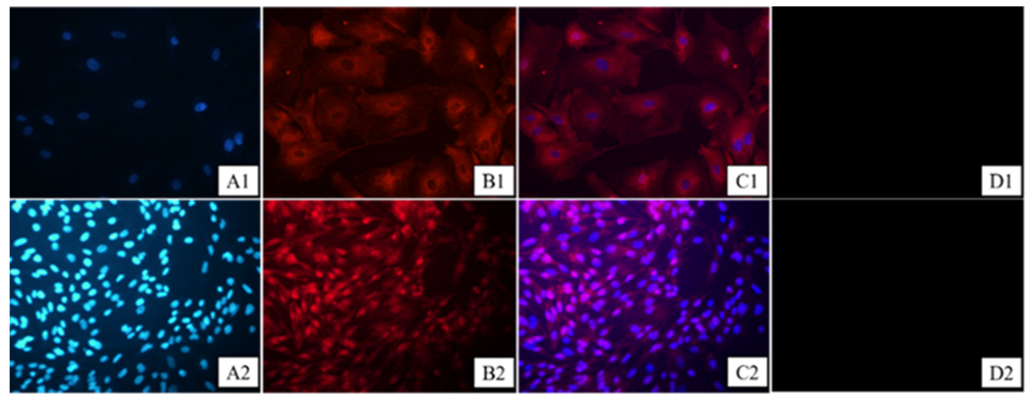
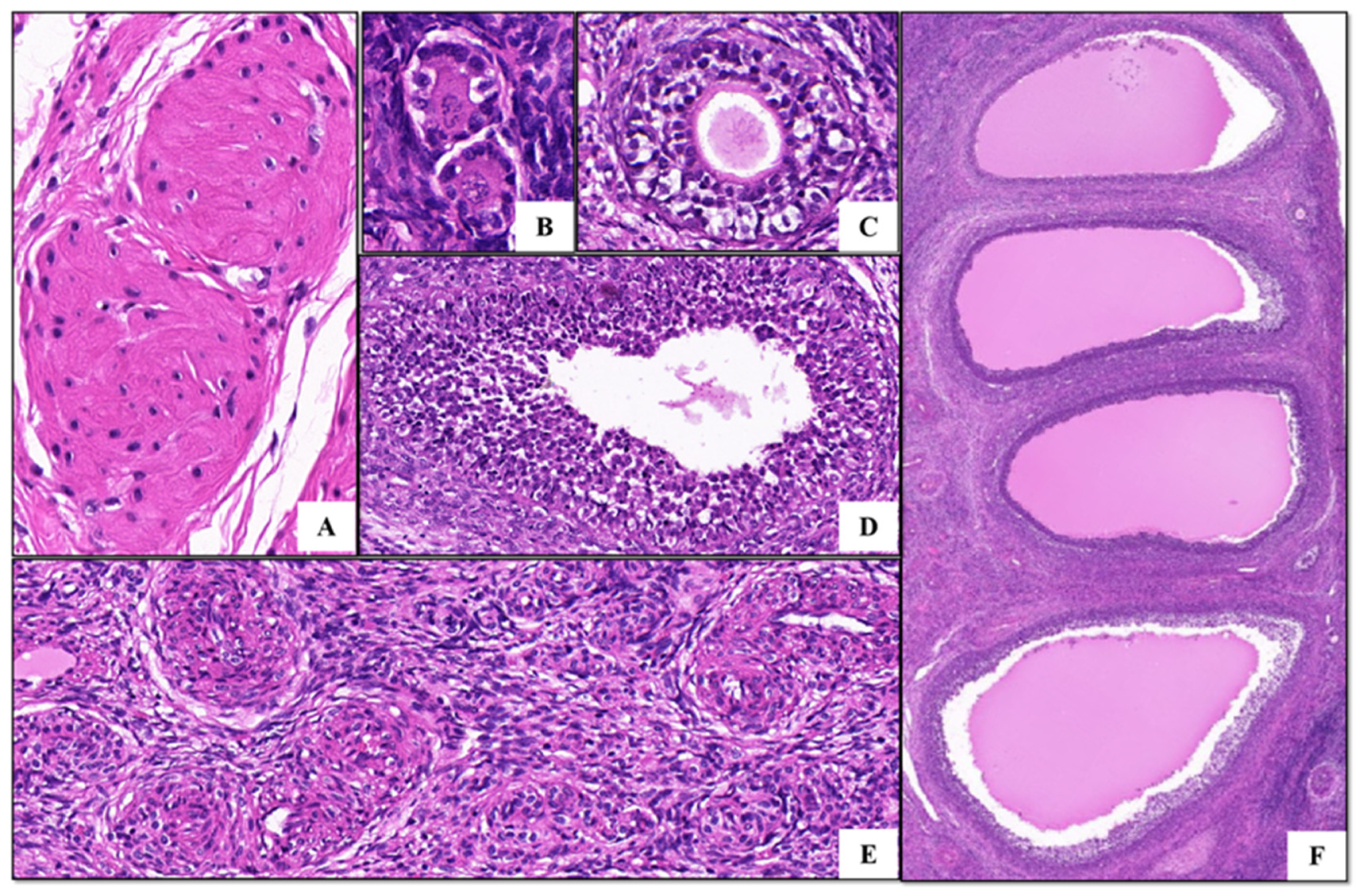
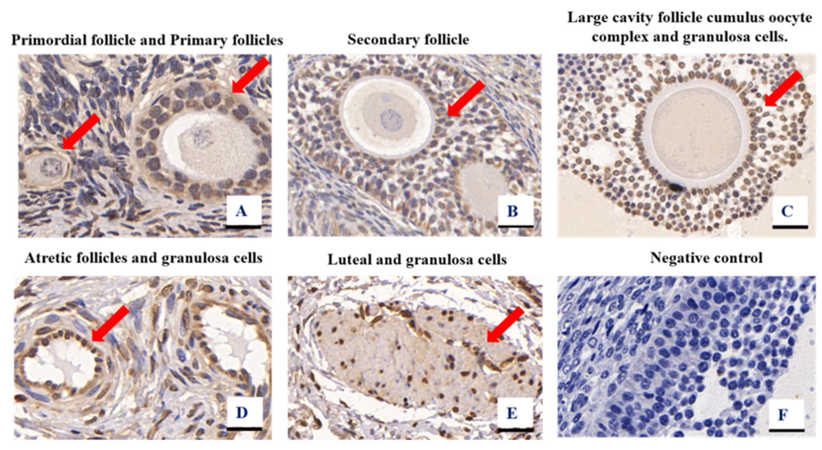
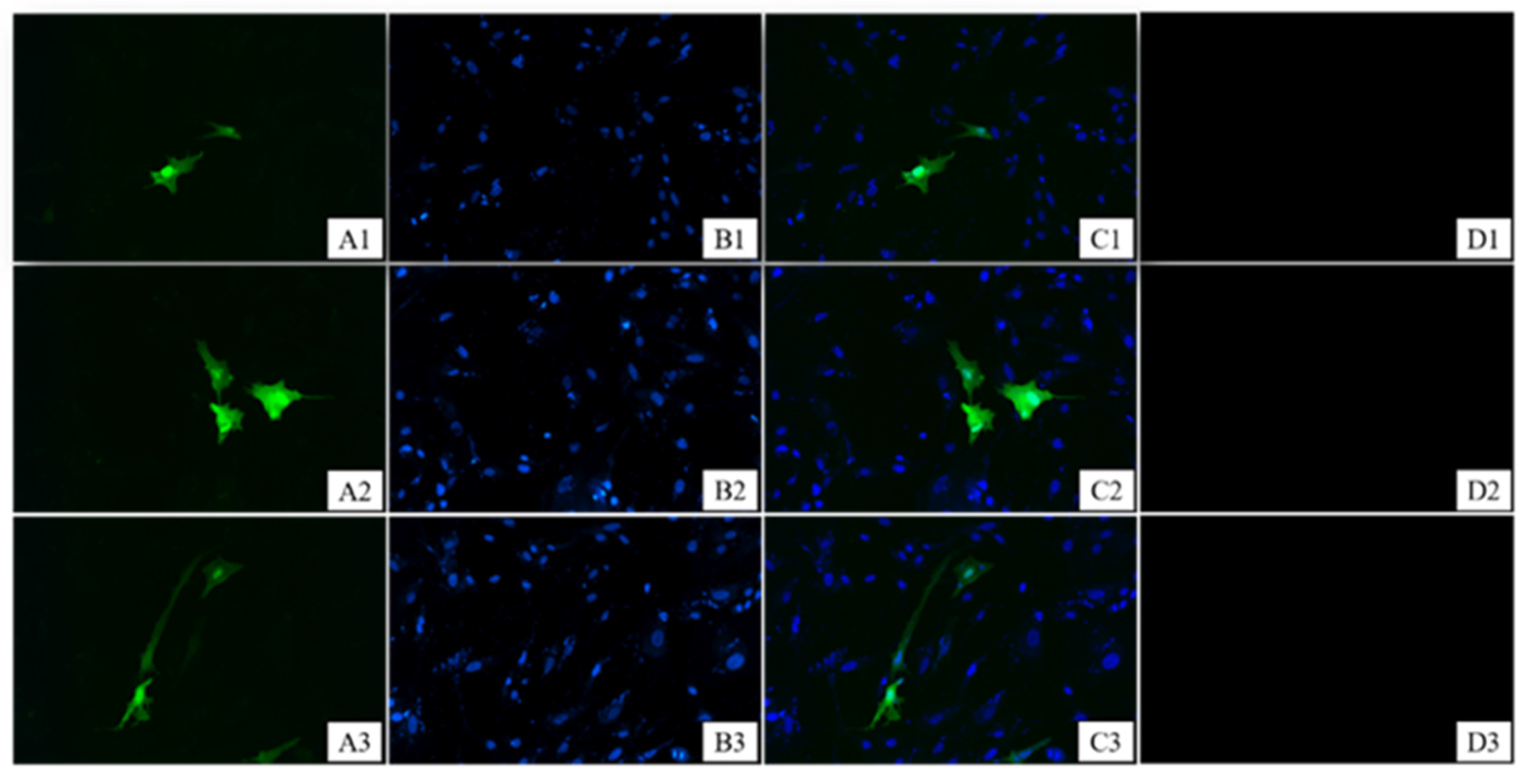
3.1. Granulosa Cell Identification and HE Staining of Ovarian Tissues

Follicle-stimulating hormone receptor (FSHR) is a transmembrane glycoprotein expressed in the granulosa cells of adult animals, so it can be used as a marker protein for granulosa cells. The assay uses indirect immunofluorescence to detect the purity of the FSHR protein. We found that more than 98% of the cells were stained and FSHR was located on the cell membrane, indicating that the goat primary cells were of high purity, confirming that the primary granulosa follicle cells of the Qianbei Ma goats were successfully cultured (Figure 1). The ovarian tissues of the Qianbei Ma goats were healthy and free of lesions, and had normal and clearly visible follicle morphology at all levels. Primordial, primary, and secondary follicles could be observed, allowing for the continuation of subsequent experiments (Figure 2).

3.2. Figures, Tables, and Schemes

The localization of CYP19A1 expression in the ovary tissues was determined using immunohistochemistry. The CYP19A1 protein was present in the primordial follicles, secondary follicles, oocyte complexes, luteal cells, and surrounding granulosa cells of developing goat follicles (Figure 3). The control reactions of the CYP19A1 antibodies tested confirmed the absence of non-specific staining (Figure 3F), suggesting that CYP19A1 is widely distributed in all parts of the ovary. The results of subcellular localization assays further revealed that the CYP19A1 protein was localized in the cell membrane and cytoplasm of granulosa cells (Figure 4).

3.3. Effects of Overexpression and Silencing of CYP19A1 on the mRNA Expressions of BMPR-IB, FSHR, and INHBA in Granulosa Cells

We investigated the effects of overexpression and silencing of the CYP19A1 gene on the mRNA levels of BMPR-IB, FSHR, and INHBA, genes related to multiple birth traits, in granulosa cells. We first verified the expression efficiencies of CYP19A1 overexpression and silencing vector in granulosa cells. Overexpression of CYP19A1 significantly increased the mRNA level of pEGFP-N3-CYP19A1 (p < 0.01) (Figure 5A). Among the four sequences that silenced CYP19A1, sh4-CYP19A1 had the best silencing efficiency (Figure 5B). Notably, overexpression of CYP19A1 significantly increased the mRNA levels of FSHR and INHBA in granulosa cells (p < 0.01) (Figure 5C) and vice versa (p < 0.01) (Figure 5D).

3.4. Effects of CYP19A1 on the Proliferation of Goat Granulosa Cells

The CCK8 assay and detection of proliferation-related genes, PCNA and Cyclin E, were used to study the effects of CYP19A1 on the biological behavior of granulosa cells. Overexpression of CYP19A1 gene in granulosa cells significantly increased cell proliferation (Figure 6A) and promoted the expressions of PCNA and Cyclin E genes (p < 0.01) (Figure 6B). In contrast, silencing of the CYP19A1 gene in granulosa cells significantly inhibited the proliferation of granulosa cells (p < 0.05) (Figure 6C) and suppressed the expressions of PCNA and Cyclin E genes (p < 0.01) (Figure 6D).

3.5. Effects of CYP19A1 on the Secretion of Steroid Hormones from Goat Granulosa Cells

We examined the intracellular estradiol (E2) and progesterone (P4) contents 6, 12, 24, and 48 h after the introduction of CYP19A1 into the cells to investigate the effects of altering the CYP19A1 content on steroid hormone secretion. Overexpression of CYP19A1 in granulosa cells increased progesterone and estrogen secretion at 6, 12, 24, and 48 h (Figure 7A,B). Similarly, the secretion of progesterone and estrogen in sh-NC was higher than that in the sh4-CYP19A1-treated group (Figure 7D,E), especially at 12, 24, and 48 h, but did not reach significant levels (Figure 7E). Similarly, overexpression of CYP19A1 significantly down-regulated the expressions of STAR, CYP11A1, and 3βSHD (p < 0.01) (Figure 7C), while silencing of CYP19A1 significantly up-regulated the expressions of STAR, CYP11A1, and 3βSHD (p < 0.01) (Figure 7F).

4. Discussion

Female fertility in mammals is co-determined by the number and quality of mature follicles [27]. Previously, researchers have postulated that the cytochrome P450 family is mainly expressed in mammalian theca cells, mouse granulose cells, ovarian and placental tissues, and other tissues [11,28,29]. These findings are consistent with our immunohistochemistry test results, suggesting that CYP19A1 is located in goat follicular granulosa cells. Interestingly, CYP19A1 is expressed in the pre-granulosa cells of female chicken embryonic gonads, unlike in mammals, where it is expressed in ovarian granulosa cells [30], a difference attributed to species differences between birds and mammals. In this study, CYP19A1 was present in theca, luteal granulosa, and cumulus complex cells of follicles at the different stages of follicular development. The subcellular localization further revealed that CYP19A1 might play a vital role in the nucleus and cytoplasm of granulosa cells. These findings collectively suggest that CYP19A1 is involved in the whole process of goat follicular development, including follicular maturation, normal ovulation, and periodic follicular derivation.
It is important to understand the molecular basis of follicular development and maturation because of its significant impact on the reproductive ability of goats [3]. The medium and high single expressions of the FSHR protein receptor in granulosa cells were identified, and follicular granulosa cells were successfully cultured to analyze the effects of changes in the CYP19A1 gene on the biological functions of granulosa cells. The growth and development of follicles in the ovary re dependent on the proliferation of granulosa cells [30]. Proliferating cell nuclear antigen (PCNA) is a proliferation marker widely distributed in various cells. The content of PCNA in cells generally reflects the proliferative rate of cells [31]. Cyclin E can also reflect the cell proliferative rate because it regulates the transition from the G0/G1 phase to the S phase in cells [32]. CYP19A1 was upregulated and silenced to investigate whether the changed expression of CYP19A1 would affect the proliferation of granulosa cells. Up-regulation of CYP19A1 improved the proliferation of granulosa cells and increased PCNA and Cyclin E expressions (p < 0.01). However, silencing of CYP19A1 decreased the proliferative rate of granulosa cells and the mRNA levels of PCNA and Cyclin E genes (p < 0.01). These findings indicated that the alteration in the CYP19A1 content in granulosa cells significantly affected the proliferative rate of granulosa cells and the mRNA expressions of INHBA and FSHR.
FSHR is a member of the transforming growth factor-β (TGF-β) family and regulates biological behaviors together with multiple cytokines, including regulating cell proliferation, apoptosis, and differentiation. It is mainly expressed in granulosa cells [33]. Relevant FSHR studies have indicated that mutation of the 5′UTR region in this gene can improve the litter size of both small-tailed Han sheep and Hu sheep [34,35]. The FSHR gene is a member of the G protein-coupled receptor family and is only expressed in mammalian follicular granulosa cells [36]. Its expression is increased with the growth of healthy follicles [37], playing a core role in promoting follicular maturation and increasing ovulation. The INHBA gene (Inhibin (INH) βA subunit) is a marker gene of granulosa cell development. It is expressed in oocytes of all follicle types, granulosa cells after the primary follicular phase, theca cells of sinus follicles, and surface epithelial cells of corpus luteum and ovaries. The expression of INHBA is also detected in the cumulus and mural granulosa cells in sinus follicles [38]. Li et al. postulated that INHBA is expressed at different follicular stages and has extremely high expression in the GCs growth period in multiparous-lamb goat population [39]. Notably, mutations in the INHBA gene have a significant effect on the litter size of sheep [34], thus further verifying that it can affect the lambing performance of goats by regulating the development of follicles.
Androstenedione and androgen can be transformed into estradiol and estrone with the action of aromatase in granulosa cells. Small amounts of the secreted estrogen act on ovary development, and the balance is released into the blood circulation and enters the target tissues, including the uterus, breast, and kidneys [40]. The destruction of the aromatase gene CYP19A1 in mice results in estrogen deficiency, leading to the normal development of ovarian follicles [21]. CYP19A1 is the key signal enzyme for estradiol synthesis [41]. The content of CYP19A1 has thus been determined in many studies to reflect the expression levels of steroid hormones. The higher the CYP19A1 content, the higher the hormone secretion. Padmanabhan et al. postulated that a decrease in CYP19A1 expression in the follicular granulosa cells of adult female sheep might destroy the balance between androgen and estrogen in follicles [22]. This imbalance potentially causes long-lasting follicles, which seriously endanger the periodic derivation of follicles and normal ovulation, causing low fecundity. The cholesterol side-chain lyase, which is encoded by CYP11A1, can catalyze the synthesis of pregnenolone, the common precursor of all steroid hormones, which plays a key role in the initial and rate-limiting steps of transformation [42]. The steroidogenic acute regulatory protein (STAR), located in the mitochondrial membrane, regulates the speed of cholesterol transport from the outer mitochondrial membrane to the inner membrane. CYP11A1 then converts the cholesterol on the inner membrane into pregnenolone, which is the substrate for progesterone synthesis [43]. Steroidogenic acute regulatory protein (STAR) and 3-β-hydroxysteroid dehydrogenase (3βHSD) are jointly involved in the development of oocytes and significantly affect oocyte quality and pregnancy results [44,45,46]. Notably, the secretion of P4 and E2 can be improved or inhibited to a certain extent by up-regulating the CYP19A1 gene in granulosa cells. In this study, up-regulation of CYP19A1 in granulosa cells increased the secretion of estrogen and progesterone but down-regulated the mRNA level of enzymes related to steroid hormone synthesis, including STAR, CYP11A1, and 3βSHD (p < 0.01). STAR, CYP11A1, and 3βSHD are the precursors for the synthesis of testosterone and progesterone in the steroid hormone synthesis pathway, and CYP19A1 is the key enzyme for the downstream hormone synthesis [47]. CYP19A1 inhibits the expression of STAR, CYP11A1, and 3βSHD after excessive secretion of estrogen and progesterone in the body through negative feedback regulation, thus stabilizing hormone synthesis in the body. Similarly, the mRNA levels of the STAR, CYP11A1, and 3βSHD genes are enhanced through negative feedback regulation (p < 0.01) when there is insufficient secretion of estrogen and progesterone in the body, further stabilizing hormone secretion. The findings reported herein will help us to understand the effects of the important pathway of CYP19A1 on hormone synthesis in granulosa cells.

5. Conclusions

Overexpression of CYP19A1 significantly increased the mRNA levels of CYP19A1, FSHR, and INHBA, which are candidate genes for multiple birth traits in goats. We found that overexpression of CYP19A1 in granulosa cells can promote the proliferation of granulosa cells; promote the expression of INHBA and FSHR genes in granulosa cells; promote the secretion of steroid hormones (E2 and P4), inhibit the expression of the key steroid synthesis genes STAR, CYP11A1, and 3βSHD; and can promote the expression of INHBA and FSHR in granulosa cells. Interfering with the expression of CYP19A1 can have the opposite result of overexpression. Therefore, we speculate that the CYP19A1 gene can regulate granulosa cell proliferation, possibly through negative feedback regulation of steroid hormone synthesis, and indirectly regulate goat follicle development and ovulation by promoting the expression of INHBA and FSHR.

Author Contributions

Methodology, Y.Z. and Z.Z.; software, Y.Z. and P.Y.; validation, Y.Z. and K.F.; formal analysis, Y.Z. and Z.Z.; resources, X.C.; data curation, Y.Z.; writing—original draft preparation, Y.Z.; writing—review and editing, Z.Z. and X.T. All authors have read and agreed to the published version of the manuscript.

Funding

This study was supported by the National Key R&D Program of China (No. 2021YFD1200403), the National Natural Science Foundation of China (No. 32060753), and the Guizhou Science and Technology Support Program Project (Qiankehe Support [2021] General 151).

Institutional Review Board Statement

The study was conducted according to the Regulations for the Administration of Affairs Concerning Experimental Animals approved by the State Council of the People’s Republic of China (No. 588, revised 2017). The use of experimental animals in this study was approved by the Laboratory Animal Ethics of Guizhou University (No. EAE-GZU-2021-P018, Guiyang, China; 11 October 2021).

Informed Consent Statement

Not applicable.

Data Availability Statement

The data presented in this study are available on request from the corresponding author.

Conflicts of Interest

The authors declare no conflict of interest.

References

  1. Tajima, K.; Orisaka, M.; Yata, H.; Goto, K.; Hosokawa, K.; Kotsuji, F. Role of granulosa and theca cell interactions in ovarian follicular maturation. Microsc. Res. Tech. 2006, 69, 450–458. [Google Scholar] [CrossRef]
  2. Vanderhyden, B.C.; Telfer, E.E.; Eppig, J.J. Mouse oocytes promote proliferation of granulosa cells from preantral and antral follicles in vitro. Biol. Reprod. 1992, 46, 1196–1204. [Google Scholar] [CrossRef] [PubMed] [Green Version]
  3. Havelock, J.C.; Rainey, W.E.; Carr, B.R. Ovarian granulosa cell lines. Mol. Cell. Endocrinol. 2004, 228, 67–78. [Google Scholar] [CrossRef]
  4. Sanchez, A.M.; Somigliana, E.; Vercellini, P.; Pagliardini, L.; Candiani, M.; Vigano, P. Endometriosis as a detrimental condition for granulosa cell steroidogenesis and development: From molecular alterations to clinical impact. J. Steroid. Biochem. Mol. Biol. 2016, 155, 35–46. [Google Scholar] [CrossRef] [PubMed]
  5. Drummond, A.E. The role of steroids in follicular growth. Reprod. Biol. Endocrinol. 2006, 4, 16. [Google Scholar] [CrossRef] [Green Version]
  6. Jain, P.; Jain, M.; Haldar, C.; Singh, T.B.; Jain, S. Melatonin and its correlation with testosterone in polycystic ovarian syndrome. J. Hum. Reprod. Sci. 2013, 6, 253–258. [Google Scholar] [CrossRef]
  7. Mahmoodi, M.; Soleimani Mehranjani, M.; Shariatzadeh, S.M.; Eimani, H.; Shahverdi, A. N-acetylcysteine improves function and follicular survival in mice ovarian grafts through inhibition of oxidative stress. Reprod. Biomed. Online 2015, 30, 101–110. [Google Scholar] [CrossRef] [PubMed] [Green Version]
  8. Macklon, N.S.; Fauser, B.C. Aspects of ovarian follicle development throughout life. Horm. Res. 1999, 52, 161–170. [Google Scholar] [CrossRef] [PubMed]
  9. Zhang, Y.; Zhang, Y.; Zhao, C.; Yu, T.; Liu, Y.; Shi, W.; Shi, F.; Liu, X.; Sheng, J.; Huang, H.; et al. Reduced alternative splicing of estrogen receptor alpha in the endometrium of women with endometriosis. Oncotarget 2017, 8, 110176–110186. [Google Scholar] [CrossRef] [Green Version]
  10. Siquara De Sousa, A.C.; Capek, S.; Amrami, K.K.; Spinner, R.J. Neural involvement in endometriosis: Review of anatomic distribution and mechanisms. Clin. Anat. 2015, 28, 1029–1038. [Google Scholar] [CrossRef]
  11. Zhang, W.; Lu, H.; Jiang, H.; Li, M.; Zhang, S.; Liu, Q.; Zhang, L. Isolation and characterization of cyp19a1a and cyp19a1b promoters in the protogynous hermaphrodite orange-spotted grouper (Epinephelus coioides). Gen. Comp. Endocrinol. 2012, 175, 473–487. [Google Scholar] [CrossRef]
  12. Vega, W.H.O.; Quirino, C.R.; Bartholazzi-Junior, A.; Rua, M.A.S.; Serapiao, R.V.; Oliveira, C.S. Variants in the CYP19A1 gene can affect in vitro embryo production traits in cattle. J. Assist. Reprod. Genet. 2018, 35, 2233–2241. [Google Scholar] [CrossRef]
  13. Bao, B.; Garverick, H.A. Expression of steroidogenic enzyme and gonadotropin receptor genes in bovine follicles during ovarian follicular waves: A review. J. Anim. Sci. 1998, 76, 1903–1921. [Google Scholar] [CrossRef] [PubMed] [Green Version]
  14. Juengel, J.L.; Hudson, N.L.; Heath, D.A.; Smith, P.; Reader, K.L.; Lawrence, S.B.; O’Connell, A.R.; Laitinen, M.P.; Cranfield, M.; Groome, N.P.; et al. Growth differentiation factor 9 and bone morphogenetic protein 15 are essential for ovarian follicular development in sheep. Biol. Reprod. 2002, 67, 1777–1789. [Google Scholar] [CrossRef] [PubMed] [Green Version]
  15. Kwon, J.; McAndrew, B.; Penman, D. Cloning of brain aromatase gene and expression of brain and ovarian aromatase genes during sexual differentiation in genetic male and female Nile tilapia Oreochromis niloticus. Mol. Reprod. Dev. 2001, 59, 359–370. [Google Scholar] [CrossRef] [PubMed]
  16. Hess, M.F.; Roser, J.F. Immunocytochemical localization of cytochrome P450 aromatase in the testis of prepubertal, pubertal, and postpubertal horses. Theriogenology 2004, 61, 293–299. [Google Scholar] [CrossRef]
  17. Albrecht, B.A.; Daels, P.F. Immunolocalization of 3 beta-hydroxysteroid dehydrogenase, cytochrome P450 17 alpha-hydroxylase/17,20-lyase and cytochrome P450 aromatase in the equine corpus luteum of dioestrus and early pregnancy. J. Reprod. Fertil. 1997, 111, 127–133. [Google Scholar] [CrossRef] [Green Version]
  18. Zhou, R.; Yang, Y.-l.; Liu, Y.; Chen, Q.-M.; Chen, J.; Li, K. Association of CYP19A1 gene polymorphisms with reproductive traits in pigs. J. Integr. Agric. 2017, 16, 1558–1565. [Google Scholar] [CrossRef]
  19. Logan, K.A.; Juengel, J.L.; McNatty, K.P. Onset of steroidogenic enzyme gene expression during ovarian follicular development in sheep. Biol. Reprod. 2002, 66, 906–916. [Google Scholar] [CrossRef] [Green Version]
  20. Quirke, L.D.; Juengel, J.L.; Tisdall, D.J.; Lun, S.; Heath, D.A.; McNatty, K.P. Ontogeny of steroidogenesis in the fetal sheep gonad. Biol. Reprod. 2001, 65, 216–228. [Google Scholar] [CrossRef]
  21. Luo, M.; Li, L.; Xiao, C.; Sun, Y.; Wang, G.L. Heat stress impairs mice granulosa cell function by diminishing steroids production and inducing apoptosis. Mol. Cell. Biochem. 2016, 412, 81–90. [Google Scholar] [CrossRef] [PubMed]
  22. Padmanabhan, V.; Salvetti, N.R.; Matiller, V.; Ortega, H.H. Developmental programming: Prenatal steroid excess disrupts key members of intraovarian steroidogenic pathway in sheep. Endocrinology 2014, 155, 3649–3660. [Google Scholar] [CrossRef] [Green Version]
  23. Wu, P.; Wang, X.; Gao, F.; Du, W. Cyp19a1Role of in the female pathway of a freshwater turtle species (Mauremys reevesii) with temperature-dependent sex determination. Zool. Res. 2022, 43, 81–84. [Google Scholar] [CrossRef] [PubMed]
  24. Zhou, Z.; Chen, X.; Zhu, M.; Wang, W.; Ao, Z.; Zhao, J.; Tang, W.; Hong, L. Cathepsin D knockdown regulates biological behaviors of granulosa cells and affects litter size traits in goats. J. Zhejiang Univ. Sci. B 2021, 22, 893–905. [Google Scholar] [CrossRef] [PubMed]
  25. Peng, J.Y.; Gao, K.X.; Xin, H.Y.; Han, P.; Zhu, G.Q.; Cao, B.Y. Molecular cloning, expression analysis, and function of decorin in goat ovarian granulosa cells. Domest. Anim. Endocrinol. 2016, 57, 108–116. [Google Scholar] [CrossRef]
  26. Gong, T.; Wang, W.; Xu, H.; Yang, Y.; Chen, X.; Meng, L.; Xu, Y.; Li, Z.; Wan, S.; Mu, Q. Longitudinal Expression of Testicular TAS1R3 from Prepuberty to Sexual Maturity in Congjiang Xiang Pigs. Animals 2021, 11, 437. [Google Scholar] [CrossRef]
  27. Storbeck, K.H.; Swart, A.C.; Slabbert, J.T.; Swart, P. The identification of two CYP17 alleles in the South African Angora goat. Drug. Metab. Rev. 2007, 39, 467–480. [Google Scholar] [CrossRef]
  28. Storbeck, K.H.; Swart, A.C.; Snyman, M.A.; Swart, P. Two CYP17 genes in the South African Angora goat (Capra hircus)—The identification of three genotypes that differ in copy number and steroidogenic output. FEBS J. 2008, 275, 3934–3943. [Google Scholar] [CrossRef] [PubMed]
  29. Guo, Y.; Cheng, L.; Li, X.; Tang, S.; Zhang, X.; Gong, Y. Transcriptional regulation of CYP19A1 expression in chickens: ESR1, ESR2 and NR5A2 form a functional network. GEN. Comp. Endocr. 2022, 315, 113939. [Google Scholar] [CrossRef]
  30. Chou, C.H.; Chen, M.J. The Effect of Steroid Hormones on Ovarian Follicle Development. Vitam. Horm. 2018, 107, 155–175. [Google Scholar] [CrossRef]
  31. Strzalka, W.; Ziemienowicz, A. Proliferating cell nuclear antigen (PCNA): A key factor in DNA replication and cell cycle regulation. Ann. Bot. 2011, 107, 1127–1140. [Google Scholar] [CrossRef] [Green Version]
  32. Pradeep, P.K.; Li, X.; Peegel, H.; Menon, K.M. Dihydrotestosterone inhibits granulosa cell proliferation by decreasing the cyclin D2 mRNA expression and cell cycle arrest at G1 phase. Endocrinology 2002, 143, 2930–2935. [Google Scholar] [CrossRef]
  33. Shimizu, K.; Nakamura, T.; Bayasula; Nakanishi, N.; Kasahara, Y.; Nagai, T.; Murase, T.; Osuka, S.; Goto, M.; Iwase, A.; et al. Molecular mechanism of FSHR expression induced by BMP15 in human granulosa cells. J. Assist. Reprod. Genet. 2019, 36, 1185–1194. [Google Scholar] [CrossRef]
  34. Zi, X.D.; Xu, H.W.; Wang, Y. Variation in sequences and mRNA expression levels of inhibin subunits alpha (INHA) and betaA (INHBA) genes between prolific and nonprolific goat breeds. Mol. Reprod. Dev. 2012, 79, 238. [Google Scholar] [CrossRef]
  35. Chu, M.X.; Guo, X.H.; Feng, C.J.; Li, Y.; Huang, D.W.; Feng, T.; Cao, G.L.; Fang, L.; Di, R.; Tang, Q.Q.; et al. Polymorphism of 5′ regulatory region of ovine FSHR gene and its association with litter size in Small Tail Han sheep. Mol. Biol. Rep. 2012, 39, 3721–3725. [Google Scholar] [CrossRef]
  36. Xia, Y.; Wang, Q.; He, X.D.; Chen, Y.; JiGe, M.T.; Zi, X.D. Cloning and expression analysis of the follicle-stimulating hormone receptor (FSHR) gene in the reproductive axis of female yaks (Bos grunniens). Domest. Anim. Endocrinol. 2020, 70, 106383. [Google Scholar] [CrossRef]
  37. Saint-Dizier, M.; Malandain, E.; Thoumire, S.; Remy, B.; Chastant-Maillard, S. Expression of follicle stimulating hormone and luteinizing hormone receptors during follicular growth in the domestic cat ovary. Mol. Reprod. Dev. 2007, 74, 989–996. [Google Scholar] [CrossRef]
  38. Silva, J.R.; van den Hurk, R.; van Tol, H.T.; Roelen, B.A.; Figueiredo, J.R. Gene expression and protein localisation for activin-A, follistatin and activin receptors in goat ovaries. J. Endocrinol. 2004, 183, 405–415. [Google Scholar] [CrossRef] [Green Version]
  39. Li, Z.; Wang, J.; Zhao, Y.; Ma, D.; Zhao, M.; Li, N.; Men, Y.; Zhang, Y.; Chu, H.; Lei, C.; et al. scRNA-seq of ovarian follicle granulosa cells from different fertility goats reveals distinct expression patterns. Reprod. Domest. Anim. 2021, 56, 801–811. [Google Scholar] [CrossRef]
  40. Klymiuk, M.C.; Neunzig, J.; Bernhardt, R.; Sanchez-Guijo, A.; Hartmann, M.F.; Wudy, S.A.; Schuler, G. Efficiency of the sulfate pathway in comparison to the Delta4- and Delta5-pathway of steroidogenesis in the porcine testis. J. Steroid. Biochem. Mol. Biol. 2018, 179, 64–72. [Google Scholar] [CrossRef]
  41. Li, Q.; Du, X.; Liu, L.; Liu, H.; Pan, Z.; Li, Q. Upregulation of miR-146b promotes porcine ovarian granulosa cell apoptosis by attenuating CYP19A1. Domest. Anim. Endocrinol. 2021, 74, 106509. [Google Scholar] [CrossRef]
  42. Miller, W.L. Androgen biosynthesis from cholesterol to DHEA. Mol. Cell. Endocrinol. 2002, 198, 7–14. [Google Scholar] [CrossRef]
  43. Miller, W.L. Disorders in the initial steps of steroid hormone synthesis. J. Steroid. Biochem. Mol. Biol. 2017, 165, 18–37. [Google Scholar] [CrossRef]
  44. Feuerstein, P.; Cadoret, V.; Dalbies-Tran, R.; Guerif, F.; Bidault, R.; Royere, D. Gene expression in human cumulus cells: One approach to oocyte competence. Hum. Reprod. 2007, 22, 3069–3077. [Google Scholar] [CrossRef] [Green Version]
  45. Hamel, M.; Dufort, I.; Robert, C.; Gravel, C.; Leveille, M.-C.; Leader, A.; Sirard, M.-A. Identification of differentially expressed markers in human follicular cells associated with competent oocytes. Hum. Reprod. 2008, 23, 1118–1127. [Google Scholar] [CrossRef] [Green Version]
  46. Wathlet, S.; Adriaenssens, T.; Segers, I.; Verheyen, G.; Janssens, R.; Coucke, W.; Devroey, P.; Smitz, J. New candidate genes to predict pregnancy outcome in single embryo transfer cycles when using cumulus cell gene expression. Fertil. Steril. 2012, 98, 432–439.e4. [Google Scholar] [CrossRef]
  47. Cui, J.; Shen, Y.; Li, R. Estrogen synthesis and signaling pathways during aging: From periphery to brain. Trends Mol. Med. 2013, 19, 197–209. [Google Scholar] [CrossRef] [Green Version]
Figure 1. Follicular granulosa cell culture identification. (A1D1) Immunofluorescence result of granulosa cells observed at 400×. (A2D2) Immunofluorescence result of granulosa cells observed at 200×. (C1) Map combining (A1 + B1), (C2) map combining (A2 + B2), and (D1,D2) blank controls without FSHR antibody staining. (A1,A2) Granule nuclear staining, (B1,B2) cytoplasmic staining, and (C1,C2) nuclear and cytoplasmic synthesis maps.
Animals 12 01911 g001
Figure 2. HE staining of ovarian tissue: (AD) 400×, (E) 200×, (F) 50×. (A) Corpus luteum; (B) primordial follicle cells; (C) secondary follicle; (D) tertiary follicle; (E) atretic follicle; (F) mature large follicle group.
Animals 12 01911 g002
Figure 3. CYP19A1 protein subcellular localization results, 400×.
Animals 12 01911 g003
Figure 4. CYP19A1 protein localization, 400×. C (C1C3) Diagram combining A (A1A3) + B (B1B3); (D1D3) blank control (n = 3).
Animals 12 01911 g004
Figure 5. Validation of CYP19A1 overexpression and silencing expression vector efficiency and results of genetic assays for reproduction-related indicators. (A) pEGFP-N3-CYP19A1 efficiency validation; (B) sh1-4-CYP19A1 efficiency validation; (C) results of overexpression of CYP19A1; (D) results of silencing CYP19A1. ** indicates highly significant difference (p < 0.01). The mRNA levels of target genes were normalized with housekeeping gene (β-actin, 139 bp) and expressed as 2−∆∆CT using samples on pEGFP-N3 or sh-NC as calibrator. Results are indicated by means ± SD (n = 3).
Animals 12 01911 g005
Figure 6. Effects of altering CYP19A1 on proliferation of granulosa cells. (A) Overexpression of CYP19A1 promotes PCNA and Cyclin E mRNA expression levels; (B) overexpression of CYP19A1 promotes granulosa cell proliferation; (C) silencing of CYP19A1 inhibits PCNA and Cyclin E mRNA expression levels; (D) silencing of CYP19A1 inhibits granulosa cell proliferation. The mRNA levels of target genes were normalized with housekeeping gene (β-actin, 139 bp) and expressed as 2−∆∆CT using samples on pEGFP-N3 or sh-NC as a calibrator. Results are indicated by means ± SD (n = 3). ** indicates highly significant difference (p < 0.01), * indicates significant difference (p < 0.05).
Animals 12 01911 g006
Figure 7. Effects of altering CYP19A1 gene on granulosa cell proliferation. (A) Overexpression of CYP19A1 promotes progesterone secretion in granulosa cells; (B) silencing CYP19A1 inhibits progesterone secretion in granulosa cells; (C) overexpression of CYP19A1 promotes mRNA expression levels of steroid-related genes; (D) overexpression of CYP19A1 promotes estrogen secretion in granulosa cells; (E) silencing of CYP19A1 inhibits estrogen secretion in granulosa cells; (F) silencing of CYP19A1 suppression inhibits the mRNA expression level of steroid-related genes. ** indicates highly significant difference (p < 0.01), * indicates significant difference (p < 0.05).
Animals 12 01911 g007
Table 1. CYP19A1 shRNA and CYP19A1 pEGFP primer information.
Gene NamePrimer Sequences (5′-3′)
sh1-CYP19A1F: CACCGCGGTCACCAACATAATCAGCTTCAAGAGAGCTGATTATGTTGGTGACCGCTTTTTTG
R: GATCCAAAAAAGCGGTCACCAACATAATCAGCTCTCTTGAAGCTGATTATGTTGGTGACCGC
sh2-CYP19A1F: CACCGACCAGAATATAGGTTTCAATTTCAAGAGAATTGAAACCTATATTCTGGTCTTTTTTG
R: GATCCAAAAAAGACCAGAATATAGGTTTCAATTCTCTTGAAATTGAAACCTATATTCTGGTC
sh3-CYP19A1F: CACCGAGGCAATGATGAGGGAAATCTTCAAGAGAGATTTCCCTCATCATTGCCTCTTTTTTG
R: GATCCAAAAAAGAGGCAATGATGAGGGAAATCTCTCTTGAAGATTTCCCTCATCATTGCCTC
sh4-CYP19AF: CACCGCTGTGCAGAAAGTATGAAAATTCAAGAGATTTTCATACTTTCTGCACAGCTTTTTTG
R: GATCCAAAAAAGCTGTGCAGAAAGTATGAAAATCTCTTGAATTTTCATACTTTCTGCACAGC
sh-NCF: CACCGTTCTCCGAACGTGTCACGTTTCAAGAGAACGTGACACGTTCGGAGAATTTTTTG
R: GATCCAAAAAATTCTCCGAACGTGTCACGTTCTCTTGAAACGTGACACGTTCGGAGAAC
pEGFP-N3-CYP19A1(BglII) F: CGTCAGATCCGCTAGCGCTACCGGACTCAGATCTATGCTTTTGGAAGTGCTGAAC
(SacIIR): CACCATGGTGGCGATGGATCCCGGGCCCGCGGCGCACTCGAGGCACTTGTCTGAATTTCTC
The pEGFPpEGFP-N3-CYP19A1 vector sequence is shown in red for the homology arm, bold black for the digestion site, and black for the target fragment.
Table 2. Related gene real-time fluorescent primer information.
Gene NamePrimer Sequences (5′-3′)Login IDFragment Size (bp)
BMPR-1BF: GCTCTTGGTCCTCATCATTTTATTC
R: ATGTAAGTTTCGTCCTGTTCTAACC
NC_022298.2143
FSHRF: TGTTATGTCCCTCCTTGTGCTC
R: CGCTTGGCTATCTTGGTGTCA
NM_001285644.1122
PCNAF: GTAGCCGTGTCATTGCGACTCC
R: GCTCTGTAGGTTCACGCCACTTG
XM_005688167.3145
INHBAF: AAGGTGGTGGATGCTCGAAA
R: GTCTCCTGACACTGCTCACA
NM_001285581.1125
CYP19A1F: CCCAAGGCATTACAATGT
R: TAAGGGTTTCCTCTCCAC
NM_001314145.1169
STARF: GGTCCCCGAGACTTTGTGAG
R: AATCCACTTGGGTCTGCGAG
XM_013975437.2262
3βHSDF: AGACCAGAAGTTCGGGAGGAA
R: TCTCCCTGTAGGAGTTGGGC
NM_001285716.1292
CYP11A1F: CTCCAGAGGCAATAAAGAA
R: TCAAAGGCAAAGTGAAACA
NM_001287574.1145
Cyclin EF: GATGTCGGCTGCTTAGAAT
R: CACCACTGATACCCTGAAAC
XM_018062248.1104
β-actinF: AGATGTGGATCAGCAAGCAG
R: CCAATCTCATCTCGTTTTCTG
NM_001297986.1139
Publisher’s Note: MDPI stays neutral with regard to jurisdictional claims in published maps and institutional affiliations.

